Clinical and molecular determinants of arterial thrombus structure
PhD thesis booklet
András Kovács, MD
Semmelweis University
Doctoral School of Molecular Medicine
Supervisor: Krasimir Kolev, MD, Dsc
Official reviewers: Emese Tóth-Zsámboki, MD, PhD Gábor Széplaki, MD, PhD
Chairman of the final examination comittee:
Péter Sándor, MD, DSc
Members of the final examination comittee:
Róbert Gábor Kiss, MD, PhD Klára Gadó, MD, PhD
Budapest, 2015
1
INTRODUCTION
Cardiovascular disease, as the leading cause of mortality in “developed countries” is often, if not always, related to arterial thrombosis. Although variations of arterial thrombus structure are strongly influenced by hemodynamic conditions and the general composition of blood, the interrelations of clinical parameters and thrombus architecture are poorly elucidated. Conversely, it also remains to be clarified how structural properties of thrombi affect the clinical picture and the course of disease. During my PhD work, I studied the determinants of arterial thrombus structure mainly from the aspect of enzymatic thrombus dissolution, a therapeutic modality which, considering the limitations of its effectiveness and frequent side effects, is yet to be refined. Even though chemical clot dissolution is almost exclusively replaced by percutaneous intervention in the treatment of acute myocardial infarction, thrombolysis might be a useful complementary tool to catheter interventions, given the fact that remnants of the aspirated thrombus might cause distal embolization and that implanted coronary stents are potential sources of rethrombosis. Safe and effective thrombolysis is even more crucial in acute ischaemic stroke in the treatment of which catheter intervention techniques are in initial stage of application.
Basically, thrombi consist of cellular elements (platelets, red blood cells, white blood cells) and fibrin
2
network. In the present work I focus on the determinants of fibrin structure and the correlations between clinical parameters and the ultrastructural architecture of thrombi removed from arterial thrombosis patients.
Although thrombin activatable fibrinolysis inhibitor (TAFI) is classically regarded as a prothrombotic agent, elevated levels of the enzyme can be accompanied by both high and low thrombotic risk and it shows contradictory in vitro behavior as well. At the end of my thesis I present our results on the influence of TAFI on clot structure and fibrinolysis.
3
OBJECTIVES
In the present thesis I sought to answer the following questions:
- What structural differences can be observed between coronary and peripheral arterial thrombi?
- Are there any interactions between thrombus components in the final determination of clot architecture?
- How does ischaemic time (from onset of symptoms to intervention) affect clot structure?
- What are the determinants of arterial thrombus structure in general?
- How does TAFI / carboxypeptidase B (CPB) influence the architecture of the fibrin network?
- Does arginine play a role in TAFI / CPB function?
- Do structural differences related to TAFI / CPB carry any functional consequences for fibrinolysis?
4 METHODS Thrombus samples
Fifty consecutive peripheral artery thrombosis patients and 101 consecutive acute myocardial infarction (AMI) patients of various age and sex were recruited over 18 months. Coronary patients were referred to primary percutaneous coronary intervention with thromboaspiration after developing symptoms of AMI and all patients were pre-treated with aspirin, Na-heparin, abciximab (unless a contraindication was present) and clopidogrel. Peripheral artery thrombosis patients were treated by thrombendarterectomy, during which systemic Na-heparin was administered before clamping. Some of the thrombendarterectomized patients received antiplatelet drugs (aspirin, clopidogrel or their combination) preoperatively.
Patient characteristics
The following clinical parameters were registered: age, sex, complete blood count, hemoglobin, hematocrit, C- reactive protein level, ECG findings (STEMI or NSTEMI, localization of myocardial lesion), accompanying diseases (atherosclerosis, diabetes, hypertension, dyslipidaemia), smoking, vascular localization of thrombi and the diameter of the vessel, ischaemic time (from onset of symptoms to intervention), anti-platelet premedication.
5
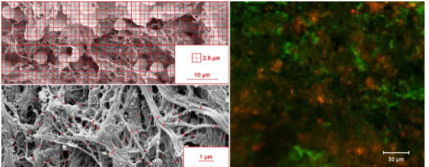
Scanning electron microscopic imaging of thrombi Thrombus samples were stored in Na-cacodylate, fixed in glutaraldehyde, dehydrated in ethanol and acetone, dryed in CO2 critical point dryer. Specimens mounted on carbon discs were sputter coated with gold to enhance contrast. Images (15 per thrombus) were taken with scanning electron microscope and morphometrically analysed in two ways: 1) Images were divided in 432 squares (8.4 µm2 each) and these areas were classified as fibrin, red blood cells, platelets, white blood cells or their combinations depending on the observed coverage by the respective component based on morphological characteristics (Figure 1, upper left panel). Quantity of the components was then calculated as percentage. 2) The diameter of the fibrin fibers were measured manually and analyzed using self-designed scripts (Figure 1, bottom left panel).
Figure 1. Microscopic analysis of thrombus composition.
6
Immunofluorescent visualization of platelets
Before the immunofluorescent examination of ex vivo clots, in order to establish a reliable method and to characterize the behavior of the antibody, we performed immunofluorescent staining of platelets. A flow chamber was set up to model platelet adhesion on a thromogenic surface. We used human iliac artery cross sections and collagen coated slides as thrombogenic surfaces. The perfusion chamber was constructed using a double-sided tape as the sidewalls of the chamber, which was sandwiched to a methacrylate cover containing the inlet and outlet tubing. A pneumatic pump was used to maintain perfusion with a shear rate of 3350 s-1, at which shear thrombocyte adhesion is strictly von Willebrand factor-dependent. After perfusion, slides were treated with mouse monoclonal anti-GpIIb/IIIa primary and green fluorescent goat-anti-mouse IgG secondary antibodies and were evaluated by confocal laser microscopy. A threshold intensity was determined in order to filter out background fluorescence, thereby optimizing the signal-to-noise ratio. The same settings were then used to analyse ex vivo thrombus specimens (see below).
Immunofluorescent imaging of arterial clots, calculation of fibrin/platelet ratio
Thrombus samples were frozen immediately upon removal and stored at -80°C. Cryosections were made
7
from 3 different parts of the thrombi for indirect immunofluorescent visualization of platelets and fibrin using anti-human GpIIb/IIIa or anti-human fibrin mouse monoclonal primary antibodies and green or red fluorophore-labelled goat-anti-mouse IgG as secondary antibodies. Fluorescent images (15/thrombus) were taken using a confocal laser scanning microscope (Figure 1, right panel). Quantification of platelets and fibrin on thrombus sections was performed, selecting the region of interest, calculating its surface area in pixels and setting threshold intensity values for identification of the areas covered by platelets and fibrin. The ratio of the two percentage coverage value was calculated and used thereafter as fibrin/platelet ratio (FP).
Turbidimetric fibrinolytic assays
The size of polymers generated during fibrin production falls into the wavelength range of visible light, therefore as light passes through the fibrin clot, it loses some of its intensity. A loss of intensity measured at 340 nm (turbidity) carries information about the structure of fibrin as well, as it is proportional to fiber diameter.
Based on this principle, a large number of parallel measurements can be performed, both purified fibrin and blood plasma clots can be studied and fibrinolysis initiated from either inside or the surface of the clot can be measured. In 96-well microtiter plates, fibrinogen and arginine or CPB were mixed with thrombin. In the assays
8
when lysis was initiated by surface tPA, fibrinogen also contained plasminogen and following 30 min clotting, tPA was applied to the surface of clots. In the assays when lysis was initiated by tPA dispersed in the clot, fibrinogen contained plasminogen and tPA was added together with thrombin. When lysis was initiated by active plasmin, the enzyme was either uniformly dispersed in the clot or applied to the clot surface. Clot formation and dissolution was followed by measuring the light absorbance at 340 nm and 37 °C. The time needed to reduce the turbidity of the clot to 50% or 10% of the maximal value was used as a quantitative parameter of fibrinolytic activity. In certain cases recalcified citrated blood plasma was clotted with thrombin instead of purified fibrinogen.
Confocal laser microscopy
Fibrin clots were prepared from fibrinogen, 2% of which was red fluorophore-labelled, plasminogen and CPB or arginine with thrombin for 30 min at room temperature in microfluidic slides containing a preformed channel of 0.4 mm height and 30 µl volume, Thereafter yellow fluorophore-labelled tPA was added to the edge of the clot and the fluorescence was monitored with a confocal laser microscope taking sequential images of the fluid- fibrin interface.
9 Plasminogen activation assay
In microtiter plates, fibrinogen containing plasminogen and arginine or CPB was clotted with thrombin. After 30 min at 37 °C tPA and synthetic Spectrozyme-PL plasmin substrate were placed on the surface of the clot. The forming plasmin generated p-nitroaniline, the absorbance of which was continuously recorded at 405 nm.
Statistical analysis
Five Bootstrap statistical tests were applied to detect differences between the characteristics of pairs of independent and identically distributed populations represented by experimentally gained one-dimensional continuous samples: two-tail and one-tail tests for means and medians and a Kuiper test for distributions. Their algorithms are modifications of established procedures.
Linear regression models and outlier rejection were based on classical regression assumptions. The distribution of the data on fiber diameter was analyzed according to an algorithm used previously: theoretical distributions were fitted to the empirical data sets and compared using Kuiper test and Monte Carlo simulation procedures.
10 RESULTS
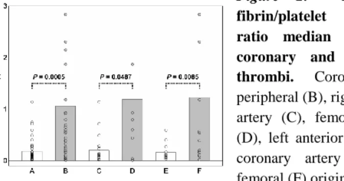
Composition of coronary and peripheral thrombi Both imaging techniques revealed significant differences in the relative platelet and fibrin content of coronary and peripheral thrombi. The median fluorescent fibrin/platelet ratio (FP50) values of peripheral thrombi were more than five-fold higher (P<0.05) than the corresponding values of the coronary thrombi. Subgroups of thrombi, defined according to the vascular location within the coronary and peripheral arteries, showed the same trend of FP50, i.e. FP50 was consistently lower in coronary than in peripheral arterial locations (e.g. lower in right coronary artery or left anterior descendent artery thrombi than in femoro-popliteal or ilio-femoral thrombi, Figure 2).
Figure 2. Fluorescent fibrin/platelet coverage ratio median (FP50) of coronary and peripheral thrombi. Coronary (A), peripheral (B), right coronary artery (C), femoro-popliteal (D), left anterior descendent coronary artery (E), ilio- femoral (F) origin.
Regression analysis revealed that above a coronary vessel diameter of 2.6 mm, larger diameter correlated with smaller relative intrathrombotic platelet occupancy (Radj2
11
=0.51, P=1×10⁻⁷). Scanning electron microscopic assessment indicated lower fibrin content in coronary than in peripheral thrombi (P<0.01). While coronary thrombi contained a fine fibrin network made up of thinner fibers, peripheral thrombi were composed of a coarse fibrin net with thicker fibers and larger pores. The median fiber diameter in coronary thrombi was 123 nm vs 135 nm in peripheral thrombi (P<0.05).
Relationship of systemic cell counts and thrombus composition, effects of anti-platelet medication
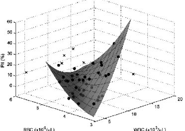
In coronary thrombi, regression analysis showed that at high and low hematocrit values, platelet content was higher than at intermediate values (Radj2 >0.7). Parabolic regression models revealed a J-shaped dependence of thrombus platelet content on the systemic platelet count for peripheral thrombi (P<0.05): above a platelet count of 250,000/µL the thrombus platelet content increased in parallel with the systemic count. Anti-platelet medication strengthened this dependence primarily in peripheral thrombi. Accompanying dyslipidaemia and smoking enhanced the association between blood and clot platelet content. More associations between systemic and intrathrombotic cell counts were revealed by multivariable regression models: the systemic white and red blood cell count influenced the clot platelet content in
12
female (Radj2 =0.70), atherosclerotic (Radj2 =0.59, Figure 3) and non-diabetic (Radj2 =0.68) patients.
Figure 3. Joint impact of systemic cell counts on platelet content in peripheral thrombi on atherosclerotic plaques.
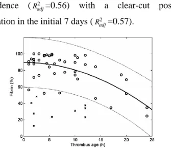
Effect of thrombus age
In coronary thrombi, a U-shaped dependence of intrathrombotic fibrin content on thrombus age was found (Radj2 =0.41), where very old (>300 h) thrombi had a fibrin content approximately equal to that in fresh thrombi. Limiting the analysis to fresh thrombi (<24 h) resulted in a negative correlation between fibrin content and thrombus age (Radj2 =0.46, Figure 4). In peripheral thrombi a parabolic regression model of clot platelet content versus thrombus age showed an inverse U-shaped
13
dependence (Radj2 =0.56) with a clear-cut positive correlation in the initial 7 days (Radj2 =0.57).
Figure 4. Impact of time on coronary clot fibrin content
Smoking as a determinant of clot structure
A considerable difference of clot fibrin content was found between smoking and non-smoking coronary patients. The mean value of the clot fibrin content for non-smokers was 62.2% versus 78.14% for smokers (P<0.05). In line with this trend of fibrin content, the fluorescent FP50 values were significantly lower in non- smokers (mean 0.24) than in active smokers (0.86) for the whole patient group (P<0.01).
Structural modifications of fibrin by CPB and arginine The presence of CPB during fibrinogen clotting modified the turbidity of the clots before initiation of lysis, which
14
suggested changes in the structure of fibrin. Scanning electron microscope images revealed that the fiber diameter decreased by 21 – 25 % in CPB-treated clots prepared from fibrinogen (Figure 5). Because the native fibrinogen does not contain any C-terminal lysine or arginine for CPB, the single target of CPB action in this purified system could be the arginine residues in the fibrinopeptides newly cleaved by thrombin. Addition of arginine caused changes in the structure of fibrin comparable to those induced by CPB.
Figure 5. Modification of fibrin structure by arginine and CPB.
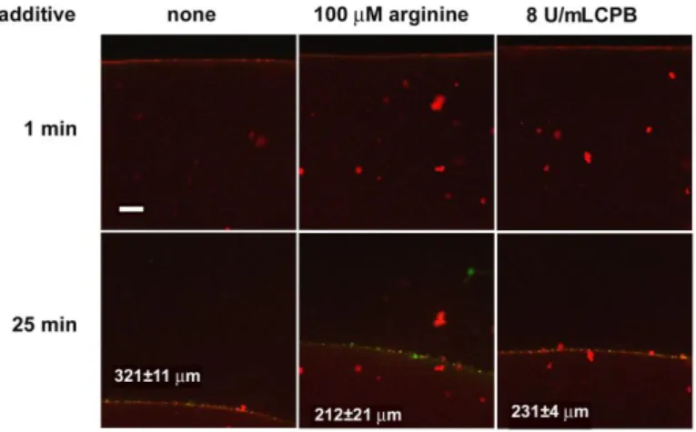
Kinetics of plasminogen activation and fibrinolysis On a microscopic scale, tPA-induced lysis of fibrin modified by CPB was significantly slowed down compared to native fibrin (Figure 6). On a macroscopic scale of lytic kinetics, CPB had a maximal inhibitory effect at 8 U/mL in the assay when tPA was added to the surface of pre-formed clots containing plasminogen, and a similar level of inhibition of lysis was also seen in clots formed in the presence of 2 µM arginine.
15
Figure 6. Effects of arginine and CPB on the penetration of tPA- YFP into fibrin in the course of lysis
Despite the identical time to complete dissolution, the lysis kinetics were somewhat different with CPB or arginine, suggesting a different mechanism of action. To address this difference two assays were used; one that monitored solely the generation of plasmin by adding synthetic plasmin substrate and a second one that bypassed the stage of plasminogen activation. As expected, plasmin generation was inhibited in the presence of CPB, however, fibrin formed in the presence of arginine proved to be a better template for plasminogen activation than the native fibrin structure, despite the overall inhibiting effect of arginine on tPA- induced fibrin degradation. When plasmin was added to the surface of fibrin, slower lysis was observed similarly to the tPA-induced lysis, if clots were formed in the
16
presence of arginine. Quite unexpectedly, CPB rendered fibrin more susceptible to lysis by plasmin added to the surface of the clot. However, if plasmin was homogeneously dispersed within the clot, neither inhibitory effect of arginine, nor stimulatory effect of CPB could be observed. The discordant effects of the modulators in the two assay formats indicate that at least in part their effects arise from the variations in the penetration of plasmin in fibrin of different structure.
CPB inhibited plasma clot lysis at lower concentrations than used in the purified systems above. If tPA was uniformly dispersed in the plasma prior clotting, 0.1 U/mL CPB prolonged the half lysis time more than two- fold, whereas the effect of arginine at physiologically relevant concentration (100 µM) was minimal (Figure 7, right panel). If tPA was applied to the surface of pre- formed clots, the effect of this arginine was equivalent to 0.4 U/mL CPB (Figure 7, left panel).
Figure 7. Effects of arginine and CPB on fibrinolysis in plasma environment
17
CONCLUSIONS
Several structural differences can be observed between coronary and peripheral arterial thrombi: the former contain less and thinner fibrin fibers and more platelets.
Smaller vessel diameter is associated with platelet- rich and fibrin-poor thrombi; the phenomenon might be related to higher shear forces in smaller arteries.
Thrombi obtained from AMI patients and smokers exhibit more resistant fibrin structure
Hemodynamic properties and systemic cell counts influence thrombus structure. Both at high and low hematocrit, clots contain more platelets than at intermediate values.
Anti-platelet medications suppress the role of local factors, thereby enhancing the contribution of systemic factors to platelet deposition and the same effect was related to statin administration in dyslipidaemic patients.
The structure of fibrin nets is influenced in vivo by the amount of available fibrin and platelets, since platelets promote the generation of thrombin, which in turn, is a well-known determinant of fiber diameter.
The abovementioned correlations are enhanced by accompanying diseases, such as atherosclerosis or dyslipidaemia.
18
Evaluation of clot structure is a pivotal step to predict therapeutic responsiveness. The presented immunofluorescent analysis that was established in a flow chamber platelet adhesion model and proved to be applicable on human ex vivo thrombus specimens as well might be a useful tool in clinical and pathological routine diagnostics.
The presence of CPB, a stable analog of TAFI or arginine promotes the formation of thin fibered, fine fibrin that is more resistant to fibrinolysis, despite being a better template for plasminogen activation.
However, CPB facilitates the penetration of plasmin into the intrathrombotic space, therefore it can exert profibrinolytic effects as well,
The latter results underline the complex activity of TAFI, which has to be considered when thinking of the enzyme as a potential drug target.
19
LIST OF PUBLICATIONS
Kovács A, Sótonyi P, Nagy AI, Tenekedjiev K, Wohner N, Komorowicz E, Kovács E, Nikolova N, Szabó L, Kovalszky I, Machovich R, Szelid Zs, Becker D, Merkely B, Kolev K. (2015) Ultrastructure and composition of coronary and peripheral arterial thrombi: correlations with clinical and laboratory findings. Thromb Res, 135: doi: 10.1016/j.thromres.2015.02.004.
Kovács A, Szabó L, Longstaff C, Tenekedjiev K, Machovich R, Kolev K. (2014) Ambivalent roles of carboxypeptidase B in the lytic susceptibility of fibrin. Thromb Res, 133: 80-87.
Wohner N, Kovács A, Machovich R, Kolev K. (2012) Modulation of the von Willebrand factor-dependent platelet adhesion through alternative proteolytic pathways. Thromb Res, 129: e41-46.