DETECTION AND FIRST MOLECULAR CHARACTERISATION OF THREE PICORNAVIRUSES FROM DIARRHOEIC CALVES
IN TURKEY
Hakan IŞIDAN1*, Turhan TURAN1, Mustafa Ozan ATASOY1, Ibrahim SÖZDUTMAZ2 and Bünyamin IREHAN3
1Department of Virology, Faculty of Veterinary Medicine, Cumhuriyet University, 58140 Sivas, Turkey; 2Department of Virology, Faculty of Veterinary Medicine, Erciyes University, Kayseri, Turkey; 3Veterinary Control Institute, Elazığ, Turkey
(Received 13 February 2018; accepted 2 May 2019)
The involvement of picornaviruses in calf diarrhoea was evaluated by the analysis of 127 faecal samples collected from diarrhoeic calves during 2014–
2016. Virus detections were carried out by PCR using generic or specific primer pairs. One-third of the faecal samples (33.86%) were found to be positive for one or more of the studied viruses. Bovine kobuvirus was detected in 22.83%, bovine hungarovirus in 11.02%, while bovine enterovirus 1 in 5.51% of the samples. The sequences of the PCR products indicated the existence of novel variants in all the three virus species. When comparing the partial sequences, the nucleotide se- quence identities between our newly detected viruses and those previously depos- ited to the GenBank ranged between 76 and 99%. Phylogenetic analyses revealed a novel lineage within the species Hunnivirus A. Our findings suggest that these viruses should be regarded as possible aetiological agents of calf diarrhoea. Based on the newly determined sequences, we designed and tested a new generic PCR primer set for the more reliable detection of bovine hungaroviruses. This is the first report on the molecular detection of the presence of bovine hungarovirus, bovine kobuvirus and bovine enterovirus 1 in the faecal samples of diarrhoeic calves in Turkey.
Key words: Bovine hungarovirus, bovine kobuvirus, bovine enterovirus 1, diarrhoea, calf, Turkey
Neonatal diarrhoea of calves is one of the most significant health problems in the cattle industry. Numerous aetiological agents including parasites (Cryptos- poridium parvum, etc.), bacteria (Salmonella enterica, Escherichia coli, Clostrid- ium perfringens, etc.) and viruses [bovine rotavirus (BRV), bovine coronavirus (BCoV), bovine viral diarrhoea virus (BVDV), etc.] have been shown to be re- sponsible for the diarrhoea of young calves (Bartels et al., 2010; Cho and Yoon, 2014). In addition, newly emerging viruses, including bovine torovirus (BToV),
*Corresponding author; E-mail: hisidan@cumhuriyet.edu.tr; Phone: 0090 (541) 610-2995;
Fax: 0090 (346) 219-1812
bovine norovirus and nebovirus, as well as bovine picornaviruses from the spe- cies Enterovirus E, Aichivirus B and Hunnivirus A have also been reported as en- teric disease agents in calves (Pham et al., 2007; Reuter et al., 2011; Cho and Yoon, 2014).
Together with Dicistroviridae, Iflaviridae, Marnaviridae, Secoviridae and some unassigned viruses, the family Picornaviridae is a member of the order Pi- cornavirales (MacLachlan and Dubovi, 2017). Members of the family Picor- naviridae share basic characteristics such as the shape and the small size (ap- proximately 30 nm in diameter) of the non-enveloped virion, as well as the sin- gle-stranded, positive sense RNA genome of approximately 7.5 to 8.5 kb in size (Zell, 2018). Picornaviridae is a large virus family currently containing more than 35 distinct genera according to the International Committee on Taxonomy of Viruses, ICTV (http://www.ictvonline.org). Three classified genera of the family are Hunnivirus, Kobuvirus and Enterovirus, members of which have been reported to cause gastrointestinal infections in cattle (Zell et al., 2017).
The genus Enterovirus contains about 15 species (Enterovirus A to L and Rhinovirus A to C) at present (Zell et al., 2017). These occur in humans and dif- ferent animal species including cattle and swine. The species Enterovirus E and F which contain bovine enterovirus 1 and 2 (BEV-1 and 2), respectively, are re- garded as aetiological agents in bovine gastrointestinal, respiratory and reproduc- tive diseases (Knowles and Mann, 1990; Blas-Machado et al., 2007).
The genus Kobuvirus, being also a member of the family Picornaviridae, currently includes six species named as Aichivirus A to E, out of which Aichivirus B contains bovine kobuvirus (BKV) (Zell et al., 2017). Kobuviruses/
Aichiviruses were first detected in human diarrhoeic stool specimens obtained from an oyster-associated gastroenteritis outbreak in Japan (Yamashita et al., 1991). Since then, kobuviruses have been detected worldwide in humans as well as in many domestic animal species including cattle, sheep, ferret and goat (Yamashita et al., 2003; Di Martino et al., 2012; Smits et al., 2013; Melegari et al., 2016; Otomaru et al., 2016; Pankovics et al., 2016). Since BKV has been de- tected in connection with calf diarrhoea, it can be considered an emerging bovine virus (Khamrin et al., 2008; Park et al., 2011). More recent studies have revealed that aichiviruses may also occur in different laboratory-bred or free-living rats (Rattus norvegicus, R. tanezumi, R. argentiventer and Bandicota indica) in the USA (Firth et al., 2014) as well as in China (Du et al., 2016).
Hunnivirus A containing bovine hungarovirus (BHuV) is the only species in the genus Hunnivirus. It was first detected from sheep and cattle in Hungary in 2008 and 2009 and had been named initially as bovine and ovine hungarovirus 1, respectively (Reuter et al., 2012).
Evidence for the presence of BEV-1 in Turkey has already been reported based on serological surveys without direct virus detection (Yilmaz et al., 2011).
Our aim was to screen faecal samples of diarrhoeic calves for selected picorna-
viruses. Partial sequence characterisation of any newly detected viruses was also planned.
Materials and methods
Samples and RNA isolation
A total of 127 faecal samples (rectal swabs) from diarrhoeic calves, up to one month old, on 76 small farms (with less than 20 animals) were collected dur- ing 2014–2016 from three provinces (Sivas, Malatya and Elazığ) located in the central Turkish inland. The collected faecal samples were transported to the la- boratory as soon as possible, then stored in an ultra-deep freezer at –80 °C until the RNA isolation step. The samples had been tested for the presence of common viral enteric pathogens (BRV, BCoV, BVDV and BToV) previously.
To remove large cellular debris, faecal samples were diluted in 1 M phos- phate buffered saline solution (1/10) and centrifuged at 5000 rpm for 5 min. The supernatants were submitted to the nucleic acid extraction procedure by using the Vivantis GF-1 Viral Nucleic Acid Extraction Kit according to the manufacturer’s instructions (Vivantis Technologies, Malaysia). Eluted nucleic acids were stored in –80 °C until use.
Genomic data analysis and primer design
Because of the genetic diversity of picornaviruses, we designed new PCR primers in order to broaden the detection limits for BEVs, BKV and BHuV. Nu- cleotide sequences of these viral agents (94 for BEV-1 and 2 entries, 20 BHuV entries and 158 individual BKV sequences) were downloaded from the GenBank, and multiple alignments were prepared by using Cluster W plugin of Unipro UGENE software version 1.21. (Okonechnikov et al., 2012). A generic detection primer pair which covers all the sequences deposited in GenBank, for BEV-1 5' UTR region was designed using primer3 plugin of BENCHLING online molecu- lar analysis software (http://www.benchling.com). A new detection primer pair amplifying the highly conserved 5' UTR region of the BHuV genome was also designed using the same software. For the detection of BKV, we used the primer pair ‘UNIV-kobu-F/R’ designed and published previously (Reuter and Egyed, 2009). All primers used in this study are listed in Table 1.
Reverse transcription polymerase chain reaction (RT-PCR)
The cDNA synthesis was carried out in a 25 µl final volume containing 4 µl of RNA extract, 10 mM deoxy-nucleoside triphosphate (dNTP), 2.5 µl 10 × RT buffer (50 mM Tris-HCl, pH 8.3 at 25 °C, 75 mM KCl, 3 mM MgCl2 and 10 mM DTT), 50 ng of random hexamer, 40 U RNasin, 200 U M-MuLV Re- verse-Transcriptase (Vivantis, Germany). The reverse transcription was per-
formed at 37 °C for 1 h. The PCR was conducted in a 50 µl final volume using 5 µl of the RT reaction mixture as template, along with 5 µl 10 × PCR buffer, 10 mM dNTP, 10 pmol/µl of each sense/antisense primer, and 5 U of Taq DNA Polymerase (Vivantis, Germany). The following PCR program was used: an ini- tial step at 95 °C for 2 min followed by 40 cycles of denaturation at 94 °C for 40 s, primer annealing (49 °C for BKV, 57 °C for BHuV and 59 °C for BEV) for 30 s, and elongation at 72 °C for 1 min. The final elongation step was at 72 °C for 10 min.
Table 1
PCR primers used in this study
Primer name Target Sequences (5'-3') Amplicon
length References Entero1-F 5' UTR region
of bovine enterovirus
GTACCTTTGTACGCCTGTT 488 bp Knowles and Mann, 1990 Entero1-R AGGATTAGCCGCATTCA Entero274-F 5' UTR region
of bovine enterovirus
TCAAGCACTYCTGTYTCCCCGG
291 bp This study Entero564-R CTCGGAGGTTRGGATTAGCAGC
UNIV-kobu-F 3D Region of bovine kobuvirus
TGGAYTACAAGTGTTTTGATGC 216 bp Reuter and Egyed, 2009 UNIV-kobu-R ATGTTGTTRATGATGGTGTTGA
Hungaro-3D-F 3D Region of bovine hungarovirus
GAYTATTCKGGATTTGATGC 465 bp Reuter et al., 2012 Hungaro-3D-R CATYACYGGGCGAACAAG
Hunni166-F 5' UTR region of bovine hungarovirus
TCAGTCGAAGCCGCTTGGAATA 312 bp This study Hunni477-R GTGCTGTWAACACCGTGGCTTT
Sequencing and phylogenetic analysis
The PCR amplicons were purified with a Wizard SV Gel and PCR Clean- Up System (Promega, Madison, WI) and sequenced using the BigDye Termina- tor Cycle Sequencing Kit (Applied Biosystems, Foster City, CA) on an automat- ed sequencer (ABI 3100; Applied Biosystems, Foster City, CA). All of the se- quence outputs were aligned with use of MUSCLE algorithm.
Phylogenetic trees were reconstructed in Unipro UGENE package version 1.21. (Okonechnikov et al., 2012) using neighbour-joining tree building method and Tamura-Nei’s genetic distance model with 1,000 bootstrap replicates. The sequences used in the phylogenetic analyses are listed in Tables 2, 3 and 4.
Table 2
Description of bovine kobuviruses used in the phylogenetic analysis (Fig. 2)
No. Designation Country Year Host Accession number
01 D40 China 2010 Bovine KF728677.1
02 GN1155 China 2010 Bovine KF728693.1
03 B1213 China 2007 Bovine KF728676.1
04 SZ7 China 2008 Bovine KF728698.1
05 T0158 China 2010 Bovine KF728700.1
06 X1-07078 China 2007 Bovine KF728703.1
07 X3-23 China 2007 Bovine KF728708.1
08 H18 China 2010 Bovine KF728689.1
09 CMB02 Thailand – – EF659450.1
10 U-1 Japan – – NC_004421.1
11 N-2 Japan – Bovine AB097157.1
12 K-38 Japan – Bovine AB097157.1
13 K-49 Japan – Bovine AB097159.1
14 K-3 Japan – Bovine AB097152.1
15 K-44 Japan – Bovine AB097158.1
16 K-60 Japan – – AB097161.1
17 K-55 Japan – Bovine AB097160.1
18 08KB680 South Korea 2008 Bovine JQ026109.1 19 bovine/CPF3419/2008/Korea South Korea 2008 Bovine HQ234905.1 20 bovine/CPF3423/2008/Korea South Korea 2008 Bovine HQ234906.1 21 bovine/CPF7030/2009/Korea South Korea 2009 Bovine HQ234910.1 22 KB40 South Korea 2010 Bovine HQ650176.1
23 KB8 South Korea 2008 Bovine HQ650166.1
24 KB96 South Korea 2010 Bovine HQ650196.1 25 BoKoV-164-BRA-BU Brazil 2012 Bovine KP164581.1 26 1830 Northern Ireland 2011 Bovine KC928134.1 27 17497 Northern Ireland 2009 Bovine KC928132.1 28 17511 Northern Ireland 2009 Bovine KC928130.1 29 17518 Northern Ireland 2009 Bovine KC928133.1 30 20021 Northern Ireland 2009 Bovine KC928131.1 31 104TE-11 ITA Italy 2011 Bovine JX070084.1
32 106R/IT Italy 2014 Roe deer KP864078.1
33 109TE-11-ITA Italy 2011 Bovine JQ900632.1 34 118TE-11-ITA Italy 2011 Bovine JQ900633.1 35 21TE-11-ITA Italy 2011 Bovine JQ900630.1 36 81TE-11-ITA Italy 2011 Bovine JQ900631.1
37 8TE-11-ITA Italy 2011 Bovine JQ900629.1
38 95R/IT Italy 2014 Roe deer KP864077.1
39 HC818 UK 2013 Bovine KR911601.1
40 HC832 UK 2013 Bovine KR911608.1
41 SC1 UK 2013 Bovine KT003671.1
42 SC840 UK 2013 Bovine KR911609.1
43 SC848 UK 2013 Bovine KR911611.1
44 SC863 UK 2013 Bovine KR911617.1
45 AIV-BOLAT2-16-TUR Turkey 2016 Bovine KY695136.1 46 AIV-BOLAT8-16-TUR Turkey 2016 Bovine KY695137.1 47 AIV-BOLAT13-16-TUR Turkey 2016 Bovine KY695138.1 48 AIV-BOLAT14-16-TUR Turkey 2016 Bovine KY695139.1 49 AIV-BOLAT16-16-TUR Turkey 2016 Bovine KY695140.1 50 AIV-BOLAT33-16-TUR Turkey 2016 Bovine KY695141.1 51 AIV-BOLAT55-16-TUR Turkey 2016 Bovine KY695142.1 52 AIV-BOLAT69-16-TUR Turkey 2016 Bovine KY695143.1 53 AIV-BOLAT110-16-TUR Turkey 2016 Bovine KY695144.1 54 AIV-BOLAT112-16-TUR Turkey 2016 Bovine KY695145.1 55 AIV-BOLAT113-16-TUR Turkey 2016 Bovine KY695146.1 56 AIV-BOLAT114-16-TUR Turkey 2016 Bovine KY695147.1 57 AIV-BOLAT117-16-TUR Turkey 2016 Bovine KY695148.1 58 AIV-BOLAT125-16-TUR Turkey 2016 Bovine KY695149.1
Table 3
Description of hunniviruses used in the phylogenetic analysis (Fig. 3)
No. Designation Country Year Host Accession number 01 83GR-70-RAT106 Vietnam 2012 Rattus argentiventer KT944212.1
02 83GR-70-RAT130 Vietnam 2012 Rattus argentiventer KT944213.1 03 05VZ-75-RAT099 Vietnam 2013 Rattus tanezumi KT944214.1 04 NrHuV/NYC-E21 USA 2012 Rattus norvegicus NC_025675.1 05 BHUV1/2008/HUN Hungary 2008 Bovine NC_018668.1
06 OHUV1/2009/HUN Hungary 2009 Ovine HM153767.3
07 HUV-BOLAT37-16-TUR Turkey 2016 Bovine KY974326.1 08 HUV-BOLAT55-16-TUR Turkey 2016 Bovine KY974327.1 09 HUV-BOLAT95-16-TUR Turkey 2016 Bovine KY974328.1
10 HUV-BOLAT89-16-TUR Turkey 2016 Bovine KY974329.1
11 HUV-BOLAT83-16-TUR Turkey 2016 Bovine KY974330.1
12 HUV-BOLAT75-16-TUR Turkey 2016 Bovine KY974331.1
13 HUV-BOLAT58-16-TUR Turkey 2016 Bovine KY974332.1
14 HUV-BOLAT1-16-TUR Turkey 2016 Bovine KY974333.1
Results and Discussion
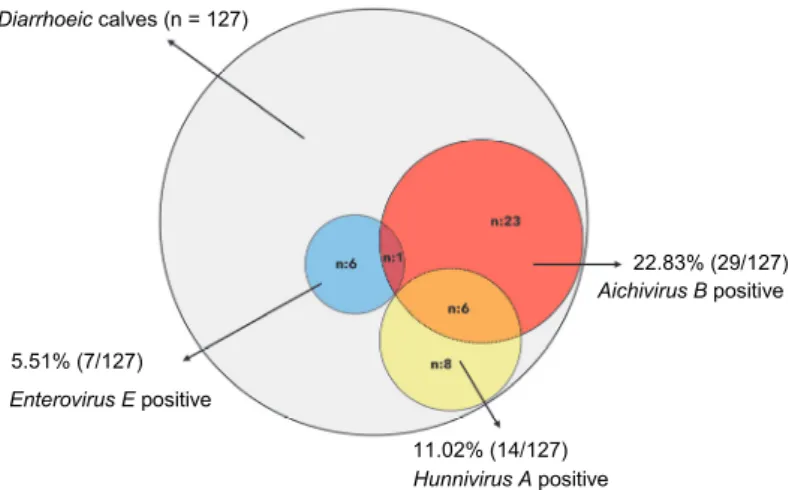
The PCR screening allowed the detection of some representatives of all the three picornavirus species in the faecal samples of diarrhoeic calves tested in this study. The most prevalent was BKV, present in 29 samples (22.83%). We found seven samples (5.51%) positive for the RNA of BEV-1 by both primer sets. Interestingly, for BHuV, 6 (4.72%) and 14 samples (11.02%) were found positive by the use of the Hungaro-3D-F/R or the Hunni166-F/477-R primer sets, respectively. The overall detection results are summarised in Fig. 1.
The aim of this study was to clarify the potential role of picornaviruses (namely members of the species Enterovirus E, Hunnivirus A, and Aichivirus B) in clinical disease. More than one-third (33.86%) of the 127 samples collected from calves with diarrhoea was found to contain one or more of the three agents.
We designed two generic primer sets for the detection of BEVs and BHuV, based on alignments of the large sequence data available in the GenBank at pre- sent. For the detection of BEV-1, both primer pairs gave identical results in our study. However, for the detection of BHuV, the newly designed primers (Hun- ni166-F/477-R) that target the 5’ UTR region, gave more positive results than the Hungaro-3D-F/R primer set (Reuter and Egyed, 2009). Nonetheless, for phylo- genetic studies, this primer set that targets the 3D region of the virus is perhaps a better choice than the novel primer set. For screening faecal samples for the presence of BHuV, the use of our more robust primer set can be recommended.
Table 4
Description of bovine enteroviruses used in the phylogenetic analysis (Fig. 4)
No. Designation Country Year Host Accession number 01 BEV/Thailand/E5 Thailand 2013 Bovine KT992109.1
02 BEV/Thailand/D5 Thailand 2013 Bovine KT992103.1 03 BEV/Thailand/E18 Thailand 2013 Bovine KT992111.1 04 BEV/Thailand/F11 Thailand 2013 Bovine KT992114.1 05 BEV/Thailand/F17 Thailand 2013 Bovine KT992116.1 06 BEV/Thailand/F22 Thailand 2013 Bovine KT992117.1
07 HY12 China 2012 Bovine KF748290.1
08 BEV IS1/Bos taurus/JPN/1990 Japan 1990 Bovine LC150009.1 09 BEV IS2/Bos taurus/JPN/1991 Japan 1991 Bovine LC150010.1 10 BEV Ho12/Bos taurus/JPN/2014 Japan 2014 Bovine LC150008.1
11 VG-5-27 – – – NC_001859.1
12 Vir 404/03 – – – DQ092771.1
13 D 8/01 Germany – – DQ092782.1
14 E 6-82 Germany – – DQ092776.1
15 SD 1182 II Germany – – DQ092784.1
16 D 14/3/96 Germany – – DQ092786.1
17 D 58/96-V2130 Germany – – DQ092772.1
18 VD 2860/1-99 Germany – – DQ092774.1
19 56/59/1 Germany – – DQ092778.1
20 Jena 38/02 Germany – – DQ092788.1
21 D 3/98 Germany – – DQ092790.1
22 D 14/1/96 Germany – – DQ092780.1
23 RM-2 – – – X79369.2
24 PA12-24791 USA 2012 Bovine KC667561.1
25 LC-R4 USA – Bovine DQ092769.1
26 PS 42 USA – Bovine DQ092792.1
27 PS 83 USA – Bovine DQ092793.1
28 IL/alpaca USA 2007 Alpaca KC748420.1
29 MexKSU/5 Mexico 2015 Bovine KU172420.1
30 BEV/Egypt/1 Egypt 2014 Bovine KM887136.1
31 BEV/Egypt/2 Egypt 2014 Bovine KM887137.1
32 BEV/Egypt/3 Egypt 2014 Bovine KM887138.1
33 BEV/Egypt/4 Egypt 2014 Bovine KM887139.1
34 BEV/Egypt/5 Egypt 2014 Bovine KM887140.1
35 BEV/Egypt/6 Egypt 2014 Bovine KM887141.1
36 BEV-Bolat12-16-TUR Turkey 2016 Bovine MF667937.1
37 BEV-Bolat20-16-TUR Turkey 2016 Bovine MF667938.1
38 BEV-Bolat42-16-TUR Turkey 2016 Bovine MF667939.1
39 BEV-Bolat63-16-TUR Turkey 2016 Bovine MF667940.1
40 BEV-Bolat64-16-TUR Turkey 2016 Bovine MF667941.1
41 BEV-Bolat86-16-TUR Turkey 2016 Bovine MF667942.1
In several previous reports, it has been demonstrated that BKV can be de- tected from clinically healthy animals as frequently as from diarrhoeic ones (Reuter and Egyed, 2009; Jeoung et al., 2011). Some other studies have reported a higher rate of positivity in diarrhoeic calves (Barry et al., 2011; Jeoung et al., 2011; Park et al., 2011; Candido et al., 2017). BKV has been detected in 5%
(2/40) of adult cattle, and in 20.9% (38/182) of diarrhoeic calf samples in Brazil
(Blas-Machado et al., 2011; Ribeiro et al., 2014). It has also been detected in 16.7% (12/72) and 6.2% of healthy cattle in Japan and in Hungary, respectively (Khamrin et al., 2008; Reuter and Egyed, 2009), as well as in 25.8% (16/62) of diarrhoeic cattle in South Korea (Park et al., 2011). On the other hand, the high- est proportion of samples from diarrhoeic cattle has been found to be positive for BKV, namely 34.6% (37/107) and 77.8% (7/9) in South Korea (Park et al., 2011) and in the Netherlands (Barry et al., 2011), respectively. In our study also, BKV was detected in the highest proportion of faecal samples collected from young diarrhoeic calves up to 1 month of age.
Fig. 1. Overall detection results of three picornaviral agents (Bovine enterovirus E, Aichivirus B and Hunnivirus A) by RT-PCR analysis
Enteric, respiratory and reproductive diseases have been described in as- sociation with BEV infections in cattle (Jiménez-Clavero et al., 2005). As the main route of transmission is the faecal-oral route, BEV has been proposed as an indicator of faecal pollution originating from animals (Ley et al., 2002; Li et al., 2012). According to a study conducted in Spain in 2004, 78.0% (78 of 100) of the tested faecal samples were BEV positive (Ley et al., 2002). In China, 24.6%
of faecal samples (17/69) from diarrhoeic and healthy cattle have been found positive for the presence of BEV RNA (Li et al., 2012). Although there is no re- port on direct virus detection, different epizootiological studies have indicated the presence of BEV-specific antibodies in 41.8–67.7% of goats, in 3.9% of wa- ter buffalos and 64.8% of cattle, detected by virus neutralisation assay in Turkey (Acar and Gur, 2009; Gür et al., 2006; Gür et al., 2008). In this study, we detect- ed the presence of BEV-1 RNA for the first time in Turkey.
We submitted 14 novel partial BKV sequences to the GenBank (accession numbers: KY695136–KY695149). According to the results of blast, our newly acquired sequences of the 217-nt-long fragment shared 89.4–99.0% identity
Aichivirus B positive 22.83% (29/127)
11.02% (14/127) Hunnivirus A positive 5.51% (7/127)
Enterovirus E positive Diarrhoeic calves (n = 127)
among each other, and 74.8–97.1% nucleotide identity with those previously de- posited in the GenBank. Phylogenetic analysis was applied to the data and these clustered on distinct branches among the 182 other sequences retrieved from the GenBank. We furthermore limited the phylogenetic tree to 58 strains to simplify (Fig. 2), which revealed that some of them appeared close to the Brazilian se- quences, while others were closer to those found in Japan or South Korea. This result indicates that a moderate level of genetic diversity exists among Turkish strains.
Fig. 2. Phylogenetic analysis based on the 217-bp partial nucleotide sequence of the 3D region of bovine kobuviruses (NJ tree, Tamura-Nei genetic distance model). The bootstrap values were generated with 1,000 pseudoreplicates. The name and description of viruses are listed in Table 2.
Novel virus sequences, acquired in the present study, are highlighted in bold
Bovine hungarovirus (species Hunnivirus A) is a recently discovered virus (Reuter et al., 2012). According to the phylogenetic analysis, the eight Turkish BHuV sequences (GenBank accession numbers: KY974326–KY974333) clus- tered with the Hungarian bovine hungaroviruses and some them formed a sepa- rate, novel lineage (Fig. 3). Considering the 301-bp PCR fragment, the nucleo- tide identity among the Turkish hunnivirus sequences ranged between 76.02 and 99.02%. The identity ranged between 75.4 and 91.8% when comparing them to the Hungarian bovine hunnivirus sequence. The nucleotide identity was 75.1–
82.3% between the Turkish BHuV and the Hungarian ovine sequence. The par- tial rat hunnivirus sequences shared 66.4 to 77.9% identity with the Turkish BHuV sequences.
Fig. 3. Phylogenetic analysis based on the 306-bp partial nucleotide sequence of the 5' UTR region of hunniviruses (NJ tree, Tamura-Nei genetic distance model). The bootstrap values were generated with 1,000 pseudoreplicates. The name and description of viruses are listed in Table 3.
Novel virus sequences, acquired in the present study, are highlighted in bold
Rat strains Bovine strains Ovine strain
On the other hand, the six novel BEV-1 sequences (GenBank accession numbers: MF667937–MF667942) occupied the same branch of the phylogenetic tree (Fig. 4). The nucleotide identity of the 293-bp partial sequence of the 5' UTR region was found to be 90.14–92.81% among the novel Turkish sequences, and ranged between 72.28 to 92.18% between the Turkish and other, previously se- quenced fragments.
Fig. 4. Phylogenetic analysis based on the 293-bp partial nucleotide sequence of the 5' UTR region of bovine enteroviruses (NJ tree, Tamura-Nei genetic distance model). The bootstrap values were
generated with 1,000 pseudoreplicates. The name and description of viruses are listed in Table 4.
Novel virus sequences, acquired in the present study, are highlighted in bold
In conclusion, this study indicates that these viral agents can be detected in diarrhoeic calves in Turkey. Based on the PCR results we suggest that the Hun-
ni166-F/477-R primer set should be used instead of Hungaro3D-F/R for the de- tection of BHuV. The impact of BEV and BHuV in the problem of calf diarrhoea remains questionable unless experimental studies can confirm it. Nonetheless, our results provide a strong evidence for the role of BKV as an enteric pathogen that can cause diarrhoea in calves. We report here a more reliable detection tool for BHuV and also a novel lineage of BHuV. Furthermore, this is the first report on the molecular detection of these three picornaviruses in Turkey.
Acknowledgement
This research was supported by the Cumhuriyet University Scientific Research Project Foundation (CÜBAP) project no: VET-029.
References
Acar, A. and Gur, S. (2009): Seroprevalence of bovine enterovirus type 1 (BEV1) in goats in Tur- key. J. Anim. Vet. Adv. 8, 1075–1078.
Barry, A. F., Ribeiro, J., Alfieri, A. F., van der Poel, W. H. and Alfieri, A. A. (2011): First detec- tion of kobuvirus in farm animals in Brazil and the Netherlands. Infect. Genet. Evol. 11, 1811–1814.
Bartels, C. J. M., Holzhauer, M., Jorritsma, R., Swart, W. A. and Lam, T. J. (2010): Prevalence, prediction and risk factors of enteropathogens in normal and non-normal faeces of young Dutch dairy calves. Prev. Vet. Med. 93, 162–169.
Blas-Machado, U., Saliki, J. T., Boileau, M. J., Goens, S. D., Caseltine, S. L., Duffy, J. C. and Welsh, R. D. (2007): Fatal ulcerative and hemorrhagic typhlocolitis in a pregnant heifer as- sociated with natural bovine enterovirus type-1 infection. Vet. Pathol. 44, 110–115.
Blas-Machado, U., Saliki, J. T., Sánchez, S., Brown, C. C., Zhang, J., Keys, D., Woolums, A. and Harvey, S. B. (2011): Pathogenesis of a bovine enterovirus-1 isolate in experimentally in- fected calves. Vet. Pathol. 48, 1075–1084.
Candido, M., Batinga, M. C., Alencar, A. L., de Almeida-Queiroz, S. R., da Gloria Buzinaro, M., Livonesi, M. C., Fernandes, A. M. and de Sousa, R. L. (2017): Molecular characterization and genetic diversity of bovine Kobuvirus, Brazil. Virus Genes 53, 105–110.
Cho, Y. I. and Yoon, K. J. (2014): An overview of calf diarrhea – infectious etiology, diagnosis, and intervention. J. Vet. Sci. 15, 1–17.
Di Martino, B., Di Profio, F., Di Felice, E., Ceci, C., Pistilli, M. G. and Marsilio, F. (2012): Molec- ular detection of bovine kobuviruses in Italy. Arch. Virol. 157, 2393–2396.
Du, J., Lu, L., Liu, F., Su, H., Dong, J., Sun, L., Zhu, Y., Ren, X., Yang, F., Guo, F., Liu, Q., Wu, Z. and Jin, Q. (2016): Distribution and characteristics of rodent picornaviruses in China.
Sci. Rep. 6, 34381.
Firth, C., Bhat, M., Firth, M. A., Williams, S. H., Frye, M. J., Simmonds, P., Conte, J. M., Ng, J., Garcia, J., Bhuva, N. P., Lee, B., Che, X., Lan Quan, P. and Lipkin, W. I. (2014): Detec- tion of zoonotic pathogens and characterization of novel viruses carried by commensal Rat- tus norvegicus in New York City. MBio 5, e01933-14-e01933-14.
Gür, S., Akça, Y. and Burgu, İ. (2006): Serological investigation of bovine enterovirus type-1 in buffaloes in Turkey [in Turkish]. Ankara Üniv. Vet. Fak. Derg. 53, 191–194.
Gür, S., Yapkiç, O. and Yilmaz, A. (2008): Serological survey of bovine enterovirus type 1 in dif- ferent mammalian species in Turkey. Zoonoses Public Health 55, 106–111.
Jeoung, H. Y., Lim, J. A., Jeong, W., Oem, J. K. and An, D. J. (2011): Three clusters of bovine kobuvirus isolated in Korea, 2008–2010. Virus Genes 42, 402–406.
Jiménez-Clavero, M. A., Escribano-Romero, E., Mansilla, C., Gomez, N., Cordoba, L., Roblas, N., Ponz, F., Ley, V. and Saiz J. C. (2005): Survey of bovine enterovirus in biological and en- vironmental samples by a highly sensitive real-time reverse transcription-PCR. Appl. Envi- ron. Microbiol. 71, 3536–3543.
Khamrin, P., Maneekarn, N., Peerakome, S., Okitsu, S., Mizuguchi, M. and Ushijima, H. (2008):
Bovine kobuviruses from cattle with diarrhea. Emerg. Infect. Dis. 14, 985–986.
Knowles, N. and Mann, J. (1990): Bovine enteroviruses. In: Virus Infections of Vertebrates. Vol- ume 3 – Virus Infections of Ruminants. Elsevier Scientific Publishers Ltd., Amsterdam.
pp. 513–516.
Ley, V., Higgins, J. and Fayer, R. (2002): Bovine enteroviruses as indicators of fecal contamina- tion. Appl. Environ. Microbiol. 68, 3455–3461.
Li, Y., Chang, J., Wang, Q. and Yu, L. (2012): Isolation of two Chinese bovine enteroviruses and sequence analysis of their complete genomes. Arch. Virol. 157, 2369–2375.
MacLachlan, J. and Dubovi, J. E. (2017): Picornaviridae. In: Fenner’s Veterinary Virology, 5th edition. Elsevier, New York. pp. 477–495.
Melegari, I., Di Profio, F., Sarchese, V., Martella, V., Marsilio, F. and Di Martino, B. (2016): First molecular evidence of kobuviruses in goats in Italy. Arch. Virol. 161, 3245–3248.
Okonechnikov, K., Golosova, O., Fursov, M. and UGENE Team (2012): Unipro UGENE: A uni- fied bioinformatics toolkit. Bioinformatics 28, 1166–1167.
Otomaru, K., Naoi, Y., Haga, K., Omatsu, T., Uto, T., Koizumi, M., Masuda, T., Yamasato, H., Takai, H., Aoki, H., Tsuchiaka, S., Sano, K., Okazaki, S., Katayama, Y., Oba, M., Furuya, T., Shirai, J., Katayama, K., Mizutani, T. and Nagai, M. (2016): Detection of novel kobu- like viruses in Japanese black cattle in Japan. J. Vet. Med. Sci. 78, 321–324.
Pankovics, P., Boros, Á., Bíró, H., Horváth, K. B., Phan, T. G., Dewalt, E. and Reuter, G. (2016):
Novel picornavirus in domestic rabbits (Oryctolagus cuniculus var. domestica). Infect.
Genet. Evol. 37, 117–122.
Park, S. J., Kim, H. K., Song, D. S., Moon, H. J. and Park, B. K. (2011): Molecular detection and genetic characterization of kobuviruses in fecal samples collected from diarrheic cattle in Korea. Infect. Genet. Evol. 11, 1178–1182.
Pham, N. T., Khamrin, P., Nguyen, T. A., Kanti, D. S., Phan, T. G., Okitsu, S. and Ushijima, H.
(2007): Isolation and molecular characterization of Aichi viruses from fecal specimens col- lected in Japan, Bangladesh, Thailand, and Vietnam. J. Clin. Microbiol. 45, 2287–2288.
Reuter, G. and Egyed, L. (2009): Bovine kobuvirus in Europe. Emerg. Infect. Dis. 15, 822–823.
Reuter, G., Boros, Á. and Pankovics, P. (2011): Kobuviruses – a comprehensive review. Rev. Med.
Virol. 21, 32–41.
Reuter, G., Boros, Á., Pankovics, P. and Egyed, L. (2010): Kobuvirus in domestic sheep, Hungary.
Emerg. Infect. Dis. 16, 869–870.
Reuter, G., Pankovics, P., Knowles, N. J. and Boros, Á. (2012): Two closely related novel picorna- viruses in cattle and sheep in Hungary from 2008 to 2009, proposed as members of a new genus in the family Picornaviridae. J. Virol. 86, 13295–13302.
Ribeiro, J., Lorenzetti, E., Alfieri, A. F. and Alfieri, A. A. (2014): Kobuvirus (Aichivirus B) infec- tion in Brazilian cattle herds. Vet. Res. Commun. 38, 177–182.
Smits, S. L., Raj, V. S., Oduber, M. D., Schapendonk, C. M. E., Bodewes, R., Provacia, L., Stittelaar, K. J., Osterhaus, A. D. M. E. and Haagmans, B. L. (2013): Metagenomic analy- sis of the ferret fecal viral flora. PLoS One 8, 8-e71595.
Yamashita, T., Ito, M., Kabashima, Y., Tsuzuki, H., Fujiura, A. and Sakae, K. (2003): Isolation and characterization of a new species of kobuvirus associated with cattle. J. Gen. Virol. 84, 3069–3077.
Yamashita, T., Kobayashi, S., Sakae, K., Nakata, S., Chiba, S., Ishihara, Y. and Isomura, S. (1991):
Isolation of cytopathic small round viruses with BS-C-1 cells from patients with gastroen- teritis. J. Infect. Dis. 164, 954–957.
Yilmaz, H., Turan, N., Altan, E., Bostan, K., Helps, C. R. and Cho, K. O. (2011): First report on the phylogeny of bovine norovirus in Turkey. Arch. Virol. 156, 143–147.
Zell, R. (2018): Picornaviridae – the ever-growing virus family. Arch. Virol. 163, 299–317.
Zell, R., Delwart, E., Gorbalenya, A. E., Hovi, T., King, A. M. Q., Knowles, N. J., Lindberg, A.
M., Pallansch, M. A., Palmenberg, A. C., Reuter, G., Simmonds, P., Skern, T., Stanway, G., Yamashita, T. and ICTV Report Consortium (2017): ICTV Virus Taxonomy Profile:
Picornaviridae. J. Gen. Virol. 98, 2–3.