PhD Thesis
PROTEIN CHANGES OF VARIOUS TYPES OF MILK AS AFFECTED BY HIGH HYDROSTATIC PRESSURE
PROCESSING
Klára Pásztor-Huszár
Supervisor:
Prof. József Farkas MHAS
Corvinus University of Budapest Faculty of Food Science
Department of Refrigeration and Livestock Products Technology
Budapest, 2008.
PhD School/Program
Name: PhD School of Food Science Field: Food Science
Head: Prof. Péter Fodor, D.Sc.
Department of Applied Chemistry Faculty of Food Science
Corvinus University of Budapest
Supervisor: Prof. József Farkas, MHAS Department of Refrigeration and Livestock Products Technology Faculty of Food Science
Corvinus University of Budapest
The applicant met the requirement of the PhD regulations of the Corvinus University of Budapest and the thesis is accepted for the defence process.
……….………. .………...
Signature of Head of School Signature of Supervisor
According to the Doctoral Council of Life Sciences of Corvinus University of Budapest on 10th June 2008, the following committee was designated for defence.
Committee:
Chair:
András Fekete, D.Sc.
Members:
Tibor Deák, D.Sc.
Emőke Németh-Szerdahelyi, Ph.D József Fenyvessy, C.Sc.
Referees:
András Szabó S., D.Sc.
Andrea Lugasi, C.Sc.
Secretary:
Zsuzsanna Cserhalmi, Ph.D.
1 CONTENTS
1 Contents ... 5
2 Notation ... 7
3 Introduction ... 8
4 Literature survey ... 10
4.1 Milk ... 10
4.2 Milk Proteins ... 11
4.2.1 Caseins ... 12
4.2.2 Whey Proteins ... 15
4.2.3 Tryptophan in Milk Proteins ... 17
4.2.4 Retinol in Milk ... 19
4.2.5 Immunoreactivity of Milk Proteins ... 20
4.2.6 Microbiology of Milk ... 21
4.3 High Pressure Processing ... 23
4.3.1 Short History ... 23
4.3.2 General Overview of High Hydrostatic Pressure ... 24
4.3.3 High Pressure Equipment ... 26
4.3.4 Principles of High Pressure Processing ... 27
4.3.5 Effect of High Hydrostatic Pressure on Proteins, with Special Regard to Milk Proteins ... 30
4.4 Polyacrylamide Gel Electrophoresis ... 34
4.4.1 SDS Polyacrylamide Gel Electrophoresis (SDS-PAGE) ... 35
4.4.2 Discontinuous SDS Polyacrylamide Gel Electrophoresis ... 35
4.4.3 Native Polyacrylamide Gel Electrophoresis (Native PAGE) ... 36
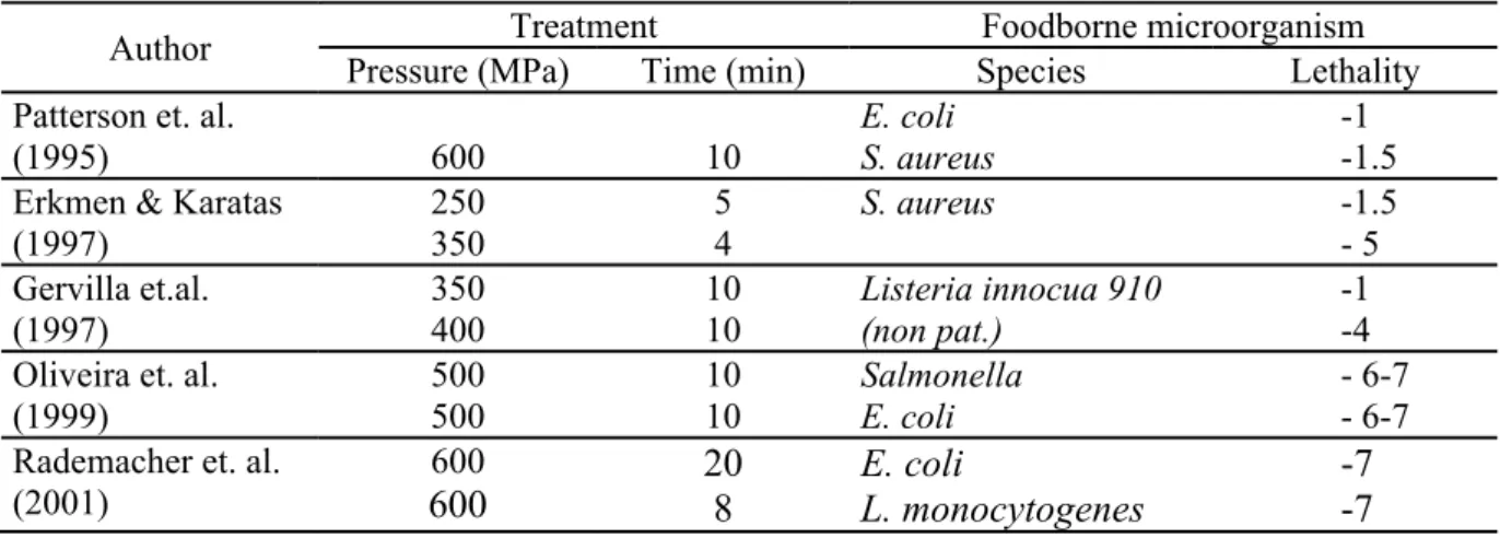
4.5 Two-dimensional Polyacrylamide Gel Electrophoresis (2D PAGE) ... 37
4.5.1 Isoelectric Focusing (IEF) ... 37
4.6 Immunoblotting ... 38
4.7 Fluorescence Spectroscopy ... 39
5 Objectives ... 44
6 Materials and Methods ... 45
6.1 Milk Types and Whey ... 45
6.2 Treatment by High Hydrostatic Pressure ... 45
6.3 Heat Treatment ... 46
6.4 SDS- and Native PAGE ... 48
6.4.1 Sample Preparation ... 48
6.4.2 Methodology ... 48
6.5 Gradient Gel ... 48
6.6 2D-PAGE ... 49
6.7 Electrophoretic Immunoblotting ... 49
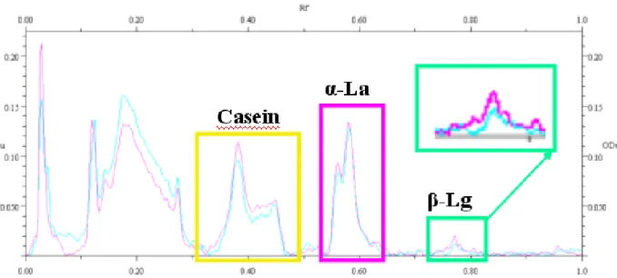
6.8 Evaluation of Electrophoretograms ... 50
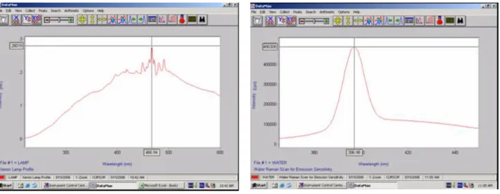
6.9 Fluorescence Spectroscopy ... 50
6.9.1 Instruments and Principal Functions ... 50
6.9.2 Calibration ... 51
6.9.3 Software ... 52
6.9.4 Settings for Recording the Fluorescence Spectra ... 52
7 Results and Discussion ... 55
7.1 Comparision of Protein Composition of Different Milk Types by Electrophoretic Methods ... 55
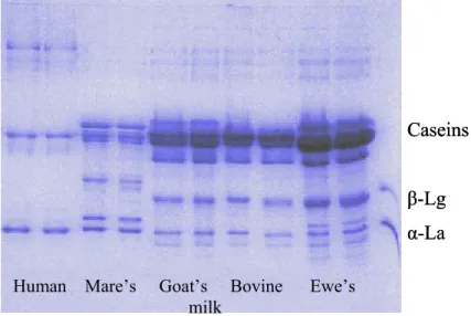
7.1.1 Comparision of Protein Composition of Different Milk Types by SDS-PAGE ... 55
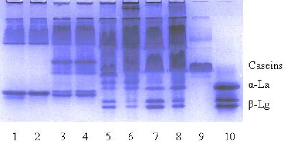
7.1.2 Effect of High Hydrostatic Pressure on the Composition of Different Milk Types . ... 58
7.1.3 More Detailed Investigation of the Effect of HHP on Proteins in Bovine Milk ... 62
7.2 Immunoreactivity of Milk Proteins ... 69
7.2.1 Immunoreactivity of Untreated Milk Samples ... 70
7.2.2 Immunoreactivity of Pressurized Milk Samples ... 71
7.3 Fluorescence Investigations ... 74
7.3.1 Changes in Tryptophan Emission ... 74
7.3.2 Tryptophan Emission Spectra of Bovine Milk ... 78
7.3.3 Tryptophan Fluorescence Emission of Bovine and Goat Milk as Affected by Heat, and HHP Treatment ... 82
7.3.4 Effects of High Pressure and Heat Processing on Fluorescence of Retinol in Milk . ... 84
7.3.5 Mathematical Statistical Comparison of the Two Treatment Methods in the Materials Investigated ... 92
7.4 New Scientific Results ... 93
7.5 Új tudományos eredmények ... 93
8 SUMMARY AND CONCLUSIONS ... 95
9 References ... 103
10 Appendix ... 114
11 ACKNOWLEDGEMENTS ... 118
2 NOTATION
HHP high hydrostatic pressure
HP high pressure
PAGE polyacrylamide gel electrophoresis
SDS-PAGE sodium-dodecyl-sulphate polyacrylamide gel electrophoresis
2D-PAGE two-dimensional polyacrylamide gel electrophoresis
β-Lg beta-lactoglobulin
α-La alfa-lactalbumin
Trp tryptophan
Phe phenilalanin
Glu glutamin
Asp asparagin
IgE immunoglobulin E
IEF isoelectric focusing
pI isoelectric point
OD optical density
Odu optical density unit
Rf relative front
3 INTRODUCTION
In the last few decades the concept of “minimal processing” of foods has arisen. Consumers increasingly demand foods which retain their natural, fresh-like flavour, colour and texture and contain fewer additives such as preservatives. In response to these needs minimal processing technologies have been developed. They are designed to limit the effect of processing on nutritional and sensory quality and to preserve food without the use of synthetic additives.
Traditional thermal processing techniques can be beneficial to foods in such areas as preservation and flavour formation but detrimental in damaging other sensory and nutritional properties. Minimising undesirable changes can be achieved in a number of ways, whether through more effective process control, the use of High Temperature Short Time (HTST) techniques such as aseptic processing, or newer thermal technologies such as volume heating methods. Infrared heating and dielectric methods e.g. the use of microwaves, and ohmic heating belong to the thermal minimal processing methods. Alternatives to processing by heat have been developed, ranging from irradiation to the use of pulsed electric fields. One of these novel non- thermal techniques is the application of high hydrostatic pressure for food preservation, that is currently receiving considerable attention from both researchers and producers.
High hydrostatic pressure with regard to food is in the range of 100-1000 MPa. The possibility of using high pressure to process and preserve foods has been known and studied for almost a century. Hite, a researcher of the West Virginia Agricultural Experimental Station published his findings in 1899 about “The effects of pressure on the preservation of milk”.
Progress, though, has been relatively slow since it was not commercially feasible to subject foods to the pressures necessary to either preserve them or to considerably modify and improve their quality. However, in the last two decades there have been important developments in the engineering aspects of high pressure equipment so that it is now both economically and technically feasible to subject foods to the pressures deemed desirable. Although the technology is now available, or at least is rapidly becoming available, because of the cost of the equipment it is only currently applied to produce high quality, relatively expensive foods. High pressure processed foods such as jams and fruit drinks, became available in Japan around 1990. As the technology develops and becomes accepted by the consumer, the number of countries marketing and manufacturing high pressure processed foods is likely to increase. Although the capital costs may be high, production costs are not excessive, and the technology is seen to be environmentally friendly, i.e. clean and “natural” (Ledward, 1995).
Milk is an important food across the globe. Its pasteurisation is common practice in the dairy industry. Given the unassailable position of heat treatment and the diverse response of
microorganisms to pressure, particularly that milk may have a baroprotective effect for certain microorganisms, it seems unlikely that pressure will ever replace heat for the safe production of large volumes of liquid milk. Then what is the use of investigating the effects of pressure on milk and dairy products? There might be some niche products for which heat treatment could be inappropriate, and for which microbiological quality could be improved by high pressure treatment. But the main reason is that pressure brings about modifications to milk components (especially proteins), that can lead to altered functionality and the possibility of novel or improved dairy products (Needs, 2002). As a result of this it is not surprising that the dairy and food industry shows increasing interest in high pressure milk processing.
However, heat and pressure have rather different effects on the structure, interactions and properties of milk proteins. For example, during manufacture their immunoreactivity may be altered. The allergenic activity of foods may be unchanged, decreased or even increased by food processing. The molecular basis of changes in the allergenic activity is the inactivation or destruction of epitope structures or the formation of new epitopes or better access to cryptic epitopes by denaturation of the native allergen. In the model studies available in the literature the effects of heating and enzymatic digestions were investigated (Besler et al., 2001) and there is very little information about the influence of high hydrostatic pressure from this point of view.
To show possible structural changes in milk proteins polyacrylamide gel electrophoresis (SDS, native, and two-dimensional PAGE) can be used very well. Immunoblotting is a time- honoured method for the detection of immunoreactivity of proteins. Although fluorescence was one of the earliest instrumental techniques available to the analytical chemists, only recent developments in instrumentation and sample handling have made it possible for its full potential to be used in everyday analysis. Thus fluorescence is becoming one of the most promising techniques in biology, medicine, and food research as well (Deshpande, 2001).
Spectrofluorometry, as a new analytical tool, was tested in the present study to observe differences between heat treated and high pressure processed milk samples, and changes in certain milk components.
4 LITERATURE SURVEY
4.1 Milk
Milk is exclusively the normal mammary gland secretion obtained from one or more milkings without either addition thereto or extraction therefrom. The natural function of milk is to nurture the young of the species. Milk has a nutritional role and hence must contain readily available sources of energy, essential fatty acids, amino acids and vitamins, all of which may be unavailable to the infant from any source other than milk. A further function of milk is to assist in combating disease, both in the mammary gland and in the infant. This is achieved by proteins such as lysozyme, peroxidase, lactoferrin and immunoglobulins. Proteinases, such as plasmin and lipases, and serum albumin or β-lactoglobulin (β-Lg), may aid digestion and nutrient absorption (Creamer, 1996).
The origin of the milk shall be indicated if it is not bovine.
Milk composition of mammalian species varies widely with reference to genetic, physiological and nutritional factors and environmental conditions (Malacarne et al., 2002).
Milk coming form cattle, goats, ewes, mares and humans was investigated in this study, their average composition is shown in Table 1.
Table 1. Average composition of different milk types (Szakály, 2001)
Component
Concentration of the given component (%) Human
milk Ewe’s
milk Mare’s
milk Goat’s
milk Bovine milk
Protein 1,3 5,5 2,15 3,9 3,3
Fat 4,5 8,2 0,6 4,0 3,8
Lactose 6,3 5,0 6,75 4,5 4,6
Minerals 0,2 0,9 0,3 0,8 0,8
Solids 12,3 19,6 9,8 13,2 12,5
Milk solids non fat 7,8 11,4 9,2 9,2 8,8
Water 87,7 80,4 90,8 86,8 87,5
Regarding their composition, human and mare’s milk belong to the so called albumin milk group, while goat, ewe’s and bovine milk to the casein milk group. Albumin milks are characterised by the relatively high albumin and globuline content, whereas about 80% of the total protein content are caseins in the casein milk types (Table 2.).
Table 2. Protein composition of albumin milks and casein milks (Császár and Unger, 2005)
Denomination
Protein (%) Total
protein Casein Whey protein Casein milks
Ewe’s milk 5,35 4,3 1,05
Goat milk 3,6 2,6 1,0
Bovine milk 3,3 2,7 0,6
Albumin milks
Human milk 1,3 0,8 0,5
Mare’s milk 2,15 1,3 0,85
4.2 Milk Proteins
In milk, there are two major protein types which in bovine milk are defined by acid precipitation: the caseins, which precipitate as a group at pH 4.6, and the whey proteins, which can be subdivided into the major mammary synthesized proteins and the minor, usually blood, proteins. Each of the mammary-synthesized proteins exists in several forms, known as genetic variants, which have slightly different amino acid sequences (Creamer, 1996).
The particular sequence of amino acids in a protein determines its structure, conformation and properties. The structure of protein is categorised as primary, secondary, tertiary or even quaternary, depending on the state of spatial arrangement of polypeptide chains. The primary structure of proteins consists of a polypeptide chain of amino acid residues joined together by peptide linkages, which may also be cross-linked by disulphide bridges. The primary polypeptides in a nascent protein in an aqueous environment tend to coil in a characteristic way to form localised secondary structures, i.e. α-helix and β-pleated sheet.
They are examples of secondary structures arising from regular and periodic steric relationships. The secondary structure is stabilised by hydrogen bonds. In aqueous medium Van der Waals interactions between adjacent residues, and hydrophobic interactions between long or bulky apolar sidechains, may contribute to stabilisation of the α-helix structure. β-pleated sheets are formed where interpolypeptide chain interactions are possible, because β-turns or folds permit adjacent polypeptide chains to associate mostly via hydrogen bonding, and to a lesser extent, via hydrophobic and electrostatic interactions. The tertiary structure refers to the spatial arrangement of amino acid residues that are far apart in the linear sequence, giving rise to further coiling and folding. In a typical tertiary structure, the polypeptides are tightly folded to give a compact molecule, in which most of the polar groups of the amino acids are located on the outer surface and are hydrated. Most of the apolar groups are internal in the hydrophobic region from
which water is essentially excluded. If the protein is tightly coiled and folded into a somewhat spherical shape, it is called a globular protein. If the protein consists of long polypeptide chains which are intermolecularly linked, they are called fibrous proteins. Many globular proteins with molecular weights exceeding 50 kilodaltons are oligomeric, consisting of two or more individual (protomer)associated polypeptides. This is called quaternary structure. A protein will tend to self-associate if it contains more than 28 mol% of the particular hydrophobic amino acids, e.g, caseins (Kinsella, 1984; Goff, 1995).
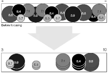
Characteristics of different milk protein fractions are shown in Table 3.
Table 3. Protein fractions of bovine milk and some of their characteristics (Farrell et al., 2004)
Protein fraction Amount in skim
milk (g/l) Genetic
variants Molecular
weight (kDa) Isoelectric point (pH) Caseins
αs1-casein 12-15 B 23.615 4,44-4,76
C 23.542 …
αs2-casein 3-4 A 25.226 …
β-casein 9-11
A1 24.023 … A2 23.983 4,83-5,07
B 24.092 -
κ-casein 2-4 A 19.037 5,45-5,77
B 19.006 5,3-5,8 Whey proteins
β-lactoglobulin 2-4 A 18.363 5,13
B 18.277 5,13
α-lactalbumin 0,6-1,7 B 14.178 4,2-4,5
Serum albumin 0,4 A 66.399 4,7-4,9
Immunoglobulin G1 0,3-0,6 … 161.000 5,5-6,8
Immunoglobulin G2 0,05 … 150.000 7,5-8,3
Immunoglobulin A 0,01 … 385.000-417.000
Immunoglobulin M 0,09 … 1000.000 …
Secretory components 0,02-0,1 … 63.750 …
Lactoferrin 0,02-0,1 … 76.110 8,81
4.2.1 Caseins
Caseins are phosphoproteins precipitated from raw milk at pH 4.6 at 20°C. They comprise approximately 80% of the total protein content in milk. The principal proteins of this group are
classified according to the homology of their primary structures into αs1-, αs2-, β- and κ-caseins (Wong et al., 1996).
Caseins are conjugated proteins, most of them with phosphate group(s) esterified to serine residues. Calcium binding by the individual caseins is proportional to the phosphate content. The conformation of caseins is similar to denatured globular proteins. The high number of proline residues in caseins causes particular bending of the protein chain and inhibits the formation of close-packed, ordered secondary structures. The lack of tertiary structure accounts for the stability of caseins against heat denaturation, because there is very little structure to unfold.
Without a tertiary structure there is considerable exposure of hydrophobic residues. This results in strong association reactions of the caseins and renders them insoluble in water. Within the group of caseins, there are several distinguishing features, based on their charge distribution and sensitivity to calcium precipitation (Wong et al., 1996; Goff, 1995; Farrell et al., 2004).
4.2.1.1 αs1 caseins
αs1 caseins have five genetic variants, A, D, B, C and E. The B variant consists of 199 amino acid residues with a calculated molecular weight of 23,614 Da. The protein contains more acidic amino acids than basic ones. It has 17 proline residues which prevent the formation of certain types of secondary structures. Three hydrophobic regions are identified that contain all the proline residues. Seven of the eight phosphate groups are located in the hydrophilic region. αs1
caseins are calcium sensitive, they can be precipitated at very low levels of calcium (Wong et al., 1996; Goff, 1995; Farrell et al., 2004).
4.2.1.2 αs2 caseins
Four variants of αs2 are known, A, B, C and D. The amino acid sequence of αs2-CN A-11P consists of 207 residues, among them ten prolines and two cysteins, and the calculated molecular weight is 24,350 Da. Concentrated negative charges are found near N-terminus and positive charges near C-terminus. It can also be precipitated at very low levels of calcium (Wong et al., 1996; Goff, 1995; Farrell et al., 2004).
4.2.1.3 β-casein
β-casein constitutes 30-35% of the total caseins. Seven genetic variants of β-casein are known. The molecular weight of β-CN A1-5P is 23,982 Da; it is composed of 209 residues, among them 35 prolines. High negative net charge is around the N-terminal region, and the C-
terminal region is highly hydrophobic. β-casein is a very amphiphilic protein, and that’s why it acts like a detergent molecule. The protein’s self-association depends on temperature. It will form a large polymer at 20° C, but not at 4° C. This type of casein is less sensitive to calcium precipitation (Wong et al., 1996; Goff, 1995; Farrell et al., 2004).
4.2.1.4 κ-casein
κ-casein constitutes about 15% of the total caseins. The major casein, κ-CN B-1P contains 169 amino acid residues (twenty prolines) and its molecular weight is 19,023 Da. This protein is positioned on the outside of the casein micelle. Unlike the other caseins κ-casein is very resistant to calcium precipitation, stabilizing other caseins. Rennet cleavage at the Phe105-Met106 bond eliminates the stabilizing ability, leaving a hydrophobic portion, para-κ-casein, and a hydrophilic portion called κ-casein glycomacropeptide (GMP), or caseinomacropeptide (CMP). Cleavage of this bond is the first step in the coagulation of milk by aggregation of the casein micelles after the loss of the hydrophilic, negatively charged surface from the micelle (Farrell et al., 2004;
Goff, 1995; Wong et al., 1996).
4.2.1.5 Casein Micelles
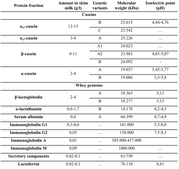
The major part of milk proteins, together with calcium phosphate, occurs in the form of large colloidal particles, the casein micelles. An average micelle contains about 104 caseins and its size ranges between 50 and 300 nm (Huppertz et al, 2006). Various different models have been proposed for micelle structure. One of them is the “sub-micelle model”. This model suggests that casein micelles are built of roughly spherical subunits or sub-micelles. The composition of sub- micelles is variable and the size is within the range of 12-15 nm in diameter, and each sub- micelle has 20-25 casein molecules. The sub-micelles are kept together by hydrophobic interactions between proteins, and by calcium phosphate linkages. There are two main types of sub-micelles. One mainly consists of αs- and β-caseins, hydrophobic regions buried in the center of the sub-micelle. The other type consists of αs- and κ-caseins. The latter are more hydrophilic because of the sugar residues on them. The κ-caseins are located near the outside of the micelle with the hydrophilic part of the C-terminal end protruding from the micelle surface to form a 'hairy' layer that will avoid further aggregation of sub-micelles by steric and electrostatic repulsion. Consequently, micelles are stable, and they do not usually flocculate (Figure 1.) (Phadungath, 2005).
Another model has been evolved recently, especially from work by Carl Holt. This “internal structure” model shows a more or less spherical, highly hydrated, and fairly open particle. Holt’s model of the casein micelle shows a tangled web of flexible casein networks that form a gel-like
structure with micro-granules of colloidal calcium phosphate through the casein phosphate center. The C-terminal region of κ-casein extends to form a „hairy layer” (Figure 2.). The two main features of this model are the cementing role of colloidal calcium phosphate and the surface location of hairy layer, which confers steric and/or charge stability to native casein particles (Phadungath, 2005).
Figure 1. The sub-micelle model Figure 2. The internal structure model
4.2.2 Whey Proteins
The following proteins belong to the whey proteins: β-lactoglobulin (β-Lg), α-lactalbumin (α-La), lesser amounts of serum albumin, immunoglobulins, and proteose peptones. Whey proteins give 20% of total protein content in bovine milk. They are globular and are present in milk as discrete molecules with varying numbers of disulfide crosslinks. These proteins are more heat sensitive, and less sensitive to calcium than caseins. They can form disulfide linked dimers or polymers via thiol disulfide interchange e.g. with κ-casein
4.2.2.1 β-lactoglobulin
β-lactoglobulin (Fig. 3.) is the major whey protein, about 54% of whey proteins is β- lactoglobulin. Five genetic variants have been characterised. It is a globular protein with a molecular weight of 18,362 Da for variant A and 18,276 for variant B. Variant B consists of 162 amino acids. A comparison of the sequences of β-Lg in bovine, ewe’s and goat milk shows, that the three proteins are highly homologous. They contain two intrachain disulfides and one sulfhydryl group. Horse β-Lg A has no free thiols, because cisteine is replaced by a tyrosine residue. Variant B consists of 166 amino acids (Wong et al., 1996).
The secondary structure of bovine β-Lg is 15% α-helix, 50% β-sheet and 15-20% reverse turn. The protein is a typical lipocalin whose structure thus contains a β-barrel with eight antiparallel β-strands, labelled A–H and a three-turn α-helix that lies parallel to three of the β
strands. Strands A–D form one surface of the barrel while strands E–H form the other. A significant feature in all lipocalins is the bend in strand A that allows it to interact with strand H.
The three-turn-α-helix follows strand H and lies on the outer surface of the barrel between the C- terminal end of the A strand and the H strand. A ninth β-strand, I, antiparallel to the first strand, A, and on the other side of H, is used in dimer formation (Kontopidis et al., 2004).
Figure 3. Structure of β-lactoglobulin (Qi et al., 1997)
The molecule contains two disulfide bonds, which are found between cisteins 106. and 19., and the cisteins 6. and 160., respectively. There is one free sulfhydril group in β-Lg, but there is no phosphorus present in this protein.
β-Lg is very acid stable. It is generally in dimer form at the isoelectric pH of 5.2 and alkaline pH range. Bovine β-Lg denatures at temperatures above 65°C at pH 6.7, typically at 70.4±0.5°C, followed by aggregation. Denaturation temperature of β-Lg depends on pH. It is most heat sensitive near pH 4.0 and most stable at pH 6.0.
Temperature affects the three dimensional structure of β-Lg. Although β-Lg is found mainly in the dimer form in milk, monomers appear when temperature is increased up to 65°C.
Critical conformational change occurs around 63°C, where there is 19% net reduction in the β- sheet content, as shown by circular dichroism (Prabakaran, Damodaran, 1997). This reduction in β-sheet content seems to be critical for initiating sulfhydryl disulfide-induced aggregation.
Above this temperature, unfolding of β-Lg structure leads to irreversible denaturation in the following order: D-E strand (55-60°C); C-D strand and α-helix (60-65°C); A-B, A-I and E-F strands (65- 70°C); and A-H, B-C and F-G strands (75-80°C). Thermal unfolding of β-Lg is almost complete at 80°C except for the G-H pair of disulfide-linked strands which are the most heat-resistant feature of the structure (Edwards et al., 2002; Doucet, 2004).
β-Lg was found to bind retinol and enhance its fluorescence. One molecule of retinol is bound per β-Lg monomer. Binding of retinol by β-Lg occurs in the interior of the hydrophobic
barrel with tryptophan19 at the bottom of the calyx interacting with the β-ionine ring of the retinol molecule (Wong et al., 1996).
β-Lg is is one of those milk proteins that are responsible for milk protein intolerance or allergy in humans (Bonomi et al., 2003; Clement et al., 2002).
4.2.2.2 α-lactalbumin
Bovine α-La (Fig. 4.) is a small globular protein that is relatively stable. It constitutes 21% of whey proteins. Its genetic variant A has a molecular weight of 14,147 Da. Variant B has a molecular weight of 14,175 Da. α-La is composed of 123 amino acid residues. The molecule has an ellipsoid shape with a deep cleft dividing the protein in two parts. Four helices form one side of the cleft and two β-sheets together with a loop-like chain make up the other one. Four disulfide bonds make this protein relatively heat stable.
α-La was found to be a cofactor in lactose synthesis and the concentrations of this protein and of lactose in milk are correlated. It is a strong binder of calcium and other ions, including Zn(II), Mn(II), Cd(II), Cu(II), and Al(III), and changes conformation markedly on calcium binding (Wong et al., 1996). One interesting feature of α-La is that it seems to exist in three different structures: the calcium-bound, the calcium-free and the low pH or A form. Recently, this latter form has been studied intensely as it may constitute a new protein structure. This
‘molten globule’ structure may be intermediate between the native and denatured forms of the protein (Creamer, MacGibbon, 1996).
Figure 4. The structure of α-lactalbumin
4.2.3 Tryptophan in Milk Proteins
The folding of a polypeptide chain to form a relatively compact globular protein inevitably results in the burial of certain amino acid residues from the external, aqueous environment. Other
residues, either by choice or chance, will lie on the surface, exposed to the polar solvent. A strategy often employed in studying the solution structure of proteins is to map out those residues which are exposed, versus those which are buried. Since most proteins contain a relatively small number of tryptophanyl residues, this amino acid has received considerable attention in such topographical studies (Puyol et al., 1991).
The fluorescence of a folded protein is a mixture of the fluorescence from individual aromatic residues. Most of the intrinsic fluorescence emissions of a folded protein are due to excitation of tryptophan (Trp) (Fig. 5.) residues with some emissions due to tyrosine and phenylalanine. Trp is an important derivative of indole, whose photophysical properties have been extensively studied because of its importance in fluorescence investigations of proteins (Royer, 1995).
Typically, tryptophan has a wavelength of maximum absorption of 280 nm and an emission peak, that is solvatochromic, ranging from ca. 300 to 350 nm depending on the polarity of the local environment. Hence, protein fluorescence may be used as a diagnostic of the conformational state of a protein. Trp has much stronger fluorescence and higher quantum yield than the other two aromatic amino acids (Tyr and Phe). The intensity, quantum yield, and wavelength of maximum fluorescence emission of Trp is very solvent dependent. The fluorescence spectrum shifts to shorter wavelength and the intensity of fluorescence increases as the polarity of the solvent surrounding the Trp residue decreases. Trp residues that are buried in the hydrophobic core of proteins can have spectra that are shifted by 10 to 20 nm compared to Trp-s on the surface of the protein. Trp fluorescence can be quenched by neighbouring protonated acidic groups such as Asp or Glu.
Figure 5. Chemical structure of tryptophan
Also, energy transfer between Trp and the other fluorescent amino acids is possible, which would affect the analysis. In addition, Trp is a relatively rare amino acid; many proteins contain only one or a few Trp residues. Therefore, Trp fluorescence can be a very sensitive indicator of
the conformational state of individual Trp residues. The advantage compared to extrinsic probes is that the protein itself does not change. In pratice the use of intrinsic fluorescence for the study of protein conformation is in practice limited to cases with few (or perhaps only one) Trp residues, since each experience is conducted in a different local environment, which gives rise to different emission spectra (Mocz, 1999).
The quantum yields for all three aromatic amino acids decrase when they are incorporated into a polypeptide chain. The fluorescence of the aromatic residues varies in somewhat unpredictable manner in various proteins. Comparing to the unfolded state, the quantum yield may be either incraesed or decreased by the folding. Accordingly, a folded protein can have either greater or less fluorescence than the unfolded form. The intensity of fluorescence in itself is not very informative. The magnitude of intensity, however, can serve as a probe of perturbations of the folded state. The wavelenght of the emitted light is a better indicator of the environment of the fluorophore. Trp residues that are exposed to water have maximal fluorescence at a wavelength of about 340-350 nm, whereas totally buried residues fluoresce at about 330 nm.
As for caseins, there are two Trp residues in αs1-CN B-8P and αs2-CN A-11P, and one Trp residue in β-CN A1-5P and κ-CN B-1P. Among whey proteins β-Lg has two tryptophanyl residues, and α-La has three (Wong et al., 1996). Papiz and co-workers (1986) noted about the crystal structure of β-Lg that Trp-19 is at the bottom of the central hydrophobic calyx of the protein, while Trp-61 is part of an external loop. The intrinsic fluorescence of β-Lg is therefore almost exclusively attributed to Trp-19, positioned in a more apolar environment than Trp-61. β- Lg exhibits structural and binding properties, that vary widely, depending on the medium. These properties of β-Lg are reflected in fluorescence intensities, steady-state anisotropies and phase lifetimes of β-Lg Trp residues.
4.2.4 Retinol in Milk
Retinol (Fig. 6.), the dietary form of vitamin A1, is a yellow, fat-soluble, antioxidant vitamin.
It belongs to the family of chemical compounds known as retinoids.
Figure 6. Structure of retinol.
Retinol (about 1μmol/l, in bovine milk) is located in the core and in the membrane of the fat globules. Due to its four conjugated double bonds, retinol is a good fluorescent probe with excitation and emission wavelengths at about 330 and 450 nm, respectively. The fluorescence properties of retinol change as a function of the environment. A very weak fluorescence is observed in aqueous solutions of retinol, but its quantum yield is drastically enhanced in an apolar environment (Dufour, Riaublanc, 1997).
4.2.5 Immunoreactivity of Milk Proteins
Allergy is a hypersensitivity reaction to macromolecules (generally proteins). They are commonly mediated by a specific class of antibodies, known as immunoglobulin E (IgE), which is normally generated as part of immune reactions to parasitic infections. But for reasons that are only partly understood, they can also be generated after exposure to environmental agents, such as pollen, dust, and foods. Only about eight types of foods are reponsible for causing the majority of food allergies, cow’s milk being one of them (Mills, Breiteneder, 2005). Cow’s milk allergy has become a common health condition in early childhood, its prevalence ranging from 1.6 to 2.8% in children younger than 2 years of age. Although most infants with IgE mediated cow’s milk allergy outgrow their sensitivity by they third year, 15% retain their sensitivity into their second decade (Natale et al., 2004). The major allergens in milk are caseins, whey proteins β-Lg, α-La and serum albumine (Besler et al., 2001), but most cow’s milk proteins are potential allergens, even proteins that occur in very low concentrations (Wal, 2002).
There are antigen determinant groups (epitopes) on the surface of allergen proteins that enable linkage with the IgE antibody. There are both conformational and linear epitopes widely spread all along the protein molecules. They may be short fragments located in hydrophobic parts of the molecule which comprise highly conserved sequences responsible for IgE cross reactivity with corresponding milk proteins of other mammals, including human beinngs (Wal, 2002). Structural studies of milk allergens have revealed that the conformations of the allergenic loops are very similar in α-La and β-Lg. It is suggesting a characteristic conformation for the
allergic sites in the proteins (Sharma, 2000). At the same time no specific structure nor function is associated with allergenicity of cow’s milk proteins. Variability and heterogeneity of the human IgE response preclude the feasibility of predicting the allergenic potential of any cow’s milk protein or its fragment.
Patients suffering from cow’s milk allergy very often show cross-reactivity to goat’s and ewe’s milk, respectively. This is not surprising, since αs1- and αs2-caseins of these animals share 87% to 98% identical amino acids. This biochemical similarity is connected to the same phylogenetic origin of these species (Bellioni-Businco et al., 1999). The amino acid composition of proteins in mare’s milk widely differs from that of the above mentioned animals. This explains very likely why cross-allergy occurs less frequently in reaction to mare’s milk (Businco et al., 2000).
4.2.6 Microbiology of Milk
In addition to being a nutritious food for humans, milk provides a favourable environment for the growth of microorganisms. The temperature of freshly drawn milk is about 38°C. Yeasts, moulds and a broad spectrum of bacteria can grow in milk, particularly at temperatures above 16°C.
Microbes can enter milk via the cow, air, feedstuffs, milk handling equipment and the milker. Once microorganisms get into the milk their numbers increase rapidly. The initial bacterial count of milk may range from less than 1000 cells/ml to 106/ml. High counts (more than 105/ml) are evidence of poor production hygiene (International Livestock Research Institute, 2008). Owing to the different properties of the various bacteria the question of contamination is not only limited to the total number of bacteria but in many respects even more to the bacteria species. Storage of milk at low temperatures will also result in a change in the microbial balance in favour of those multiplying at low temperature, such as the psychrotrophic bacteria. The most common Gram negative psychrotrophic bacteria belong to Pseudomonas, Alcaligenes, Achromobacter and Flavobacterium and 60% of the Gram positive non- sporeforming psychrotrophic bacteria pertain to the genus Arthrobacter.
As Pseudomonas strains are often rather proteolytic and lipolytic, the milk will easily deteriorate even though it is stored at a rather low temperature. Proteolysis of the milk will not only result in off flavour but also in a smaller yield when the milk is used for cheese production.
Lipolysis causes the milk to become rancid in a very short time, a flavour which is easily transferred to various milk products such as butter and cheese.
As special requirements, milk must contain only a few thermoduric bacteria if it is to be used for the production of liquid milk or milk powder, since these bacteria survive the normal heat treatment of milk for this purpose.
If the milk is used for cheese production, care must be taken to prevent the milk being contaminated with gas-producing bacteria since they may result in swelling of the cheese.
Coliform bacteria are destroyed by the normal heat treatment of milk but recontamination is rather common.
More severe is contamination with anaerobic sporeformers since the spores are not destroyed by the heat treatment (Cross, Overby, 1988).
Bacterial types commonly associated with milk are given in Table 4.
Table 4. Bacterial types commonly associated with milk.
Pseudomonas Spoilage Brucella Pathogenic Enterobacteriaceae Pathogenic and spoilage
Staphylococci
Staphylococcus aureus Pathogenic Streptococci
S. agalactiae Pathogenic
S. thermophilus Acid fermentation
S. lactis Acid fermentation
S. lactis-diacetylactis Flavour production
S. cremoris Acid fermentation
Leuconostoc lactis Acid fermentation
Leuconostoc lactis Acid fermentation
Lactobacilli
L. lactis Acid production
L. bulgaricus Acid production
L. acidophilus Acid production
Propionibacterium Acid production
Mycobacterium tuberculosis Pathogenic
Natural souring of milk may be advantageous. The low pH retards growth of lipolytic and proteolytic bacteria. The acidity of the milk also inhibits the growth of pathogens. It does not, however, retard the growth of moulds.
Fermented milk is used to make many products, e.g. yoghurt, sour cream, ripened buttermilk and cheese. These products provide ways of preserving milk and are also pleasant to consume.
They are produced by the action of fermentative bacteria on lactose and are more readily digested than fresh milk (International Livestock Research Institute, 2008).
4.3 High Pressure Processing 4.3.1 Short History
Most likely Certes was the first researcher to publish data in 1883 about the effects of high hydrostatic pressure (HHP) on organisms. He found viable bacteria in water obtained from 5100 m depth. HHP treatment of food was applied for the first time by Bert Hite in 1899 (Knorr, 1995). He inoculated milk samples with pure cultures of “anthrax, typhoid, tuberculosis, Proteus vulgaris, and bubonic plague”. Some of each of the organisms survived pressure treatment and despite of a catastrophic pressure vessel failure involving typhoid, so too did the investigating workers the accident (Johnston, 1995). Chlopin and Tamman (1903) used pressures of about 300 MPa and reported, that some of the tested micoorganisms changed under high pressure into “a condition of faint from which they do not recover until some time”.
After a long pause in 1965 appeared the next publication by Timson and Short. They showed that spores had higher resistance to pressure than vegetative cells. In 1970 Gould and Sale demonstrated that pressure induced germination of spores. After more than one decade high pressure research on food systems was resumed in 1982 by Hoover and his co-workers at the University of Delaware. At Kyoto University food-related high pressure activities were begun in 1989, and the Japanese Society for High Pressure was formed. The first commercial products preserved by high pressure appeared on the Japanese market as early as 1990. Since then academic and industrial research and interest in the application of high pressure is uninterrupted, first of all in Japan, the U.S.A. and Europe (Knorr, 1995; Farkas, Hoover, 2000).
The main reason of the long intermission in the field of high pressure investigation was the lack of appropriate equipment. High pressure technology has depended largely on the development of guns or cannon. High pressure equipment has to be designed to effectively generate and hold the desired pressure while remaining controllable. Operational safety (“leak before fracture”), protection of personnel must be ensured, and an acceptable economic lifetime must be achieved (Crossland, 1995). The status of technology today is such that capacity, operating, process control, and safety requirements can readily be met. Commercial high- pressure food processing must meet specific requirements with regard to sanitation and cleaning, material handling, package design, and operational cost effectiveness (Mertens, Deplace, 1993).
4.3.2 General Overview of High Hydrostatic Pressure
High pressure (HP) has been used effectively for decades in several industrial branches, such as the oil and metallurgical industries and in the production of special ceramics and plastics.
According to Pascal’s law, pressure acts instantly, isostatically and homogenously, independently of the size and shape of the material. In high pressure treatment of foods, pressures between 100 and 1,000 MPa are used. This is higher than pressures present in deep sea.
During HHP treatment the food packed in a flexible packaging material is put in a high pressure cylindrical vessel where it is surrounded by a non-compressible pressure-transmitting medium, usually water. The transmitting medium is pressurized up to the treatment pressure. This pressure is kept constant from a few minutes to multiples of times 10 minutes.
It has been established for both solid and liquid foods with moderate or high water content that the pressure is equal at each point of the treated product to the pressure of the transmitting medium (Mermelstein, 1998).
In the food industry the main field of application of HHP is food preservation. Food spoilage is very often caused by microorganisms and biochemical processes catalyzed by ezymes. With HHP a great part of microorganisms can be destroyed and most of the enzymes can be inactivated (Mertens, Deplace, 1993). Using HHP treatment, undesirable changes and thermal degradation of heat-sensitive food components can be avoided, a major advantage. The treatment is effective at ambient or moderate temperatures. Tests show that this treatment affects only the non-covalent bonds (i. e. hydrogen, ionic and hydrophobic) bonds, and impacts taste, colour and nutritional value of foods to a negligible degree. Thermal treatment, on the other hand, changes the covalent bonds and significant changes are also observed in food components.
Depending on the kind of food the effects can be beneficial or undesirable (colour, texture, structure etc.). In general, components with low molecular weight remain intact while macromolecules (proteins, complex carbohydrates) undergo changes (Datta, Deeth, 1999).
HHP also affects biochemical reactions. Pressure reduces the size of the molecules and promotes bond formation between side-chains (Hoover et al., 1989). Protein molecules are denatured under high pressure. This is a complex phenomenon: it depends on the structure of the proteins, the extent of the pressure, the temperature and the pH (Zamyatnin, 1972; Hinrichs and Rademacher, 2002). The effect of HHP on microorganisms depends on the composition of the foodstuffs and the physiological condition of the microorganisms.
HHP also affects the morphology of microorganisms. Survival of the microorganisms depends on the extent of pressure, holding time and temperature, composition of the food and the condition and growth phase of microbes (Patterson et al., 1995). Pressures between 300-600 MPa inactivate yeasts, moulds and most of the vegetative bacteria. Bacterial spores can be
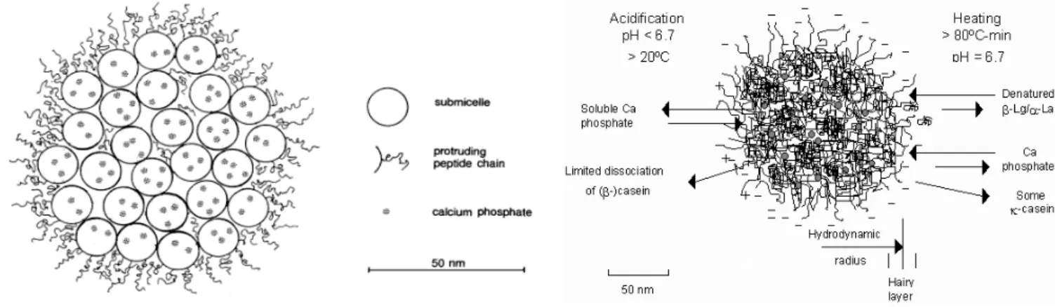
destroyed substantially only with pressures higher than 1000 MPa. Pressures between 50 and 300 MPa may even stimulate spore germination. It is known from the literature, how differently pathogens react to HHP in milk. Selected data are shown in Table 5 (Koncz et al., 2007).
Table 5. Effect of high hydrostatic pressure on foodborne microorganisms in milk according to data from the literature (Koncz et al., 2007)
Author Treatment Foodborne microorganism Pressure (MPa) Time (min) Species Lethality Patterson et. al.
(1995) 600 10
E. coli S. aureus
-1 -1.5 Erkmen & Karatas
(1997)
250 350
5 4
S. aureus -1.5
- 5 Gervilla et.al.
(1997)
350 400
10 10
Listeria innocua 910 (non pat.)
-1 -4 Oliveira et. al.
(1999)
500 500
10 10
Salmonella E. coli
- 6-7 - 6-7 Rademacher et. al.
(2001)
600
600 20
8
E. coli
L. monocytogenes
-7 -7 The advantages of HHP processing can be summarised in the followings:
High retention of colour, aroma, and nutritional value;
Potential to form novel texture;
The food is pressurized in packaged form (no re-contamination);
Positive consumer acceptance.
Perhaps the greatest hindrance of the broader application of HHP in food industry is the high investment costs of the equipment, that can be compensated by the smaller energy and running costs. Other problems that should be solved on the introduction of this technology: continuous processing is difficult; destruction of spores needs combined treatment; enzyme inactivation is not complete; flexible packaging materials are necessary; food has to contain min. 40% water to achieve antimicrobial effect. Legislation is still lacking, although according to the “Novel Food Regulation” (EC No.258/97) food treated at pressures higher than 150 MPa can be considered as
“novel” food (Behsilian et al., 2003).
There are still some other aspects to be taken into consideration regarding commercial HHP processing, and all in all the general view is among the experts of the food industry, that HHP technology is too risky at the moment for the major companies, and its users tend to be small or medium-size food producers (Corkindale, 2006).
Fifty-five companies used HHP technology in 2005, that meant about 90 pieces of industrial- scale equipment. Most of them work in America (U.S.A., Mexico, Canada: 56 pcs). In Europe 19
HHP equipment have been installed, and 14 pcs in Asia. The total production amounted to 100- 120 thousand tons in that year.
However, spreading of HHP technology is supposed to grow gradually, and in spite of the higher product prices once consumers have tried HHP food, they keep choosing it, no matter what it costs.
4.3.3 High Pressure Equipment
A schematic diagram of basic equipment design used for HHP processing is presented in Fig.7.
Figure 7. Schematic diagram of basic equipment design for high pressure processing of foods (Barta, 2007)
A typical HHP system consists of four main parts: a high pressure vessel and its closure, a pressure-generating system, a temperature-control device and a material-handling system (Mertens, Deplace, 1993; Mertens, 1995). The pressure vessel is usually a forged monolithic cylinder made of low-alloy steel of high tensile strength. The wall thickness is determined by the maximum working pressure, the vessel diameter and the number of cycles the vessel is designed to perform; this thickness can be reduced by using multi-layer, wire reinforced or other pre- stressed designs (Mertens, Deplace, 1993). Once loaded and closed, the vessel is filled with a pressure-transmitting medium. In food processing generally potable water or ethanol are used (Myllymäki, 1996). Air must be removed from the vessel, by compressing or heating the medium, before pressure is generated (Deplace, 1995). In the food industry, vessels with a volume of several thousand litres are used, with typical operating pressures in the 100 MPa – 500
MPa range, and holding times of about 5–10 minutes (Myllymäki, 1996). Laboratory-scale HHP equipment capable of reaching pressures up to 1000 MPa is also available.
4.3.4 Principles of High Pressure Processing
Pressure and temperature determine many properties of inorganic and organic substances. In food preservation, thermal processing is commonplace. If, however, a substance is exposed to increasing pressure, many changes will occur, especially at pressures of several hundred MPa (Buchheim, Prokopek, 1992). The behaviour of biological macromolecules under pressure is important for understanding the effects of HHP on milk. Under pressure, biomolecules obey the Le Chatelier-Braun principle, i.e., whenever stress is applied to a system in equilibrium, the system will react so as to counteract the applied stress; thus, reactions that result in reduced volume will be triggered under HHP. Such reactions may result in inactivation of microorganisms or enzymes and in textural changes in foods (Balci, Wilbey, 1999).
If the conditions for equilibrium or isokineticity are plotted against temperature and pressure, a stability phase diagram is obtained with an elliptical shape. Of particular interest in food processing are effects of HHP on proteins. Figure 8. shows a schematic pressure- temperature diagram of proteins. Proteins can be denatured using heat, pressure, and low temperatures.
Figure 8. Typical phase transition curve of proteins in the pT-diagram. The relation between heat-, cold and pressure-denaturation of proteins is presented by the sign of enthalpy changes
(ΔH) and volume changes (ΔV) (Heremans, 2002)
Denaturation of single-chain proteins may be regarded as a two-component system, where the native and denatured forms of protein are interchanging. From Fig. 8. it is apparent that denaturation temperature rises initially as the pressure rises. At maximum transition temperature
the sign of volume (∆V) changes. From this point on the the protein denatures at lower temperatures at the given pressure. At the maximal transition temperature the sign of entropy (∆S) changes and from this point on the protein denatures at lower pressures at the given temperature.
4.3.4.1 The Two-state Model and the Phase Transition
The folding–denaturing transition in proteins is a highly cooperative process. In certain cases, as a rule for smaller proteins, it suffices to describe this transition within a two-state approach involving the native state N, and the denatured state D, only. All those states are associated here in which the protein is working with the native state N, and all those states in which the protein is not working with the denatured state D. Despite the large structural manifolds involved, the two-state approach seems to work well in case that the two phase space areas can be lumped together to form two effective states. A prerequisite for this kind of state lumping is that thermodynamic equilibrium is established, an assumption which is itself quite severe and not always easily proved.
Provided that all these assumptions hold, the simplest approach to model protein stability is to consider the folding–denaturing transition as a phase transition. If in the D-N two-phase system the phases are in equilibrium, while material of a certain weight transfers from one phase to the other, then the Clausius-Clapeyron equation is valid:
dP/dT=∆S/∆V Equation 1.
Note that Eq. (1.) is an immediate consequence of the condition for the phase boundary,
∆G=0. ∆S and ∆V are the entropy and volume changes associated with the transition. Both quantities depend on the actual pressure P, and temperature T, where the transition takes place.
The boundaries of the stability phase diagram, i.e. the area in a pressure–temperature plane where the protein is stable in its native state, can then be determined from a solution of Eq. (1.).
This equation is readily solved by resorting to a further approximation.
In Eq. (1.) ΔS, and ΔV represent the differences in entropy, and volume, respectively, in the individual phases. These quantities are in close relation to the specific heat capacity and the thermal expansion. These are system parameters which we assume to be well defined, i.e. to be roughly independent on pressure and temperature as mentioned above. If so, ∆S and ∆V in Eq.
(1.) depend only linearly on T and P, and, hence, the equation can easily be integrated. The result is a general 2nd order curve in P and T whose shape may be elliptic, parabolic or hyperbolic:
aP2 + bT2 + 2cPT + 2fP + 2gT + const = 0 Equation 2.
4.3.4.2 Stability Against Temperature
During the temperature-induced denaturing transition, a protein changes from a rather well- organized structure into a random coil-like structure in which the hydrophobic amino acids come into contact with water. As a consequence, water forms locally ordered structures around the hydrophobic molecules, the so-called iceberg. These local structures are characterized by a low entropy as well as by a low enthalpy due to the wellaligned hydrogen bonds. The change of the specific heat, ∆CP=CpD - CpN, associated with the transition, is generally assumed to be positive, in agreement with the experimental findings. The difference in enthalpy, ∆H, between the native and the denatured state increases as the temperature is raised, according to ∆H(T) =H(T1) +
∆CP[T-T1]. At the same time, the respective difference in entropy, ∆S, increases as well, since the ordered solvent structures melt away. At some critical temperature T =T0, the enthalpic term,
∆H, and the entropic term, -T∆S, cancel, rendering a free energy change ∆G of zero. At this temperature, namely at T = T0, the transition to the denatured state takes place because it is energetically more favorable.
The same arguments can be used to understand, on a qualitative level, the phenomenon of cold denaturation: Lowering the temperature decreases the enthalpic term (note that, in this case T <T1) so that it eventually becomes negative and may compensate the entropy term, T∆S, which is positive due to a decreasing entropy. The actual transition temperatures into the denatured state depend of course on pressure: High pressure at low temperature may destabilize the locally ordered structures („iceberg”) because it counteracts an optimum alignment of the hydrogen bonds.
4.3.4.3 Stability Against Pressure
The free energy change associated with protein denaturation, becomes lower as pressure is increased, at least above some threshold pressure. We can use similar arguments as above, namely that ∆G(T)= ∆G(T1)+ ∆V[P-P1]. ∆V =VD-VN is the volume change in going from the native to the denatured state. As a rule, ∆V is negative because the structure of the native state has voids, for instance in the protein pockets, which are squeezed away in the denatured state so that its volume is smaller and, hence, the transition into the denatured state becomes favored under high pressure. Increasing the pressure up to some critical level P=P0, the protein may eventually cross the boundary ∆G=0, and the transition to the denatured state takes place.
The respective transition at low pressure is less straightforwardly to understand. First of all, we note that, in a large temperature range, the low pressure denaturation regime would require negative pressure, a condition which has, so far, not been realized experimentally. There is indeed a temperature range in which high pressure leads to a stabilization of the native state, and, consequently, low pressure to a destabiliation associated with denaturation. At rather high pressure (i.e. outside this range) the denatured state is far from being a random coil state. It is plausible that unfolding to a random coil against high pressure is severely hindered. Instead, the high pressure denatured state is still kind of a globular state where the voids in the protein are squeezed to a high degree so that VD < VN. On the other hand, in the lower pressure range and at sufficiently high temperature, unfolding to a random coillike state is still possible. Accordingly, the protein acquires a larger surface and, concomitantly, a larger volume. In addition, compression is much harder than in the native state because the compressible voids have vanished and the hydration shell is harder to compress than bulk water due to the ordered structures induced by the hydrophobic amino acids (Scharnagl et al., 2005).
4.3.5 Effect of High Hydrostatic Pressure on Proteins, with Special Regard to Milk Proteins
In their native state, proteins are stabilised by covalent bonds (including disulphide bridges) plus electrostatic interactions (ion pairs, polar groups), hydrogen bridges and hydrophobic interactions. Covalent bonds are almost unaffected by HHP, at least at relatively low temperatures (0–40°C), and so the primary structure of proteins remains intact during HHP treatment (Mozhaev et al., 1994). High pressure affects:
a.) the quaternary structure (e.g. through hydrophobic interactions);
b.) the tertiary structure (e.g. through reversible unfolding);
c.) the secondary structure (e.g. through irreversible unfolding) (Balci and Wilbey, 1999).
Stabilising hydrogen bonds are enhanced at low pressures and ruptured only at very high pressures. Significant changes to the tertiary structure of proteins, which is maintained chiefly by hydrophobic and ionic interactions, are observed at >200 MPa (Hendrickx et al., 1998).
Multimeric proteins, held together by non-covalent bonds, dissociate at relatively low pressures (~150 MPa), thereby disrupting quaternary structures. The exposure of protein surfaces, that formerly interacted with each other, to a solvent (hydrophobic solvation), results in the binding of water molecules, thereby reducing the volume of the system; thus, increasing pressure moves
the equilibrium between monomeric and multimeric states of proteins towards monomerisation (Gross, Jaenicke, 1994; Hendrickx et al., 1998).
Exposed to pressures above 400 MPa most of the proteins denature. Sensitivity to pressure or temperature varies with the type of bonds maintaining the structure. Measurements showed that structures with β-sheets are more stable against pressure than those with α-helices. The former is nearly incompressible while the latter can be deformed more easily. Oligomeric proteins dissociate to subunits while volume decreases. After dissociation subunits may reaggregate or denature. At pressures above 200 MPa chains begin to unfold and subunits of dissociated oligomers start reassociating. However, small molecules that have little secondary, tertiary and quaternary structure, such as amino acids, vitamins, flavour and aroma components, remain unaffected (Balci, Wilbey, 1999).
4.3.5.1 Effect of High Hydrostatic Pressure on Whey proteins
The behaviour of whey proteins under HHP is particularly important for milk and dairy products.
Johnston et al. (1992) were among the first researchers, who investigated the effects of HHP on whey proteins. The authors found that the amount of non-casein nitrogen decreased in milk serum with increasing pressure, that suggested denaturation and insolubilisation of whey proteins.
It was published in several studies that β-Lg is more sensitive to pressure than α-La.
Denaturation of whey proteins is usually determined by a loss in solubility at pH 4,6. With this method α-La was denatured at pressures higher than 400 MPa, and β-Lg at pressures higher than 100 MPa. The higher barostability of α-La is related to its more rigid molecular structure because there are four intra-molecular disulphide bonds in the protein, while in β-Lg there are only two.
Besides, β-Lg contains a free sulphydril-group which can participate in sulphydril oxydation or sulphydril-disulphide interchange reactions (López-Fandiño et al., 1996; Hinrichs et al., 1996;
Felipe et al., 1997; López-Fandiño, 1998; López-Fandiño, Olano, 1998; Garcia-Risco et al., 2000; Scollard et al., 2000; Huppertz et al., 2004; Hinrichs, Rademacher 2004; Huppertz et al., 2004b; Zobrist et al., 2005). After treatment at 400 MPa denaturation of β-Lg reached 70-80%.
At higher pressures, at 400-800 MPa, relatively little further denaturation occurs (Scollard, 2000).
The extent of HHP-induced denaturation of α-La and β-Lg increases with increasing holding time, temperature, and pH of milk (López-Fandiño et al., 1996; Felipe et al., 1997; López- Fandiño, 1998; López-Fandiño, Olano, 1998; Garcia-Risco et al., 2000; Scollard et al., 2000;
Huppertz et al., 2004; Hinrichs, Rademacher 2004; Huppertz et al., 2004b; Gaucheron et al., 1997; Arias et al., 2000).
Under HHP β-Lg unfolds and thus its free sulphydril group gets exposed. During HHP treatment of milk, denatured β-Lg may form small aggregates (Felipe et al., 1997) or interact with casein micelles (Needs et al., 2000a; Scollard et al., 2000). Dumay et al. (1994) and Van Camp et al. (1997) suggested that HHP-induced aggregation of β-Lg may be partially reversible on subsequent storage.
In HHP treated whole milk, some α-La and β-Lg are also found associated with the milk fat globule membrane (Ye et al., 2004).
The mechanism for high pressure induced denaturation of α-La and β-Lg in milk as well as in whey might be as follows (Huppertz, 2006):
β-Lg unfolds under high pressure, which results in the exposure of the free sulphydryl group in β-Lg. This free sulphydryl-group can interact with other milk proteins (κ-casein, α-La or β-Lg, and perhaps αs2-casein), through sulphydryl-disulphide interchange reactions. On release of pressure, unfolded α-La and β-Lg molecules, that have not interacted with another protein, may refold to a state closely related to that of native form of these proteins. The close structural similarity of monomeric untreated, and HHP treated β-Lg indicates that the sulphydryl- disulphide interchange reactions occur during HHP treatment, since the free sulphydryl-group of β-Lg is not available for interaction after high pressure treatment.
β-Lg exists in several isoforms. Isoforms A and B are the most abundant ones. Pressure stability of these variants were compared by Botelho et al (2000). Pressure denaturation experiments revealed different stabilities of the two isoforms. β-Lg B had higher pressure sensitivity than β-Lg A. It was proposed by the authors that the existence of of a core cavity in β- Lg B may explain its higher pressure sensitivity compared to β-Lg A.
4.3.5.2 Effect of High Hydrostatic Pressure on Caseins
Casein micelles are influenced considerably by HHP treatment. In one of the first studies Schmidt and Buchheim (1970) used electronmicroscopy to examine the size of casein micelles after HHP treatment. Since then several methods have been used to detect changes in casein micelles during or following pressurization, such as transmission electron microscopy, laser granulometry, photon correlation spectroscopy, and turbidimetry.
Casein micelle size is affected only slightly by HHP treatment at pressures below or at 200 MPa at 20°C. HHP treatment at 250 MPa increases average micelle size by ~30% and pressures higher than 300 MPa reduce micelle size by ~50% (Desobry- Banon et al., 1994; Gaucheron et
al., 1997; Needs et al., 2000b; Huppertz et al., 2004b; Huppertz et al., 2004c). Increase in the average size of casein micelles after treatment at 250 MPa is reversible during storage. Increased storage time and temperature enhance the reversibility (Huppertz et al., 2004b). Pressurization at 400 MPa or at 600 MPa broke up all large micelles into smaller fragments (Needs et al., 2000b).
Any decreases in micellar size after treatment at higher pressure (300-800 MPa) are irreversible during storage.
Fragmentation of casein micelles under pressure is caused partly by the solubilisation of colloidal calcium phosphate, and partly by the dissociation of hydrophobic and electrostatic interactions (Schrader, Buchheim, 1998; Needs et al., 2000b). Micellar calcium phosphate (MCP) is believed to play an important role in maintaining the integrity of casein micelles. The framework of the casein micelles is formed by so-called nanoclusters, that consist of an amorphous MCP core, which is surrounded by a multilayer of caseins. Solubilisation of MCP leads to the disruption of calcium phosphate nanoclusters, and thus weakens the integrity of the micelles. HHP readily disrupts electrostatic interactions that further promote micellar disruption.
Micellar caseins may re-associate under prolonged pressurization at 200-300 MPa, because hydrophobic bonds are favoured over hydrophobic solvation. Re-association doesn’t take place at higher pressure (Huppertz et al., 2006). Upon increasing the calcium concentration in a calcium caseinate suspension, micelles become more resistant to pressure-induced disruption (Lee et al., 1996; Anema et al., 1997). Introduction of calcium to the system most likely shift the calcium equilibrium from the soluble to the colloidal phase.
HHP treatment increases the hydration of casein micelles. This is partly due to the association of denatured β-Lg with the casein micelles. Thus, the net negative charge on the micelle surface increases and enhances micellar solvation. HHP induced disruption of micelles further increases micellar hydration, which increases with decreasing micelle size, and is higher for irregularly-shaped than spherical particles (Huppertz et al., 2006).
High hydrostatic pressure (100-400 MPa) significantly increased the transfer of individual caseins from the colloidal to the soluble phase of milk from several species (López-Fandiño et al., 1998). The order of the dissociation of casein variants in bovine milk was as follows:
β>κ>αs1>αs2. In goat’s, and ewe’s milk the order was different: κ>β>αs2>αs1 casein (Huppertz et al., 2002).
Temperature affects micelle size of HHP treated milk. For example when reconstituted skim milk was pressurized at 250 MPa, 20°C, HHP treatment didn’t cause significant effect on micelle size. When HHP treatment was carried out at 40°C, micelle size increased, at 4°C micelle size decreased (Gaucheron et al., 1997).