Foreign body impaction in the sigmoid colon: A twenty euro bet
Katalin E Müller, András Arató, Péter László Lakatos, Mária Papp, Gábor Veres Katalin E Müller, András Arató, Gábor Veres, 1st Department
of Pediatrics, Semmelweis University, 1083 Budapest, Hungary Péter László Lakatos, 1st Department of Medicine, Semmelweis University, 1083 Budapest, Hungary
Mária Papp, 2nd Department of Medicine, University of Debre- cen, 4032 Debrecen, Hungary
Author contributions: Müller KE collected data and prepared the manuscript; Arató A critically revised the manuscript; Lakatos PL interpreted clinical data and critically revised the manuscript;
Papp M interpreted clinical data and revised the manuscript;
Veres G collected data, supervised preparation of the manuscript and critically revised the manuscript.
Supported by János Bolyai Research Grant, to Veres G; A Hun- garian Scientific Research Fund Grant, No. OTKA-K 105530 Correspondence to: Gábor Veres, MD, PhD, 1st Department of Paediatrics, Semmelweis University, 53 Bokay Street, 1083 Budapest, Hungary. vergab@gyer1.sote.hu
Telephone: +36-20-8258163 Fax: +36-1-3036077 Received: July 23, 2012 Revised: October 26, 2012 Accepted: November 14, 2012
Published online: June 28, 2013
Abstract
Foreign body ingestion is a common clinical problem in early childhood. However, it may occur even in adults, unknowingly. Most ingested foreign bodies entering the stomach pass through the gastrointestinal tract uneventfully. Here we report on a 13-year-old boy who presented with chronic abdominal pain, weight loss and occult gastrointestinal bleeding for 6 mo.
Colonoscopy was negative; however, a ballpoint pen was impacted in the sigmoid region. Subsequently, the child admitted swallowing a pen as a 20-euro bet 6 mo previously. Crohn’s disease is a chronic relaps- ing inflammatory gastrointestinal disease. It is often difficult to diagnose due to the fact that there is no single pathognomonic sign or symptom. This case is a description of an adolescent with chronic gastrointes- tinal symptoms due to a foreign body. Therefore, an ingested foreign body should be included in the differ-
ential diagnostic procedure related to gastrointestinal symptoms.
© 2013 Baishideng. All rights reserved.
Key words: Foreign bodies; Pen; Differential diagnosis;
Crohn’s disease; Child
Müller KE, Arató A, Lakatos PL, Papp M, Veres G. Foreign body impaction in the sigmoid colon: A twenty euro bet. World J Gastroenterol 2013; 19(24): 3892-3894 Available from: URL:
http://www.wjgnet.com/1007-9327/full/v19/i24/3892.htm DOI:
http://dx.doi.org/10.3748/wjg.v19.i24.3892
INTRODUCTION
Foreign body ingestion is a common problem in everyday emergency practice, primarily in children. Fortunately, the majority of the ingested foreign objects pass through the gastrointestinal tract without complications. Foreign body ingestion may present without symptoms[1], and in some cases results in perforation with gastrointestinal bleeding or an obstruction[1]. Rarely, an abscess or a fistula oc- curs[1]. It has been reported to mimic other diseases (renal stone or irritable bowel syndrome)[1]; however, there are no literature data concerning differential diagnostic dif- ficulties of Crohn’s disease (CD) and a foreign body.
CDis a chronic relapsing inflammatory gastrointestinal disease. It is often difficult to diagnose due to the fact that there is no single pathognomonic sign or symptom. Here we report on a case with chronic abdominal symptoms, weight loss and occult bleeding suggesting CD. However, at colonoscopy, a swallowed pen was impacted in the sig- moid region causing the aforementioned symptoms.
CASE REPORT
A 13-year-old boy was admitted to the First Depart- CASE REPORT
Online Submissions: http://www.wjgnet.com/esps/
wjg@wjgnet.com
doi:10.3748/wjg.v19.i24.3892
3892 June 28, 2013|Volume 19|Issue 24|
WJG|www.wjgnet.com
World J Gastroenterol 2013 June 28; 19(24): 3892-3894 ISSN 1007-9327 (print) ISSN 2219-2840 (online)
© 2013 Baishideng. All rights reserved.
ment of Paediatrics at Semmelweis University with 10-kg weight loss in the last 6 mo and intermittent colicky abdominal pain. Past medical history revealed that 6 mo previously he was admitted to hospital for 2 d due to acute abdominal pain and vomiting. He was given intra- venous fluids and his complaints improved. However, an- orexia persisted, abdominal pain returned intermittently, and he lost 10 kg during this period. Stool blood test was positive twice, while stool culture, parasite and assays for Clostridium difficile toxins A and B were all negative. Consid- ering the age of the patient, weight loss, chronic abdomi- nal pain and positive stool blood test, as well as negative stool culture, inflammatory bowel disease was suspected and he was referred to our clinic.
Physical examination revealed normal vital signs with- out any clinical abnormalities. His abdomen was soft, non-tender, without guarding or palpable masses. There was normal sphincter tone, no perianal abscess, skin tag or fistula at rectal examination. Laboratory tests showed normal red blood cell count, white blood cell count and thrombocyte count. C-reactive protein, total protein and albumin were within the normal limits. Abdominal ultrasound showed slight wall thickening in the descend- ing colon. There was no family history of inflammatory bowel disease.
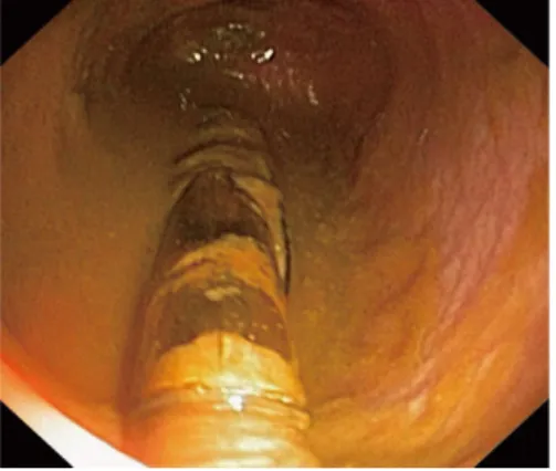
The upper endoscopy was negative. There were no ulcers, no sign of bleeding, and no antral or bulbar lymphonodular hyperplasia. The terminal ileum and the colon appeared normal, confirmed by multiple mucosal biopsies at histology. Surprisingly, at withdrawal of the colonoscope, an impacted foreign body (plastic half-ball pen, Figure 1) was observed embedded in the sigmoid region. The surrounding mucosa was inflamed, with no visible mucosal vessels. The plastic, numbered ballpoint pen could not be removed by Dormia set and polypec- tomy snare despite several attempts. After the diagnostic procedures, the patient admitted swallowing a half-plastic pen as a 20-euro bet with his friend 6 mo ago. At first, he thought there might have been a connection between the swallowed pen and his symptoms as he had a hospital admission for 2 d due to acute abdominal pain and vom- iting (see above). Later, he was embarrassed and hoped that these two events were just a coincidence. He never disclosed his bet.
Plain abdominal X-ray and abdominal ultrasound were performed the next day after endoscopy, but the foreign body could not be visualized. The pen passed through the colon spontaneously hence the control colo- noscopy showed no foreign body after 5 d. The patient had no symptoms during 21 mo of follow-up; his weight gain is normal, and there is no occult bleeding.
DISCUSSION
We report on a case of an ingested foreign body as a twenty-euro bet. To our knowledge, this is the first description of a swallowed object causing chronic gas- trointestinal symptoms in a paediatric patient. However,
ingested foreign bodies are a common event in the paedi- atric population.
The first recorded paediatric foreign body ingestion was described by Frederick the Great in 1692[2]. Most accidentally ingested foreign bodies go undetected and pass through without any incident. However, 10%-20%
require endoscopic removal and 1% or less require surgi- cal intervention[3]. In general, the passing of an ingested foreign body depends on the anatomic conditions of the gastrointestinal tract and on factors related to the ingest- ed foreign body. Long, thin objects as seen in our case are less likely to pass the pylorus or the duodenum[2]. The presenting features vary according to the site and include pancreatitis, hepatic abscess, appendicitis, intussuscep- tions and irritable bowel syndrome[1,2].
There are only a few reports of foreign bodies imitat- ing CD in the English literature. Ioannidis et al[4] reported on a case of incidental toothpick ingestion which caused an ileum fistula and mimicked CD. In addition, a patient presented with recurrent, subacute obstruction and right iliac mass mimicking the presentation of CD[5]. Subse- quent lower endoscopy revealed small bowel phytobezoar which passed spontaneously.
To our knowledge, only one paediatric case of an in- gested foreign body mimicking CD has been reported[6]; nevertheless, this was an acute event. A 7-year-old boy pre- sented with a 2-wk history of cramping abdominal pain and low grade fever. Colonoscopy revealed an oedema- tous, friable rectosigmoid junction with a solitary fistula or ulcer. Hydrocortisone enemas were prescribed with mini- mal improvement. Seven days later his condition became more serious, and a computed tomography scan revealed a right iliopsoas abscess. Repeated colonoscopy showed a toothpick in the lumen of the rectosigmoid colon.
Foreign bodies usually cause acute symptoms of per- foration, obstruction or gastrointestinal bleeding. How- ever, our patient had chronic symptoms. This is probably due to the lack of perforation or obstruction. Meanwhile, his symptoms were similar to the reported cases, in which perforation or fistula was diagnosed.
We were presented with a healthy, young adolescent with chronic abdominal pain, weight loss and occult bleeding - classic presenting features of paediatric inflam-
3893 June 28, 2013|Volume 19|Issue 24|
WJG|www.wjgnet.com
Muller KE et al. Foreign body impaction: A twenty euro bet
Figure 1 Half-plastic pen impacted in the sigmoid region.
matory bowel disease. Our working hypothesis was CD, though the laboratory tests were negative. Colonoscopy revealed a plastic, half-ball pen embedded in the sigmoid mucosa. The patient concealed swallowing a half-pen as a 20-euro bet with his friend. His subsequent hospital ad- mission due to vomiting and acute abdominal symptoms may be explained as due to gastric irritation. Later the foreign body passed through the upper gastrointestinal tract and impacted in the sigmoid region. We speculate that occult bleeding was caused by the chronic mucosal irritation around the embedded pen. Anorexia and ab- dominal pain can be explained by the increased bowel peristalsis around the logged pen[7].
The value of imaging studies for an ingested for- eign body seems to be questionable based on our case.
Hence, plain abdominal X-ray (plastic pen) and abdomi- nal ultrasound (sigmoid localization) could not identify the pen. Nevertheless, the role of imaging studies is crucial to determine the inflammatory reaction in and around the bowel wall and to exclude findings requiring surgical intervention[7].
The majority of ingested foreign bodies that reach the stomach pass through the alimentary tract without complication. If not, the management of ingested for- eign bodies is dependent on their size, shape, material and location. After imaging studies, endoscopy should be considered as the crucial step in management since it is a potent and safe diagnostic tool. On the other hand, surgical treatment is mandatory in the presence of com- plications such as abscesses and fistulas[7]. In our patient, we attempted the removal of the embedded pen, but due to the increased possibility of perforation, we did not prevail. Subsequently, the pen was spontaneously passed after the first endoscopy as a result of the previous at- tempts of removal.
In summary, we report on an adolescent patient with
chronic gastrointestinal symptoms due to a swallowed plastic pen that mimicked CD. Therefore, an ingested foreign body should be included in the differential di- agnostic procedure related to chronic gastrointestinal symptoms.
ACKNOWLEDGMENTS
We thank Alexandra Horvath for her language editing.
REFERENCES
1 Komninos ID, Tsiligianni IG. Foreign body ingestion mim- icking irritable bowel syndrome: a case report. J Med Case Rep 2010; 4: 244 [PMID: 20684775 DOI: 10.1186/1752-1947-4-244]
2 Antao B, Foxall G, Guzik I, Vaughan R, Roberts JP. Foreign body ingestion causing gastric and diaphragmatic perfora- tion in a child. Pediatr Surg Int 2005; 21: 326-328 [PMID:
15645252]
3 Eisen GM, Baron TH, Dominitz JA, Faigel DO, Goldstein JL, Johanson JF, Mallery JS, Raddawi HM, Vargo JJ, War- ing JP, Fanelli RD, Wheeler-Harbough J. Guideline for the management of ingested foreign bodies. Gastrointest Endosc 2002; 55: 802-806 [PMID: 12024131 DOI: 10.1016/
S0016-5107(02)70407-0]
4 Ioannidis O, Kakoutis E, Sakkas L, Konstantara A, Chatzo- poulos S, Kotronis A, Makrantonakis N. Ingested toothpick fistula of the ileum mimicking Crohn’s disease. Acta Gastro- enterol Belg 2010; 73: 527-529 [PMID: 21299167]
5 Prior A, Martin DF, Whorwell PJ. Small bowel phytobezoar mimicking presentation of Crohn’s disease. Dig Dis Sci 1990;
35: 1431-1435 [PMID: 2226106 DOI: 10.1007/BF01536753]
6 O’Gorman MA, Boyer RS, Jackson WD. Toothpick foreign body perforation and migration mimicking Crohn’s disease in a child. J Pediatr Gastroenterol Nutr 1996; 23: 628-630 [PMID:
8985858 DOI: 10.1007/BF0153675]
7 Zezos P, Oikonomou A, Souftas V, Gkotsis D, Pitiakoudis M, Kouklakis G. Endoscopic removal of a toothpick perforat- ing the sigmoid colon and causing chronic abdominal pain:
a case report. Cases J 2009; 2: 8469 [PMID: 19918434 DOI:
10.4076/1757-1626-2-8469]
P- Reviewer Devanarayana NM S- Editor Gou SX L- Editor Logan S E- Editor Li JY
3894 June 28, 2013|Volume 19|Issue 24|
WJG|www.wjgnet.com
Muller KE et al. Foreign body impaction: A twenty euro bet