Neural mechanisms of predatory and abnormal intraspecific forms of aggression induced by chronic
glucocorticoid-deficiency or early social isolation Doctoral theses
Áron Tulogdi
Semmelweis University
János Szentágothai PhD School of Neurosciences
Supervisor: Dr. József Haller scientific advisor, D.Sc.
Official reviewers: Dr. Szilvia Mezey research fellow, Ph.D.
Dr. Péter Kabai university docent, Ph.D.
Head of the Final Examination Committee:
Dr. Béla Halász professor emeritus,
member of the Hungarian Academy of Sciences Members of the Final Examination Committee:
Dr. György Nagy professor, D.Sc.
Dr. György Lévay scientific advisor, Ph.D.
Budapest
2014
Introduction
Pathological aggression is a serious medical condition with significant burden for the society, however, the underlying mechanisms are still hardly known. Proactive (cold-blooded) and reactive (emotional) forms of these abnormal behaviors had been differentiated. Mechanisms of these distinct forms of aggression could be better understood by investigating animal models based on etiological factors of pathological human aggression. A laboratory model of abnormal aggression was first developed by our research group based on qualitative alterations: low, non-reactive glucocorticoid levels induced by the surgical removal of the adrenals led to abnormal bites that were aimed at vulnerable body parts of the opponent.
These bites were less preceded by normal offensive signals, and were associated with decreased autonomic reactivity. Thus this paradigm was suggested to model the proactive, cold-blooded form of aggression. Later, our group also reported that post-weaning social isolation of laboratory rats leads to a similar phenotype. Since disturbed early social life is an important etiological factor of pathological human aggression, this model may help to understand the underlying mechanisms.
If isolation-induced abnormal aggression was associated with increased stress and autonomic functions (as it is suggested by earlier data), this could be the first animal model showing the etiological factors of the reactive, emotional form of pathological aggression. Here we investigated this question. We also examined if abnormal aggression could be reversed by resocialization in adulthood.
We investigated the neuronal activation of hypothalamic, amygdalar, prefrontal, midbrain and other brain regions in the above mentioned models of aggression (chronic glucocorticoid-deficiency- and social isolation-induced aggression) using c-Fos immunohistochemistry.
We focused our analysis on regions relevant to the control of normal and abnormal aggression.
Since similarities were suggested between cold-blooded and predatory forms of aggression, we assessed brain activation patterns during predatory aggression in rats and compared it to activation expressed during abnormal forms mentioned above. To date predatory aggression was mostly examined in cats by means of electrical stimulation.
In addition to their relevance for human behavior, these data are important for an ethological point of view to better understand the effect of stress on aggression and the physiological background of aggressive behavior in animal populations.
Aims
Characterization of early social isolation-induced abnormal aggression
1. What kind of stress and autonomic functions are associated with early social isolation-induced abnormal aggression?
2. How does early social isolation affect aggression and social behavior shown in a social group in adulthood? Can early social isolation- induced abnormal aggression be reversed by resocialization in adulthood?
Neural mechanisms of different forms of aggression
3. Brain activation patterns associated with chronic glucocorticoid- deficiency-induced abnormal aggression: do brain regions involved in predatory aggression play a role in this model?
4. Which brain regions are activated during early social isolation-induced abnormal aggression? Do the activated brain regions show overlap with those activated during chronic glucocorticoid-deficiency-induced abnormal aggression or with those activated during natural aggression?
5. Which brain regions are activated during predatory aggression in rats?
Do the activated brain regions show overlap with those implicated in feline predatory aggression based on electrical stimulation methods or with those activated during chronic glucocorticoid-deficiency-induced abnormal aggression?
Methods
Animals and housing conditions
Rats involved in these experiments were male Wistar rats (Charles River Laboratories). In experiments which involved social isolation, rats were housed in a reversed cycle of 12:12 hours. In experiments which involved glucocorticoid-deficiency or predatory aggression, rats were moved to the reversed cycle at least two weeks before behavioral testing.
The weight of subjects was 400-500 g in the beginning of behavioral testing.
Intruders used in resident-intruder tests weighed approximately 30% less than the residents (300-350 g). Mice were 50-70 day-old male CD1 mice.
Tap water and standard laboratory food were available ad libitum throughout. In the experiment which involved adrenalectomy, rats had free access to 0.9% saline solution as well. Experiments were carried out in accordance with the European Communities Council Directive of 24
November 1986 (86 ⁄ 609 ⁄ EEC), and were reviewed and approved by the Animal Welfare Committee of the Institute of Experimental Medicine.
Post-weaning social isolation
Pups were weaned on the 21st postnatal day and were either housed individually (isolated group), or in groups of 4 rats (social, control group) for 7 weeks. Rats were not handled except for handling associated with regular cage cleaning. Behavioral testing started on the 8th post- weaning week i.e. at 11 weeks of age.
Surgical techniques
In vivo biotelemetry measurements
Changes in heart rate and locomotor activity during the aggressive interaction were monitored by means of biotelemetry e-mitters implanted in the abdominal cavity of rats (HR EMitter, PDT-4000; Mini Mitter Company, Bend, OR, USA). E-mitters contained the positioning locators and two heart rate leads that were attached to the chest. Signals transmitted by the e-mitter were received by a receiver unit under the cage of the subjects. The receiver transmitted the signals to the VitalView Data Acquisition System software (Mini Mitter Company, Bend, OR, USA). Heart rates and locomotor activity were registered during baseline conditions and during the resident- intruder test.
Adrenalectomy and implantation of the glucocorticoid pellet
Low, non-reactive glucocorticoid levels were generated by the surgical removal of the adrenals, followed by the implantation of a subcutaneous pellet containing 25 mg of corticosterone. According to our earlier experiments, this procedure provides low and stable plasma levels of corticosterone for at least three weeks. Sham operated animals were used as controls.
Behavioral methods
Resident-intruder test
Aggressive behavior was assessed by means of the resident- intruder test. In this paradigm an intruder of smaller size was placed into the home-cage of the respective rat (resident), and left there for 20 min. Attack bites were analyzed in details by low speed video analysis. Bites aimed at vulnerable (head, throat, belly) and non-vulnerable body parts (back and flanks) of the opponent were differentiated. Another qualitative parameter was the ratio of non-signaled attacks. Our earlier results suggest that these qualitative aspects of aggression characterize pathological alterations much more reliably than quantitative changes. To assess the behavioral signs of arousal we conducted a detailed behavior analysis by means of the H77 event recorder software.
Behavior in a social group
Two types of behavior were examined within a social group:
‘group aggression’ in the active (dark) phase, and huddling behavior in the inactive (light) phase. To assess ‘group aggression’, we made one-hour long video recordings. We quantified the frequency of offensive, dominant, defensive (provoked and unprovoked), submissive behavior and that of attack bites for each animal.
Huddling behavior during sleep was assessed during the early phase of the light (inactive) period. Two consecutive days were analyzed together. Huddling behavior was characterized by sleeping in direct physical contact on at least one of the two days with at least one cage mate.
If no such contacts were established, rats were considered separated.
Predatory aggression
To assess predatory aggression, an adult male mouse was placed in the home-cage of the rat in the early hours of the dark phase. If the rat killed the mouse within 20 min, the mouse was removed immediately and at the same time, another, uninjured mouse was removed from the home cage of a randomly chosen rat. The former rat was assigned to the muricidal, while the latter rat was assigned to the non-muricidal group. To assess the baseline neuronal activity, control rats that had not been exposed to mice were examined.
Hormone measurements
Blood was sampled by means of tail incision. The whole procedure lasted less than 1 min which allowed blood sampling without significant changes in basal corticosterone levels. Blood was sampled in tubes containing ethylenediaminetetraacetic acid (EDTA), blood samples were centrifuged. Plasma corticosterone concentrations were measured by radioimmunoassay. Prior to assay, corticosterone was separated from corticosterone-binding globulin (CBG). 125I-labeled carboxymethyloxime–
tyrosine–methyl ester derivative was used as tracer. All samples were measured in the same assay.
Histological procedures
Perfusion
Perfusion was timed 60-120 min after the behavioral test (similar delays within specific experiments). Within this time window the concentration of the c-Fos protein product is stable and maximal. Rats were anesthetized and perfused with 150 ml 4 °C phosphate-buffered saline (PBS) followed by 300 ml paraformaldehyde (4 %, diluted in PBS). The brains were removed, post-fixed in paraformaldehyde for 3 h and stored at 4 °C in PBS-azide. Sections were cryoprotected by 20 % sucrose (diluted in
PBS, 48 h). 30 µm frozen sections were cut in the frontal plane on a sliding microtome. Sections were stored at 4 °C in PBS-azide until the immunohistochemical labeling.
Immunohistochemical labeling and quantitative analysis of c-Fos Sections were incubated in Triton-X-100 and hydrogen peroxide, and afterwards in a blocking medium (normal horse serum). The primer antibody was a rabbit polyclonal antibody raised against the amino terminus of c-Fos p62. This antibody is a reliable marker of acute neuronal activity.
Sections were incubated in biotinylated FAB chains, and in streptavidin- conjugated horseradish peroxidase or in avidin-biotin complex. The peroxidase reaction was developed in the presence of diaminobenzidine tetrahydrochloride (DAB), nickel–ammonium sulphate and hydrogen peroxide.
As c-Fos protein is located in the nucleus and fills it, stained nuclei can be easily differentiated from their environment. Counting the stained cell nuclei makes quantitative analysis possible. In these experiments we focused on brain regions relevant to aggression control (hypothalamus, amygdala, midbrain, prefrontal cortex). Digital photomicrographs were taken by means of an Olympus microscope and the number of c-Fos positive nuclei was assessed my means of the ImageJ software.
Statistical analyses
Statistical analyses were conducted in the Statistica software. In the case of the resident-intruder paradigm, Kruskal-Wallis tests and Mann- Whitney U post-hoc tests were applied. Group aggression, corticosterone levels, heart-rates, body temperature, locomotor activity and c-Fos counts were analyzed by means of ANOVA models. In these cases Newman-Keuls post-hoc analyses were applied if needed (with Holm-Bonferroni correction). Huddling behavior was analyzed by means of the Chi-square test. Correlations between behavioral measures and the activations of specific brain regions were examined by the Spearman method.
Significance level was set at p < 0.05.
Results and Discussion
Different forms of aggression observed in the investigated paradigms
We confirmed earlier results by showing that the forms of abnormal aggression shown by adrenalectomized and isolated rats are similar on one hand as rats exhibit an increased share of vulnerable bites and decreased intention signaling in both paradigms. On the other hand,
there are important differences: (1) induction of these forms (i.e. chronic glucocorticoid-deficiency is a disrupted physiological function; the lack of early social experience is an environmental effect), (2) quantitative increase of attack counts and increased defensiveness were only observed in isolated rats.
Muricidal rats showed a species-typical behavioral pattern: after a sudden attack, they broke the neck of the mouse without any threat signals.
The characteristics of abnormal aggression induced by social isolation
Glucocorticoid levels, autonomic functions and behavioral patterns induced by social isolation
Under baseline conditions corticosterone secretion and the diurnal rhythm of heart rate were similar in isolated and social rats. However, during the aggressive interaction (resident-intruder test) both corticosterone level and heart rate were excessively increased in isolated rats as compared to the control (social) group. These data suggest that the abnormal aggression of isolated rats is associated with increased stress and autonomic reactivity. Excessive stress reactivity of isolated animals was shown earlier in response to different (non-aggressive) social challenges. Increased autonomic (heart rate) reactivity shown here might be the consequence of increased adrenaline reactivity shown earlier in isolated rats.
In the resident-intruder test, isolated rats switched from one behavior to another frequently. Our hypothesis is that this fragmented pattern of behavior may be considered a behavioral sign of hyperarousal which has been shown in several non-social contexts. However, according to our best knowledge, the present data are the first ones showing behavioral hyperarousal in a social context.
Excessive autonomic reactivity, stress hyperreactivity and behavioral hyperarousal render the aggressiveness of isolated rats more similar to psychopathologies induced by early social adversities, as earlier data showed increased autonomic and stress reactivity under these conditions. Moreover, increased autonomic and stress reactivity was also strongly implicated in violent behavior.
Our results suggest that abnormal aggression induced by post- weaning social isolation is similar to the reactive (emotional) form of human aggression in phenomenological and etiological terms as well. The factors that induce these alterations (early social adversities) and the phenomenological alterations of the behavior (vulnerable attacks, disturbed social signaling, defensiveness, behavioral hyperarousal) are similar.
Furthermore, physiological mechanisms of these behaviors also show analogies (increased glucocorticoid and autonomic reactivity). In contrast, abnormal aggression of adrenalectomized rats resembles the proactive
(cold-blooded, instrumental) form of pathological human aggression as these show similarities in both phenomenology (vulnerable attacks, disturbed social signaling) and physiological mechanisms (low, non- reactive glucocorticoid levels and decreased autonomic reactivity). Besides the previously discussed overlaps and differences, these assumptions reveal an important physiological difference between these two paradigms (i.e.
adrenalectomy and social isolation): decreased and increased stress and autonomic reactivity, respectively.
Effects of resocialization on social isolation-induced abnormal aggression and social anxiety
When isolated rats were placed in a social group in adulthood, their social behavior was abnormal: isolated rats hardly showed offensive and dominant (i.e. threatening) behaviors, while bite counts were similar to controls (lack of signals). Isolated rats reacted with defensiveness to non- threatening behaviors of the social partners. These features suggest that the aggression of isolated rats is ambiguous: intention signaling of attacks is decreased while defensiveness is increased. Similar phenomena were previously noticed in isolated chimpanzees and rhesus macaques.
During the first week of resocialization isolated rats showed decreased direct physical contacts with cage mates, i.e. decreased huddling behavior. This behavior was normalized by the end of the first week. This alteration is reminiscent of the social withdrawal that was shown in isolated and later resocialized rhesus macaques and chimpanzees.
In contrast, three-week long resocialization period was unable to reverse the abnormal features exhibited in the resident-intruder test, i.e.
increased share of vulnerable bites and non-signaled bites. The latter suggests that isolation has a marked long-term impact on aggressive behavior. In quantitative terms, aggression showed a mild improvement at the end of the three-week long resocialization period which is in line with earlier findings reporting resocialization normalizing qualitative alterations of aggression in the social interaction test.
Similar findings were also reported in human studies. Childhood neglect and abuse are important risk factors both for violence and social withdrawal. Psychotherapy showed promising results in the treatment of social phobia, however, it is considerably less successful in the treatment of pathological aggression.
Taken together, our results suggest that different symptoms induced by social isolation show different sensitivities to resocialization.
While social problems shown in a group of conspecifics were reversed in a social group, unprovoked defense and abnormal aggression were resilient to the same treatment. These features render this model more similar to human psychopathologies induced by early social disturbances.
Neuronal activation patterns observed in different forms of aggression
The aggression of adrenalectomized rats show similarities with predatory aggression regarding phenomenology and physiological mechanisms: adrenalectomized rats deliver a considerable amount of vulnerable bites, their attacks are less signaled, and their aggressiveness is associated with a blunted autonomic reactivity. This similarity motivated the investigation of brain regions that had previously been implicated in the control of predatory aggression, and we found that these brain regions (lateral hypothalamus, central amygdala) were overactivated during the abnormal aggression of adrenalectomized rats. Based on our correlational analyses and the known anatomical connections one could hypothesize that the central amygdala affects behavior through the lateral hypothalamus, and abnormal attack patterns emerge accordingly. Blunted autonomic reactivity might be associated with the shift of periaqueductal gray activation towards the ventral regions, which might also be a consequence of the excessive activation of the central amygdala. The hypothalamic attack area and the medial amygdala -brain regions playing crucial roles in the control of natural territorial aggression- seem not to play a role in the control of abnormal aggression induced by adrenalectomy.
During the abnormal aggression of isolated rats, brain regions responsible for predatory aggression (which could also be responsible for the abnormal aggression of adrenalectomized rats, i.e. central amygdala, lateral hypothalamus, ventrolateral column of the periaqueductal gray) did not show excessive activation. Therefore, the mechanisms underlying abnormal attack patterns of isolated rats remains unclear. One could assume that the altered prefrontal function causes abnormal aggression via disturbed evaluation of social contexts; however, further investigation is needed to clarify this issue. In contrast, abnormal aggression of isolated rats was associated with the overactivation of brain regions responsible for natural aggression (hypothalamic attack area, medial amygdala), which was not observed in adrenalectomized rats. Based on these data one could hypothesize that the excessive activation of brain regions responsible for natural aggression plays a role in the isolation-induced quantitative increase in aggression.
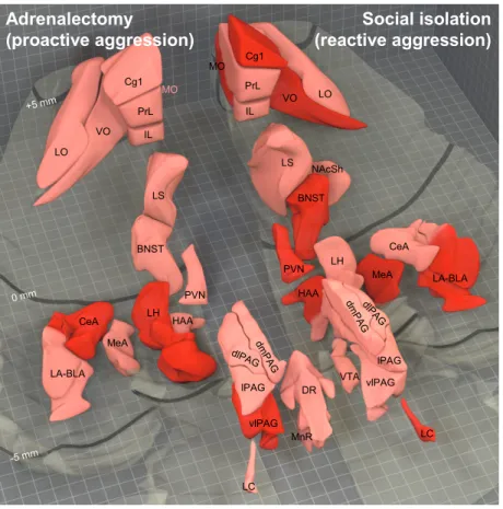
Brain activation patterns observed in adrenalectomy-induced and social isolation-induced abnormal aggression are shown in Figure 1.
Adrenalectomy
(proactive aggression)
Social isolation (reactive aggression)
LO
VO IL
PrL Cg1
Cg1 PrL IL
VO LO MO
LA-BLA
LA-BLA CeA
MeA
MeA
CeA LH
HAA
HAA PVN LH
PVN
dlPAG dmP
AG
lPAG
vlPAG
vlPAG
LC
LC MO
dmP AG
dlPAG
lPAG VTA BNST
BNST LS
LS
NAcSh
DR
MnR +5 mm
0 mm
-5 mm
Adrenalectomy
(proactive aggression)
Social isolation (reactive aggression)
LO
VO IL
PrL Cg1
Cg1 PrL IL
VO LO MO
LA-BLA
LA-BLA CeA
MeA
MeA
CeA LH
HAA
HAA PVN LH
PVN
dlPAG dmP
AG
lPAG
vlPAG
vlPAG
LC
LC MO
dmP AG
dlPAG
lPAG VTA BNST
BNST LS
LS
NAcSh
DR
MnR +5 mm
0 mm
-5 mm
Figure 1. Brain activation patterns observed in abnormal aggression induced by adrenalectomy (left hand-side) and social isolation (right hand-side). Based on phenomenological and etiological similarities these forms of aggression can be considered as animal models of proactive (cold-blooded, instrumental) and reactive (emotional) forms of abnormal aggression, respectively. The ventral surface of the rat brain is transparent in the figure, dark lines indicate the distance from Bregma. Brain regions shown in pink were activated during natural aggression (in control animals), brain regions shown in red showed excessive activation in the respective model of abnormal aggression. In the case of adrenalectomy those brain regions that were not investigated in the present experiment were illustrated based on previous results that were obtained by our research group in the same paradigm using similar methodology. BNST, bed nucleus of the stria terminalis, medial part; CeA, central amygdala; Cg1, anterior cingulate cortex;
dlPAG, periaqueductal gray, dorsolateral column; dmPAG, periaqueductal gray, dorsomedial column; DR, dorsal raphe; HAA, hypothalamic attack area; IL, infralimbic cortex; LA-BLA, lateral-basolateral amygdala complex; LO, lateral orbitofrontal cortex;
LC, locus coeruleus; LH, lateral hypothalamus; lPAG, periaqueductal gray, lateral column; LS, lateral septum; MeA, medial amygdala; MnR, median raphe; MO, medial orbitofrontal cortex; NAcSh, nucleus accumbens shell; PrL, prelimbic cortex; PVN, hypothalamic paraventricular nucleus, parvocellular region; lPAG, periaqueductal gray, ventrolateral column; VO, ventral orbitofrontal cortex; VTA, ventral tegmental area.
Taken together, these data show that the abnormal aggression of adrenalectomized and isolated rats is different regarding not only phenomenology and associated autonomic functions, but brain mechanisms as well. Thus the term “pathological aggression” covers a heterogeneous behavioral category regarding its mechanisms. This can be one reason of the poor efficacy of the currently available treatment methods. These types of aggression should be differentiated in both human and animal research.
The neural background of predatory aggression was not studied earlier in rats except for identifying the main locus of control i.e. the lateral hypothalamus. Our results suggest that the neural background of predatory aggression in rats -as revealed by c-Fos immunohistochemistry- is markedly similar to the neural background of predatory aggression in cats, which was revealed by stimulation studies. This indicates that the mechanisms underlying predatory aggression are highly conservative. The mechanisms of predatory aggression that were revealed here are markedly different from those of intra-specific aggression studied by highly similar techniques. It occurs that the key elements of intra-specific aggression are the medial amygdala and the hypothalamic attack area, the activation of which is associated with equilibrated periaqueductal gray activation. By contrast, the key brain areas involved in predatory aggression are the central amygdala and the lateral hypothalamus. The activation of these regions is associated with a ventral shift in periaqueductal gray activation. Highly similar differences were obtained when affective aggression (serving both self and territory defense) and predatory aggression were compared in cats.
By investigating predatory aggression we aimed at verifying another hypothesis as well. According to this hypothesis, brain regions responsible for predatory aggression play a role in the control of hypoarousal-associated abnormal aggression. This hypothesis derived from an experiment conducted in rats, but was mainly based on findings obtained in cats due to paucity of rat data. The present findings confirm this hypothesis: brain regions playing key roles in the control of predatory aggression (lateral hypothalamus, central amygdala, ventral part of the periaqueductal gray) were considerably activated in both muricidal and adrenalectomized rats. This suggests that these two different types of behavior are based on similar neural mechanisms.
Conclusions
Post-weaning social isolation of rats leads to abnormal aggression in adulthood (vulnerable, non-signaled bites), and this abnormal aggression is associated with excessive behavioral, glucocorticoid and autonomic reactivity. Based on etiological and phenomenological similarities, the aggressiveness of isolated rats seems to be a feasible and -according to our best knowledge- the first model of reactive (emotional) abnormal human
aggression. We hypothesize that -from an etiological point of view- differentiating between abnormal forms of aggression in rats based on the associated stress and autonomic functions is in line with the differentiation of human aggression based on its emotional background. According to this hypothesis, the abnormal aggressions of adrenalectomized and isolated rats can be considered as models for proactive (cold-blooded, instrumental) and reactive (emotional) aggression, respectively.
While resocialization in adulthood reversed some of the isolation- induced social problems (i.e. huddling behavior), the isolation-induced unprovoked defense and abnormal aggression was not affected. Different symptoms induced by isolation therefore -as well as the respective human symptoms- reacted differently to the same treatment. These results render this animal model more similar to early social adversity-induced (especially neglect-induced) social incompetence in humans (e.g. social phobia, violence).
The comparison of brain activation patterns associated with different forms of aggression led to the following conclusions. Neural mechanisms of muricidal behavior in rats (which can be considered as predatory aggression in this species) are considerably similar to that of feline predatory aggression. Moreover, neural mechanisms of different forms of intra-specific aggression -natural aggression, hypoarousal- associated and hyperarousal-associated abnormal aggression- are considerably different. Thus these forms of behavior are different in regards of phenomenology, stress and autonomic functions, and neural mechanisms.
While the hyperarousal-associated aggression of isolated rats is associated with excessive activation of brain regions responsible for natural aggression (medial amygdala, hypothalamic attack area), the hypoarousal-associated aggression of adrenalectomized rats seems to be associated with excessive activation of brain regions responsible for predatory aggression (central amygdala, lateral hypothalamus, ventral part of the periaqueductal gray).
Based on the above findings, and considering the phenomenological and etiological similarities between the investigated animal models and different types of human aggression, one could assume that neural mechanisms of proactive (cold-blooded, instrumental) and reactive (emotional) forms of human aggression show profound differences.
While reactive aggression might be associated with the hyperactivation of brain regions responsible for normal aggression, proactive aggression seems to be associated with brain mechanisms that are not involved in normal aggression, i.e. those involved in the control of predatory aggression.
Summary
Pathological aggression was divided into proactive (hypoarousal- driven, cold-blooded) and reactive (hyperarousal-driven, emotional) forms.
Mechanisms of these forms of aggression could be better understood by investigating suitable animal models. Abnormal aggression in laboratory rats based on qualitative behavioral alterations was first described by our research group: surgical removal of the adrenals led to abnormal bites that were aimed at vulnerable body parts, were less signaled, and were associated with decreased autonomic reactivity. The authors suggested this paradigm as a model of proactive (hypoarousal-driven) aggression. Later it was discovered by our research group that post-weaning social isolation also leads to abnormal attack patterns in rats.
We showed in the present work -using hormone and in vivo biotelemetry measurements- that abnormal aggression of isolated rats was associated with increased glucocorticoid and autonomic reactivity.
Therefore this paradigm shows etiological similarities to reactive pathological aggression. Similarly to human findings, abnormal aggression of isolated rats was resilient to resocialization in adulthood, although certain social deficits were readily reversed by the same treatment.
We investigated the neural mechanisms of different forms of abnormal aggression by means of c-Fos immunohistochemistry. During the aggression of isolated rats -associated with increased stress and autonomic functions- brain regions responsible for natural aggression (medial amygdala, hypothalamic attack area) were overactivated, while during the aggression of adrenalectomized rats -associated with decreased stress and autonomic functions- the overactivated brain regions were those which had been involved in feline predatory aggression in studies based on electrical stimulation (central amygdala, lateral hypothalamus, ventral parts of the central gray). To better understand this similarity, we investigated the brain activation patterns of predatory aggression in rats by means of c-Fos immunohistochemistry. We found that the mechanisms of this behavior are similar in these species.
Based on phenomenological and etiological similarities, our results suggest that abnormal aggression patterns of adrenalectomized and isolated rats are suitable models for proactive and reactive forms of human pathological aggression, respectively. We suggest that these forms of aggression are different as it regards phenomenology, and hormonal, autonomic and neuronal mechanisms.
Publications of the author
Publications that form the basis of the Ph.D. dissertation
1. Tulogdi Á, Tóth M, Halász J, Mikics É, Füzesi T and Haller J (2010) Brain mechanisms involved in predatory aggression are activated in a laboratory model of violent intra-specific aggression. Eur J Neurosci, 32: 1744-1753. IF: 3.658
2. Tóth M, Mikics É, Tulogdi Á, Aliczki M and Haller J (2011) Post- weaning social isolation induces abnormal forms of aggression in conjunction with increased glucocorticoid and autonomic stress responses. Horm Behav, 60: 28-36. IF: 3.865
3. Tóth M, Tulogdi Á, Biró L, Sörös P, Mikics É and Haller J (2012) The neural background of hyper-emotional aggression induced by post-weaning social isolation. Behav Brain Res, 233: 120-129. IF:
3.327
4. Tulogdi Á, Tóth M, Barsvári B, Biró L, Mikics É and Haller J (2014) Effects of resocialization on post-weaning social isolation-induced abnormal aggression and social deficits in rats. Dev Psychobiol, 56:
49-57. IF: 2.595
5. Tulogdi Á, Biró L, Tóth M, Barsvári B, Stankovic M and Haller J.
The neural background of spontaneous mouse-killing behavior in rats - a c-Fos study. Manuscript.
Other publications of the author
6. Halász J, Zelena D, Tóth M, Tulogdi Á, Mikics É and Haller J (2009) Substance P neurotransmission and violent aggression: the role of tachykinin NK(1) receptors in the hypothalamic attack area. Eur J Pharmacol, 611: 35-43. IF: 2.585
7. Tóth M, Füzesi T, Halász J, Tulogdi Á and Haller J (2010) Neural inputs of the hypothalamic "aggression area" in the rat. Behav Brain Res, 215: 7-20. IF: 3.393
8. Tulogdi Á, Sörös P, Tóth M, Nagy R, Biró L, Aliczki M, Klausz B, Mikics É and Haller J (2012) Temporal changes in c-Fos activation patterns induced by conditioned fear. Brain Res Bull, 88: 359-370. IF:
2.935
9. Aliczki M, Balogh Z, Tulogdi Á and Haller J (2012) The temporal dynamics of the effects of monoacylglycerol lipase blockade on locomotion, anxiety, and body temperature. Behav Pharmacol, 23:
348-357. IF: 2.301
10. Klausz B, Haller J, Tulogdi Á, Zelena D. Genetic and Epigenetic Determinants of Aggression. In: Minárovits, J and Niller, HH (eds.), Patho-Epigenetics of Disease. Springer, New York, USA, 2012: 227- 280. Book chapter.
Acknowledgements
First of all, I thank my supervisor, Dr. József Haller, who never spared time and effort to help my doctoral work with his professional support. This helped me a lot to develop my scientific mind.
I am grateful to my colleagues, Dr. Éva Mikics, Dr. Máté Tóth, Beáta Barsvári, László Biró, and to my co-authors who not only provided invaluable theoretical and practical support and a friendly work environment, but without their work these results could have never been arisen.
I thank Dr. Manó Aliczki and all the other fellows and students of the Department of Behavioral Neurobiology that we could work together productively in a friendly atmosphere.
I am thankful to all other fellows of the Institute of Experimental Medicine of the Hungarian Academy of Sciences for their unselfish help in case of necessity.
I thank the staff of the Doctoral Secretariat of the Semmelweis University for their devoted work in organizing the doctoral training, and for their willing availability in case of any administrative issue.
I thank Terézia Gulyás for the language editing.
Last but not least I thank my wife, my parents, my brothers, all my relatives and friends for their love and encouragement which saw me through my work.