Acute and long-term effects of a single dose of MDMA on aggression in Dark Agouti rats
Eszter Kirilly1, Anita Benko1, Linda Ferrington2, Romeo D. Ando1, Paul A. T. Kelly2 and Gyorgy Bagdy1
1Laboratory of Neurochemistry and Experimental Medicine and Department of Vascular Neurology, Semmelweis University, National Institute of Psychiatry and Neurology, Budapest, Hungary
2Division of Clinical Neurosciences, University of Edinburgh, Western General Hospital, Edinburgh EH4 2XU, UK
Abstract
MDMA causes selective depletion of serotonergic terminals in experimental animals and the consequent decrease in synaptic 5-HT may, inter alia, increase impulsivity. To study the effects of MDMA upon brain function, the behaviour of male Dark Agouti rats exposed to MDMA (15 mg/kg i.p.), two 5-HT1Bagonists (CGS-12066A and CP-94,253, both 5 mg/kg i.p.) or saline were investigated in the resident-intruder test. Studies were performed in drug-naive rats and also in rats exposed to MDMA (15 mg/kg i.p.) 21 d earlier. In parallel experiments the functional neuroanatomy of MDMA effects were assessed using 2-deoxyglucose imaging of local cerebral metabolic rate of glucose utilization (LCMRGlu) and neurotox- icity was assessed by measuring [3H]paroxetine binding. There was no significant difference in aggressive behaviour (biting, boxing, wrestling and their latencies) between drug-naive rats and rats previously exposed to MDMA 21 d earlier, despite reduced social behaviour, decreased LCMRGluin several brain areas involved in aggression, and reductions in paroxetine binding by 30–60 % in the forebrain. CGS- 12066A, CP-94,253 and acute MDMA produced marked decreases in aggressive behaviours, especially in biting, boxing and kicking found in drug-naive rats. In animals previously exposed to the drug, acute anti- aggressive effects of MDMA were, in general, preserved as were MDMA-induced increases in LCMRGlu. Our studies provide evidence that in the resident-intruder test, where social isolation is a requirement, aggressive behaviour and acute anti-aggressive effects of MDMA and 5-HT1Breceptor agonists remain intact 3 wk after a single dose of the drug despite significant damage to the serotonergic system.
Received 8 December 2004 ; Reviewed 25 January 2005 ; Revised 13 May 2005 ; Accepted 18 May 2005 Key words: Aggression, ecstasy, local cerebral metabolic rate of glucose utilization, 5-HT1B, serotonin transporter.
Introduction
The ring-substituted amphetamine derivative (¡)- 3,4-methylendioxymethamphetamine (MDMA ; ec- stasy) has become a widely abused psychoactive drug among young people and is popular for its mood-enhancing effects (Bond et al., 2004 ; Parrott, 2001 ; Steele et al., 1994). In the central nervous system (CNS), MDMA has characteristic, and well-docu- mented two-phase effects upon monoaminergic sys- tems. First, shortly after administration, there is an
acute release of serotonin (5-HT) (Green et al., 2003 ; Mechan et al., 2002 ; Shankaran and Gudelsky, 1998), dopamine (DA) and norepinephrine (Colado et al., 1999 ; Fitzgerald and Reid, 1990 ; Rothman et al., 2001 ; Yamamoto and Spanos, 1988). Second, in the brains of rodents and non-human primates, MDMA causes a long-term reduction in 5-HT, 5-hydroxyindolacetic acid (5-HIAA) concentration and 5-HT transporter density. Most investigators interpret the latter as a neurotoxic effect that involves the serotonergic axons and depletion of terminals (Battaglia et al., 1987 ; Colado et al., 1993 ; Green et al., 2003 ; Ricaurte et al., 2000 ; Schmidt, 1987).
There is strong evidence that 5-HT plays a crucial role in the regulation of impulsivity and aggression (Berman et al., 1997 ; Morgan, 1998), although other neurotransmitters, notably DA and GABA have also been linked to human and animal aggression (Miczek
Address for correspondence : Dr G. Bagdy, Laboratory of Neurochemistry and Experimental Medicine and Department of Vascular Neurology, Semmelweis University, National Institute of Psychiatry and Neurology, Huvosvolgyi ut 116, 1021 Budapest, Hungary.
Tel.:+36-1-3915407 Fax:+36-1-3915408 E-mail: bag13638@mail.iif.hu
International Journal of Neuropsychopharmacology(2006),9, 63–76. Copyrightf2005 CINP doi:10.1017/S146114570500581X
ARTICLE
et al., 2002 ; Nelson and Chiavegatto, 2001). 5-HT re- lease, or high 5-HT levels, are generally correlated with low aggressiveness (Nelson and Chiavegatto, 2001 ; van der Vegt et al., 2003). In contrast, serotonergic dysfunction, as defined by low cerebrospinal fluid (CSF) levels of 5-HT and 5-HIAA, has been associated with increased aggression (Clark and Neumaier, 2001 ; Scearce-Levie et al., 1999a).
Acute administration of MDMA has anti-aggressive effects both in humans and in mice (Curran et al., 2004 ; Miczek and Haney, 1994), presumably due to 5-HT release although increased impulsivity and aggression has been found amongst humans who report previous chronic heavy ecstasy use (Bond et al., 2004 ; Gerra et al., 1998 ; Morgan, 1998 ; Parrott et al., 2000).
Although it is tempting to speculate that increased aggression in human users is a symptom of MDMA- induced neurotoxicity, the supporting evidence, whilst growing (Curran, 2000 ; Reneman et al., 2001 ; Ricaurte et al., 2000) is not yet totally compelling. It is possible that symptomatic neurotoxicity may be limi- ted to heavy users, and/or to particularly susceptible individuals (McCann and Ricaurte, 1991), in which vulnerability may be genetically (Roiser et al., 2005) or environmentally determined. This may go some way towards explaining why MDMA-induced seroton- ergic neurotoxicity is not fully established in humans and shows great inter-individual variation. None- theless, it is perhaps timely to assess whether MDMA treatment in rats, within the dose range which causes a well-established long-term serotonergic deficit, is likely to elicit changes in aggressive behaviour and impulsivity which may parallel similar behavioural changes in humans.
The drug-induced long-term serotonergic loss may evoke functional changes to 5-HT receptors which, as well as other physiological functions in the brain, may play a role in modulating aggression and related be- haviours. The 5-HT1Breceptor in particular is thought to have such a function, and a number of pharmaco- logical studies have suggested that 5-HT1B receptor activation reduces aggression (Clark and Neumaier, 2001 ; Muehlenkamp et al., 1995 ; Scearce-Levie et al., 1999a). Moreover, it is known that CGS-12066 and CP- 94,253, two selective 5-HT1Breceptor agonists, have an anti-aggressive effect (Bell et al., 1995 ; Clark and Neumaier, 2001 ; Fish et al., 1999).
In our previous studies, we have reported that acute MDMA results in a generalized increase in metabolic activity in many brain areas, while in the longer term, chronic hypoactivity has been described (Balogh et al., 2004 ; Quate et al., 2004). These effects are more widespread than would be predicted solely on the
basis of serotonergic terminal distributions, reflecting the highly integrated nature of cerebral function, and may help in identifying brain areas that are involved in the control of aggressive behaviour both directly and indirectly. Despite the evidence of these and other studies on the acute effects, few studies have investigated the effects of MDMA on behaviour in animals previously exposed to the drug. In this study, we aimed to reassess previous results showing that acute MDMA decreases aggressive-type behaviour, and to test the hypothesis that 3 wk after a single, neurotoxic dose of MDMA, there would be a signifi- cant increase in aggression and impulsivity, an attenuation of the acute effects of further MDMA exposure, and changes in 5-HT1Breceptor function.
To these ends, the aim of this work was to examine the acute and long-term effects of a single dose of MDMA upon aggression, in drug-naive rats and rats previously exposed to the same dose 3 wk earlier, using the resident-intruder aggression test. A second set of experiments was designed to assess possible MDMA-evoked changes in 5-HT1B receptor function in rats previously exposed to the drug, by measuring the acute effects of the 5-HT1Bselective agonist CGS- 12066 (and its congener CP-94,253) upon aggression.
In parallel to the behavioural studies we examined the acute and long-term effects of MDMA upon brain metabolism, in drug-naive and MDMA-pretreated animals, by measuring the local cerebral metabolic rate of glucose utilization (LCMRGlu), in brain areas relevant to the regulation of aggression. In addition, [3H]paroxetine binding to 5-HT transporter sites was measured by autoradiography in order to provide evidence for long-term serotonergic changes in fore- brain areas. These transporter sites are localized to 5-HT terminals and reductions in radioligand binding have been shown to correlate with anatomical evi- dence of terminal depletion (Battaglia et al., 1987).
Methods Animals
All animal experiments were carried out in accordance with the European Communities Council Directive of 24 November 1986 (86/609/EEC) and the National Institutes of Health ‘Principles of Laboratory Animal Care’ (NIH Publication no. 85-23, revised 1985). In addition, specific national laws were adhered to [the Hungarian Governmental Regulation about animal studies of 31 December 1998 and the United Kingdom Animals (Scientific Procedures) Act, 1986] ; permission was also obtained from local ethical committees.
Male Dark Agouti (DAg) rats (Harlan, Olac Ltd, Shaw’s Farm, Blackthorn, Bicester, Oxon, UK, aged 4–5 wk and weighing 50–80 g upon arrival), were used in the experiments. Animals were housed under stan- dard 12-h light–dark conditions (lights on 06:00 hours), with standard rat chow and water freely available (Balogh et al., 2004). Ambient temperature in the animal house was maintained at 21¡1xC. MDMA or saline pretreatment was administered at the age of 7 wk and the resident-intruder test was carried out 3 wk later when the resident animals were 10 wk old (weight 215¡2.50 g) and the intruders were 5–6 wk old (weight 80.65¡1.35 g). To establish the resident status of the rats, these animals were housed singly for the final 2 wk prior to the test (Stuerenburg et al., 2002). The same conditions were replicated as closely as possible for animals used in the measurement of LCMRGlu, with the exception that animals were group-housed until the day of the final experiment.
Drugs
(¡)3,4-methylenedioxymethamphetamine hydrochlor- ide (MDMA, certified reference compound, purity
>99.5 %) was provided by Sanofi-Synthelabo-Chinoin, Budapest, Hungary. The MDMA was dissolved in saline and animals injected with a dose equivalent to 15 mg/kg free base. 5-propoxy-3-(1,2,3,6-tetrahydro- 4-pyridinyl)-1H-pyrrolo[3,2-b]pyridine hydrochloride (CP-94,253 ; Tocris, Bristol, UK) was dissolved in dis- tilled water at a dose of 5 mg/kg. 7-trifluoromethyl- 4(4-methyl-1-piperazinyl)-pyrrolo[1,2-a]quinoxaline maleate (CGS-12066A ; Sigma RBI, Steiheim, Germany) was dissolved in 10 % HPCD (2-hydroxypropyl-b- cyclodextrin ; Fluka, Buchs, Switzerland).
Control rats received an injection of 0.9 % NaCl (saline) or 10 % HPCD (vehicle for CGS-12066A) in a volume of 1 ml/kg. All drugs were administered intraperitoneally (i.p.).
Experimental procedures Resident-intruder test
Resident rats were pretreated with MDMA (15 mg/
kg) or saline on a single occasion, 3 wk prior to the acute drug administrations (MDMA, CGS-12066, CP-94,253), which occurred 20 min prior to the intro- duction of the intruder to the home cage of the resi- dent rat (Fish et al., 1999). Treatment groups consisted of 6–10 animals in the resident-intruder test. Each rat was tested only once.
The resident-intruder aggression test was conduc- ted by introducing a small (intruder) rat into the cage
of a larger (resident) rat (Muehlenkamp et al., 1995).
The behaviour of the animals was recorded on video- tape for 15 min immediately after the introduction of the smaller animal into the cage (Holmes et al., 2002).
The record was scored manually at a later time using a computer program designed for the purpose, as pre- viously described by us (Kantor et al., 2000). The data were calculated from the raw scores made by a trained observer and these were included in the results.
The following behavioural elements were scored : biting, boxing, kicking were scored and classified as impulsive aggressive behaviours, and the incidence of each parameter, as well as their sum were calculated.
The latency of each behaviour (the time elapsed until the first event) was also recorded. An additional par- ameter, namely the latency of first impulsive aggress- ive behaviour (biting, boxing, and kicking) was also recorded. This was defined as the time elapsed until the appearance of the first element irrespective of whether this constituted biting or boxing or kicking.
Duration and latency of wrestling were also scored and calculated. Within this parameter pushing and dominant posture were also included.
Duration of anogenital sniffing, grooming of the intruder, over-crawling and under-crossing were included and summarized within the social behav- iours. From these behaviours, grooming the intruder was also analysed separately, scored and calculated.
The motor activity of each resident animal was observed and scored in each of the three 5-min periods and a single numerical score was assigned to each rat, for each period. Scoring was based on a five-point scale (0–4) ; 0 indicating lack of movement, and 4 indicating continuous motor activity. The three scores from each of the periods were summed, yielding a score in the range 0–12 for each resident animal.
Exploratory behaviour was scored in a similar way to locomotor activity, using a five-point (0–4) scale.
Scoring recorded the frequency of the following events : rearing, sniffing, exploration of the home cage.
For statistical analysis, STATISTICA6.0 (Statsoft Inc., Tulsa, OK, USA) and GraphPad Prism (GraphPad Software Inc., San Diego, CA, USA) software were used. Tests for homogeneity of variances indicated that variances for duration of wrestling, social behav- iour and grooming were not homogenous and thus, the Kruskal–Wallis non-parametric test was performed for these parameters while incidences and latencies of events, scores of motor activity and exploratory be- haviour were analysed using non-parametric tests.
Where statistical differences were detected, further comparisons were performed by Mann–Whitney U tests. For the analysis of pretreatmentrtreatment MDMA and aggression 65
interactions two-way analysis of variance (ANOVA) was used for all parameters.
Measurement of LCMRGlu
On the day of the experiment, 21 d after pretreatment with either MDMA or saline, rats were anaesthetized with halothane (maintained at 1 % in a gas mixture of 70 % nitrous oxide, 30 % oxygen) and polythene can- nulae were inserted into both femoral arteries (to allow sampling of arterial blood and monitoring of arterial blood pressure), and both femoral veins (for the injection of radiolabelled tracers and barbiturate at the termination of the experiment). An additional cannula was inserted i.p. via a trocar and held in place with a fine suture to allow the i.p. injection of MDMA.
The incision sites were infiltrated with local anaes- thetic and the wounds sutured closed. A loose-fitting, individually moulded plaster cast was applied around the hindquarters and pelvis and secured to a weighted platform. Thus, lightly restrained and supported, a rectal temperature probe was inserted and anaesthesia was discontinued. Animals were allowed to recover from anaesthesia for at least 2 h prior to further experimental manipulation.
The measurement of LCMRGlu was performed using the fully quantitative [14C]-2-deoxyglucose (Sokoloff et al., 1977) autoradiographic technique. The measurement protocols were in complete accordance with the methodology as originally published (Sokoloff et al., 1977) and as previously detailed from this laboratory (Kelly et al., 1995 ; Quate et al., 2004).
On the final experimental day, rats from both pre- treatment groups were injected with MDMA (15 mg/
kg ; saline pretreatment+acute MDMA group and MDMA pretreatment+acute MDMA) or saline as control (saline group) via the in-dwelling i.p. cannula.
Fifteen minutes later, the measurement of LCMRGlu
was initiated with the intravenous injection of [14C]-2- deoxyglucose (30mCi in 0.75 ml saline) administered at a constant rate over the first 30 s of the experiment.
Over the subsequent 45 min, a total of 14 timed arterial blood samples were collected at pre-determined in- tervals which were centrifuged in order to separate the plasma. Aliquots of plasma were removed for the determination of plasma14C concentrations (20ml) and glucose levels (10ml) by liquid scintillation analysis and semi-automated glucose oxidase assay (Beckman, High Wycombe, Bucks, UK) respectively. At the end of the measurement period the rats were killed with a rapid intravenous injection of sodium pentobarbitone and the brains were immediately dissected out intact, and rapidly frozen in pre-cooled 2-methylbutane
(x45xC). Frozen brains were mounted onto specimen holders and stored at x70xC until sectioned in a cryostat for the preparation of autoradiograms.
Sections adjacent to those used for autoradiographic measurement of LCMRGluwere thaw-mounted onto gelatine-covered glass slides and stored atx70xC for subsequent [3H]paroxetine autoradiographic binding analysis of 5-HT uptake sites according to the protocol described by De Souza and Kuyatt (1987) but with the addition of two 5-min prewashes in buffer [50 mM Tris–HCl, 120 mMNaCl, 5 mMKCl (pH 7.7) at 23xC] to remove 14C tracers from the tissue (De Souza and Kuyatt, 1987 ; Sharkey et al., 1991). Sections were then incubated in the same buffer containing a saturating concentration (250 pM) of [3H]paroxetine (specific activity 23.1 Ci/mmol ; New England Nuclear DuPont, Stevenage, UK). Non-specific binding was defined in adjacent sections by [3H]paroxetine binding in the presence of 4mMcitalopram. Following incubation, the sections were washed in buffer, dipped in deionized water and rapidly dried under a stream of cold air.
The slides, together with a set of [3H]-containing standards (Amersham Microscales, Amersham, Bucks, UK) were apposed to X-ray film (Amersham Hyperfilm) in a light-tight cassette and stored at x70xC for 4–6 wk. The films were processed accord- ing to the manufacturer’s instructions.
Analysis of [14C]-2-deoxyglucose and [3H]parox- etine autoradiograms was performed using a com- puter-based image analysis system (MCID/M5+).
Local tissue isotope concentrations were derived from the optical density of autoradiographic images of brain tissue, relative to appropriate [14C]- or [3H]- containing standards. LCMRGluwas calculated using the operational equation for the technique (Sokoloff et al., 1977), and radioligand binding was calculated from the tissue concentrations and the specific activity of the [3H]paroxetine. Data were analysed using Student’sttest for grouped data with acceptable levels of significance set atp<0.05.
Results
Long-term effects of MDMA
In rats exposed to MDMA 3 wk prior to the exper- iment there were no significant differences in the incidence of biting, boxing, the sum of biting, boxing and kicking or in the latencies of the three combined measures, and neither was there any difference in the duration of wrestling, or its latency compared to the control, drug-naive group (Figure 1). There was, however, a significant increase in kicking (H=18.912,
*
(a)
Incidence of biting
2
1
0 Sal
+ Sal
Sal + MDMA
MDMA + Sal
MDMA + MDMA
* *
8 9 (d)
6 7 5
Total incidence of biting, boxing, kicking 3 4 2 1 0
Sal + Sal
Sal + MDMA
MDMA + Sal
MDMA + MDMA
*
*
(g)
Latency of wrestling (s)
800 900
600 700 500 300 400 200 100 0
Sal + Sal
Sal + MDMA
MDMA + Sal
MDMA + MDMA
* *
(h) #
Duration of social behaviour (s)
200 Interaction
p=0.0008
100
0 Sal
+ Sal
Sal + MDMA
MDMA + Sal
MDMA + MDMA
* *
#
Interaction p=0.0184
(i)
Duration of grooming (s)
125 100
25 50 75
0 Sal
+ Sal
Sal + MDMA
MDMA + Sal
MDMA + MDMA
* *
800 900 (e)
600 700 500 Latency of biting, boxing, kicking (s)
300 400 200 100 0
Sal + Sal
Sal + MDMA
MDMA + Sal
MDMA + MDMA
* *
(f) 60 50
Duration of wrestling (s)
30 40
20 10 0
Sal + Sal
Sal + MDMA
MDMA + Sal
MDMA + MDMA
* *
(b) 6 7 5
Incidence of boxing
3 4 2 1 0
Sal + Sal
Sal + MDMA
MDMA + Sal
MDMA + MDMA
*
(c) # 2.5
Incidence of kicking
1.5 2.4
1.0 0.5 0.0
Sal + Sal
Sal + MDMA
MDMA + Sal
MDMA + MDMA
Figure 1.Acute effects of MDMA (15 mg/kg) upon incidence of biting (a), boxing (b), kicking (c), total incidence (d) and latency (e) of biting, boxing, kicking, duration (f) and latency (g) of wrestling, duration of social behaviour (h) and grooming (i) in drug-naive rats and in rats exposed to MDMA 3 wk earlier. Each group consisted of seven animals.
Pretreatmentrtreatment interactions of ANOVA are significant where noted. * Significantly different from the appropriate control with the same pretreatment (p<0.05).
# Significantly different from the saline-pretreated group with the same acute treatment (p<0.05). Sal, Saline.
MDMAandaggression67
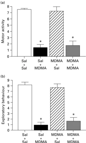
p<0.05, Figure 1) and a small decrease in grooming and social behaviour (H=24.473, p<0.05 and H=24.340,p<0.01, for grooming and social behaviour respectively ; Figure 1). Locomotor activity and exploratory behaviour were also unchanged in rats pretreated with MDMA (Figure 2).
Acute effects of MDMA in drug-naive rats and in rats previously exposed to MDMA
In drug-naive rats, acute MDMA treatment caused massive decreases in all measures of aggressive behaviour, namely, incidence of biting (H=12.749, p<0.01), boxing (H=19.829, p<0.001), kicking (H=18.912, p>0.05), and both the sum (H=21.602, p<0.001) and latency of the three measures combined (H=15.832, p<0.001). The duration of wrestling (H=21.237, p<0.01) was also reduced and subs- equently, its latency was increased (H=20.350, p<0.01 ; Figure 1). Furthermore, a marked reduction
was also observed in other social behaviours (H=24.340, p<0.001) and grooming (H=24.473, p<0.001, Figure 1) When young intruder animals were placed into the residents’ home cages, both loco- motor activity and exploratory behaviour of saline- pretreated resident rats was markedly increased. This was not exclusively restricted to exploration of the intruder animal, but also the home cage itself was intensively explored. Increases in locomotor activity (H=20.541, p>0.05) and exploratory behaviour (H=20.957, p>0.05, Figure 2) were attenuated in saline-pretreated animals after acute exposure to MDMA. In the MDMA-pretreated rats, all acute effects of MDMA were preserved (Figures 1 and 2). The pre- treatmentrtreatment interaction was significant only for the duration of social behaviours (F1,24=6.394, p=0.0008) and grooming (F1,24=14.824,p=0.018).
Acute effects of CGS-12066 and CP-94,253 in drug-naive rats and in rats previously exposed to MDMA
Acute treatment with CGS-12066 reduced the inci- dence of biting (H=8.029, p<0.01) and boxing (H=12.372, p<0.01), the sum of biting, boxing and kicking (H=15.792, p<0.01), and the duration of wrestling (H=8.298, p<0.01) in drug-naive animals (Figure 3). Latencies of biting, boxing and kicking (H=12.022, p<0.01) were increased by the drug.
Interestingly, the effects of the drug on biting, boxing and kicking were more robust than the effects upon wrestling. The compound also significantly decreased grooming (H=11.617,p<0.01) and the sum of all non- aggressive social behaviour (H=6.107,p<0.05 ; Figure 3). In contrast to the effects of acute MDMA, CGS- 12066 significantly increased the motor activation elicited by the presence of the intruder (H=14.938, p<0.01 ; Figure 4) and exploratory behaviour was also increased (H=22.471, p>0.05). The locomotor scores presented here reflect the time spent in locomotor activity. It was observed, however, that all motor activities, including exploration, social behaviour and different manifestations of aggressive behaviour were all performed at a noticeably increased speed in the presence of CGS-12066.
In rats previously exposed to MDMA, the acute effects of CGS-12066 were preserved with respect to most parameters (biting, boxing, duration of wrestling and grooming) although some of these, such as the effects on kicking, the sum of biting, boxing and kick- ing, as well as their latencies were more weakly expressed (Figure 3). In contrast, CGS-12066 treat- ment enhanced locomotor activity (H=14.938,p<0.05) 8
(a)
6 7
5
Motor activity
3
* *
4
2 1 0
Sal + Sal
Sal + MDMA
MDMA + Sal
MDMA + MDMA
*
* 8
9 (b)
6 7 5
Exploratory behaviour
3 4 2 1 0
Sal + Sal
Sal + MDMA
MDMA + Sal
MDMA + MDMA Figure 2.Acute effects of MDMA (15 mg/kg) on motor activity and exploratory behaviour in drug-naive rats and in rats exposed to MDMA 3 wk earlier. Each group consisted of seven animals. * Significantly different from the appropriate control with the same pretreatment, (p<0.05). Sal, Saline.
*
(a)
Incidence of biting
1.5
1.0
0.0 0.5
Sal + Veh
Sal + CGS
MDMA + Veh
MDMA + CGS
* *
(b)
Incidence of boxing
3 4
2 1 0
Sal + Veh
Sal + CGS
MDMA + Veh
MDMA + CGS
#
# (c)
2
Incidence of kicking
1
0 Sal
+ Veh
Sal + CGS
MDMA + Veh
MDMA + CGS
#
*
*
(d)
6 7
5
Total incidence of biting, boxing, kicking 3 4
2 1 0
Sal + Veh
Sal + CGS
MDMA + Veh
MDMA + CGS
#
*
800 900 (e)
600 700 500 Latency of biting, boxing, kicking (s)
300 400 200 100 0
Sal + Veh
Sal + CGS
MDMA + Veh
MDMA + CGS
*
(f) 60 50
Duration of wrestling (s)
30 40
20 10 0
Sal + Veh
Sal + CGS
MDMA + Veh
MDMA + CGS (g)
Latency of wrestling (s)
60 70
50
30 40
20 10 0
Sal + Veh
Sal + CGS
MDMA + Veh
MDMA + CGS
*
(h)
Duration of social behaviour (s) 200
100
0 Sal
+ Veh
Sal + CGS
MDMA + Veh
MDMA + CGS
*
(i)
Duration of grooming (s)
90 80 70
20 30 10 50 40 60
0 Sal
+ Veh
Sal + CGS
MDMA + Veh
MDMA + CGS
Figure 3.Acute effects of CGS-12066 (5 mg/kg) upon incidence of biting (a), boxing (b), kicking (c), total incidence (d) and latency (e) of biting, boxing, kicking, duration (f) and latency (g) of wrestling, duration of social behaviour (h) and grooming (i) in drug-naive rats and in rats exposed to MDMA 3 wk earlier. Each group consisted of 6–10 animals [Sal+Sal (n=8), Sal+CGS (n=10), MDMA+Sal (n=6), MDMA+CGS (n=10)]. * Significantly different from the appropriate control with the same pretreatment (p<0.05).
# Significantly different from the saline-pretreated group with the same acute treatment (p<0.05). Sal, Saline ; Veh, vehicle.
MDMAandaggression69
and exploratory behaviour (H=22.471, p<0.001) in MDMA-pretreated animals (Figure 4). Significant pretreatmentrtreatment interaction was found in exploratory behaviour (F1,32=4.947,p=0.033).
The effects of CP-94,253 were, in general, compar- able to those of CGS-12066, with similar interactions with MDMA pretreatment (data not shown).
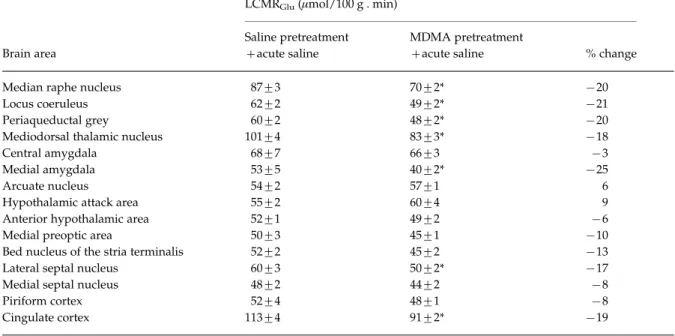
Acute and long-term effects of MDMA on LCMRGlu The effects of acute MDMA on LCMRGluin those brain areas thought to be involved in aggression were meas- ured in both drug-naive rats and in rats treated with MDMA 3 wk earlier. In rats pretreated with MDMA, and treated acutely with saline, there was a general trend towards decreased LCMRGlu(Table 1), ranging
from x3 % in central amygdala (not significant) to x25 % in medial amygdala (p<0.05). Other significant decreases were observed in cingulate cortex, lateral septal nucleus, mediodorsal thalamic nucleus, peri- aqueductal grey matter, locus coeruleus and median raphe nucleus. In two areas of the brain, the hypo- thalamic attack area and the arcuate nucleus, LCMRGlu was found to be increased in MDMA- pretreated rats, but these effects were small (9 % and 6 % respectively) and not statistically significant.
In keeping with previous observations, acute treat- ment with MDMA resulted in widespread increases in LCMRGlu in drug-naive rats (Figure 5). The only exceptions were found in the hypothalamic attack area (x9 %), anterior hypothalamic area (x4 %) and median raphe (x5 %) but the decreases in LCMRGluin these areas were relatively small and not significant.
However a more substantial decrease in the cingulate cortex (x20 %) was significantly different from that in rats treated acutely with saline.
The increases in LCMRGluelicited by acute MDMA in rats previously exposed to the drug were more widespread, and included areas in which the drug had been shown to decrease LCMRGluin previously drug- naive rats (most notably median raphe, anterior hypothalamus and cingulate cortex). Only in the hypothalamic attack area were decreases in LCMRGlu
(of similar magnitude) found in response to acute MDMA, irrespective of pretreatment. Elsewhere in the brain, there was evidence that the response to acute MDMA was potentiated by MDMA-pretreatment (Figure 5). This potentiation was most marked in the lateral septal nucleus (+30 % vs. +70 %) and locus coeruleus (+15 % vs.+55 %).
Long-term effects of MDMA on[3H]paroxetine binding
[3H]paroxetine binding was measured in brain tissue from animals exposed to saline or MDMA 3 wk pre- viously (Table 2). No significant effects of MDMA were observed in the median raphe nucleus (2 %), although significant reductions in binding were observed in the piriform cortex (x38 %), cingulate cortex (x59 %), septal nucleus (x20 %), amygdala (x31 %).
Discussion and conclusion
Acute MDMA treatment decreased aggressive-type behaviour as measured with the resident-intruder test, and the anti-aggressive effects of 5-HT1Bagonists were similar to those of MDMA. Acute MDMA treatment 12.5
(a)
* *
10.0 7.5
Motor activity
5.0 2.5 0.0
Sal + Sal
Sal + CGS
MDMA + Sal
MDMA + CGS
*
# 12.5
(b)
10.0 7.5
Exploratory behaviour
5.0 2.5 0.0
Sal + Sal
Sal + CGS
MDMA + Sal
MDMA + CGS Figure 4.Acute effects of CGS-12066 (5 mg/kg) on motor activity and exploratory behaviour in drug-naive rats and in rats exposed to MDMA 3 wk earlier. Each group consisted of 6–10 animals [Sal+Sal (n=8), Sal+CGS (n=10),
MDMA+Sal (n=6), MDMA+CGS (n=10)]. * Significantly different from the appropriate control with the same pretreatment (p<0.05). # Significantly different from the saline-pretreated group with the same acute treatment, (p<0.05). Sal, Saline.
increased brain metabolism in several areas including those involved in the regulation of aggression.
Significant long-term decreases in paroxetine binding
and brain metabolism were found in many brain areas, 3 wk after MDMA administration, although at this time-point aggressive-type behaviour was unaltered. Contrary to expectation, increases in brain metabolism induced by acute MDMA were not diminished in rats previously exposed to the drug.
The anti-aggressive effects of 5-HT1B-agonists in MDMA-pretreated rats were, in general, preserved and only modest changes were observed.
Table 1.Local cerebral metabolic rate of glucose utilization (LCMRGlu) in saline and MDMA (15 mg/kg i.p.) pretreated animals
Brain area
LCMRGlu(mmol/100 g . min)
Saline pretreatment +acute saline
MDMA pretreatment
+acute saline % change
Median raphe nucleus 87¡3 70¡2* x20
Locus coeruleus 62¡2 49¡2* x21
Periaqueductal grey 60¡2 48¡2* x20
Mediodorsal thalamic nucleus 101¡4 83¡3* x18
Central amygdala 68¡7 66¡3 x3
Medial amygdala 53¡5 40¡2* x25
Arcuate nucleus 54¡2 57¡1 6
Hypothalamic attack area 55¡2 60¡4 9
Anterior hypothalamic area 52¡1 49¡2 x6
Medial preoptic area 50¡3 45¡1 x10
Bed nucleus of the stria terminalis 52¡2 45¡2 x13
Lateral septal nucleus 60¡3 50¡2* x17
Medial septal nucleus 48¡2 44¡2 x8
Piriform cortex 52¡4 48¡1 x8
Cingulate cortex 113¡4 91¡2* x19
Data presented as mean¡S.E.M. Percentages are differences from saline control.
* Significantly different from saline control group (p<0.05). Each group consisted of seven animals.
Median raphe nucleus Locus coeruleus Periaqueductal grey Mediodorsal thalamic nucleus Central amygdala Medial amygdala Arcuate nucleus Hypothalamic attack area Anterior hypothalamic area Medial preoptic area Bed nucleus Lateral septal nucleus Medial septal nucleus Piriform cortex Cingulate cortex
Percentage change from control
–40 0 40 80
∗
∗
∗
∗
∗
∗
∗
∗
∗
∗
∗
Figure 5.Acute effects of MDMA upon LCMRGluin drug- naive animals and in animals previously exposed to MDMA.
Data are presented as mean percentage change from control¡S.E.M. * Significantly different from saline pretreated animals (p<0.05, Mann–WhitneyUtest). Each group consisted of seven animals.&, MDMA pretreatment+acute MDMA ;%, saline pretreatment+acute MDMA.
Table 2.[3H]Paroxetine binding in animals exposed to either saline or MDMA (15 mg/kg) 3 wk previously
Brain area Saline MDMA Lesion %
Neocortex
Piriform cortex 64¡5 40¡7* x38 Cingulate cortex 66¡7 27¡4* x59
Septal nucleus 64¡4 51¡3* x20
Amygdala 210¡15 144¡10* x31
Median raphe nucleus 170¡16 173¡21 2
Data are presented as mean binding density (fmol/mg tissue)¡S.E.M.
Each group consisted of seven animals.
* Significantly different from saline control group (p<0.05).
MDMA and aggression 71
Acute effects of MDMA on aggression and LCMRGlu The data from the present study support previous observations that acute MDMA treatment decreases aggressive-type behaviour in rats, as measured in the resident-intruder test (Miczek et al., 1994 ; Navarro et al., 1999 ; Verheyden et al., 2002), as well as in human abusers (Curran et al., 2004). However, in the present study there were some differences in the effect of MDMA upon the separate parameters of aggression, for example in the frequency of biting, boxing and kicking. Closer examination of the data reveals that this is due, in the main, to differences in the manifestation of attack between individuals, with some animals preferring to kick, while others showed a greater tendency to box or bite.
Interestingly, changes were also found in other parameters not closely related to aggression. Intro- ducing an intruder to the home cage usually results in marked increases in locomotor activity and explora- tory behaviour from the resident animal, but this hyperlocomotion was significantly attenuated by acute MDMA treatment (Figure 2). Similarly, signifi- cant decreases were found in social behaviour (Figure 1). Thus, it appears that if the resident animal is treated with MDMA in advance of the introduction of the intruder, it is less affected by the presence of the intruder animal. In parallel studies on animals outwith the behavioural paradigm, i.e. where no stimulus or target for aggressive behaviour is available, MDMA- induced increases in local cerebral metabolic rate were found in several brain regions that regulate (either activate or inhibit) aggression, and also in other regions that regulate many other physiological func- tions. This is perhaps not surprising given the wide- spread anatomical distribution of serotonergic projections, and the possible involvement of both dopaminergic and noradrenergic systems in the pharmacological response to MDMA.
The long-term effects of MDMA on aggression, LCMRGluand paroxetine binding
In our study, MDMA was administered to Dark Agouti rats at a dose of 15 mg/kg. Although there are considerable strain differences in the susceptibility to MDMA-induced long-term serotonergic depletion, many authors suggests that a single, 15 mg/kg injec- tion of MDMA in rats may more accurately model the possible exposure to the drug in humans, particularly when pharmacokinetic scaling is applied to body size and basal metabolic rate (Colado and Green, 1995 ; Malpass et al., 1999). In contrast, it is less likely that previously used models in which, typically, rats are
treated with a higher dose (20 mg/kg) twice a day for four consecutive days adequately reflect human experience. Moreover, Dark Agouti rats show de- creased microsomal CYP2D6 isoenzyme activity, which plays an important role in the metabolism of MDMA. As a consequence, the Dark Agouti rat is considered to be a poor or moderate metabolizer of the drug with MDMA pharmacokinetics that are similar to those which may be found in particularly vulner- able human users (Balogh et al., 2004 ; O’Shea et al., 1998 ; Quate et al., 2004). Such users might represent a genetically defined human sub-population in which clinical complications are more likely to occur.
In this study, significant decreases in paroxetine binding of between 30 % and 60 % were found in the neocortex, septal nucleus and amygdala, but not in the median raphe nucleus, 3 wk after a single exposure to MDMA. This finding is totally in agreement with previous data, which showed that a similar MDMA treatment caused long-term serotonergic depletion in the forebrain of Dark Agouti rats (Colado et al., 1998 ; O’Shea et al., 1998). Of particular interest in the context of our behavioural data, is the observation of decreased binding in some areas of the brain known to be involved in the regulation of aggression and impulsivity. However, despite the deleterious effect of MDMA on the serotonergic system, and previous evidence that depletion of 5-HT in the amygdala facilitates aggressive behaviour (Morgan, 2000), the present study indicates that manifestations of aggressive behaviours was not abnormal in rats pretreated with MDMA 3 wk earlier. For example the animals did not exhibit attacks to vulnerable areas on the target intruder, which is considered a pathological form of aggression (Halasz et al., 2002a,b). There was, however, evidence from our metabolic mapping studies that, in the resting state at least, MDMA pretreatment did result in altered functional activity in some elements of the amygdala, indicating that the treatment was not without effect.
The lack of evidence for increased aggression in this test may be explained in many ways. First, the level of aggression exhibited by rats in this test is normally relatively high. It could be difficult to detect further small increases in an already high aggressive state, which is described as an essential factor in this test paradigm (Navarro and Maldonado, 1999). Further- more, chronic social isolation, which is necessary to confer resident status, has been reported to decrease 5-HT levels in forebrain areas in the long-term (Dalley et al., 2002 ; Fulford and Marsden, 1998 ; Robbins et al., 1996), and indeed this could be one explanation for the increased baseline aggression in the resident-intruder
test. A further decrease in 5-HT content as a result of MDMA pretreatment may not elicit any further increase in aggression. Moreover, in keeping with these behavioural findings, LCMRGlu was signifi- cantly decreased 3 wk after MDMA administration, albeit in the absence of aggression-inducing stimuli.
The decrease in metabolism was found in several brain areas, including those thought to mediate aggressive behaviour, and also in those that inhibit aggression.
It is conceivable that the integration of these inhibitory and disinhibitory effects could confound our measures of aggression, or alternatively represent a compensatory mechanism to normalize function.
Human studies that have sought to identify MDMA-induced abnormalities are difficult to inter- pret due to inherent methodological complications with retrospective studies. These complications in- clude inaccurate reporting of the doses MDMA taken, the duration and frequency of drug use, and non- reporting of the concurrent use of other substances, licit or illicit (Green et al., 1995 ; Morgan, 2000).
However, some studies have demonstrated an in- crease in aggression and impulsivity in heavy ecstasy users (Bond et al., 2004 ; Gerra et al., 1998, 2000), which may suggest that in the long term MDMA might in- crease aggression in humans. However, no significant increase was found in light/moderate users (Morgan, 1998 ; Parrott, 2000) and earlier studies in humans have found that, although aggression increases 4 d after exposure to MDMA, there is no significant increase in aggressive behaviour 7 d after the exposure (Curran et al., 2004). Only McCann et al. (1994) reported that heavy ecstasy users had lower aggression than control participants.
Effects of a previous MDMA treatment on MDMA-induced increases in LCMRGluand on the behavioural effects of acute MDMA, CGS-12066 and CP-94,253 administration
Our results indicate that all of the acute behavioural effects of MDMA were preserved when a neurotoxic MDMA pretreatment was applied 3 wk before the test.
The MDMA-induced depletion of serotonergic ter- minals, for which our [3H]paroxetine-binding data provide positive evidence in this study, could be assumed to reduce the availability of releasable 5-HT, and might be expected to result in an attenuated anti-aggressive response to acute MDMA treatment.
However, these anti-aggressive effects or any possible changes caused by 5-HT depletion, may possibly be masked by the general inhibitory behaviour which is unaffected by neurotoxic MDMA pretreatment.
Acute MDMA caused a significantly greater increase in brain metabolism in pretreated rats compared to saline-treated animals. This could be explained in part by the lower baseline activity in the brain areas examined (as stated previously), so that metabolic rates after acute MDMA administration were largely similar in drug-naive rats and rats pre- viously exposed to the drug. However, this cannot explain the divergent response found, for example in the cingulate cortex, where an MDMA-induced de- crease in drug-naive rats is replaced by a significant MDMA-induced increase in rats previously exposed to the drug.
The acute anti-aggressive effects of 5-HT1Bagonists in drug-naive rats were more selective than those of MDMA, namely, changes in impulsive and aggressive behaviours such as biting, boxing, kicking were very marked. Furthermore, the effects of 5-HT1Bagonists on motor activation and exploratory behaviour induced by the presence of the intruder animal were quite dif- ferent from those of MDMA, such that 5-HT1Bagonists caused increases in this motor activation, while MDMA caused a reduction. 5-HT1B agonists evoked generally similar effects in MDMA-pretreated rats, compared to drug-naive rats. These data do not, therefore, indicate a significant sensitization of 5-HT1B
receptor function as a consequence of a long-term serotonergic depletion. A possible explanation is that despite the partial axonal loss, some, or all, of the serotonergic axons and terminals involved in anti- aggressive behaviour may remain intact. Furthermore, 5-HT1B receptors involved in this action may be located post-synaptically (Barnes and Sharp, 1999 ; Clark and Neumaier, 2001 ; Scearce-Levie et al., 1999b) or are heteroreceptors, located not only on sero- tonergic terminals, but also on terminals where they might inhibit the release of other neurotransmitters including GABA, acetylcholine or glutamate (Clark and Neumaier, 2001 ; Sexton et al., 1999). Alternatively, there is the possibility that non-serotonergic mechan- isms are also involved in the acute effects of MDMA upon aggression, including dopamine, glutamate or GABA (Sexton et al., 1999). It is also possible that anx- iety, reactivity and impulsivity confound the overt behavioural expression of MDMA-induced effects, as has previously been described after partial 5-HT depletion (Green et al., 2003 ; Harro, 2002). There was, however, a difference in drug-induced motor activation. The increase in locomotor activity and ex- ploration was significantly enhanced in MDMA pre- treated rats, suggesting that the involvement of 5-HT1B
receptors in locomotor activation differs from that in the regulation of aggression and impulsivity.
MDMA and aggression 73
In conclusion, acute MDMA administration abol- ished aggressive behaviour in the resident-intruder test as expected. Unexpectedly, MDMA pretreatment and the consequent depletion of serotonergic ter- minals did not increase aggression. This may be ex- plained by the confounding effects of social isolation, which is known to induce decrease in brain 5-HT concentration. Thus, the lack of increase in aggression may be explained by the neurochemical effects of social isolation that might be similar to the long-term effects exerted by MDMA. This suggestion is also supported by the fact that a previous exposure to MDMA failed to affect 5-HT1B receptor function in these animals.
Acknowledgements
This study was supported by the Fifth Framework Programme of the European Community, QLG3-CT- 2002-00809, by the Sixth Framework Programme, LSHM-CT-2004-503474, the Hungarian Research Fund Grant T020500, the Ministry of Welfare Research Grant 058/2003, the Fund Management of the Ministry of Education, OMFB 01926/2002 and Ph.D. Fellowship Program of the Semmelweis University, Ministry of Education, Hungary.
Statement of Interest None.
References
Balogh B, Molnar E, Jakus R, Quate L, Olverman HJ, Kelly PA, Kantor S, Bagdy G(2004). Effects of a single dose of 3,4-methylenedioxymethamphetamine on circadian patterns, motor activity and sleep in drug-naive rats and rats previously exposed to MDMA.Psycho- pharmacology(Berlin)173, 296–309.
Barnes NM, Sharp T(1999). A review of central 5-HT receptors and their function.Neuropharmacology 38, 1083–1152.
Battaglia G, Yeh SY, O’Hearn E, Molliver ME, Kuhar MJ, De Souza EB(1987). 3,4-Methylenedioxymethamphetamine and 3,4-methylenedioxyamphetamine destroy serotonin terminals in rat brain : quantification of neurodegeneration by measurement of [3H]paroxetine-labeled serotonin uptake sites.Journal of Pharmacology and Experimental Therapeutics 242, 911–916.
Bell R, Donaldson C, Gracey D(1995). Differential effects of CGS 12066B and CP-94,253 on murine social and agonistic behaviour.Pharmacology, Biochemistry and Behavior 52, 7–16.
Berman ME, Tracy JI, Coccaro EF(1997). The serotonin hypothesis of aggression revisited.Clinical Psychology Review 17, 651–665.
Bond AJ, Verheyden SL, Wingrove J, Curran HV(2004).
Angry cognitive bias, trait aggression and impulsivity in substance users.Psychopharmacology(Berlin)171, 331–339.
Clark MS, Neumaier JF(2001). The 5-HT1B receptor : behavioral implications.Psychopharmacology Bulletin 35, 170–185.
Colado MI, Granados R, O’Shea E, Esteban B, Green AR (1998). Role of hyperthermia in the protective action of clomethiazole against MDMA (‘ecstasy’)-induced neurodegeneration, comparison with the novel NMDA channel blocker AR-R15896AR.British Journal of Pharmacology 124, 479–484.
Colado MI, Green AR(1995). The spin trap reagent alpha- phenyl-N-tert-butyl nitrone prevents ‘ecstasy’-induced neurodegeneration of 5-hydroxytryptamine neurones.
European Journal of Pharmacology 280, 343–346.
Colado MI, Murray TK, Green AR(1993). 5-HT loss in rat brain following 3,4-methylenedioxymethamphetamine (MDMA), p-chloroamphetamine and fenfluramine administration and effects of chlormethiazole and dizocilpine.British Journal of Pharmacology 108, 583–589.
Colado MI, O’Shea E, Granados R, Esteban B, Martin AB, Green AR(1999). Studies on the role of dopamine in the degeneration of 5-HT nerve endings in the brain of Dark Agouti rats following 3,4-methylenedioxymethamphetamine (MDMA or
‘ ecstasy’) administration.British Journal of Pharmacology 126, 911–924.
Curran HV(2000). Is MDMA (‘ ecstasy’) neurotoxic in humans? An overview of evidence and of methodological problems in research.Neuropsychobiology 42, 34–44.
Curran HV, Rees H, Hoare T, Hoshi R, Bond A(2004).
Empathy and aggression : two faces of ecstasy? A study of interpretative cognitive bias and mood change in ecstasy users.Psychopharmacology(Berlin)173, 425–433.
Dalley JW, Theobald DE, Pereira EA, Li PM, Robbins TW (2002). Specific abnormalities in serotonin release in the prefrontal cortex of isolation-reared rats measured during behavioural performance of a task assessing visuospatial attention and impulsivity.Psychopharmacology(Berlin)164, 329–340.
De Souza EB, Kuyatt BL(1987). Autoradiographic localization of 3H-paroxetine-labeled serotonin uptake sites in rat brain.Synapse 1, 488–496.
Fish EW, Faccidomo S, Miczek KA(1999). Aggression heightened by alcohol or social instigation in mice : reduction by the 5-HT(1B) receptor agonist CP-94,253.
Psychopharmacology(Berlin)146, 391–399.
Fitzgerald JL, Reid JJ(1990). Effects of methylene- dioxymethamphetamine on the release of monoamines from rat brain slices.European Journal of Pharmacology 191, 217–220.
Fulford AJ, Marsden CA(1998). Conditioned release of 5-hydroxytryptamine in vivo in the nucleus accumbens following isolation-rearing in the rat.Neuroscience 83, 481–487.
Gerra G, Zaimovic A, Ferri M, Zambelli U,
Timpano M, Neri E, Marzocchi GF, Delsignore R,
Brambilla F(2000). Long-lasting effects of
(¡)3,4-methylenedioxymethamphetamine (ecstasy) on serotonin system function in humans.Biological Psychiatry 47, 127–136.
Gerra G, Zaimovic A, Giucastro G, Maestri D, Monica C, Sartori R, Caccavari R, Delsignore R(1998). Serotonergic function after (¡)3,4-methylene-dioxymethamphetamine (‘Ecstasy’) in humans.International Clinical
Psychopharmacology 13, 1–9.
Green AR, Cross AJ, Goodwin GM(1995). Review of the pharmacology and clinical pharmacology of 3,4-methylenedioxymethamphetamine (MDMA or
‘ Ecstasy’).Psychopharmacology(Berlin)119, 247–260.
Green AR, Mechan AO, Elliott JM, O’Shea E, Colado MI (2003). The pharmacology and clinical pharmacology of 3,4-methylenedioxymethamphetamine (MDMA,
‘ ecstasy’).Pharmacological Reviews 55, 463–508.
Halasz J, Liposits Z, Kruk MR, Haller J(2002a). Neural background of glucocorticoid dysfunction-induced abnormal aggression in rats : involvement of fear- and stress-related structures.European Journal of Neuroscience 15, 561–569.
Halasz J, Liposits Z, Meelis W, Kruk MR, Haller J(2002b).
Hypothalamic attack area-mediated activation of the forebrain in aggression.Neuroreport 13, 1267–70.
Harro J(2002). Long-term partial 5-HT depletion : interference of anxiety and impulsivity?Psycho- pharmacology(Berlin)164, 433–434.
Holmes A, Murphy DL, Crawley JN(2002). Reduced aggression in mice lacking the serotonin transporter.
Psychopharmacology(Berlin)161, 160–167.
Kantor S, Anheuer ZE, Bagdy G(2000). High social anxiety and low aggression in Fawn-Hooded rats.Physiology &
Behavior 71, 551–557.
Kelly PA, Ritchie IM, Collins FM(1995). Cerebrovascular consequences of repeated exposure to NG-nitro-L-arginine methyl ester.British Journal of Pharmacology 116, 2771–2777.
Malpass A, White JM, Irvine RJ, Somogyi AA, Bochner F(1999). Acute toxicity of
3,4-methylenedioxymethamphetamine (MDMA) in Sprague–Dawley and Dark Agouti rats.Pharmacology, Biochemistry and Behaviour 64, 29–34.
McCann UD, Ricaurte GA(1991). Lasting neuropsychiatric sequelae of (¡)methylenedioxymethamphetamine (‘ecstasy’) in recreational users.Journal of Clinical Psychopharmacol 11, 302–305.
McCann UD, Ridenour A, Shaham Y, Ricaurte GA (1994). Serotonin neurotoxicity after (¡)3,4-methylene- dioxymethamphetamine (MDMA ; ‘Ecstasy’) : a controlled study in humans.Neuropsychopharmacology 10, 129–38.
Mechan AO, Esteban B, O’Shea E, Elliott JM, Colado MI, Green AR(2002). The pharmacology of the acute hyperthermic response that follows administration of 3,4-methylenedioxymethamphetamine (MDMA,
’ecstasy’) to rats.British Journal of Pharmacology 135, 170–180.
Miczek KA, Fish EW, De Bold JF, De Almeida RM (2002). Social and neural determinants of aggressive
behavior : pharmacotherapeutic targets at serotonin, dopamine and gamma-aminobutyric acid systems.
Psychopharmacology(Berlin)163, 434–458.
Miczek KA, Haney M(1994). Psychomotor stimulant effects of d-amphetamine, MDMA and PCP : aggressive and schedule-controlled behavior in mice.Psychopharmacology (Berlin)115, 358–365.
Morgan MJ(1998). Recreational use of ‘ecstasy’
(MDMA) is associated with elevated impulsivity.
Neuropsychopharmacology 19, 252–264.
Morgan MJ(2000). Ecstasy (MDMA) : a review of its possible persistent psychological effects.Psychopharmacology(Berlin) 152, 230–248.
Muehlenkamp F, Lucion A, Vogel WH(1995). Effects of selective serotonergic agonists on aggressive behavior in rats.Pharmacology, Biochemistry and Behavior 50, 671–674.
Navarro JF, Maldonado E(1999). Behavioral profile of 3,4-methylenedioxy-methamphetamine (MDMA) in agonistic encounters between male mice.Progress Neuropsychopharmacology & Biological Psychiatry 23, 327–334.
Nelson RJ, Chiavegatto S(2001). Molecular basis of aggression.Trends in Neurosciences 24, 713–719.
O’Shea E, Granados R, Esteban B, Colado MI, Green AR (1998). The relationship between the degree of
neurodegeneration of rat brain 5-HT nerve terminals and the dose and frequency of administration of MDMA (‘ecstasy’).Neuropharmacology 37, 919–926.
Parrott AC(2000). Human research on MDMA
(3,4-methylene-dioxymethamphetamine) neurotoxicity : cognitive and behavioural indices of change.
Neuropsychobiology 42, 17–24.
Parrott AC(2001). Human psychopharmacology of Ecstasy (MDMA) : a review of 15 years of empirical research.
Human Psychopharmacology 16, 557–577.
Parrott AC, Sisk E, Turner JJ(2000). Psychobiological problems in heavy ‘ecstasy’ (MDMA) polydrug users.
Drug and Alcohol Dependence 60, 105–110.
Quate L, McBean DE, Ritchie IM, Olverman HJ, Kelly PA (2004). Acute methylenedioxymethamphetamine administration : effects on local cerebral blood flow and glucose utilisation in the Dark Agouti rat.Psycho- pharmacology(Berlin)173, 287–295.
Reneman L, Lavalaye J, Schmand B, de Wolff FA, van den Brink W, den Heeten GJ, Booij J(2001). Cortical serotonin transporter density and verbal memory in individuals who stopped using 3,4-methylenedioxy- methamphetamine (MDMA or ‘ ecstasy’) –
preliminary fundings.Archives of General Psychiatry 58, 901–906.
Ricaurte GA, Yuan J, McCann UD(2000).
(¡)3,4-Methylenedioxymethamphetamine (‘ Ecstasy’)- induced serotonin neurotoxicity : studies in animals.
Neuropsychobiology 42, 5–10.
Robbins TW, Jones GH, Wilkinson LS(1996). Behavioural and neurochemical effects of early social deprivation in the rat.Journal of Psychopharmacology 10, 39–47.
MDMA and aggression 75