COMPREHENSIVE EVALUATION OF NOVEL TREATMENT POSSIBILITIES FOR PERIODONTAL
HARD- AND SOFT TISSUE RECONSTRUCTION PhD Thesis
Bálint Molnár
School of Clinical Medicine Semmelweis University
Supervisors:
Péter Windisch, DMD, PhD Official reviewers:
Attila Szűcs DMD, PhD Vilmos Tóth DMD, PhD Final Examination Board:
Head: Tamás Divinyi DMD, PhD Members: György Simon, PhD
Márta Radnai, DMD, PhD
2013
1 1. TABLE OF CONTENTS
1. TABLE OF CONTENTS ... 1
2. LIST OF ABBREVIATIONS ... 3
3. PREAMBLE ... 6
4. INTRODUCTION ... 8
5. OBJECTIVES ... 14
6. METHODS ... 15
6.1 Literature review on the application of enamel matrix proteins in periodontal regenerative therapy ... 17
6.2 In vitro isolation and differentiation of periodontal ligament stem cells ... 17
6.2.1. Cell isolation and culturing of periodontal ligament stem cells ... 17
6.2.3. Cell viability studies and treatment with enamel matrix derivative ... 18
6.2.4. Immunocytochemistry ... 19
6.2.5. FACS analysis ... 19
6.2.6. Osteogenic induction ... 20
6.2.7. Neuronal induction ... 20
6.2.7.1 Protocol 1. ... 20
6.2.7.2. Protocol 2. ... 21
6.2.7.3. Protocol 3. ... 21
6.2.8. Real-time PCR ... 22
6.2.9. Statistical analysis ... 22
6.3 Clinical studies ... 23
6.3.1 Hard tissue regeneration following treatment with rhGDF-5/β-TCP ... 23
6.3.1.1 Subject selection, preoperative protocol ... 25
6.3.1.2 Study material ... 25
6.3.1.3 Surgical procedures ... 26
6.3.1.4 Postoperative care ... 26
6.3.1.5 Clinical assessment ... 27
6.3.1.6 Safety assessment ... 28
6.3.1.7 Statistical analysis ... 28
6.3.2 Soft tissue regeneration following treatment with Mucograft® ... 29
6.3.2.1 Subject selection, preoperative protocol ... 29
6.3.2.2 Study material ... 30
6.3.2.3 Surgical procedures ... 32
6.3.2.4 Postoperative care ... 33
6.3.2.5 Clinical assessments ... 35
6.3.2.6 Evaluation of patients’ satisfaction ... 35
6.3.2.7 Statistical analysis ... 36
7. RESULTS ... 37
7.1 Literature review on the application of enamel matrix proteins in periodontal regenerative therapy ... 37
7.2 Isolation and in vitro differentiation of periodontal ligament stem cells ... 38
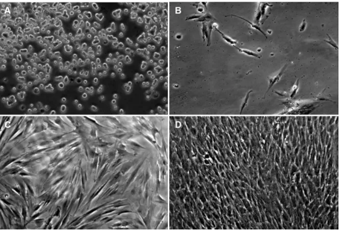
7.2.1 Isolation and primary cultures ... 38
7.2.2 Cell viability studies and treatment with enamel matrix derivative ... 39
7.2.3 Immunocytochemistry and FACS analysis ... 41
7.2.4 Osteogenic differentiation ... 42
2
7.2.5 Neuronal differentiation ... 43
7.2.5.1 Partial differentiation induced by Protocols 1 and 2 ... 43
7.2.5.2 Robust neuronal differentiation induced by Protocol 3 ... 44
7.3 Healing following rhGDF-5/β-TCP treatment ... 47
7.3.1 Patient demographics and baseline defect distribution ... 47
7.3.2 Clinical assessments ... 47
7.3.3 Safety findings ... 49
7.3.4 Adverse events ... 50
7.3.5 Protocol deviations ... 50
7.4 Healing following treatment with Mucograft® ... 51
7.4.1 Patient demographics and baseline defect distribution ... 51
7.4.1.1 Pilot study ... 51
7.4.1.2 Split mouth randomised controlled study ... 51
7.4.2 Clinical assessments ... 51
7.4.2.1 Pilot study ... 51
7.4.2.2 Split mouth randomised controlled study ... 52
8. DISCUSSION ... 56
9. CONCLUSIONS ... 64
10. SUMMARY ... 66
11. ÖSSZEFOGLALÁS ... 67
12. BIBLIOGRAPHY ... 68
13. BIBILIOGRAPHY OF THE CANDIDATE’S PUBLICATIONS ... 81
13.1 Related publications ... 81
13.2 Not related publications ... 82
14. ACKNOWLEDGEMENTS ... 83
3 2. LIST OF ABBREVIATIONS
ALT – alanine transaminase ADM – acellular dermal matrix AST – aspartate transaminase
bFGF – basic fibroblast growth factor BHA - butylated hydroxyanisole BID – twice a day
BMSC – bone marrow stem cells BSA – bovine serum albumine β-TCP – beta tricalcium phosphate CAF – coronally advanced flap CAL – clinical attachment level
cAMP – cyclic adenosine monophosphate CD-34 - human cluster of differentiation 34 CEJ - cementoenamel junction
c-KIT – cellular transmembrane receptor tyrosine kinase CM – collagen matrix
CRC- complete root coverage
DMEM - Dulbecco's modified Eagle's medium DMSO – dimethyl sulfoxide
DNA - deoxyribonucleic acid DPSCs – dental pulp stem cells
EDTA – ethylenediaminetetraacetic acid ELISA - enzyme-linked immuno sorbent assay EMD – enamel matrix derivative
ETT – Egészségügyi Tudományos Tanács FACS – fluorescence activated cell sorting FAM – carboxyfluorescein
FBS – fetal bovine serum
FMBS – full mouth bleeding score FMPS – full mouth plaque score
4 FSH – folliculus stimulating hormone GFAP - glial fibrillary acidic protein GGT – gamma glutamil transferase GR – gingival recession
GRD – gingival recession depth GRW – gingival recession width GT – gingival thickness
GTR – guided tissue regeneration Hb1AC - glycated hemoglobin
HIV – human immunodeficiency virus HRP - horseradish peroxidase
IBMX - 3-isobutyl-1-methylxanthine IgG – immunoglobuline G
ITS - insulin-transferrin-sodium KCl – calcium-chloride
KT – keratinised tissue
KTW – keratinised tissue width
MAGR – multiple adjacent gingival recessions MCAF – modified coronally advanced flap MCAT – modified coronally advance tunnel
MedDRA - medical dictionary of regulatory activities MEM – Eagle’s Minimum Essential Medium
MGB - tripeptide minor groove binder MGJ – mucogingival junction
MRC – mean root coverage MTT - microculture tetrazolium NGF – neural growth factor NSE – neurospecific enolase NT-3 – neurotubulin 3
OFD – open flap debridement PD – probing depth
PDLSCs – periodontal ligament stem cells
5 PFA - paraformaldehyde
PKC – protein kinase C
PBS – phosphate buffered saline PCR – polymerase chain reaction RCT – randomised controlled trial
rhGDF – recombinant human growth and differentiation factor rhPDGF – recombinant human platelet derived growth factor RPLPO - large ribosomal protein
RNA – ribonucleic acid RT – room temperature
SCTG subepithelial connective tissue graft SD – standard deviation
S.E.M. – standard error of the mean SOC – system of classes
STRO-1 - stromal cell surface marker-1 TID – three times a day
TMB – tetramethyl benzydine TPA – tissue plasminogen activator
TUKEB – Tudományos Kutatásetikai Bizottság VAS – visual analogue scale
VIM – vimentin
6 3. PREAMBLE
During my undergraduate academic years I had been fascinated by the recent progress of periodontal research on the field of tissue regeneration. After graduation receiving a residency status at the Department of Periodontology my professional interest turned to the biological background of tissue regeneration.
Having been accepted to the Dental Research Programme lead by Professor Gabor Varga of the Semmelweis University School of Clinical Medicine the initial objective of my PhD research was to investigate the cellular events contributing to periodontal tissue regeneration. While preparing for the first in vitro studies I got the opportunity to participate in a literature search and collect data for a literature review on the application of Enamel Matrix Derivatives (EMD) (Study I). That time it became clear to me that the EMD’s effect on several cell types in the periodontium had been studied but there had only been relatively limited data available on cellular mechanisms of EMD on pluripotent mesenchymal cells of periodontal ligament origin. It was therefore a challenging opportunity for me to study how EMD act on stem cells of periodontal ligament (Study II). That time stem cell culturing techniques had been elaborated at the Department of Oral Biology, Semmelweis University. According to the later published results of our team it was shown that isolation and culturing of periodontal ligament and pulp derived cell colonies was a suitable approach to study the regenerative and differentiating potential of multipotent adult stem cells. This study lead us to show that stem cells of periodontal origin have the capacity to differentiate not only into different cell lineages of the attachment apparatus but also into cells with neuronal characteristics (Study III).
Several signalling factors might play a crucial role in this differentiation process.
The first phase of clinical research was to investigate those modulating/differentiation factors that, like EMD may guide cell differentiation during wound healing and regeneration. At the Department of Periodontology, led by Professor Istvan Gera, an opportunity was given when Professor Peter Windisch invited me to a clinical and histological study by Prof. Ulf Wikesjö and Prof. Anton Sculean on the safety and efficacy of a recently discovered growth and differentiation factor (rhGDF-5). I was involved as study coordinator in the first human clinical and histological data on the regenerative capacity of rhGDF-5 (Study IV)
7
Periodontal destruction not only affects the attachment apparatus but might also result in soft tissue defects. Reconstruction of these anomalies is often required to resolve severe aesthetic problems as well as for long-term hard- and soft tissue stability. As a result of this aesthetic mucogingival surgery became again a main focus of clinical research during the turn of this century.
The next phase of my research project was to investigate how connective tissue grafting could be substituted with biomaterials. Since the first studies on Guided Tissue Regeneration (GTR) several attempts had been made to utilise GTR techniques and biological membranes for soft tissue augmentation and root coverage, but without any convincing success. Development of a novel biocompatible xenograft matrix provided a good opportunity to conduct clinical studies in the field of periodontal plastic surgery.
Our research group was led by Professor Anton Sculean and Sofia Aroca, we wanted to investigate if the application of the new material to cover denuded root surfaces and improve the biotype of the patients might be comparable to the success rate by the autogenous connective tissue graft. This resulted in two publications (Studies V and VI) in peer reviewed journals, our group was the first to conduct a randomised split mouth clinical trial to evaluate the use of the novel collagen matrix for correction of multiple recessions.
It has always been a great honour for me that I had had the chance to participate in several research projects and to be member of clinical multi-centre studies. Thus I could participate in providing new and relevant data either on adult stem cell research or on clinical studies on the safety and efficacy of certain new and promising growth and differentiation factors as well as some novel biomaterials. In my thesis I would like to give an overview of the relevant literature data on tooth derived stem cell research, novel approaches of periodontal regeneration and also periodontal plastic surgery. I am going to chronologically present the evolution of my doctorate work based on the six phases of my research projects, starting with the summary of the systemic review of the literature on the application of EMD followed by adult stem cell studies. In the clinical part of my studies I am going to summarize and discuss the clinical and histological results from a randomised controlled study on the application of rhGDF-5 and finally finishing with the most recent studies on the clinical applicability of collagen xenograft matrices in periodontal plastic surgery.
8 4. INTRODUCTION
/Review of the related literature/
The goal of regenerative therapy for periodontal hard tissue reconstruction is the recreation of the lost periodontal structures (i.e. new formation of root cementum, periodontal ligament and alveolar bone). The approximately 30 years research on GTR and the 60 years history of research on mucogingival surgery and clinical techniques provided tremendous amount of information and knowledge. Nevertheless several questions are still unanswered and there are numerous controversies in the current opinions and trends. It is still not clear what kind of biological factors play crucial roles in wound healing and particularly in complex periodontal regeneration, where three different cell lineages should be re-established.
Many treatment modalities, such as various types of bone grafts, GTR and EMD have been used with varying success during the past to accomplish this goal (Sallum et al. 2003, Donos et al. 2003, Palioto et al. 2004). Results from basic and clinical research have pointed to the predictability, safety and efficacy of the application of EMD and GTR (World Workshop in periodontology 1996, Rincon et al. 2003, Donos et al. 2003) in the periodontal wound healing and regeneration. Nevertheless, current surgical techniques and available biomaterials for hard tissue reconstructions have some well-known limitations in case of advanced attachment loss and unfavourable defect configurations.
Beyond hard tissue reconstruction and gain of new attachment, correction of periodontal soft tissue defects has again become a main focus of clinical research during the past decade. (Hofmänner et al. 2012) Main goals of surgical approaches include prevention of advanced periodontal defects related to mucogingival anomalies as well as fulfilling the increasing aesthetic demands of patients. Periodontal plastic surgery aims at the reconstruction of soft tissue deformities as well as at the modification of unfavourable anatomic conditions, such as thin gingival biotype. Various surgical techniques have been suggested for changing the gingival biotype as well as correction of recession defects.
According to literature to achieve optimal predictability and long-term stability connective tissue grafting has been suggested as the standard adjunctive therapy for periodontal plastic surgery (Cairo et al 2008, Chambrone et al 2010, Hoffmänner et al.
9
2012). Nevertheless, increased patient morbidity and duration of surgery related to a donor surgical site and tissue harvesting are well known drawbacks of autogenous soft tissue grafting (Cairo et al. 2008).
The limitations of currently applied techniques for periodontal hard- and soft tissue reconstruction related to treatment efficacy and patient morbidity have raised a demand to introduce novel treatment approaches as well as biomaterials aiming at increased treatment efficacy as well as reducing duration of treatment and patient morbidity. During the last decade, emerging new research fields have investigated the possibilities of tissue engineering related to the isolation and differentiation of human adult tooth derived stem cells (Gronthos et al. 2000, Miura et al. 2003, Seo et al. 2004) and application of different recombinant growth-factors (Morotome et al. 1998, Sena et al. 2003) for periodontal hard tissue reconstruction as well as application of novel xenogenic materials (Vignoletti et al. 2011) for reconstruction of soft tissue anomalies.
Stem cell research and possibly related tissue engineering applications have become a promising field for tissue regeneration and implementation of regenerative medicine. Since the discovery and characterization of multipotent mesenchymal stem cells from bone marrow, similar populations from other tissues have now been characterized. Postnatal stem cells have been isolated from a variety of tissues including bone marrow, brain, skin, skeletal muscle and the gastrointestinal tract (Kuehnle and Goodell 2002, Javazon et al. 2004, Le Blanc and Pittenger 2005). This obviously influenced and inspired basic research possibly related to future dental applications.
Recent studies have revealed the presence of adult stem cells in tissues of dental origin as well. Namely, primary cell cultures containing progenitor cells originating from both adult and deciduous dental pulp as well as periodontal ligament were described (Gronthos et al. 2000, Miura et al. 2003, Seo et al. 2004). Recently, an extraordinary plasticity of postnatal stem cells has been suggested. Bone marrow stem cells may contribute to muscle, liver, and neuronal tissue formation (Miura et al. 2003, Clarke 2003, Seo et al.
2004, Grove et al. 2004). To utilize this potential, it is necessary to gain further insight into the characteristics of postnatal stem cells of dental origin and examine their full developmental potential first in vitro. Since the stem cell cultures of dental origin exhibit mesenchymal stem cell characteristics (Gronthos et al. 2000, Miura et al. 2003, Seo et al.
2004), one of the most plausible direction for differentiation and potential utilization of
10
these cells in periodontal regeneration is the osteogenic one. Indeed, one important feature of both pulp and periodontal cells is their mineralization potential in response to appropriate pharmacological induction (Gronthos et al. 2000, Miura et al. 2003, Seo et al.
2004). Cells can be induced in vitro to differentiate into cells of osteogenic/odontogenic phenotype, characterized by polarized cell bodies and accumulation of mineralized nodules (Tsukamoto et al. 1992, About et al. 2000, Couble et al 2000). Nevertheless, the exact molecular signalling mechanism for this transition, and also the interaction of various pathways being involved is not completely understood. The dental pulp and the periodontal ligament have also been suggested to harbour cells that are able to differentiate into neuronal direction (Miura et al. 2003, Nosrat et al 2004, Shi et al. 2003, Shi et al. 2005, d’Aquino et al. 2007, Techawattanawisal et al 2007, Widera et al. 2007, Arthur et al. 2008, Koyama et al 2009).
While stem cells research and tissue engineering techniques are not yet available for human application, utilising human recombinant growth factors presents a novel promising treatment option for periodontal hard tissue reconstruction (Morotome et al.
1998, Sena et al. 2003). Nevertheless, literature data are still sparse on treatment safety and efficacy. The 1996 American Academy of Periodontology World Workshop (World Workshop in Periodontology 1996) formulated the following criteria for a treatment modality to be considered a periodontal regenerative procedure: a) controlled histological animal studies demonstrating formation of new cementum, periodontal ligament, and alveolar bone; b) controlled clinical studies demonstrating gain of clinical attachment and alveolar bone; and c) human biopsies demonstrating formation of new cementum, periodontal ligament, and alveolar bone onto a previously “plaque-infected root surface”.
Fulfilling the first criterion, preclinical studies have pointed to a role of growth/differentiation factor -5, -6, and -7 in the formation of the periodontal ligament (Morotome et al. 1998, Sena et al. 2003). rhGDF-5 exhibits osteoinductive properties in vitro and in vivo (Spiro et al. 2000). Moreover, rhGDF-5 may provide an environment conducive to periodontal wound healing/regeneration affecting extracellular matrix metabolism (Nakamura et al. 2003). Still other studies have shown significant periodontal regeneration in discriminating large animal models following surgical implantation of both rhGDF-5 and rhGDF-7 (Wikesjö et al. 2004, Kim et al. 2009, Lee et al. 2010). An rhGDF-5/β-TCP device has been shown to enhance periodontal regeneration in deep one-
11
wall intrabony defects in dogs (Lee et al. 2010). The beta-tricalcium-phosphate (β-TCP) carrier matrix exhibits a resorption profile that apparently minimally interferes with bone formation/remodelling and periodontal regeneration; β-TCP, being biocompatible, resorbs and is replaced by bone within weeks of implantation. Indeed, standalone β-TCP technologies have been used for orthopaedic and craniofacial indications for more than 20 years as implantable bone substitutes (Galois et al. 2002). A recent study has shown that rhGDF-5/β-TCP implanted in a rat calvarial defect model enhances local bone formation (Pöhling et al. 2006). Taken together, preclinical data suggest that rhGDF-5 may have a significant potential not only to induce/support periodontal wound healing/regeneration but also to support regeneration elsewhere in the axial and appendicular skeleton (Moore et al. 2010). Beyond preclinical studies, controlled clinical pilot studies are needed to demonstrate clinical potential and safety. However, although rhGDF-5 appears to be promising for enhancing periodontal regeneration, until now, it has not been used in humans to treat periodontal defects and thus, the safety and the clinical potential of the material are unknown.
Mucogingival deformities are often associated to advanced periodontal hard tissue defects but may also occur without the presence of periodontitis. For the correction of periodontal soft tissue defects, application of xenogenic grafting materials has been suggested as a promising alternative for connective tissue grafting (Vignoletti et al. 2011).
Since utilising xenografts in regenerative periodontal therapy has been performed on a regular basis in the past, introducing novel xenogenic biomaterials is easily applicable for human use in periodontal plastic surgery, the most important goal being gingival recession coverage. Gingival recession is defined as the exposure of the root surface due to the displacement of the gingival margin apical to the cemento-enamel junction (CEJ).
(Wennström 1996, Armitage 1999) As a result, root surface exposure to the oral cavity is frequently associated with aesthetic complaints, root hypersensitivity and difficulties to achieve optimal plaque control (Serino et al. 1994, Lovegrove et al. 2004, Susin et al.
2004, Daprile et al. 2007).
The aetiology of gingival recession is complex, commonly related to over contoured tooth shape and malposition in the dental arch, alveolar bone dehiscence, thin biotype, muscle attachment, obsessive tooth brushing, localized or generalized periodontal disease, iatrogenic dental treatments (Serino et al. 1994, Susin et al. 2004,
12
Lovegrove et al. 2004, Daprile et al. 2007). As one of the most significant predeterminants, a thin gingival biotype is considered to be the most relevant anatomical factor of gingival recession (Müller et al. 1998), although controversial data have been published on the minimally sufficient width and thickness of keratinised gingiva, needed for long-term stability of marginal soft tissue contours (Kennedy et al. 1985, Aguido et al. 2009). Therefore, most soft tissue augmentation procedures aim not only to obtain complete root coverage (CRC) and natural tissue blending of the exposed surfaces and but also to increase gingiva width and thickness to ensure long-term stability.
Results from systematic reviews indicate that at single Miller (Miller 1985) class I and II gingival recessions CRC can predictably be obtained using different surgical techniques mainly including coronally advanced flap (CAF) with and without soft tissue grafting and/or biologic agents such as an enamel matrix derivative (Cairo et al 2008;
Chambrone et al 2010).
On the other hand, predictable coverage of multiple adjacent gingival recessions (MAGR) still represents a challenge for the clinician due to difficulties in managing the soft tissues and poorer wound healing related to factors such as the large avascular surface, blood supply, differences in recession depth and position of the teeth (Hofmänner et al. 2012). From a clinician’s point of view treatment of MAGR is a very demanding situation due also to the extent and duration of surgery and patient morbidity. A very recent systematic review evaluating the predictability of various surgical techniques used for the treatment of MAGR has indicated that the modified coronally advanced flap (MCAF) with and without soft tissue grafting and the modified coronally advanced tunnel (MCAT) using soft tissue grafting are the most predictable methods to obtain CRC in Miller Class I and II MAGR (Hofmänner et al. 2012). It is, however, important to point out that on a long-term basis (i.e. up to five years), the use of connective tissue grafts in combination with MCAF yielded more stable outcomes compared to the use of MCAF alone (Pini-Prato et al. 2010). The MCAT has been proposed for the surgical treatment of MAGR since it has several advantages such as: a) it avoids vertical releasing incisions and does not incise the papillae thus improving blood supply, b) due to its coronal displacement, it covers and protects the soft tissue graft thus improving graft survival (Azzi and Etienne, 1998, Zuhr et al. 2007, Aroca et al. 2010). Interestingly, according to the best of our knowledge, at present MCAT in combination with subepithelial connective
13
tissue grafting is the only technique which has been shown to result in predictable improve coverage of Miller Class III MAGR (Aroca et al. 2010; Hofmänner et al.2012).
Connective tissue graft harvesting is often associated with increase patient morbidity, prolonged surgical time and the possibility of postoperative complications such as bleeding and numbness in the donor area (Hofmänner et al.2012). In order to overcome these inconveniences, attempts are made to develop new materials aiming to replace connective tissue grafts thus, improving patient acceptance and minimizing morbidity. Both the MCAF and the MCAT techniques have been reported applied in combination with biological adjuncts, such as EMD (Pilloni et al. 2006), acellular dermal matrix (ADM) (Modaressi 2009) and platelet rich fibrin (PRF) (Aroca 2009).
Nevertheless, according to a recently published systematic review, none of these alternative biological factors have reached or surpassed the effecacy and predictability of connective tissue grafting (Cairo et al. 2008).
A newly developed porcine derived bioresorbable collagen matrix (CM) (Mucograft®, Geistlich Pharma, Wolhusen, Switzerland) has been recently introduced proposed as an alternative to the subepithelial connective tissue graft (SCTG) in periodontal plastic surgery procedures. The safety and efficacy of the CM in root coverage procedures was reported in a histological study of the minipig (Vignoletti et al.
2011), as well as in controlled human clinical studies comparing treatment of Miller Class I and II single recessions by means of CAF with CM or SCTG (McGuire et al. 2010, Cardaropoli et al. 2012). Both randomized controlled clinical studies have indicated that in Miller Class I and II single recessions, CM may yield comparable outcomes in terms of root coverage and tissue blending to that obtained with SCTG. Furthermore, the use of CM was associated with significantly reduced surgical time and patient morbidity compared to the use SCTG (McGuire et al. 2010, Cardaropoli et al. 2012). Taken together, the available data appear to suggest that CM might represent an alternative to SCTG thus warranting further investigations. However, according to the best of our knowledge, until now no prospective, randomized, controlled, clinical studies have compared treatment of MAGR by means of MCAT using either CM or SCTG.
14 5. OBJECTIVES
The goal of my PhD dissertation was to evaluate - based on the existing evidences available in literature related to adult stem cells, human recombinant growth factors and novel xenogenic biomaterials - the currently available treatment options and the novel materials and techniques that might be the future in periodontal hard- and soft tissue reconstruction.
Available data related to in vitro and clinical research on periodontal regenerative therapy have raised a number of fundamental questions dealing with possible future clinical impact of the above mentioned novel regenerative procedures and biomaterials.
These goals focus on establishing the methodological basis to develop future tissue engineering applications, as well as safety and efficacy of currently available prototype biomaterials for human periodontal application. During my PhD research in vitro and clinical studies were conducted to find answers to the main question: how periodontal wound healing and complete regeneration can be improved beyond current therapeutical approaches.
The performed in vitro and clinical studies aimed at:
• Establishing cell cultures of periodontal origin, investigating the effect of EMD on cell proliferation; characterisation of adult stem cells in vitro
• Developing in vitro protocols for osteogenic differentiation of periodontal ligament stem cells (PDLSCs) for future tissue engineering applications
• Investigating the safety and efficacy of a human recombinant growth factor on a β-TCP carrier (rhGDF-5) designed for periodontal hard tissue reconstruction in a pilot clinical study
• Investigating the safety and efficacy of a novel collagen matrix (Mucograft®) for gingival recession coverage of MAGR in a pilot clinical case series
• Comparing the clinical outcome and patient satisfaction related to the application of Mucograft® compared to connective tissue grafting in the treatment of MAGR in a split mouth randomised controlled study
15 6. METHODS
In this section the literature research, experimental, surgical and analytical methodology will be described. The present thesis reports on a review article, two in vitro research articles as well as three publications reporting on clinical studies, which are summarised in Table 1.
In vitro research was carried out at the Department of Oral Biology, Semmelweis University. All patients included in the clinical studies were referred to the Department of Periodontology, Semmelweis University for treatment of periodontal soft- or hard tissue defects.
Table 1: Summary of literature review, clinical- and in vitro studies
I. A literature review was performed to collect relevant informations prior to initiating further in vitro and clinical periodontal research. To collect valuable informations, the application of EMD in periodontal regeneration compared to alternative treatment options (e.g. GTR) was analysed based on currently available literature data.
Study Description Appendix
I Literature analysis on the application of enamel matrix proteins in periodontal regenerative therapy
[I]
II Isolation and culturing of PDLSCs, investigating the effect of EMD on cell proliferation and viability
[II]
III Establishing protocols for in vitro differentiation of human periodontal and pulpal stem cells
[III]
IV Evaluating the clinical safety and efficacy of a novel recombinant growth factor for periodontal hard tissue reconstruction
[IV]
V Assessing a novel bioabsorbable collagen matrix for soft tissue reconstruction in root coverage procedures
[V]
VI Comparing the novel collagen matrix and connective tissue grafting for root coverage
[VI]
16
II. In the first in vitro study cell cultures from human periodontal ligament were established and multipotential adult stem cells (PDLSCs) were identified in these cultures. The effect of EMD was also analysed with regards to viability of cells cultures. We established the methodological basis for further in vitro research.
III. The second in vitro study described the introduction of differentiation protocols applicable for maintainable cell cultures containing PDLSCs and dental pulp stem cells (DPSCs). Using optimized pharmacological protocols the potential of periodontal and pulp derived adult stem cell cultures to form mineralized tissues and to undergo neuronal differentiation was analysed.
IV. The first clinical exploratory study was specifically designed to evaluate the clinical and histological outcomes following treatment of intrabony defects with open flap debridement alone or in combination with rhGDF-5 adsorbed onto a particulate β-tricalcium phosphate carrier. The publication reported on the study protocol, safety profile, the early healing phase and the clinical outcomes at 24 weeks while the histological outcomes were presented and discussed in great detail in a subsequent paper (Stavropoulos et al. 2011).
V. The second clinical study presented data from a prospective pilot case series, which was performed to evaluate the safety and efficacy of Mucograft® in the treatment of Miller class I and II MAGR using the MCAT technique.
VI. The third clinical study reported on a prospective, randomized, controlled, split-mouth clinical study. This was conducted to clinically evaluate the treatment of Miller class I and II MAGR using the MCAT technique either in combination with Mucograft® or SCTG.
17
6.1 Literature review on the application of enamel matrix proteins in periodontal regenerative therapy
In the literature search a protocol of review was set out with the following eligibility criteria for study inclusion to collect valuable information on the application of EMD in periodontal regeneration. A technique or a material must have fitted in the following categories to be classified as "regeneration-related article"
• In vitro studies, which investigated the cellular and molecular mechanisms of EMD
• Controlled histological animal studies, which evaluated the formation of new root cementum, periodontal ligament and alveolar bone.
• Human biopsies, which assessed the formation of root cementum, periodontal ligament and alveolar bone on a plaque-infected root surface.
• Controlled clinical studies, which measured the magnitude of gain of clinical attachment and radiographical new bone formation. In the literature overview, the existing evidence regarding the clinical use of EMD was provided.
6.2 In vitro isolation and differentiation of periodontal ligament stem cells
In our in vitro studies different methodologies were applied to establish and maintain periodontal and pulpal cell cultures and to achieve differentiation of adult stem cells into different cell lineages.
6.2.1. Cell isolation and culturing of periodontal ligament stem cells
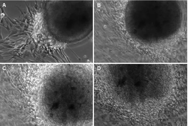
Our protocol to isolate and culture dental pulp stem cells is based on a procedure described previously (Gronthos et al. 2000), with some modifications. In brief, normal human impacted third molars were collected from adults (18-26 years of age) at the Department of Periodontology, Semmelweis University, under approved ethical guidelines set by the Ethical Committee of the Hungarian Medical Research Council.
Tooth surfaces were cleaned and the periodontal tissue was removed with a sterile scalpel and was collected. The tooth was cut around the cemento-enamel junction by sterile dental fissure burs to expose the pulp chamber, and the pulp tissue was removed from the
18
crown and root. Both pulp tissue and periodontal tissue were then separately digested in a solution of collagenase type I (3 mg/ml, Sigma-Aldrich, St. Louis, USA) and dispase (4mg/ml, Roche, Basel, Switzerland) for 1 hour at 37°C. Single-cell suspensions were obtained by passing the cells through a 70 µm strainer (Falcon). Cells were seeded into 6-well plates (Costar) in alpha modification of Eagle's medium (a-MEM, GIBCO/BRL) supplemented with 15% Fetal Bovine Serum (FBS, GIBCO/BRL), 100 µM L-ascorbic acid 2-phosphate (Sigma-Aldrich, St. Louis, USA), 2 mM L-glutamine, 100 units/ml penicillin, 100 mg/ml streptomycin (GIBCO/BRL), and then incubated at 37°C in 5%
CO2. To assess colony-forming capability, 14 day old cultures were fixed with 4%
paraformaldehyde, and then stained with 2% Giemsa. Aggregates of 50 cells were scored as colonies
6.2.3. Cell viability studies and treatment with enamel matrix derivative
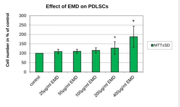
The influence of FBS and EMD (Emdogain, Straumann, Basel, Switzerland) containing media as well as osteogenic and neuronal differentiation protocols on primary cell cultures was assessed by Microculture Tetrazolium (MTT) assay. To test the effect of these conditions on culture growth, DPSCs and PDLSCs were cultured in 96-well plates for 24 hours. In each well 3 x 10³ DPSC cells or 5 x 10³ PDLSC cells were grown in their regular media for 24 hours. Afterwards, cells were serum-starved for another 24 hours. Then, 15% (PDLSC) or 20% (DPSC) FBS containing medium, or serum free medium (control) was added for 24 hours. Thereafter, 100 µl MTT solution (0.2 mg/ml, Sigma-Aldrich, St. Louis, USA) diluted in a-MEM was added into each well until formazane crystal formation occurred. 100 µl DMSO (99.5%, dimethyl-sulfoxid) was added into wells to dissolve formazane crystals. Then the intensity of staining was determined by a microplate reader (Model 3550, Biorad, Hercules, USA) at 595 nm (measurement wavelength) and 650 nm (reference wavelength). Under these circumstances, the level of optical density is proportional to the number of living cells in the culture. The proliferative effect was expressed as a ratio between optical density of treated cells and serum-free cultured control cells and given in percent.
19 6.2.4. Immunocytochemistry
To identify the mesenchymal stem cell marker “stromal cell surface marker-1“
(STRO-1) in our cultures, cells were grown on glass coverslips in 24-well plates (Costar, Cole-Parmer, Vernon Hills, Illinois, USA) (5x10⁴ cells per well) and fixed with 4% PFA in phosphate buffered saline (PBS) for 20 min. To block non-specific binding, fixed cultures were incubated in PBS containing 7.5% FBS for 90 min and incubated with an anti-STRO-1 primary antibody (1/200, a generous gift from Prof Richard Oreffo, University of Southampton, Southampton, UK) overnight at 4°C. Subsequently, the cells were incubated with Alexa 488 conjugated goat anti-mouse IgG (1:1000, Molecular Probes, Invitrogen, Carlsbad, CA, USA) for 1 hour. Nuclei were counterstained with 10 mg/ml bisbenzimide (Sigma-Aldrich, St. Louis, USA) for 30 minutes.
To evaluate protein expression during differentiation experiments, cells grown on poly-L-lysine-coated glass coverslips were fixed with 4% PFA in PBS for 20 min at room temperature (RT), then 0.1% Triton X-100 (in PBS) was added for 8 min to permeabilise them. Fixed cultures were incubated in PBS containing 4% bovine serum albumin (BSA;
90 min at RT) to block non-specific binding, then reacted with primary antibodies at 4°C overnight. Antibodies were diluted in 4% BSA as follows: anti-NSE 1/200, anti-NF-M 1/200. IgG anti-mouse and anti-rabbit Alexa Fluor 488 conjugated (Molecular Probes, Invitrogen, Carlsbad, CA, USA) secondary antibodies were diluted 1/750 and applied for 1 h at RT. Nuclei were counterstained with 10 mg/ml bisbenzimide (Sigma-Aldrich, St.
Louis, USA) for 30 minutes. Labelled preparations were examined by a fluorescent microscope (Nikon Eclipse E600, Nikon Instruments, Tokyo, Japan), and images were captured with a cooled CCD camera (SPOT RT Color 2000, Spot Imaging Solutions, Sterling Heights, Michigan, USA) connected to a PC running an image acquisition software (SPOT Advanced, Spot Imaging Solutions, Sterling Heights, Michigan, USA.).
Adobe Photoshop® was used to merge the digitized images of bisbenzimide and specific staining.
6.2.5. FACS analysis
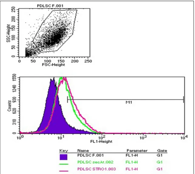
Fluorescence Activated Cell Sorting (FACS) analysis was performed to identify cells expressing STRO-1, CD34 and c-kit mesenchymal stem cell markers as described
20
previously (Gronthos et al. 1994, Laino et al. 2005). Single cell suspension were prepared from the cell cultures of 0,2% EDTA content, and subsequently incubated with STRO- 1/CD34/c-kit antibody or with isotype matching negative controls for 1 hour on ice. Cells were washed with 5% PBS solution of FBS, then fluorescent stain-conjugated secondary antibodies were added to the samples. Following repeated rinsing with 5% PBS solution of FBS, cell suspensions were fixated with 4% paraformaldehyde. FACS analysis was performed subsequently.
6.2.6. Osteogenic induction
Osteogenic differentiation was induced by modifications of a previously reported protocol (Kemoun et al. 2007). In brief, DPSCs and PDLSCs were cultured with 1% FBS, 100 mg/ml streptomycin, 100 U/ml penicillin, 2 mML-glutamine, 10-8M dexamethazone, 50 mg/ml L-ascorbic acid 2-phosphate, 10 mmol/l b-glycerophosphate in aMEM for 20 days without passaging. The medium was replaced twice a week. After 3 weeks of treatment calcium accumulation was detected by 2% Alizarin red S (pH 4.2, buffered with ammonium hydroxide) staining. Similar culture media without dexamethazone and ß- glycerophosphate was used as control condition.
6.2.7. Neuronal induction
For neuronal differentiation, cultured morphologically homogeneous DPSCs and PDLSCs, (passage 1-4) were plated (~2×10⁴ cells/well) into a 24 well plate containing poly-L-lysin coated glass coverslips. After 24 hours, cells were treated with 3 different protocols:
6.2.7.1 Protocol 1.
Cells were differentiated as previously described by Scintu et al. (Scintu et al.
2006) with 10 ng/ml FGF-1 (R&D, Minneapolis, MN), 200 nM 12-O- tetradecanoylphorbol-13-acetate (TPA; Sigma-Aldrich, St. Louis, USA), 250 µM IBMX (Sigma-Aldrich, St. Louis, USA) and 50 µM forskolin (Sigma-Aldrich, St. Louis, USA), in Dulbecco's modified Eagle's medium/F12 1:1 (DMEM/F12) (Sigma-Aldrich, St.
21
Louis, USA) supplemented with ITS Liquid Media Supplement (Sigma-Aldrich, St.
Louis, USA). The cells were fixed for immunocytochemistry right before and 24 h post- induction.
6.2.7.2. Protocol 2.
This protocol was also based on a method recently reported by Choi et al. (Choi et al. 2006). Cells were preinduced for 1 day with DMEM/F12, with 20% FBS, and 10 ng/ml basic fibroblast growth factor (bFGF; Sigma-Aldrich, St. Louis, USA). The preinduction medium was removed, cells were washed with PBS and then changed to serum-free induction medium that consisted of DMEM containing 2% DMSO, 200 µM BHA, 25 mM KCl, 2 mM valporic acid, 10 µM forskolin, 1 µM hydrocortisone and 5 µg/ml insulin (Sigma-Aldrich, St. Louis, USA). The cells were fixed for immunocytochemistry right before and 24 h post-induction.
6.2.7.3. Protocol 3.
A three-step differentiation method was developed in our own laboratory since Protocols 1 and 2 did not yield satisfactory results. DPSCs or PDLSCs were seeded onto poly-L-lysin coated glass coverslips in DMEM/F12, 2.5% FBS, 100 mg/ml streptomycin, and 100 U/ml penicillin, and cultured for 24 h. Step 1: epigenetic reprogramming was performed using 10 mM 5-azacytidine in DMEM/F12 containing 2.5% FBS and 10 ng/ml bFGF for 48 h. Step 2: neural differentiation was induced by exposing the cells to 250 mM IBMX, 50 mM forskolin, 200 nM TPA, 1 mM dbcAMP, 10 ng/ml bFGF, 10 ng/ml NGF and 30 ng/ml NT-3, supplemented with ITS Liquid Media Supplement in DMEM/F12 for 3 days. Step 3: at the end of the neural induction treatment, cells were washed with PBS and then neuronal maturation was performed by maintaining the cells in Neurobasal A media supplemented with 1 mM dbcAMP, 1% N2, 1% B27, and 30 ng/ml NT-3 for 3-8 days. Solutions 168 were freshly prepared immediately prior to use.
The cells were fixed for immunocytochemistry before treatment, on the first day neuronal induction (step 2) and on the third day of maturation (step 3).
22 6.2.8. Real-time PCR
Total RNA from DPSCs and PDLSCs was isolated using an RNeasy Plus Micro Kit (Qiagen) with on-column DNase digestion. The concentration of the RNA was determined by the Ribogreen method (Invitrogen, Carlsbad, CA, USA). The integrity of the RNA was verified by electrophoresis on a 1% agarose gel and 200 ng total RNA was used per sample for cDNA synthesis, using random primers (High-Capacity cDNAArchive Kit, Applied Biosystems, Invitrogen, Carlsbad, CA, USA) in a total volume of 50 µl. For quantitative PCR amplification, 5% of the cDNA synthesis reaction was used with real time PCR primers and a target-specific fluorescence probe (FAM- labelled MGB probe). The probes and primers were selected from the Applied Biosystem Assay on Demand database for the specific markers vimentin (VIM) and neurospecific enolase (NSE) and for the human acidic ribosomal phosphoprotein P0 (RPLP0), which was used as an internal control. Universal Mastermix (Roche, Basel, Switzerland) containing AMP-erase was used for amplification in a total volume of 20 µl. For detection of fluorescence signal during the PCR cycles, a (StepOne® Real-Time PCR System, Applied Biosystem, Invitrogen, Carlsbad, CA, USA) was used with the default setting (50°C for 2 min, 95°C for 10 min, 45 cycles: 95°C for 15 s, 60°C for 1 min). Each treatment was repeated five times and each sample was measured in duplicate. Changes in gene expression levels were estimated by calculating the relative expression values normalized to the RPLP0 level from the same sample.
6.2.9. Statistical analysis
Data were presented as means ± S.E.M. For statistical comparisons, analysis of variance was followed by Bonferroni post-hoc test (Instat, GraphPad Software).
23 6.3 Clinical studies
In our clinical studies hard- and soft tissue regenerative procedures were investigated using similar preoperative protocol and postsurgical care. Standardised clinical measurements were taken for evaluation of treatment safety and efficacy. Surgical protocols varied throughout the studies.
6.3.1 Hard tissue regeneration following treatment with rhGDF-5/β-TCP
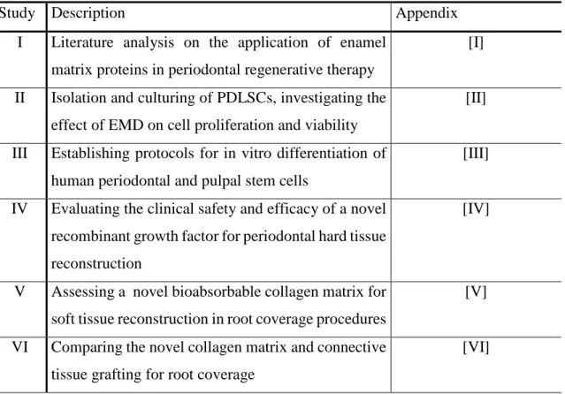
This pilot, phase IIa study used a stratified randomized, open, controlled, two- arm, parallel group design. The overall design and patient treatment allocation is summarized in Fig. 1. The study was conducted at the Department of Periodontology, Semmelweis University, Budapest, Hungary between July 2007 and August 2008. The study protocol was approved by the Hungarian National Institute of Pharmacy and the Institutional Ethics Committee (application no. 32579/40/06) of the Semmelweis University, Budapest, Hungary (TUKEB no. 20/2007). All patients received oral and written explanations of the research protocol. Patients signed a consent form providing the possibility of withdrawing from the study at any time. The study was planned and conducted in compliance with the Declaration of Helsinki of 1975 as revised in 2000, Good Clinical Practice, and relevant local laws.
The total study duration was 175–182 days, in all ten visits/patient. After screening, selected patients received flap surgery (control) or flap surgery combined with implantation of rhGDF-5/β-TCP at the qualified defect site (Visit 2). They then returned for general and oral health evaluations as well as professional tooth cleanings following a set schedule (Visits 3 through 8). Blood samples were collected at screening (Visit 1), and at weeks 2 and 24 (Visits 3 and 9) to evaluate routine haematology and clinical chemistry, rhGDF-5 plasma levels, and antirhGDF-5 antibody formation.
Randomization was performed using a computer-generated randomization list via block randomization. Ten patients were randomized to each treatment group. A separate random scheme was generated. The investigators were masked to the block length. The sponsor retained the randomization scheme for control purposes. The investigator implemented the predefined randomization by opening a randomization envelope at the appointment for surgery (Visit 2). The randomization code was opened only after the
24
defect site was fully prepared. The defects were randomly assigned to receive: rhGDF- 5/β-TCP following the manufacturer’s instructions (test), or no additional treatment (control). All randomized patients completed the study. Masking of treatment was not applicable because the test group received rhGDF-5/β-TCP whereas the control group was treated by periodontal surgery only without additional treatment.
Fig. 1 Study IV flow chart including patient enrolment, treatment allocation, follow-up and analysis (FS:
flap surgery; DD: intrasurgery defect depth).
25 6.3.1.1 Subject selection, preoperative protocol
Twenty Caucasian male and female patients, non-smokers, in good general health, volunteered to participate in this study. They all exhibited advanced chronic periodontitis with one deep intrabony defect located at a maxillary or mandibular single-rooted tooth without root concavities/ furrows or at the mesial or distal aspect of a mandibular molar without contacting teeth (Fig. 1). Mandibular incisors and teeth with furcation involvements were excluded. Only teeth with a probing depth ≥6 mm and an intrabony component ≥4 mm as estimated from long cone parallel technique radiographs confirmed during surgery were considered (Fig. 1). Moreover, the patients were expected to meet oral hygiene standards encompassing full mouth plaque and bleeding scores <20% after completion of basic periodontal therapy (O’Leary et al. 1972, Ainamo and Bay 1975).
Each patient contributed one tooth subject to the study treatment. Main exclusion criteria were: a) women of childbearing potential (FSH level <25 IU/L and menstrual bleeding within 6 months)/pregnant or lactating women; b) tobacco smoking; c) evidence of acute/chronic infection at the study site; d) previous (<2 months)/current treatment with systemic corticosteroids of a prednisone equivalent >5 mg/day; e) previous (<12 months)/current treatment with drugs influencing bone metabolism including calcitonin, parathormone, bisphosphonates, or fluoride; f) common contraindications for periodontal surgery; and g) clinically relevant cardiovascular, hepatic, and renal diseases. Due to the explorative type of this study, a sample size of ten patients/group was selected.
All patients had completed basic periodontal therapy (individual oral hygiene instructions, supra- and subgingival scaling and root planing) 8 weeks before screening.
If necessary, composite splinting of mobile teeth or eventually fixed temporary restorations were completed.
6.3.1.2 Study material
The rhGDF-5/β-TCP device (Scil Technology GmbH, Martinsried, Germany) comprises rhGDF-5 coated onto a synthetic inorganic carrier, β-TCP, at a concentration of 500 µg/g β-TCP [13]. The β-TCP carrier consists of particles of 500 to 1,000 µm in size with interconnecting porosity. It comprises microporous and macroporous irregular granules of a phase purity >95%. The results of porosity analysis have shown 43.7%
26
microporosity, an average pore diameter of 2.12 µm, and a total pore area of 0.647 m2/g.
The pore size of the macropores ranges between 100 and 400 µm. The surface area is estimated at 1.2 m2/g (Pöhling et al. 2006). The rhGDF-5 protein was coated onto the carrier using Scil Technology’s proprietary technology. One vial rhGDF-5/β-TCP contained 250 µg rhGDF-5 and 0.5 g β-TCP (Pöhling et al. 2006). In vitro analysis of the carrier used in this study has shown that almost the entire amount of rhGDF-5 was released from the carrier within the first 7 days (Pöhling et al. 2002).
6.3.1.3 Surgical procedures
One experienced periodontist (PW) performed all surgeries using local anaesthesia, microsurgical instrumentation, and appropriate magnification. (Fig. 2. Fig.
13) The surgical technique was exactly the same for both the test and control groups. An intracrevicular incision was made on the buccal and lingual aspects of the surgical site.
The flap was horizontally extended to accommodate the defect location and configuration, and ensured tension-free wound closure for primary intention healing.
Vertical releasing incisions were not used. Granulation tissue removal and root instrumentation followed elevation of the mucoperiosteal flaps. In the test group, six patients received one-half vial rhGDF-5/β-TCP, one patient received three-fourth vial rhGDF-5/β-TCP, and three patients received one vial rhGDF-5/β-TCP (one vial rhGDF- 5/β-TCP contains 250 µg rhGDF-5 and 0.5 g β-TCP). The mucoperiosteal flaps were then adapted and closed using vertical or horizontal holding mattress sutures and interrupted closing monofilament sutures (5/0 Dafilon; B. Braun Melsungen AG, Melsungen Germany).
6.3.1.4 Postoperative care
Postsurgery care included pain control (Nurofen, 200 mg, 3–4 times per day, Reckitt Benckiser, Slough, UK), systemic (Augmentin 625 mg, GlaxoSmithKline, London, UK London, UK; TID/7 days) and local (twice daily 0.2% chlorhexidine;
Curasept, Curadent International AG, Kriens, Switzerland; rinses for 1 min, BID/4 weeks) antimicrobial control. Antibiotic therapy started immediately after surgery.
Sutures were removed at day 14. A series of control and recall appointments were
27
scheduled (biweekly, the first 6 weeks and then monthly until the end of the study) including reinforcements of oral hygiene and professional supragingival tooth cleaning.
Fig. 2 Flap surgery (control): Presurgery (top left); intrasurgery defect morphology (top right); the biopsy event at 24 weeks postsurgery (bottom left); and biopsy including defect site (bottom right). Histological outcomes were published elsewhere (Stavropoulos et al. 2011).
6.3.1.5 Clinical assessment
Clinical outcomes were evaluated at baseline and at 24 weeks postsurgery.
Probing depth (PD), gingival recession (GR) and clinical attachment level (CAL) were recorded using a standard periodontal probe (UNC 15, Hu-Friedy, Chicago, IL, USA).
Intraoral radiographs were taken with the long cone parallel technique at baseline and at 24 weeks postsurgery. However, due to the design of the study (i.e. no grafting in the control group), the radiographs were not evaluated. Full mouth plaque and bleeding scores were recorded as a percentage of total surfaces (four surfaces/tooth) with the presence of plaque/bleeding on probing, respectively (O’Leary et al. 1972, Ainamo et al.
1975). One calibrated examiner, masked to the patients’ treatment protocol, performed
28
all clinical recordings. At 24 months postoperatively, biopsy removal was performed.
Histological outcomes were published elsewhere (Stavropoulos et al. 2011).
6.3.1.6 Safety assessment
Adverse events were monitored and recorded throughout the study, as well as laboratory values, vital signs, and physical status. Adverse events were coded using the
Medical Dictionary of Regulatory Activities (MedDRA)
[http://www.meddramsso.com/index.asp]. Summaries and tabulations by severity and relationship to therapy were based on the preferred terms and the primary system organ classes (SOCs). Blood samples were collected at screening (Visit 1), 2 weeks postsurgery (Visit 3), and prior to conclusion of study (Visit 9) to determine laboratory values (clinical chemistry, haematology), rhGDF-5 plasma levels, and antirhGDF-5 antibodies.
The determination of rhGDF-5 in human plasma (EDTA) samples was carried out by Elisa over a quantitation range of 40 pg/ml to 1,250 pg/ml. A monoclonal antibody specific for rhGDF-5 has been precoated on a 96-well plate. Standards/QCs and samples were then pipetted into the wells and any rhGDF-5 present was bound by the immobilized antibody. After washing away any unbound substances, a biotinylated monoclonal antibody specific for rhGDF-5 was added to the wells. After a second washing step, PolyHRP Streptavidin was added that bound to the biotinylated antibody. After a third washing step, peroxidase bound in the complex was visualized by TMB (3,3′,5,5′- Tetramethylbenzidine) substrate solution. After stopping the enzymatic reaction with sulphuric acid, the intensity of the resulting colour was determined at 450 nm. The colour intensity was proportional to the concentration of rhGDF-5 in the sample.
6.3.1.7 Statistical analysis
The statistical analysis was conducted on an intent-to-treat basis. All randomized patients with periodontal treatment were included in the intent-to-treat population. Paired sample t-test and Wilcoxon signed-rank test were used to evaluate the impact of surgical interventions on the various clinical parameters. Mann–Whitney U test (rank sum test) was used to analyse differences among the various outcome variables between treatment groups. No formal statistical comparisons were made related to safety data.
29
6.3.2 Soft tissue regeneration following treatment with Mucograft®
a) In the pilot case series, 8 adult patients (3 males and 5 females, aged from 18 to 39 years, mean 29 years) presenting Miller class I-II MAGR displaying a total of 42 recession were recruited. All patients presented MAGR defects, which were treated by means of MCAT technique using a bioresorbable collagen matrix (Mucograft®, Geistlich, Wolhusen, Switzerland). The primary outcome variable was the assessment of CRC. The secondary outcome variables included the assessment of mean root coverage (MRC), keratinised tissue width (KTW) and gingival thickness (GT).
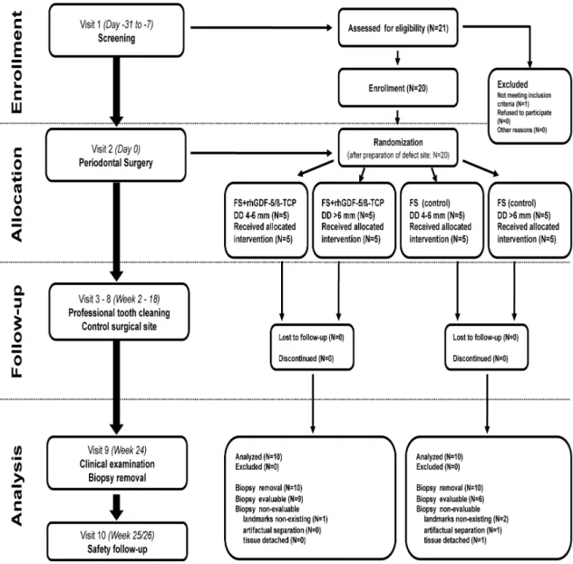
b) The randomised controlled study was performed according to a split-mouth design.
Thus, in each patient, one side of the jaw served as control while the contralateral side served as test (Fig. 3.) Randomisation was performed by using a computer-generated programme. Recessions were treated by means of MCAT technique using either a bioresorbable collagen matrix (Mucograft®, Geistlich, Wolhusen, Switzerland) (test) (Fig. 12) or SCTG harvested from the palate (control) (Fig. 13). Both surgeries (test and control site) were performed during one single session by the same experienced surgeon (S.A.). The primary outcome variable was the assessment of CRC. The secondary outcome variables included the assessment of MRC, KTW, GT and patient-centred outcomes.
6.3.2.1 Subject selection, preoperative protocol
a) In the pilot case series, patients were treated after having completed preliminary professional tooth cleaning and having received individual oral hygiene instructions. The study was performed between July 2009 and June 2010 at the Department of Periodontology, Semmelweis University Budapest, Hungary in accordance with the Helsinki Declaration of 1975, as revised in 2000 and following approval of the Regional Bioethical Committee (Approval number: ETT TUKEB/365/PI/10/). Inclusion criteria for participation in the study were as follows: (1) at least 18 years of age (2) systemically healthy without any signs of periodontal disease (3) presence of at least three adjacent gingival recessions in the maxilla or mandible, (4) a full-mouth plaque score (FMPS) <
20%18; (5) full-mouth bleeding score (FMBS) < 20%19 (6) non-smoker; (7) not pregnant.
30
Before enrolment, written informed consent forms were obtained from all patients participating in the study.
b) In the split mouth randomised, controlled study 22 patients with multiple Miller Class I and II MAGR (Miller 1985) with evidence of CEJ were enrolled in the study after having signed an informed consent. The study protocol was in accordance with the Helsinki Declaration of 1975, as revised in 2002 and was submitted to and approved by the ethical committee of the Semmelweis University Budapest, Hungary (protocol: 5242-0/2010- 101SEKU; 365/PI/10). The study was performed between July 2010 and November 2011 in the Department of Periodontology of the Semmelweis University Budapest. One month before surgery, individualized oral hygiene instructions were given for each of the included patients accompanied by full mouth supragingival scaling and polishing. The following inclusion criteria were applied: 1) Age ≥ 18 years, 2) Absence of relevant medical conditions, 3) Patients with healthy or treated periodontal conditions. 4) Presence of ≥ 3 adjacent Miller class 1 and 2 gingival recessions on both sides of the maxillary or mandibular arch with an apico-coronal extension (i.e. recession depth) > 2 mm, 5), Full- Mouth Plaque Score (FMPS) ≤ 25% (O`Leary et al. 1972). Patients were excluded on the basis of the following criteria:1) Pregnant or lactating females, 2), Tobacco smoking, 3) Uncontrolled medical conditions, 4) Untreated periodontal conditions, 5) Use of systemic antibiotics in the past 3 months, 6) Use of systemic antibiotics for endocarditis prophylaxis, 7) Patients treated with any medication known to affect gingival conditions (e.g. hyperplasia), 8)Infectious diseases such as hepatitis, tuberculosis and HIV, Drug and alcohol abuse, (9) Failure to sign written informed consent
6.3.2.2 Study material
The CM (Mucograft®, Geistlich Pharma, Wolhusen, Switzerland) has a bilaminar structure, consisting of two adherent layers: a superficial, compact, cell occlusive membrane-like layer incorporating collagen fibres, and an underlying three dimensional spongious collagen matrix designed to serve as scaffold conducing the ingrowth of blood vessels and cells and to enhance blood clot stability.
31
Fig. 3 Study VI flow chart including patient enrolment, treatment allocation, follow-up and analysis
32 6.3.2.3 Surgical procedures
a) Patients recruited to the pilot case series and MAGR defects on the test sides of patients recruited to the split mouth trial were consecutively treated as follows using the MCAT technique (Azzi, Étienne, 1998). Preoperatively, resin bonding of adjacent contact points at the operation site was performed in order to enable suspended suturing.
Following local anaesthesia (Ultracain DS Forte, Sanofi Aventis, Paris, France) root planing of the exposed root surfaces was performed with Gracey curettes (Hu-Friedy, Chicago, IL, USA). Intrasulcular incisions around involved teeth were performed using microsurgical tunnelling knives (Stoma, Liptingen, Germany). Mucoperiosteal envelope flap elevation was performed via the same instruments up to the level of the mucogingival junction at each recession site, leaving interdental papillae intact (Fig. 2). Separate mucoperiosteal envelopes were subsequently interconnected, resulting in tunnel preparation. Mucoperiosteal tunnel elevation was extended by full thickness preparation apically from the mucogingival junction utilising tunnelling knives. Attaching muscles and inserting collagen fibres were separated and released from the inner aspect of the alveolar mucosa by means of Gracey-curettes (Stoma, Liptingen, Germany). As a result of this, the tunnelled flap could be mobilised and advanced coronally without tension. To achieve complete mobilisation of the flap, interdental papillae were gently undermined using microsurgical elevators. Special attention was paid not to disrupt the interdental papillary tissues. Subsequently, the CM was trimmed and adapted to the recipient site with gentle wetting. The CM was carefully advanced into the subperiosteal tunnel through the widest recession using horizontal mattress sutures at the mesial and distal aspects of the matrix (Fig. 12). At surgical sites extending to more than 3 teeth, the CM was cut to multiple segments. The compact membrane-like layer was directed towards the inner side of the flap. The CM was gradually moisturised by sterile saline during this procedure to avoid detaching the underlying spongious layer by overwetting. Having reached the desired position with the coronal margin positioned at the level of the cemento-enamel junction (CEJ), the CM was fixed to gingiva via previously inserted horizontal mattress sutures (Fig. 12). Finally, suspended sutures (i.e. crossed horizontal mattress sutures, anchored over the preoperatively placed interproximal resin splints) were placed into interdental gingiva to coronally advance the fully mobilized mucoperiosteal tunnel,
33
resulting in complete coverage of the CM and the recessions (Fig. 12). In cases where complete CM coverage could not be obtained with the first sutures, additional vertical mattress sutures were placed interdentally to enable coronal displacement of the tunnel slightly over the CEJ.
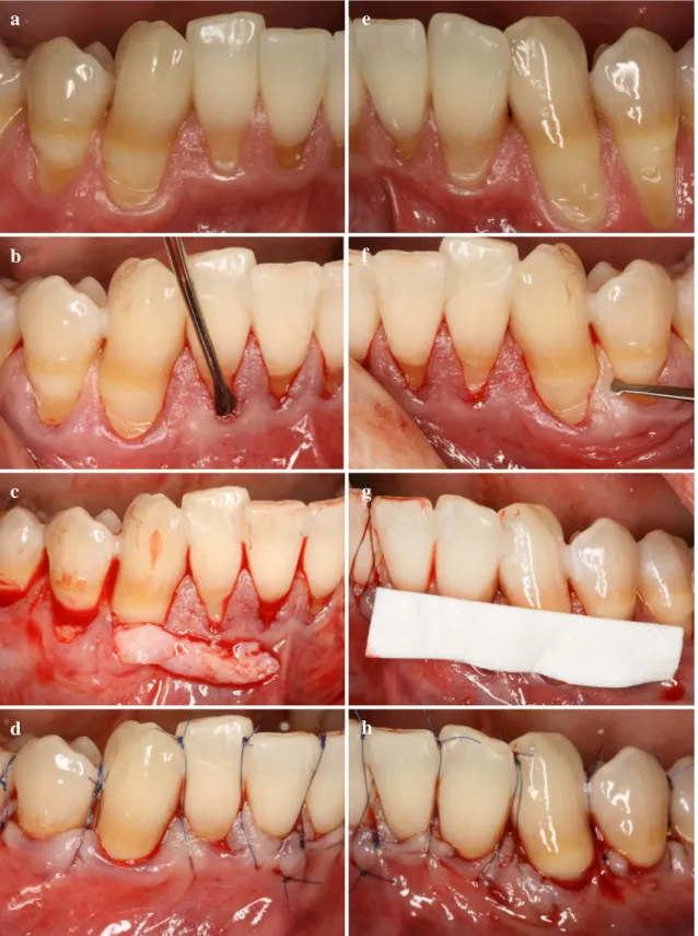
b) MAGR defects on control sides of patients recruited to the split mouth trial were treated with the MCAT technique as described above, in combination with connective tissue grafting. A SCTG was immediately harvested after tunnel preparation by using either a modified distal wedge procedure (Azzi & Etienne 1998) or the single incision technique (Hürzeler & Weng 1999) depending on anatomical considerations. If needed, the harvested graft was trimmed using a N°15 blade to achieve an optimal thickness of 1-1.5 mm. Immediately after SCTG harvesting, the donor site was closed with either a cross-mattress suture or with a modified mattress suture (Monnet-Corti et al. 2006) (5-0 polyglactin 910, Vicryl, Ethicon, Johnson & Johnson, USA). SCTG was always inserted under the tunnelled flap by starting at the deepest recession (Fig 13).
Subsequently, the grafts were pulled laterally towards each end of the tunnel by means of mattress sutures (Azzi & Etienne 1998) . Finally, the flaps were positioned coronally to the cemento-enamel junction (CEJ) by means of suspended sutures placed above the contact point (Azzi & Etienne 1998) (Fig. 4, 14, 15).
6.3.2.4 Postoperative care
Patients attending either of the trials were given postoperative analgesics (3 X 50 mg Cataflam, Budapest, Hungary) for 3 days and antibiotics (3x625 mg Augmentin, Pfizer KFT, Budapest, Hungary) for 7 days due to university regulation for implantable biological materials. Patients were instructed to rinse with a 0.2% chlorhexidine solution, two times a day for one minute for 3 weeks. Patients avoided brushing in the operated area until suture removal. Patients underwent manual supragingival tooth cleaning twice a week until suture removal. At suture removal two weeks after surgery, patients were instructed in mechanical tooth cleaning of the operated areas using a soft tooth brush and a roll technique. The interproximal resin splints were removed at 21 days. All patients were recalled after 28 days, 3, 6 and 12 months and received one session of prophylaxis, including reinforcement of oral hygiene, supragingival debridement, and tooth polishing.
34
Fig. 4 Split Mouth Modified Coronally Advanced Tunnel Technique (MCAT) in combination with either subgingival connective tissue graft (SCTG) or Mucograft® (CM) (a) Control side prior treatment (b) Control side tunnel preparation (c) Control side SCTG insertion (d) Control side suspended suturing (e) Test side prior treatment (f) Test side tunnel preparation (g) Test side CM insertion (h) Test side suspended suturing
a e
b
c
d
f
g
h
35 6.3.2.5 Clinical assessments
In both studies following measurements were made at the mid-buccal point of the involved teeth at baseline (prior to surgery) 6, and at 12 months by the same blinded investigator (B.M.) using the same type of periodontal probe (UNC 15, Hu-Friedy, Chicago, IL, USA): 1) Gingival Recession Depth (GRD in mm) measured as the distance from the CEJ to the Gingival Margin, 2) Gingival recession width (GRW in mm) measured at the CEJ 3), keratinised tissue width (KTW in mm), measured as the distance from the mucogingival junction (MGJ) to the gingival margin.
To avoid interference with wound healing, the following clinical parameters were only registered at baseline, 6 and 12 months postoperatively: 4) Gingival thickness (GT in mm) measured 3 mm apically from the free gingival margin at the mid buccal aspect of the tooth, 5) pocket probing depth (PPD in mm) at the distobuccal, midbuccal, mesiobuccal aspects of surgical sites, 6) clinical attachment level (CAL in mm). At surgery, the length of time of the full procedure was evaluated (in minutes).
Intra-examiner reproducibility: In both trials, the same calibrated investigator performed all clinical measurements using a standard periodontal probe (PCP-UNC 15, Hu-Friedy, Chicago, IL, USA). Five patients, not related to the study and each showing a pair of contralateral single-rooted teeth (with recession depth > 2 mm on the mid-buccal aspect) were used to calibrate the examiner. The examiner evaluated the patients on two occasions 24 hours apart. Calibration was accepted if 90% of the recordings could be reproduced within a difference of 1.0 mm (Pilloni et al. 2006).
6.3.2.6 Evaluation of patients’ satisfaction
In the split mouth trial, at suture removal, both procedures were evaluated by the patient for discomfort, duration and difficulty on a visual analogue scale (VAS). At 12 months, the aesthetic outcome of both treatments was appreciated by the patient on a VAS scale.