The Merit of Histological Evaluation of
Clinically Successful Reconstructive Therapies in Periodontology and Related Fields
PhD Thesis
Dr. Attila Horváth
Semmelweis University PhD School of Clinical Medicine
Consultant:
Dr. István Gera PhD
Official reviewers:
Dr. István Gorzó PhD Dr. Árpád Joób-Fancsaly PhD
Final Examination Board:
Dr. Ida Nyárasdy PhD (Head) Dr. Vilmos Tóth PhD Dr. Szabolcs Gyulai-Gaál PhD
Budapest 2014
“The more I learn, the more I learn how little I know.”
Socrates
(469-399 BC)
TABLE OF CONTENTS
1. LIST OF ABBREVIATIONS………...………...…...….4
2. INTRODUCTION………...…………...…...6
2.1. Tissue regeneration in periodontology. History and principles...6
2.2. Current regenerative considerations of the non-contained periodontal defects....11
2.3. Rationale of peri-implant dehiscence therapy...14
2.4. Provision of post-extraction alveolar sockets...18
3. OBJECTIVES.………...………...20
4. METHODS AND MATERIALS………....…....………..21
4.1 Overview of the applied methods...21
4.2 Regeneration of periodontal defects……...23
Clinical and histological evaluation of the effect of a novel biphasic synthetic bone substitute (Straumann BoneCeramic®) and enamel matrix derivative (Emdogain®) on human intrabony periodontal defects Clinical and histological evaluation of the effect of a novel unsintered nanocrystalline synthetic bone substitute (Ostim®) on human intrabony periodontal defects 4.3 Regeneration of peri-implant dehiscence defects……...30
Clinical and histological evaluation of the effect of a novel biodegradable synthetic hydrogel membrane (Straumann MembraGel®) in combination with a novel biphasic synthetic bone substitute (Straumann BoneCeramic®) on porcine peri-implant dehiscence defects 4.4 Regeneration of post-extraction alveolar sockets……...…...38
Clinical and histological evaluation of the effect of alveolar ridge preservation on human extraction sockets 5. RESULTS………...………43
5.1. Periodontal regeneration...…………...……….43
5.1.1. Treatment with EMD and SBC………....………...43
5.1.2. Treatment with nano-HA...………...….……...…………...45
5.2. Provision of peri-implant dehiscence defects with SBC and PEG...47
5.3. The effect of ARP on extraction sockets..……...…...……...56
6. DISCUSSION………...…..77
6.1. Limits of periodontal regeneration...………...77
6.1.1. Treatment with EMD and SBC………....………...77
6.1.2. Treatment with nano-HA...…...….……...………….…...80
6.2. Peri-implant dehiscence defect therapy with SBC and PEG...83
6.3. Current options for the treatment of extraction sockets by ARP...…..88
6.4. Overall considerations...96
7. CONCLUSIONS……….100
8. SUMMARY……….102
9. ÖSSZEFOGLALÁS………103
10. REFERENCES………104
11. LIST OF PUBLICATIONS……...…..………129
12. ACKNOWLEDGEMENTS………..………...131
1. LIST OF ABBREVIATIONS
3D Three-Dimensional
AAP American Academy of Periodontology ABB Autogenous Bone Block
ABP Autogenous Bone Particulates ANOVA Analysis of Variance
AR Alveolar Ridge
ARP Alveolar Ridge Preservation BCP Biphasic Calcium Phosphate
BS Bone Surface Area
β-TCP Beta-Tricalcium Phosphate
BG Bioactive Glass
BIC Bone-to-Implant Contact BMP Bone Morphogenetic Protein BoP Bleeding on Probing
cAMP Cyclic Adenosine Monophosphate CBCT Cone-Beam Computerised Tomography CEJ Cementoenamel Junction
CENTRAL The Cochrane Central Register of Controlled Trials CI Confidence Interval
CONSORT Consolidated Standards of Reporting Trials DBBM Deproteinized Bovine Bone Mineral
DFDBA Demineralized Freeze-Dried Bone Allograft EDTA Ethylene Diamine Tetraacetic Acid
EMD Enamel Matrix Derivative
e-PTFE Expanded Polytetrafluoroethylene FMBS Full Mouth Bleeding Score FMPS Full Mouth Plaque Score GBR Guided Bone Regeneration GS Graft Surface Area
GTR Guided Tissue Regeneration
HA Hydroxyapatite
IL-6 Interleukin-6
INTRA Depth of the Intrabony Component of the Periodontal Defect IOPA Intraoral Periapical Radiograph
ISRCTN International Standard Randomised Controlled Trial Number Register LILACS Literatura Latino-Americana e do Caribe em Ciências da Saúde LJE Long Junctional Epithelium
LRAP Leucine-Rich Amelogenin Peptide
Nano-HA Unsintered, Nanocrystalline, Phase-Pure Hydroxyapatite OFD Open Flap Debridement
PDL Periodontal Ligaments
PEG Polyethylene Glycol-Based Hydrogel PGPL Polyglycolide/Polylactide
PPD Pocket Probing Depth
PRGF Plasma Rich in Growth Factor
RCT Randomised Controlled Clinical Trial REC Gingival Recession
rhBMP-2 Recombinant Human Bone Morphogenetic Protein-2 ROI Region of Interest
SLA Sandblasted, Large-grit, Acid-etched
SLActive Modified (Hydrophilic) Sandblasted, Large-grit, Acid-etched TCP Tricalcium Phosphate
TGF-β Transforming Growth Factor-Beta TRAP Tyrosine-Rich Amelogenin Peptide
2. INTRODUCTION
2.1. Tissue regeneration in periodontology. History and principles
Periodontal disease has been considered as one of the major causes of tooth loss, being accountable for 30-35% of extractions [1]. A widely accepted measure to determine the severity of the disease is the periodontal pocket probing depth (PPD). Advanced periodontitis (PPD>5 mm) has been observed in 2-40% of the population in Europe [1]
and 7.4% in Hungary, according to a recent survey [2]. The treatment of periodontitis involves non-surgical as well as surgical means, beyond the inevitable enforcement of individual oral hygiene. The main goal of treatment is alleviating inflammation, arresting disease progression and establishing a stable periodontal condition that is maintenable for long run by the patient’s self-performed oral hygiene.
The principles above that were laid down by the great pioneers of periodontology in Scandinavia and North America remain valid today [3]. Nevertheless, the outcome of the conservative (i.e. non-surgical) periodontal therapy or even of the resective/explorative surgical treatment are often associated with gingival recession and very limited regeneration of the periodontium [4]. Despite the clinically stable periodontal conditions of such cases (i.e. PPD≤4 mm, negative bleeding on probing (BoP)), histological results are mostly characterised by reparative tissues. In such type of healing, the genuine attachment apparatus (acellular root cementum – inserting Sharpey fibres – bundle bone) that was lost as a result of periodontitis, has been predominantly replaced by long junctional epithelium (LJE) instead. Such epithelial attachment merely represents a secondary (inferior) connection in terms of quality and tissue adhesion [5]. Optimal treatment outcome would incorporate the reconstitution of the lost periodontal tissues (i.e. cementum, functionally oriented periodontal ligament, alveolar bone and gingiva). In the Seventies, Melcher suggested that periodontal healing might be characterized by the type of tissues first repopulating the periodontal defect following flap surgery [6]. Based on this assumption, it was hypothesized that precluding the undesired cells (epithelium, gingiva, etc.) from the healing hub of the intrabony periodontal pocket by a mechanical barrier device, would enable the cells from the remaining periodontal ligament to repopulate the defect. Consequently, the
principles of Guided Tissue Regeneration (GTR) and subsequently Guided Bone Regeneration (GBR) were conceived and described in Scandinavian research centres by Jan Gottlow, Thorkild Karring, Jan Lindhe, Sture Nyman, Christer Dahlin and many others [7] [8] [9] [10]. Thenceforth, the theory of GTR and GBR was broadly investigated and proved by sound evidence. However, the “mechanical concept” that the clue of regeneration is purely based on the cell-occlusive barrier function of the membrane is being challenged nowadays. Emerging evidence suggests a rather biological way of thinking that the intent of a membrane might be (i) to protect and stabilize the wound by neutralizing and deflecting any wound rupturing forces away from the root surface, (ii) to provide a secluded space to release the native potential for regeneration and therefore, (iii) to maintain the structural integrity of a maturing blood clot [11] [12] [5].
Over the past decades, tremendous efforts have been made by scientists and clinicians to develop novel techniques and materials in order to improve the outcome of regenerative procedures. Treatment of supra-alveolar defects or intrabony defects with missing buccal and/or lingual wall (i.e. non-containing defects) is often associated with least predictable outcome. For such augmentations a combinative approach was suggested and successfully utilized by means of different ‘bone fillers’ in combination with GTR/GBR [13]. Several grafts, bone substitutes and biomaterials were introduced and widely employed to fill such defects in order to exploit their possible osteoconductive, osteoinductive or even osteogenic potentials. However, the additional benefit of the application of these materials has not been unanimously supported by the literature [14]
[15] [16]. As described above, the volume of regenerated tissue may rest on the wound stability and the space provided by the barrier membrane beneath the tension-free approximated mucoperiosteal flaps. Therefore, the main mechanism behind the use of bone replacements seems to be blood clot stabilization through their scaffolding architecture, thus space provision, rather than osteoconduction or induction [17].
The use of barrier membranes, with or without bone substitutes, is, however, frequently associated with side effects, such as membrane exposure. It may arise from the compromised re-vascularization of the flaps above an implanted “foreign body”, as well as from the tension of the flaps caused by the increased volume of the augmentation. In
Hammarström and co-workers that promotes regeneration without the side effects and the handling difficulties of the GTR technique [18] [19] [20]. The biological active component of this regenerative gel originates from amelogenin, an enamel-specific protein of porcine tooth buds. Enamel matrix derivative (EMD), as it is predominantly quoted in the literature, refers to amelogenin fraction following a purification process.
EMD in combination with a propylene glycol alginate vehicle results in Emdogain®, the brand name of this biological active device. In the past fifteen years a large number of preclinical and clinical trials have proven its ability to regenerate periodontal ligaments (PDL), cementum and alveolar bone in some extent [21].
However, more than a decade following the first publications of EMD, the biologic cascade of the effects of EMD on wound healing at subcellular level has not yet been fully understood. According to data from in vitro studies EMD may modulate wound healing by stimulating the proliferation of preosteoblasts and the differentiation of osteoblasts via upregulating cyclic adenosine monophosphate (cAMP) levels, inducing the synthesis and secretion of transforming growth factor-beta (TGF-β) and interleukin-6 (IL-6) in gingival fibroblasts and PDL [22] [23] [24] [25]. In addition, EMD may retard epithelial cell proliferation and may confound dental plaque homeostasis [26] [27] [28].
Hence, it seems that EMD may stimulate periodontal wound healing by an indirect effect on the release of growth factors and by retarding the downgrowth of junctional epithelium. A very recent in vitro investigation of amelogenin peptides, conducted by the research group of Nikos Donos at the UCL Eastman Dental Institute, elucidated novel possible pathways of the effect of different fractions of EMD on bone repair and regeneration [29]. It was demonstrated that low- and high-molecular-weight fractions of EMD, namely leucine-rich amelogenin peptide (LRAP) upregulated osteogenic differentiation and enhanced terminal differentiation of bone-forming cells, meanwhile tyrosine-rich amelogenin peptide (TRAP) suppressed the formation of bone-like mineralized nodules. This indicates that fractions of EMD might play a therapeutic role not only in periodontal or alveolar bone regeneration, but also in orthopaedic repair (LRAP), or in the opposite, treatment of pathologic or even malignant bone formation (TRAP). A more recent publication of the same group demonstrated that EMD could modulate the differentiation of PDL cells in vitro, such as up-regulating chondrogenic, neovasculogenic and osteogenic genes, but suppressing adipogenesis, gliogenesis and neurogenesis [30]. A very recent review of the effect of EMD at cellular level concluded
that EMD elicited a regenerative response in periodontal tissues through a complex cascade of gene expression, protein production, proliferation and differentiation of various cells, particularly periodontal ligament and osteoblastic cell types [31].
Our research group at the Semmelweis University, led by Péter Windisch and Anton Sculean, has considerably contributed to the understanding of the effect of EMD on human periodontal defect, by illumination of its histological outcomes [32] [33] [34]
[35] [36]. Although the regenerative potential of EMD per se was demonstrated both at preclinical and clinical levels [21] [37], when treating large or non-containing defects, clinicians are often facing the challenge of space provision and prevention of flap collapse. For such cases, bone fillers (in combination with EMD) are suggested to overcome these problems. Data from controlled clinical trials have demonstrated greater clinical improvements following treatment of EMD combined with certain types of bone grafts, such as autogenous bone particles (ABP), or deproteinized bovine bone mineral (DBBM) compared to EMD alone [33] [38] [39]. Nevertheless, other substitutes, such as demineralized freeze-dried bone allograft (DFDBA), or bioactive glass (BG) did not demonstrate significant clinical benefit in terms of PPD reduction and CAL gain over EMD alone [40] [41]. Moreover, the histological evidence seems to be even more conflicting, since beside the formation of new cementum, periodontal ligament and alveolar bone up to a various extent, the implanted bone substitute was frequently encapsulated in connective tissue [34] [35] [42].
It has also been investigated over the decades, whether bone grafts/substitutes alone (i.e.
without the additional effect of GTR or EMD) might bear the potential to restore the lost periodontal attachment apparatus. The use of certain types of grafting materials such as ABP, DFDBA or DBBM has been shown to result not only in substantial clinical improvements, evidenced by PPD reduction, defect fill and CAL gain, but also to promote, at least to some extent, formation of a new connective tissue attachment (i.e.
new cementum with inserting collagen fibres) and of new alveolar bone [43] [44] [45]
[46] [47]. However, the use of autografts for regenerative periodontal therapy is limited by its source and increases donor site morbidity. Furthermore, most types of DBBM possess a fairly slow resorption potential, resulting in a mixed hard tissue formation in periodontal osseous defects [48]. In addition, the use of grafts from bovine (e.g. DBBM) or human origin (e.g. DFDBA), still comport the risk (at least theoretical) for
antigenicity and disease transmission [49] [50]. Furthermore, the animal origin of these materials keeps raising concerns for some patients, due to religious purposes or animal right considerations.
To overcome these drawbacks, synthetic materials in conjunction with open flap debridement were tested. The currently available data suggest that in intrabony defects, the implantation of various types of such alloplasts may lead to significant improvements of the investigated clinical parameters [51] [52] [53] [54] [55] [56]. On the other hand, the available histological evidence indicates that in human intrabony defects, the healing following implantation of alloplasts is predominantly characterized by epithelial proliferation, connective tissue encapsulation and limited periodontal regeneration [57] [58] [59] [53] [60] [61] [62] [63] [64] [65].
The above inconsistent findings on the adjunctive benefit of the application of bone replacement biomaterials highlight the current criteria of successful regeneration. In terms of clinical success, beyond arresting inflammation, we aimed at reconstituting the lost periodontal attachment apparatus. Although we are more aware nowadays that the complete reconstruction may remain a daydream, the therapist must possess certain clinical measures that support the evaluation of the treatment outcome. Such measures are PPD reduction, CAL gain, decreased furcation involvement or sometimes reduced gingival recession, discontinued tooth mobility, etc. Albeit the improvement of all of these measurements undoubtedly indicates a level success, none of them is able to demonstrate the presence of true regeneration (i.e. new cementum, inserting Sharpey- fibres and new bundle/alveolar bone). The reduction of PPD for instance may well be a result of reparative attachment (i.e. LJE) between the root surface and the bone substitute that may not bear long-term stability. In order to display the presence of new bone, standardised intraoral periapical radiograph (IOPA) is commonly used. One should bear in mind though, that based on an radiographic image, we will not be able (i) to distinguish the new bone and the implanted, non/slow resorbable radiopaque bone substitute, (ii) to find out the type of connection to the root cementum and (iii) to ascertain, whether the substitute is connected to the newly formed bone, or encapsulated in connective tissue, which is not regarded as functional bonding. Therefore, these measurements are inappropriate for demonstrating the formation of new cementum, new bone and new periodontal ligaments, merely to unveil the presence of the radiopaque
biomaterial. Thus, from the biological point of view, it seems reasonable that the selection of bone substitutes for the treatment of periodontal defects should be based on histological evidence, indicating positive biological properties of the biomaterial (with or without GTR technique or in combination with biologically active agent) [66].
In order to supply the clinician with generalizable guidelines, the World Workshop in Periodontics of the American Academy of Periodontology (AAP) summarized the criteria for a periodontal treatment to be considered a regenerative procedure [67]:
- Controlled animal histologic studies demonstrating formation of new cementum, periodontal ligament and bone (in absence of human histologic data retrieved from controlled trials)
- Human histologic specimens demonstrating formation of new cementum, periodontal ligament and bone coronal to the former defect base
- Controlled human clinical trials demonstrating improved clinical probing attachment and bone levels.
In addition to these criteria, a further requirement has been proposed, namely, the regenerative procedure should be based on a concept that explains why the treatment resulted in regeneration [68].
Nowadays, the market of regenerative materials offers an overwhelming number of products, making the right choice for the right clinical scenario extremely difficult. A meticulous analysis of the literature reveals that only a few materials meet the above cited criteria. There are, in fact, several data on clinical and radiographic success, but the histological evidence behind is limited. Furthermore, just a few studies have evaluated human histology and ultimately, there are very limited data on controlled human histological trials on periodontal regeneration, such as the one our group published recently [69] [70].
2.2. Current regenerative considerations of the non-contained periodontal defects As discussed above it has been proven that EMD alone possesses the biological properties to regenerate PDL and cementum in periodontal intrabony defect with supportive anatomy. However, in a wide, non-contained defect EMD gel cannot provide sufficient support for the flap to prevent its collapse, therefore various combinations of
EMD and different types of scaffolds (bone grafts/substitutes), such as ABP, DFDBA, DBBM, BG, or beta-tricalcium phosphate (β-TCP), have been employed [77] [38] [40]
[39] [34] [41].
Synthetic bone substitutes bear the advantage of unlimited source, lack of donor site morbidity, lack of disease transmission and lack of religious concerns. Recently a new composite bone substitute was introduced for periodontal and peri-implant reconstructions. This biphasic calcium phosphate (BCP), namely Straumann BoneCeramic®, consists of 60% hydroxyapatite (HA) and 40% β-TCP in a particulate form. Preclinical evidence suggested that this HA/β-TCP ratio may allow for optimised control of bioabsorbability, thus resulting in accelerated new bone formation [60] [78]
[50] [79].
Therefore, the use of a combination of EMD and BCP might be of clinical and biologic relevance in the treatment of advanced, non-contained periodontal defects, since the absorption pace of BCP placeholder might be in line with the regenerative process stimulated by EMD. However, as previously underscored, we cannot declare the possible regenerative features of a novel material or combination of materials, until affirmed by human histology.
To the best of our knowledge, no human histologic data have been available so far evaluating the effect of OFD combined with EMD and BCP on intrabony periodontal defects.
Apart from investigating the above treatment combination (Study 1), we wanted to go one step further to examine, whether a synthetic bone substitute per se, without the regenerative potential of EMD would be able to reconstitute periodontium. It would simplify and accelerate the surgical procedure, as well as reduce treatment cost for the sake of the patient.
Therefore, it would be valuable to seek for an osteoconductive scaffold that is able on its own to (i) function as a space-providing buttress for the flap; (ii) foster blood clot stabilization; (iii) induce proliferation, differentiation and maturation/mineralization of periodontal cells; (iv) resorb totally, in harmony with the formation of new bone.
Ideally, this material is synthetic, does not inflict allergic reaction, inexpensive and originates from an unlimited source.
Studies investigating hydroxyapatite (HA) and/or tricalcium phosphates (TCP) in reconstructive periodontal therapy reported promising clinical, but variable histological results [52]. [58] [59] [54] [60] [62] [63] [65]. Incomplete resorption and implant encapsulation in soft tissue seem to be common drawbacks of the sintered HA. In a recent case report where dense hydroxyapatite material (surface area 59 m2/g) was placed in postextraction socket, the implant particles were observable in the histologic sections even 20 years after implantation [80]. This might be the consequence of the sintering process at high temperature (1200°C). Therefore, the surface structure of the biomaterial could not attract osteoclasts, resulting in an undesirable slow resorption pace [81].
Recently, a new, fully synthetic, unsintered, nanocrystalline, phase-pure hydroxyapatite (nano-HA) has been developed (Ostim®; Heraeus Kulzer, Hanau, Germany). The nanometre sized crystalline structure results in a large, 106 m2/g surface, due to the lack of the high temperature sintering phase [82]. It may be a promising candidate for enhancing periodontal and bone regeneration, since its chemical composition and nanocrystalline structure correspond to the calcium phosphate component of natural bone. Ostim® is supplied in a ready-to-use, sterile capsule, containing the injectable, white, aqueous suspension of 35% nano-HA. According to the manufacturer’s instructions, application of biologic barrier above the implanted nano-HA is not required. Hence, the possibility of incomplete primary closure and flap adaptation, thus membrane exposure and infection is reduced to minimum. In comparison with hydroxyapatite porcelains (sintered HA), nano-HA has greater potential for resorption.
The nanocrystals can be ingested and broken down by phagocytosis, which did not appear to increase of the serum calcium level [83] [84]. Depending on the site and volume, it is resorbed within few months [85].
Results of in vitro studies indicate that nano-HA bears the potential to stimulate the migration, adhesion, differentiation and proliferation of PDL cells [86] [87] [88] [89]. In preclinical experiments nano-HA demonstrated accelerated angiogenesis,
[90] [91] [92] [93]. Rapid resorption was observed via phagocytosis by mononuclear macrophages and multinuclear giant cells [85]. Promising clinical and histological results were reported in the field of orthopaedic trauma and reconstructive surgery following nano-HA implantation [94] [95] [96] [97]. In the field of dentistry, case reports have demonstrated substantial clinical improvements following the use of nano- HA, as a filler of jaw cysts, extraction sockets, sinus and ridge augmentation, periodontal and peri-implant defects [98] [99] [100] [101] [102] [103] [104]. Human histology demonstrated the presence of the alloplast surrounded by woven bone six or even 36 months following implantation in lateral ridge augmentation or sinus floor elevation [101] [103].
Furthermore, the use of nano-HA for the surgical treatment of intrabony periodontal defects demonstrated statistically significantly higher clinical improvements compared to OFD alone [105] [106].
In spite of these promising results, to the best of our knowledge, no data are available from human histological studies on the healing of intrabony defects following perodontal surgery with nano-HA. Thus, at the time being, it is virtual y unknown to what extent this material may promote periodontal wound healing/regeneration in humans.
2.3. Rationale of peri-implant dehiscence therapy
Comprehensive restorative dentistry that includes the placement of dental implants is considered as a safe and successful therapy aimed at restoring fully or partially edentulous alveolar ridges [126] [127]. However, sufficient quantity and quality of alveolar bone is required for the longevity of a dental implant [128]. Adequate amount of bone is seldom found prior to implantation due to periodontal disease, periapical pathology and trauma before or even during tooth removal. Moreover, the jawbone undergoes atrophy as part of a natural remodelling after tooth extraction [129] [130]
[76] [131]. This resorption requires the restoration of the remaining alveolar ridge [132], in order to meet the contemporary demand of the three-dimensional, prosthetically driven implant placement, as a prerequisite of long term success in function and aesthetics [133].
Several surgical methods have been proposed over the past decades to rebuild the alveolar ridge. These procedures comprise the use of autogenous bone block (ABB), ABP, bone substitutes (allografts, xenografts, alloplasts), GBR alone or with grafting, sinus floor augmentation, forced eruption, as well as ridge expansion techniques utilizing “split” osteotomy or distraction osteogenesis [127].
Among these procedures, GBR has found to be one of the most effective according to the current scientific evidence [127] [134] [135] [136]. Successful bone regeneration has been observed when GBR was used alone or in combination with bone grafts, either prior to placement of dental implants (i.e. two stages procedure) [137] [138], or simultaneously with the placement of implants (i.e. one stage procedure) [139] [140]
[141] [142]. In addition, the survival rate of implants placed in the augmented alveolar ridge is comparable to that of implants placed in pristine sites [127] [143].
As per definition, GBR requires the placement of an occlusive barrier that prevents the invasion of non-bone-forming cells from the surrounding soft tissues into the defect. At the same time it allows sufficient time and space for bone forming cells to repopulate the defect [144] [145] [146] [147].
One of the most employed and researched non-resorbable barrier, which had proven to be effective in bone regeneration is the expanded polytetrafluoroethylene membrane (e- PTFE) [148] [149] [150] [151] [152]. However, exposure and inflammation, resulting in soft tissue dehiscence, premature membrane removal, thus compromising bone regeneration, were frequently reported [149] [150] [153] [154] [155].
The main disadvantage of non-resorbable materials is the need for a second surgical procedure to remove the device. This led to the development of bioabsorbable barrier membranes, which did not require a re-entry surgery. Resorbable membranes, such as collagen or glycolide and trimethylene carbonate, have shown improved tissue healing, decreased morbidity, complete resorption and in case of exposure, the risk of bacterial contamination is reduced [156] [157] [158] [112] [159]. On the other hand, some of the resorbable materials may elicit tissue reactions, have uncontrolled resorption rate and show poor resistance to collapse [147] [160] [161].
An ideal membrane is (i) highly biocompatible (does not elicit adverse tissue reactions);
(ii) totally resorbable in a predictable rate (reliable maturation of the newly formed tissue beneath); (iii) easy to handle (predictable result even in the hand of less experienced surgeons); (iv) inexpensive and synthetic (available for patients with less financial resources and with concern of animal or human origin).
In order to meet the above demand, a novel synthetic bio-degradable polyethylene glycol hydrogel (PEG) membrane (MembraGel®; Straumann; Basel, Switzerland) was developed recently. PEG membrane is composed of two liquid PEG compounds that react upon mixing and form a hydrogel. PEG has been shown to be highly biocompatible and it is presently approved for several pharmaceutical applications or as medical device [162] [163]. Polyethylene glycol hydrogel degrades by hydrolysis and experimental studies have shown that this process is complete within 4–6 months, therefore a second surgery to remove the membrane is not required [164] [165]. This material, applied as a membrane, has been shown to be cell-occlusive and to be able to prevent soft tissue ingrowth and collapse [165] [166]. Recent experimental studies have also demonstrated positive results in bone regeneration with PEG membranes in bone defects and for the treatment of dehiscence defects around implants [167] [168] [169]
[170] [166]. Since its biodegradation is significantly slower compared to standard resorbable collagen membranes, the required barrier function may last longer [164].
This novel barrier material is easy to handle, since the two component of the hydrogel is delivered in an automix syringe, thus the amalgamated membrane gel could simply be placed on the top of the defect or bone graft/substitute. The time of application is shown to be significantly reduced, compared to a conventional collagen membrane, hence the length of the surgery is reduced [169].
In order to stabilize the blood clot and provide space-maintenance below the barrier, the use of a recent developed synthetic biphasic bone substitute seemed to be beneficial.
The BCP we used (Straumann BoneCeramic®; Straumann; Basel, Switzerland) had been shown to accelerate bone formation in standard, dehiscence bone defects [78] [79].
There is not much evidence in the literature evaluating the response of regenerated bone to functional loading. It has been perceived that once the protection of the secluded space created by a membrane is removed and the newly formed bone is not functionally loaded, then some bone resorption might take place [171] [172]. The influence of
loading on the outcome of GBR in peri-implant dehiscence defects was investigated in a preclinical study [173]. At the loaded sites significant decrease in bone fill was occurred between the three and nine months healing period (loading), whereas no change was observed at non-loaded sites. On the other hand, the one year data of an ongoing five years RCT conducted by our group, failed to demonstrate significant differences between the loaded (immediately provisionalized) and the non-loaded (healing abutments) groups [174].
When looking into the literature for the desired histological evidences of regenerative procedures in other disciplines in the neighbourhood of periodontology, such as ridge augmentation prior to placement of a dental implant, it has to be realised that alike in periodontology, only a limited number of histological studies could be identified. The explanation is presumably the cumbersome patient and case selection, which must be under any circumstances in coherence with the current ethical guidelines. Furthermore, the concomitant cost of such histological analysis either in preclinical, or in clinical setting, prerequisites a wealthy sponsor or department. Finally, a human histological trial usually encompasses a compensative treatment for the patient that extends the overall treatment time and cost.
If we would like to investigate a regenerative method and material with simultaneous dental implant placement, which is quite a frequent clinical scenario in the daily practice, we could face enormous difficulties with a human histological trial design. For such study, an experimental narrow implant should be placed in an experimental defect that would be regenerated with the experimental biomaterial. Then, it is removed for histological evaluation with some surrounding hard and soft tissues resulting in a much larger bony defect that should be eventually restored with a corresponding size compensatory definitive implant. The chance to obtain a positive ethical authorization or to recruit the sufficient number of patients for such experiment is meagre. Thus, designing such a human histological trial could practically be beyond the bounds of possibilities.
2.4. Provision of post-extraction alveolar sockets
Various methods of alveolar bone augmentation could predictably support the prosthetically driven, three-dimensional implant placement that is a contemporary requirement for long-term success in implant therapy [133]. Simultaneous augmentation procedures can be implemented with high predictability, provided that three intact bone walls are present and the implant location is inside the alveolar envelope [139] [140]
[141] [142]. However, in case of extensive ridge resorption, simultaneous bone augmentation becomes less predictable [155]. Two-stages implant placement, which is preceded by GBR, block grafting procedure or ridge expansion techniques, could be implemented with success, although the concomitant treatment time is extended [137]
[138] [155]. Moreover, this intervention often comprises intraoral bone harvesting as well as extensive soft tissue manipulation aiming at tensionless flap approximation, which considerably increases morbidity and patient’s intra and postoperative discomfort. Therefore, it is desirable to preserve, rather than reconstruct post extraction ridge dimension, thus minimizing morbidity and discomfort.
The reduction of alveolar ridge (AR) dimension may originate in various reasons.
Firstly, periodontal disease, periapical pathology and mechanical trauma could result in bone loss prior to tooth removal [131]. Secondly, indelicate extraction has also been associated with additional bone deficit. Finally, alveolar bone undergoes additional atrophy as a result of natural bone remodeling following tooth removal [129] [130]
[197] [198] [199]. This process begins subsequently after extraction and continues for years resulting in even 50% reduction of alveolar ridge width [131]. According to a recent review, the horizontal and vertical components of this resorption may amount to 3.87 and 1.67 mm, respectively [200].
Prevention of alveolar bone resorption would apparently maintain acceptable ridge contour for pontics in areas of aesthetic concern. Nowadays, great emphasis is placed on preserving adequate dimensions of alveolar bone in order to facilitate implant placement in prosthetically driven positions [133] [192] [201]. In order to prevent alveolar ridge atrophy, thereby omit extensive augmentative surgeries, alveolar ridge preservation (ARP) procedures have been introduced. This would reduce treatment time, cost and complexity. Again, such treatment should only be regarded as a valid regenerative
therapy, if human histological results corroborated the possible positive clinical benefits.
Several methods have already been investigated for ARP in preclinical models [14] [202]
[203] [204] and clinical studies, such as socket grafting with autogenous bone [205], DFDBA [205] [206] [207], xenografts, DBBM [208], alloplasts [209]. GBR with or without bone grafts has also been evaluated [210] [211] [212] [213] [214] [192] [201]. In addition, biologically active molecules, like bone morphogenic proteins (BMP) were also tested [215] and for the improvement of early soft tissue closure socket sealing technique was also recommended [216].
Although some of the above procedures were able to limit the resorption of post-extraction alveolar ridge up to a certain extent based on clinical assessments, the quality of the new tissue in the socket varied broadly. The remnants of the graft materials often interfered with the normal healing process according to preclinical results [205] [206] [207] [217].
Due to the increasing interest in ARP technique, an overwhelming number of original articles, as well as some reviews on ARP were published in the last decade [218] [219]
[220] [221] [222] [223]. However, a systematic assessment of the nature and quality of the newly formed tissue alongside evaluation of methodological quality and risk of bias of the studies has not been carried out to the best of our knowledge. Furthermore, non-controlled prospective and retrospective studies, as well as case series and solitaire case reports were also included in most of the previous reviews without the comparison to the control group of unassisted socket healing [224] [225] [226] [227]. Conclusions from such articles might not reflect accurately the available highest evidence.
To summarize the background of the present thesis, we can conclude that several
‘regenerative’ methods and bone substitute materials have been claimed as successful in the field of reconstructive periodontology and implantology over the past decades. However, they do not always appear to promote the formation of new bone, periodontal tissue or functional attachment, according to the only reliable histological evidences. In addition, several data sets seem to be contradictory, indicating that some of these materials may even hinder tissue regeneration. Hence, it would be essential to support the clinician’s daily decision-making with evidence-based methods. Within its limits, the present thesis would like to contribute to this decision-making even with a limited extent, with illuminating the true regenerative properties of some of the available materials and methods.
3. OBJECTIVES
The aim of the studies included the present thesis was to investigate the histological appearances and properties of some representative materials and methods in the field of regenerative periodontology and implant dentistry, which had shown favourable clinical outcomes already.
In other words, are the treatments and employed materials that were claimed clinically successful in tissue regeneration really effective in the light of histology?
Regeneration of periodontal defects
The aims of the human clinical and histological case series were to evaluate the healing of periodontal intrabony defects following surgical treatment with either a combination of EMD and BCP, or with the nano-HA.
Regeneration of peri-implant dehiscence defects
The aim of the preclinical study was to evaluate histologically the effect of a (i) novel BCP (ii) with or without the application of the novel PEG barrier and (iii) the effect of functional loading on buccal peri-implant dehiscence defects.
Regeneration of post-extraction alveolar sockets
Our objective was to methodically collect, meticulously scrutinise and systematically evaluate the evidence available in the literature on the effect of ARP on the residual alveolar ridge dimensions and on histological characteristics, compared to unassisted socket healing.
4. METHODS AND MATERIALS 4.1. Overview of the applied methods
In order to answer the above questions we aimed at investigating the histological healing characteristics alongside clinical outcomes of selected materials and treatments used for reconstructive periodontal and implant therapy. Therefore, three scenarios were set up.
Firstly, we have examined clinically and histologically the efficiency of two novel bone substitute biomaterials in periodontal settings. It is based on sound grounds in the literature that the highest proof of the possible regenerative properties of a material (i.e.
human histology) could be investigated in human intrabony periodontal defects.
Furthermore, over the past years, an accepted protocol has been establish and published for such human histological investigations in our Department of Periodontology at the Semmelweis University. [34] [71] [35] [65] [69] [72] [36] [70]
Regeneration of periodontal defects STUDY 1
To examine the clinical and histological healing characteristics of EMD combined with a new alloplastic defect filler (Straumann BoneCeramic®) for the treatment of human intrabony periodontal pockets. (Clinical case series) [73]
STUDY 2
To examine the clinical and histological healing characteristics of a new synthetic nanocrystalline bone substitute (Ostim®) that may per se promote healing, in absence of any mechanical barrier, for the treatment of human periodontal intrabony pockets. (Clinical case series)[74]
Secondly, we aimed at assess the regenerative potential of the same bone substitute with a novel hydrogel barrier membrane in the field of reconstructive implant dentistry.
Practically, it is not feasible to conduct a human histological study to investigate the performance of these materials in case of human peri-implant dehiscence defect, due to
assess the regeneration of a critical size, peri-implant dehiscence defect model on Göttingen minipigs.
Regeneration of peri-implant dehiscence defects
STUDY 3
Clinical and histological evaluation of the effect of a novel biodegradable synthetic hydrogel membrane (Straumann MembraGel®) in combination with a novel biphasic synthetic bone substitute (Straumann BoneCeramic®) on critical size porcine peri-implant dehiscence defects. (Preclinical randomised controlled trial) [75]
Finally, we wanted to investigate whether or not the bone loss, associated with tooth extraction, could be prevented with any material or method, hence to limit the need for peri-implant bone augmentation, with its inherent cost, discomfort and morbidity. The pilot search in the literature resulted in overwhelming number of studies in the field of alveolar ridge preservation. However, a meticulous analysis of these data with structured methodology, especially the assessment the risks of bias and the histological healing characteristics in light of clinical results was lacking. Therefore, instead of conducting yet another human clinical and histological trial, we have decided to perform a systematic review in order to extract the available histological and clinical data on ARP, thus to obtain the highest level of evidence in this field.
Regeneration of post-extraction alveolar sockets STUDY 4
Clinical and histological evaluation of the effect of alveolar ridge preservation on human extraction sockets. (Systematic review of human controlled trials)[76]
4.2. Regeneration of periodontal defects (Study 1, 2) Experimental design and subject population
Both trials were designed as prospective, single arm, human histological study in accordance with the latest amendment of Declaration of Helsinki and were approved by the Regional Ethical Committee of Semmelweis University, Budapest, Hungary. Prior to signing the informed consent form each volunteered, enrolled patient received verbal and written explanations of the research protocol, its purpose, risks and the possibility to withdraw at any time without further consequences. All patients were recruited and treated in the Department of Periodontology, Semmelweis University.
Ten (Study 1) and six (Study 2) patients with advanced chronic periodontitis were enrolled in the trials in 2005 (Study 1) and in 2007 (Study 2). Subjects presented with one advanced intrabony defect around the teeth scheduled for extraction due to advanced destruction of the periodontal attachment apparatus and further prosthetic considerations in conjunction with the overall treatment plan (Fig. 1a, 2a). PPD of at least 6 mm and intrabony component of at least 3 mm were present as visualized on the intraoral radiographs (Fig. 1b, 2b). The selected teeth had some potential for regeneration of lost attachment apparatus as diagnosed clinically and radiographically.
In every case, the decision of inclusion was based upon agreement of two clinicians, who were entirely independent of the study.
Furthermore, the following inclusion criteria were set: (i) 20-70 years of age; (ii) completed initial phase of periodontal therapy at least six weeks prior to surgery; (iii) good level of oral hygiene as evidenced by a plaque index <1 [107] (Study 1); (iv) FMPS≤20% [108], FMBS≤20% [109] (Study 2); (v) good compliance to follow up visits and to maintain self-performed oral hygiene; (vi) legal ability to sign informed consent form; (vii) absence of untreated endodontic lesion, hypermobility and occlusal overload.
Exclusion criteria were: (i) general medical history that contraindicates elective surgery and may affect treatment outcome (e.g. uncontrolled diabetes, osteoporosis, immunodeficiency); (ii) medication that may affect treatment outcome (e.g. high dose steroid, hormone replacement therapy, bisphosphonate, chemotherapy,
current study; (iv) pregnancy during experimental period; (v) heavy smoking within the past five years (more than 10 cigarettes per day or equivalent); (vi) history of irradiation in head and neck region; (vii) previous periodontal surgery at the selected site.
Clinical parameters and outcome assessment
The primary outcome of the studies was the histological evaluation of the healing.
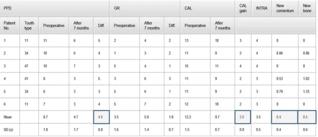
Secondary outcomes were the change in PPD, CAL and REC measured at baseline and before biopsy with the same type of periodontal probe (PCPUNC 15; Hu-Friedy;
Chicago, IL, USA) and by the same calibrated examiner. Furthermore, plaque and bleeding scores and in Study 2 the depth of the intrabony component (INTRA) were also calculated. CEJ was used as the reference point. Where CEJ was not visible, the restoration margin was considered instead. Clinical recordings were made at six sites per tooth (mesio-buccal, mid-buccal, disto-buccal, mesio-lingual, mid-lingual and disto- lingual). The studies report the measurements at the same site (deepest at baseline) of the selected defect. Measurements were rounded up to the nearest millimetre. Examiner calibration included CAL and PPD measurements on five periodontal patients with similar disease severity, but other, than the patients enrolled in the study. Data were captured from six sites per tooth from all quadrants by the same way and same type of probe as described above. Measurements were repeated alike, 90 minutes apart.
Calibration was accepted, if at least 90% of the collected figures were reproduced within a millimetre difference.
In addition, standardized long cone radiographs were taken at baseline, after three months and before biopsy for the radiological evaluation, utilizing commercial plastic film holder individualized by silicone putty impression material [110].
Reconstructive periodontal surgery
All surgeries were performed by the same, experienced periodontist in both trials, respectively. Prior to surgery clinical parameters were recorded as described above.
Patients were asked to rinse with 0.2% chlorhexidine (Curasept ADS 220; Curaden, Kriens, Switzerland) for two minutes before perioral disinfection. In local anaesthesia (articain 80 mg + epinephrine 0.024 mg; Ultracain D-S forte; Aventis Pharma, Frankfurt am Main, Germany) full thickness mucoperiosteal flaps were reflected following intracrevicular incisions at the investigated site with additional one to two teeth apart. No releasing incisions were deemed necessary in Study 2, whereas vertical
releasing incisions were implemented if necessary for improve access in Study 1.
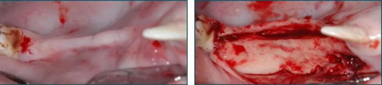
Granulation tissue was removed and the roots were meticulously debrided by means of ultrasonic and hand instruments (Fig. 1c, 2c).
The following measurements were then made in Study 1: distance from CEJ to the bottom of the defect (CEJ–BD) and distance from CEJ to the most coronal extension of the alveolar bone crest (CEJ–BC). The intrabony component of the defect was defined as (CEJ–BD) − (CEJ–BC). INTRA was measured in Study 2 thereafter. By using a round diamond bur (1 mm diameter) a notch was placed at the bottom of the defect on the test tooth which was previously scheduled for extraction (Fig. 1d, 2d). Thus, any PDL tissue which later developed coronally to this notch on the root surface would be de novo formed connective tissue and clearly distinguishable in histological sections of biopsies.
After defect debridement, in Study 1, the involved root surfaces were conditioned for 2 minutes with Ethylene Diamine Tetraacetic Acid (EDTA) gel (PrefGel; Straumann;
Basel, Switzerland) in order to remove the smear layer, according to the manufacturer’s instruction [111]. The defect, the underlying bone and the surrounding mucoperiosteal flaps were thoroughly rinsed with sterile saline to remove all EDTA residues. Following root conditioning, EMD (Emdogain®; Straumann; Basel, Switzerland) was applied on the root surfaces (Fig. 1e). The defects were then filled with a mixture of EMD + BCP (Straumann BoneCeramic®; Straumann; Basel, Switzerland) (Fig. 1f, g). Where needed, the periosteum, at the base of the mucoperiosteal flaps was incised to allow tension-free flap closure in coronal position.
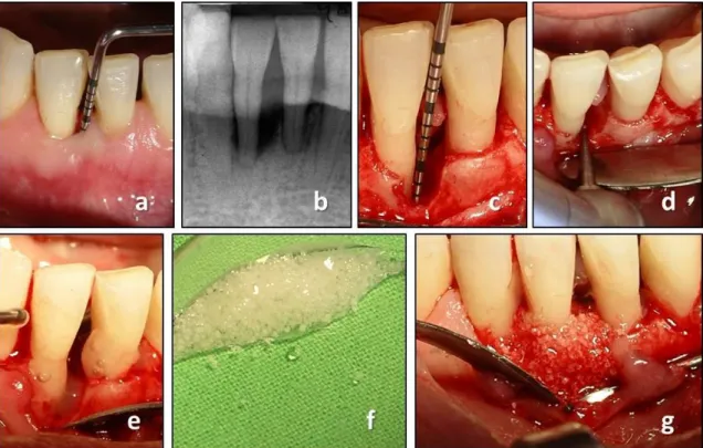
Fig. 1 Reconstructive periodontal surgery with the use of EMD + BCP. (a) preoperative clinical measurement demonstrates a PPD of 9 mm; (b) preoperative radiograph demonstrates the presence of a deep intrabony defect; (c) intraoperative measurement following debridement;
(d) placement of the notch by means of a round bur indicating the bottom of the intrabony defect; (e) application of EMD; (f) BCP mixed with EMD prior to application; (g) periodontal defect filled with EMD + BCP.
No root conditioning or any other surface modifications were applied in Study 2. The intrabony defect was subsequently filled with the nano-HA paste (Ostim®; Heraeus Kulzer; Hanau, Germany) and adjusted to the alveolar crest, according to the manufacturer’s instruction (Fig. 2e, f).
The mucoperiosteal flaps were then repositioned and secured with a combination of suspended vertical mattress and single interrupted sutures (non-resorbable, monofilament; Dafilon 5/0; Braun Aesculap; Tuttlingen, Germany) in order to achieve tensionless flap closure. Teeth were splinted in case of extreme mobility.
Postoperative care
All patients were postoperatively administered antibiotics (amoxicillin 500 mg + clavulanic acid 125 mg; Augmentin 625; GlaxoSmithKline; Brentford, Middlesex, UK)
Fig. 2 Reconstructive periodontal surgery with the use of nano-HA. (a) preoperative clinical measurement demonstrates a PPD of 10 mm; (b) preoperative radiograph demonstrates the presence of a deep intrabony defect; (c) the intraoperative situation revealed a deep one- and two-wall intrabony defect following debridement; (d) placement of the notch by means of a round bur indicating the bottom of the intrabony defect; (e) application of nano-HA; (f) periodontal defect filled with nano-HA.
three times daily for seven days and painkiller (diclofenac 75mg; Diclofenac Duo, Pharmavit, Veresegyház, Hungary) according to individual need. Subjects were advised not to brush the surgical area but rinse with 0.2 % chlorhexidine two times daily for 90 seconds during the postoperative four weeks. Subjects were asked to refrain any mechanical plaque control at the surgical site. Sutures were removed 10 to 14 days after surgery. Then the patients resumed tooth cleaning with the use of a soft brush.
Additional appointments including oral hygiene instructions and professional supragingival tooth cleaning were performed fortnightly during the first twelve postoperative weeks. After this period and until biopsy removal, recall appointments were scheduled monthly. Neither subgingival instrumentation nor periodontal probing was performed during the entire experimental period.
Re entry, biopsy and histological procedure
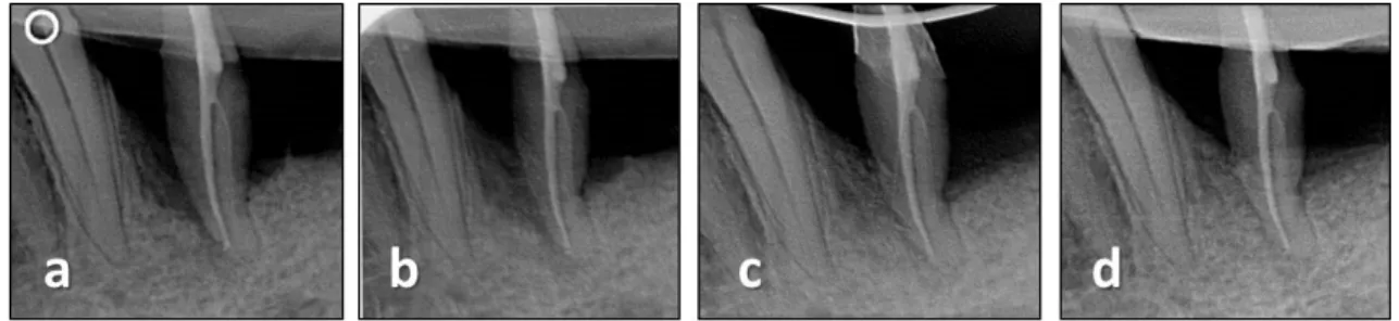
After a healing period of nine (Study 1) or seven months (Study 2) the teeth were removed together with some of their surrounding periodontal tissues following the same type of local anaesthesia and presurgical preparations (Fig. 3a-c, 4a-c).
Fig. 3 Re-entry and biopsy after nine months of healing following treatment of EMD +BCP.
(a) stable-looking soft tissue conditions; (b) radiograph indicates radiopaque tissue in the former defect; (c) decoronated tooth with some of the surrounding tissues
Fig. 4 Re-entry and biopsy after seven months of healing following treatment of nano-HA. (a) at 7 months following surgery, a substantial reduction of PPD was measured; (b) radiograph indicates mineralisation in the former defect; (c) removed tooth with some of the surrounding tissues
Specimens were subsequently placed in 10% buffered formalin for fixation. The extraction sites were then augmented with the use of various types of bone substitutes and barrier membranes (Study 1) and with DBBM (TutoDent Microchips; Tutogen;
Neunkirchen, Germany) in conjunction with a resorbable collagen membrane of bovine pericardium origin (TutoDent Membrane; Tutogen; Neunkirchen, Germany) in Study 2.
According to local need, connective tissue graft was transplanted in order to increase the volume of keratinised gingiva [112]. After the healing of the extraction sites, subjects received either implant-based crowns or fixed partial dentures as part of the prosthetic rehabilitation.
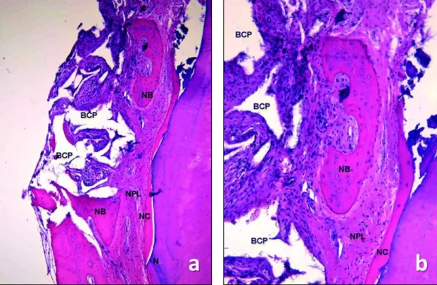
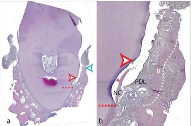
The block biopsies were fixed in 10 % buffered formalin, decalcified in EDTA for a period of 4–6 weeks (depending on tooth/root volume) and dehydrated in graded series of ethanol. Immediately prior to embedding in paraffin, the roots/teeth were split in two along their long axis either in bucco-oral or mesiodistal direction (depending on the location of the deepest site of the defect) exactly at the notch indicating the site of interest. Thus, each biopsy provided two specimen blocks without the need for extensive cutting. Twenty sections from each of the two blocks per specimen were obtained with the microtome, set at 5 to 8 μm, and subsequently stained with hematoxylin–eosin and further with the oxone-aldehyde-fuchsin-Halmi staining method in Study 2. One experienced examiner measured the parameters by means of a computer-assisted toolbox. The field of view in the light microscope (Olympus DH 50;
Olympus Denmark AS; Ballerup, Denmark) was examined on an LCD flat screen monitor through live streaming, which were also captured by a digital camera (Olympus DP 71; Olympus Denmark AS; Ballerup, Denmark). The following parameters were measured: (i) height of cementum regeneration: distance between the apical extension of the defect (bottommost point of the notch) and the coronal extension of a continuous layer of new cementum or cementum-like deposit on the planed root (millimetres); (ii) bone regeneration height: distance between the bottommost point of the notch and the coronal extension of regenerated alveolar bone along the planed root (millimetres) (The coronal extension of regenerated bone was defined as the most coronal level where the PDL space had an almost normal width); (iii) root resorption: combined linear heights of distinct resorption lacunae on the planed root (millimetres); (iv) ankylosis: combined linear heights of ankylotic union between the regenerated alveolar bone and the planed root (millimetres). Digital images were evaluated using a software program (SIS AnalySIS Auto Software 3.2; Soft Imaging System; Münster, Germany). The histomorphometric evaluation was carried out under ×25 to ×100 magnification. In the absence of a visible notch, the apical extension of instrumentation was used as landmark for the histomorphometric measurements.
Sources of support
Study 1 was supported by Institute Straumann, Basel, Switzerland. Study 2 was self- funded and supported by a research grant from Heraeus Kulzer, Hanau, Germany.
Candidate’s contribution to the studies
Fine-tuning of the protocol, study co-ordination, patient recruitment, assessment, data acquisition, patient management, compensatory treatment provision, revision and approval of the manuscript (Study 1); study design and protocol, study co- ordination, patient recruitment, surgical treatment (both stages), patient management, compensatory treatment provision, analysis and interpretation of data, preparation of the manuscript were carried out by the candidate of the present thesis. (Study 2)
4.3. Regeneration of peri-implant dehiscence defects (Study 3) Experimental model and management
In the present experiment twelve female Göttingen minipigs (20 months of age and average body weight of 35 kg) were used. Before each surgical procedure, the animals were fasted overnight, weighed and pre-medicated. Each subjects received an injection of analgesia (Temgesic®; Schering-Plough; Brussels, Belgium) on the day of the surgery and on the next 3 days. StreptocillinVet® 250 + 200 mg/ml (Boehringer Ingelheim;
Copenhagen, Denmark) was started one day before the procedure and was continued for seven days. The animals were anaesthetized according to a standard procedure using ketamine (Ketalar® 50 gm/ml; Pfizer; Sollentuna, Sweeden) and midazolam (Hameln 5 mg/ml; Pharmaceuticals GmbH; Hameln, Germany). After disinfection of the surgical site with 0.2% chlorohexidine solution (Corsodyl®; GlaxoSmithKline; Brentford, Middlesex, UK), local infiltration anaesthesia (Lidocaine 2% with 1:80,000 of epinefrine; HenrySchein Inc; Port Washington, NY, USA) was given. During the operation, additional 10 ml of ketamine and 1.5 ml of midazolam were administered when needed. Soft diet was provided daily during the whole course of the study. For documentation purposes digital photography and radiographs of the inserted implants were taken. The experimental protocol was approved by the Ethical Committee of the University of Lund (Sweden).
Study sequence
All surgical procedures were performed by two experienced periodontists. The treatment sequences in the study were:
1. Extractions and creation of “chronic” defect. Day 0/Baseline
2. Randomised implantation with/without creation of acute defect, with/without the use of test materials (4 groups: T1, T2, N, P). 3 Months
3. Uncovering of the implants and connection of abutments in different length (2 groups: loaded/non-loaded). 6 Months
4. Termination, histological analysis. 8 Months
Extraction and creation of chronic defect – Day 0/Baseline
Following intracrevicular incisions and careful elevation of a full thickness muco- periosteal flap, the premolars and the first molar were extracted in both hemi-mandibles in all animals (Fig. 9a). Particular care was taken to ensure that no root remnants were left in the extraction sockets. Following tooth extraction, bone defect was created on both sides of the mandible by removal of the buccal plate with a chisel, on a length of 40 mm and a height of 6 mm (Fig. 9b). The lingual cortical bone wall was left intact.
The surgical area was then rinsed with saline. Flap closure was achieved with absorbable sutures (Vicryl® 4–0; Ethicon GmbH; Norderstedt, Germany) (Fig. 9c) and the defect was allowed to heal for three months (Fig. 9d).
Fig. 9 (a) Day 0: Extraction of P2, P3, P4 and M1. (b) Day 0: Removal of the buccal plate by a chisel. (c) Day 0: Flap closure and suturing. (d) Three months: Healing following teeth extraction and buccal plate removal (’chronic defect’).
Implantation with/without creation of acute defect, with/without the use of test materials (first stage surgery) – 3 months
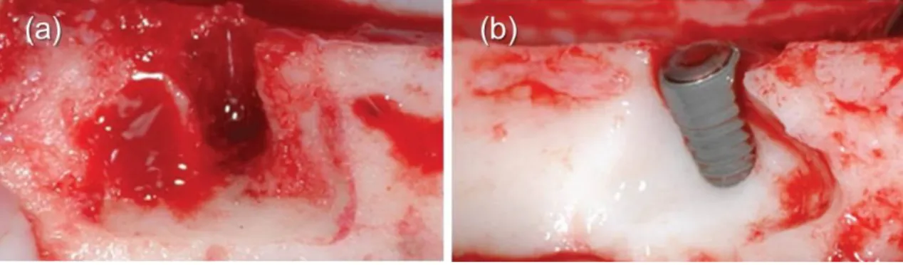
After a healing period of three months, following midcrestal incision and full thickness flap reflection, novel, titanium, bone level, 4.1x8 mm dental implants with a modified hydrophilic surface (Straumann®, Bone Level, SLActive implants; Straumann, Basel, Switzerland) were inserted either into the reduced thickness alveolar ridge (chronic defect and pristine site), or into a combined chronic and a surgically created standardised acute buccal dehiscence-type defect. Triangular shaped acute defects (height: 6 mm, apical mesio-distal width: 12 mm, bucco-lingual width: 2 mm) were prepared with a rotating surgical drill with ample saline irrigation. Defects were created in order to mimic real clinical circumstances of a partially edentulous ridge.
Consequently, the coronal 6 mm of the implants were exposed on the buccal aspect.
Only the apical 2 mm of the implants was in the alveolar bone around the whole circumference (Fig. 10). In total, 48 bone level implants with a diameter of 4.1 mm and a length of 8 mm were placed in 12 minipigs (four implants/animal; two in each hemi-mandible). All implants were inserted according to the guidelines of the manufacturer. Profile drilling (without tapping) was performed and the implants were inserted in such a way that the implant shoulder was aimed to match the bone crest level. Insertion torque was always higher than 35 Ncm.
Fig. 10 Schematic drawing of the chronic and acute, standardised, peri- implant, critical size dehiscence defect.
According to a computer-generated randomization scheme, four groups were created:
P (12 implants) – In the positive control group, twelve implants were inserted into the
chronic defects resulting from the 3-month healing following buccal bone plate removal at the time of teeth extraction (pristine sites) (Fig. 11).
Fig. 11 Three months after baseline: Implant placement in group P (pristine site). Minor buccal and/or lingual dehiscence defects were occasionally present.
N (12 implants) – In the negative control group the chronic and acute dehiscence defects were left untreated as such the buccal surface of the implant remained exposed (negative control) (Fig. 12).
Fig. 12 Three months after baseline: Creation of standardized acute dehiscence-type defect.
(a) Standardized defects were created around the osteotomy by removing part of the buccal bone. (b) The resulting dehiscence defects presented triangular-shaped base and the following dimensions: 6 mm apico-coronally, 12 mm mesio-distally, 2 mm bucco-lingually.
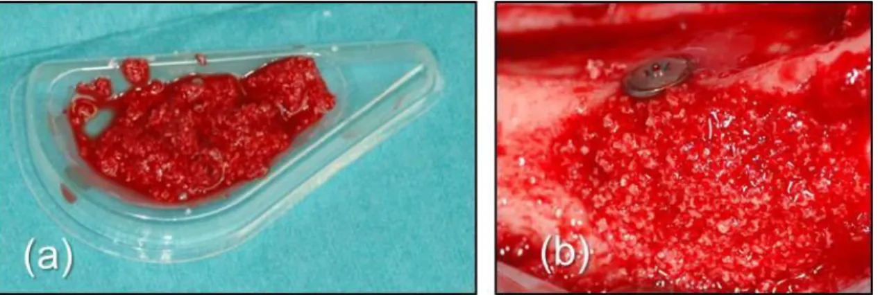
T1 (12 implants) – The exposed implant surface in the dehiscence defects was treated with BCP (Straumann BoneCeramic®; particle size 500–1000 µm; Straumann; Basel, Switzerland). The BCP particles were mixed with autologous blood collected in the surgical site. The graft material was then applied in the dehiscence defect in contact with the exposed implant threads. Care was taken to avoid overfilling or coverage of the implant head (Fig. 13).
Fig. 13 Three months after baseline: Group T1: (a) BCP was mixed with autologous blood from the surgical site; (b) The exposed implant surface in the dehiscence defects was covered with the BCP.
T2 (12 implants) – BCP and the novel PEG membrane (Straumann MembraGel®; Straumann; Basel, Switzerland) was used for the treatment of the dehiscence defects.
The synthetic bioresorbable polyethylene glycol hydrogel membrane was activated by mixing its components according to the manufacturer’s guidelines. The membrane was applied as a gel with a continuous flow over the bone substitute and the adjacent bone.
(Fig. 14a-c). After the cross-linking reaction of the membrane components was completed and the membrane set, the margins were smoothed with a sharp scalpel blade. The membrane extended on the adjacent bone surface approximately 3 mm further than the grafted area and covered completely the implant head (Fig. 14d).
Fig. 14 Three months after baseline: Group T2: (a) BCP was placed onto the defect; (b) The two component of the hydrogel was mixed prior to application; (c) The polyethylene glycol hydrogel membrane (PEG) was applied on and around the BCP and the implant; (d) The set, polimerised membrane in place.