REGULAR ARTICLE
Protective effects of the neuropeptide PACAP in diabetic retinopathy
Krisztina Szabadfi&Tamas Atlasz&Peter Kiss&
Dora Reglodi&Aliz Szabo&Krisztina Kovacs&
Balint Szalontai&Gyorgy Setalo Jr.&Eszter Banki&
Katalin Csanaky&Andrea Tamas&Robert Gabriel
Received: 22 November 2011 / Accepted: 25 January 2012 / Published online: 17 February 2012
#Springer-Verlag 2012
Abstract Pituitary adenylate cyclase activating polypeptide (PACAP) is a neuropeptide with highly potent neurotrophic and neuroprotective effects. PACAP and its receptors occur in the retina and PACAP has been applied in animal models of metabolic retinal disorders to reduce structural and functional damage. Furthermore, PACAP has been implicated as a po- tential anti-diabetic peptide. Our aim has been to investigate,
by using a complex morphological, immunochemical and molecular biological approach, whether PACAP attenuates diabetic retinopathy. Diabetes was induced in rats with a single streptozotocin injection. PACAP was injected intravi- treally into one eye (100 pmol) three times during the last week of a 3-week survival period. Retinas were processed for the following procedures: routine histology, immunohisto- chemistry (single and double labeling, whole-mount), quanti- tative reverse transcription with the polymerase chain reaction and Western blotting. Cone photoreceptors and dopaminergic amacrine and ganglion cells degenerated in diabetic retinas and glial fibrillary acidic protein were upregulated in Müller glial cells. The number of cones, the length of their outer segments and the cell number in the ganglion cell layer were decreased. PACAP ameliorated these structural changes.
Moreover, PACAP increased the levels of PAC1-receptor and tyrosine-hydroxylase as detected by molecular biological methods. Thus, PACAP has significant protective effects in the diabetic retina. PACAP treatment attenuates neuronal cell loss in diabetic retinopathy, the protective effects of PACAP probably being mediated through the activation of PAC1- receptor. These results suggest that PACAP has a therapeutic potential in diabetic retinopathy.
Keywords Retina . Diabetic retinopathy . PACAP.
Neuroprotection . PAC1-R . Rat (adult male Wistar)
Introduction
Sight-threatening neurodegenerative diseases of the retina such as glaucoma, ischemia, retinitis pigmentosa and diabetic retinopathy involve the progressive loss of retinal neurons.
Diabetic retinopathy develops in patients with both type 1 and 2 diabetes, progressing to blindness in about 5% of cases This work was supported by the Hungarian Science Research Fund
OTKA K100144, K72592, 78480, 72315, 78223, SROP-4.2.2/B-10/1- 2010-0029, Bolyai Scholarship, Lendulet Program of Hungarian Academy of Sciences and Richter Gedeon Foundation, SROP-4.2.1.B- 10/2/KONV-2010-0002.
K. Szabadfi (*)
:
R. GabrielDepartment of Experimental Zoology and Neurobiology, Institute of Biology, University of Pecs,
Ifjusag Street 6, 7624 Pecs, Hungary
e-mail: kriszta.szabadfi@gmail.com T. Atlasz
Department of Sportbiology, University of Pecs, Pecs, Hungary
P. Kiss
:
D. Reglodi:
E. Banki:
K. Csanaky:
A. Tamas Department of Anatomy, PTE-MTA PACAP Research Team, University of Pecs,Pecs, Hungary A. Szabo
:
K. KovacsDepartment of Biochemistry and Medical Chemistry, University of Pecs,
Pecs, Hungary B. Szalontai
Department of Plant Physiology, University of Pecs, Pecs, Hungary
G. Setalo Jr.
Department of Medical Biology, University of Pecs, Pecs, Hungary
(Engerman and Kern 1995; Kempen et al. 2004). As the worldwide prevalence of diabetes continues to increase, dia- betic retinopathy is a leading cause of vision loss in developed countries (Fong et al.2004). The inability of the retina to adapt to metabolic stress leads to a glucose-mediated microvascular disease together with chronic inflammation, finally causing degeneration and dysfunction in the retina (Liu et al.2008).
Diabetes induces neurodegeneration and dysfunction as evidenced by several studies demonstrating decreases in en- zyme activities and declining levels of growth and transcrip- tion factors in retinal neurons (Seki et al.2004; Park et al.
2006). The rate of neuronal loss is slow, leading to a gradual cumulative reduction mostly in amacrine and ganglion cells (Barber et al.1998; Gastinger et al.2006,2008). Functional deficits can be found in both rod and cone responses related to the loss of outer segment membranes and/or shortened outer segments (Holopigian et al.1997).
The functional loss of photoreceptors in diabetes is coupled with the failure of color vision and contrast sensitivity (Alvarez et al.2010). In animal models of diabetes, losses in the function of rod photoreceptors and the inner retina can be seen as early as 2 days after the induction of diabetes. As a consequence, some inner retinal responses and the cone response and oscil- latory potentials remain consistently depressed (Phipps et al.
2004). Hyperglycemia disrupts cone photoreceptors as evidenced by prominent pathological features at light and electron microscopic levels and dysfunctional cone-mediated electroretinograms. Kurtenbach et al. (2006) have reported that cone metabolism in hyperglycemia is significantly enhanced compared with rod metabolism. The photoreceptor functions are strongly supported by inner retinal neuromodulators, such as dopamine.
In the retina, dopamine plays a central role in the adaptation to light. Dopamine is synthesized and released by a sparse population of amacrine cells, composing less then 1% of ama- crines in mammals (Witkovsky and Schutte1991; Gustincich et al.1997; Jeon et al.1998). These cells can be labeled by tyrosine-hydroxylase (TH) immunohistochemistry and are lo- cated in the innermost sublayer of the inner nuclear layer in the rat retina (Witkovsky and Schutte 1991). Alterations in the dopaminergic system are thought to be among the first significant events in the development of diabetic retinopathy (Nishimura and Kuriyama 1985; Northington et al. 1985;
Fernstrom et al.1986). Such alterations include the degenera- tion of some dopaminergic amacrine cells during the early stage of diabetes (Seki et al.2004). In early stages of diabetes, loss of oscillatory potentials are thought to be a sign of degeneration of dopaminergic amacrine cells (Shirao and Kawasaki1998). The increase of glial fibrillary acidic protein (GFAP) expression in Müller glial cells has been reported to be another early histo- logical change in diabetic rats (Lieth et al.1998).
Several animal models of diabetes have been established, with streptozotocin being widely used to induce experimental
type I diabetes. It selectively destroys pancreatic beta cells and thus is suitable for studying the short- and long-term conse- quences of diabetes and protective strategies (Portha et al.
1974; Szkudelski2001). Several protective agents have been examined in the hope of providing a cure for diabetic retinop- athy (Mohamed et al.2007). Among the numerous potential therapeutic agents in diabetic retinopathy, growth factors are promising tools against various pathological components. For example, ciliary neurotrophic factor, pigment epithelium de- rived factor and nerve growth factor have all been shown to be protective in animal models of diabetic retinopathy (Tao2006;
Steinle 2010; Shen et al. 2010). Pituitary adenylate cyclase activating polypeptide (PACAP), a neurotrophic member of the vasoactive intestinal peptide/secretin/glucagon peptide superfamily, is a neuropeptide with highly potent neu- roprotective and general cytoprotective effects. PACAP and its receptors (PAC1-R, VPAC1 and VPAC2) occur in the retina and PACAP treatment has been demonstrated to be protective in several retinal pathologies. PACAP has been shown to attenuate retinal damage in excitotoxic, ischemic, traumatic and UV-light-induced retinal degeneration (Atlasz et al.
2010). Several lines of evidence suggest that PACAP might be of therapeutic potential in diabetes (Yamamoto et al.2003).
First, PACAP stimulates insulin secretion from beta cells in a glucose-dependent manner. Second, PACAP protects beta cell damage induced by various insults such as oxidative stress, cytokines and gluco- and lipotoxicity. In addition to the effects in pancreas islets, a few studies have indicated that PACAP also attenuates diabetes-related pathologies. Systemic PACAP treatment decreases streptozotocin-induced nephropathy in rats, as shown by laboratory and histological parameters (Li et al. 2008). PACAP also attenuates experimental neuropathy (Dickinson et al. 1999). Finally, a recent study has revealed that hyperglycemia-induced microvascular endo- thelial cell growth can be inhibited by PACAP treatment in vitro (Castorina et al.2010).
Based on the potency of PACAP in diabetes and related conditions in addition to its retinoprotective actions, our aim has been to investigate whether PACAP could attenuate diabetic retinopathy, one of the most devastating consequen- ces of diabetes. With this aim, we have used a complex morphological, immunohistochemical and molecular bio- logical approach.
Materials and methods
Animals
Experimental procedures were carried out in accordance with approved protocols (University of Pecs; BA02/2000- 24/2011). Adult male Wistar rats (n053) were housed under light/dark cycles of 12:12 h. For the induction of diabetes,
70 mg/kg streptozotocin (Sigma, Hungary) was injected intravenously (n038). Blood glucose concentration was measured weekly, before and after diabetes induction, for 3 weeks (Glucotrend Accu-Check, Roche, Hungary). Rats with glucose levels higher than 11 mmol/l were classified as diabetic; others that recovered with normalized glucose levels were excluded from our study. Most animals, however, devel- oped massive diabetes with classical symptoms such as poly- uria, polydipsia and excessive weight loss.
PACAP (100 pmol/5μl saline solution; 20μM) was injected (n038) into the vitreous body of the right eye with a Hamilton syringe under isoflurane anesthesia. Animals received PACAP treatment three times during the last week of survival: 7, 4 and 1 day before being killed. The same volume of saline was injected into the other eye to serve as untreated diabetic control (n038). The dose of PACAP was based on previous observa- tions where this dose was effective (Tamas et al. 2004). A separate group of animals without induced diabetes served as controls injected with saline (n015), including animals with PACAP injected into the vitreous of the right eye (n015). Three weeks after diabetes induction and 1 day after the last PACAP treatment, animals were killed with an overdose of anesthetic and eyes were processed for further examination.
Histology
After removal, eyes (n03 per group) were immediately dissect- ed in ice-cold phosphate-buffered saline (PBS; Spektrum3D, Hungary) and fixed in 4% paraformaldehyde (PFA; Merck, Hungary) dissolved in 0.1 M phosphate buffer (PB; Spek- trum3D). Retina histology was performed as previously de- scribed (Atlasz et al.2010). Embedded tissues were cut at a thickness of 2μm, stained with 1% toluidine blue (Sigma) and examined in a Nikon Eclipse 80i microscope. Measurements were taken with the SPOT Basic program. Six tissue blocks from at least three animals were prepared from each group.
Central retinal areas within 1 and 2 mm from the optic disc were used for measurements (n02-5 measurements from one tissue block). Sections in which the inner nuclear layer (INL) was thicker than four rows of cells or in which the ganglion cell layer (GCL) appeared to be thicker than a single cell row were excluded from evaluation. The following parameters were measured: (1) cross section of the retina from the outer limiting membrane to the inner limiting membrane; (2) the width of individual retinal layers; (3) the number of cells/100μm section length in the GCL. Statistical comparisons were made by using an analysis of variance (ANOVA) followed by Tukey-B post hoc analysis (P<0.05). Data are presented as means ± SEM.
Immunohistochemistry
For immunohistochemical analysis, eyes were dissected in ice-
cold PBS immediately after death and were fixed in 4% PFA Table1AntibodiesusedinimmunohistochemicalandWesternblottingexperiments(THtyrosine-hydroxylase,PAC1-RPACAP1-receptor,GFAPglialfibrillaryacidicprotein,GAPDH glyceraldehyde3-phosphatedehydrogenase) PrimaryantibodiesSupplierRaisedinDilutionSecondaryantibodiesSupplierDilutionMethods Anti-THMillipore,USAMouse1:1000AlexaFluor488Invitrogen,USA1:1000Immunhistochemistry (crosssectionandAnti-PAC1-RKindgiftofProf.SeijiShiodaRabbit1:100AlexaFluor568 whole-mount)Anti-GFAPSigma-Aldrich,HungaryRabbit1:500AlexaFluor568 Anti-THMillipore,USAMouse1:500Horseredish-peroxidase- conjugatedsecondary antibody BioRad,Hungary1:3000Westernblot Anti-PAC1-RKindgiftofProf.SeijiShiodaRabbit1:500 Anti-GAPDHSigma-Aldrich,HungaryMouse1:5000
for 2 h at room temperature (n06 in control group, n09 in diabetic group). Tissues were then washed in PBS and cryo- protected in 20% sucrose (Sigma) at 4°C. For cryostat section- ing, retinas were embedded in tissue-freezing medium (Tissue- Tek, Sakura Finetech, The Netherlands), cut in a cryostat (Leica, Germany) at a thickness of 10μm radially. Sections were mounted on subbed slides. At least 48 sections/eye were examined. Antibody details are presented in Table1. Anti-TH, anti-PACAP1-receptor (PAC1-R), anti-GFAP antibodies and fluorescein-isothiocyanate-conjugated peanut agglutinin (PNA, Vector) were used overnight at room temperature. Next day, the sections were incubated for 2 h at room temperature with the corresponding secondary fluorescent antibodies, namely, Alexa Fluor 488 and 568, in the dark (Table 1) and then coverslipped by using Fluoromount-G (Southern Biotech, USA). For control experiments, primary antibodies were
omitted. Cross-reactivity of the non-corresponding secondary antibodies with the primary antibodies was also checked.
Micrographs were taken via a Fluoview FV-1000 Laser Con- focal Scanning Microscope (Olympus, Japan) and further pro- cessed with Adobe Photoshop 7.0 program. Images were adjusted for contrast only; they were aligned, arranged and labeled by using the functions of the above program. Images were evaluated by an examiner blinded to the treatment.
For whole-mounts (n04 in control groups; n010 in diabetic and in diabetes + PACAP-treated groups), we removed the lens and the vitreous body and post-fixed the retinas in PFA for 12 h at 4°C. After pre-incubation with normal goat serum (Southern Biotech) for 1 h, whole-mounts were incubated with primary antibody against TH for 72 h and visualized with Alexa Fluor 488. Images from whole-mounts were used for counting TH-positive cells.
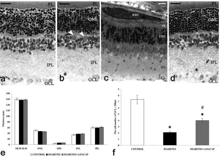
Fig. 1 Representative sections of control (a), diabetic (b, c) and diabetes + 3×PACAP-treated (d) rat retinas stained with toluidine blue for light microscopy. Morphological differences were not found be- tween the examined groups, except for some non-typical components in diabetic retinas (arrowheadsgranulocytes).Bar20μm. Significant differences could not be observed in the retinal layers between the three groups (e) in cross-sections of the retina (OLM-ILM) and widths of the individual layers (ONL, OPL, INL, IPL). The number of cells per
100 μm GCL length decreased in induced diabetes but this was attenuated by PACAP treatment (f). *P<0.05 compared with control retinas;#P<0.05 compared with diabetic retinas (RBCred blood cells, PEpigment epithelium,PLphotoreceptor layer,OLMouter limiting membrane,ONLouter nuclear layer,OPLouter plexiform layer,INL inner nuclear layer,IPLinner plexiform layer,GCLganglion cell layer, ILMinner limiting membrane)
PNA-labeling was quantified from cryosections, by counting cone terminals/100 μm retina and measuring the length of their outer segments. Data are presented as mean ± SEM (ANOVA, Tukey-B post hoc analysis;P<0.05;
P<0.01).
Analysis by quantitative polymerase chain reaction Retinas from nine animals in each group were removed for quantitative polymerase chain reaction (qRT-PCR) analysis. Total RNA was extracted from the samples by using TRI Reagent (Applied Biosystems, USA) according to the manufacturer’s instructions. RNA was quantified
by the NanoDrop quantification system (Thermo Scien- tific, USA). Reverse transcription was performed by using a First Strand cDNA Synthesis Kit (Fermentas, USA) with oligodT(18) primer. qRT-PCR was performed by using the Step One Real-Time PCR System (Applied Biosystems). Reaction mixtures consisted in: 10 μl Maxima SYBR Green 2× mix including ROX (Fermen- tas), 0.6 μl each of two primers (at 10 pmol/μl), 2 μl cDNA samples in a 20-μl final volume. Relative product quantities were determined by using Step One software and the ΔΔCt analysis method. β-Actin was used as endogenous control. Relative expression data are pre- sented (± SEM; Welch’s test; P<0.05).
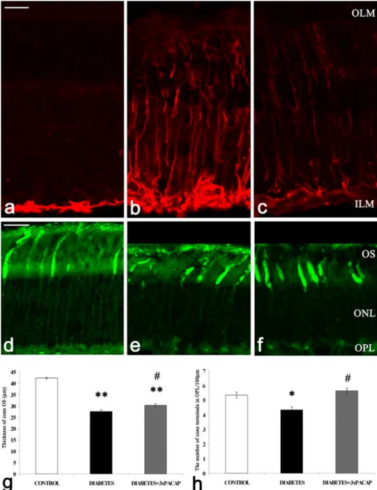
Fig. 2 Glial fibrillary acidic protein (GFAP) labeling of Müller glial cells and peanut agglutinin (PNA) labeling of cone photoreceptors. GFAP immunostaining was observed in the endfeet of Müller glial cells in the control condition (a). In diabetic retinas, GFAP immunoreactivity was strong in the processes of Müller cells and, spanning from the inner (ILM) to the outer (OLM) limiting membrane, within their endfeet at the vitreoretinal border (b).
Reduced GFAP
immunopositivity was detected in the processes of Müller glial cells in the whole retina of diabetes + PACAP-treated animals (c). Both the outer seg- ments of photoreceptors (OS) and the terminals showed PNA bind- ing in the control condition (d). In diabetic retinas, a damaged shorter OS and a decreased number of terminals were ob- served (e,g,h). The preserved structure of cones in
diabetes + PACAP-treated retinas was observed and the number of cone terminals and the thick- ness of the OS layer were also significantly increased by PACAP treatment (f,g,h) compared with diabetic retinas. *P<0.01, **P<0.001 compared with control retinas;#P<0.01 compared with diabetic retinas (ONLouter nuclear layer,OPLouter plexiform layer).Bar20μm
Western blot analysis
A separate group of retinas (n06 in control groups;n07 in diabetes and in diabetes + PACAP-treated groups) was re- moved 24 h after the last PACAP treatment. Samples were processed for Western blot analysis as described earlier (Racz et al.2006). Membranes were probed overnight at 4°C with anti-TH, anti-PAC1-R and anti-GAPDH antibodies (Table1).
Membranes were washed six times for 5 min in TRIS-buffered saline (pH 7.5) containing 0.2% Tween prior to the addition of goat anti-rabbit or anti-mouse horseradish-peroxidase- conjugated secondary antibody. The antibody-antigen com- plexes were visualized by chemiluminescence. After the scan- ning step, results were quantified by the NIH ImageJ program.
The retina from each rat was analyzed twice in two separate experiments. Data are represented by pixel density in arbitrary units (± SEM; ANOVA, Tukey-B post hoc analysis;P<0.05;
P<0.001).
Results
Histology
The characteristic layers of the mammalian retina were readily visible in normal control preparations: the photoreceptor layer (PL), outer nuclear layer (ONL), outer plexiform layer (OPL), inner nuclear layer (INL), inner plexiform layer (IPL) and GCL (Fig.1a). PACAP treatment caused no alterations in control retinas (Atlasz et al. 2010; data not shown). Light microscopic examination did not reveal gross histological differences between the four examined groups (control; con- trol + PACAP; diabetes; diabetes + PACAP; Fig.1a-d). Some non-typical components such as granulocytes (Fig.1b) and precipitated red blood cells (Fig.1c) were found in diabetic retinas. Among the examined morphometric parameters, a significant decrease was observed only in the cell number of the GCL in diabetic retinas (Fig.1f); this was attenuated by PACAP treatment (Fig.1f). No significant difference was seen in the other morphometric parameters.
Analysis of Müller glial cells and cone photoreceptors GFAP positivity was selectively localized to the endfeet of Müller cells in control retinas (Fig.2a). Increased GFAP im- munoreactivity was observed in the entire diabetic retina (Fig.2b), whereas in diabetes + PACAP-treated retinas, a slight decrease in the number of GFAP-positive processes of Müller glial cells was detected (Fig.2c).
PNA was used to label the cone photoreceptors. Staining extended up to the outer segments and down to the terminals of the cones in the OPL in control conditions (Fig.2d). In diabetic retinas, the outer segments were degenerated (Fig. 2e); the
number of cone terminals and the thickness of the outer seg- ment layer were also significantly decreased compared with the Fig. 3 Representative fields of whole-mount preparations of control, diabetic and diabetes + PACAP-treated retinas with tyrosine- hydroxylase (TH)-immunoreactive cell bodies and dendrites. The cells were at higher density in the control retinas (a,b) compared with diabetic samples in which the arborization of the TH-positive cells was also reduced (c,d). Large numbers of TH-positive neurons were present in PACAP-treated retinas than in diabetic retinas. The dendritic tree was also retained (e,f). Bars 200μm (a,c,e), 40μm (b,d,f). The decrease in cell number in diabetes was significant and was attenuated by PACA treat- ments (g). *P<0.05 compared with control retinas;#P<0.05 compared with diabetic retinas
control retinas (Fig.2g, h). In PACAP-treated retinas, terminals of the cone photoreceptors were better preserved (Fig.2f) and a significant elevation of the outer segments length and also of their number was observed (Fig.2g, h).
Immunohistochemical analysis of dopaminergic and PAC1-R-containing elements
Alterations of the retinal dopaminergic system with the loss of dopaminergic amacrine cells are among the first detect- able signs in diabetic retinopathy (Nishimura and Kuriyama 1985; Northington et al.1985; Fernstrom et al.1986). TH is the rate-limiting enzyme for dopamine synthesis and allows the direct visualization of dopaminergic amacrine cells in the retina by means of immunohistochemistry (Brecha et al.
1984). To examine the effect of diabetes and the protective action of PACAP, TH immunolabeling was used in whole- mount preparations and cross cryosections.
We discerned changes in the number and the anatomy of cells, such as in soma shape and the arborization pattern (Fig.3a-f). Not only were the dendrites fewer but also their appearance was varicose and sometimes segmented in diabetic retinas. Loss of arborization in surviving cells was also ob- served in diabetes (Fig.3c, d) compared with control retinas (Fig. 3a, b). The trophic action of PACAP could also be detected in the retained structure of the processes (Fig.3e, f).
In addition, alterations in their density as induced by diabetes
and by diabetes + PACAP treatment were also analyzed. The density of TH-positive cells was significantly reduced in dia- betes compared with controls and an increase in the cell numbers could be detected after PACAP treatment (Fig.3g).
In cross sections, dopaminergic amacrine cells and their processes were observed at the border of INL and IPL, the somas being present in the innermost part of the INL (Fig.4a1). The processes of the cells were thinner in diabetic animals (Fig. 4b1). After PACAP treatment, we observed a labeling pattern according to which most of the features of the control preparations were preserved. However, TH immuno- labeling seemed to be stronger in the proximal sublaminas of the IPL in these sections (Fig.4c1).
To analyze whether PACAP could protect dopaminergic amacrine cells involving the upregulation of its main receptor, PAC1-R, we use double-immunofluorescent staining. In nor- mal retinal sections, PAC1-Rs were found mostly in the inner retinal layers, on the cell bodies of INL and GCL and some processes showed an intense signal in distal sublaminas of the IPL (Fig.4a2). Both the diabetic and the diabetes + PACAP- treated retinas showed PAC1-R immunoreactivity (Fig. 4b2, c2), with less intense staining in the diabetic retinas (Fig.4b2).
Dopaminergic amacrine cells did not express PAC1-R under normal or diabetic conditions (Fig. 4a3, b3). However, in diabetes + PACAP-treated retinas, a few double-labeled cells were found (Fig.4c3), with the majority of TH-positive cells still not showing PAC1-R immunoreactivity (not shown).
Fig. 4 Retinal cross sections from the three different groups of animals.
Localization of dopaminergic amacrine cell with anti-TH (green) and anti-PACAP1-receptor (PAC1-R;red) antibodies. TH immunoreactivity was seen in the cell bodies and in the arborization of the dopaminergic amacrine cells (a1,b1,c1). PAC1-R-positive cells and their processes
were localized in the inner retinal layers (INL, IPL;a2,b2,c2). Coloc- alization was not found in the control and diabetic retinas (a3,b3) but a few TH/PAC1-R double-labeled cells were detected in the PACAP- treated diabetic retinas (c3,yellowcell body).Bars20μm
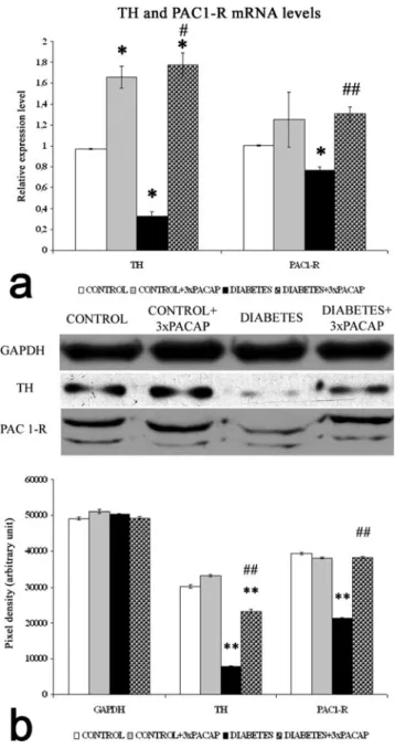
Examination of TH and PAC1-R at a molecular level The relative expression of TH and PAC1-R mRNA levels for the whole retina were measured by qRT-PCR (Fig.5a). The
relative expression values were standardized to β-actin mRNA levels in the same mRNA samples. We observed a significant reduction in the expression levels of TH and PAC1-R mRNA in diabetic retinas compared with control samples (P<0.05). In diabetic retinas, the TH transcript level was 35% of control levels, whereas PAC1-R mRNA was 76%
of control levels. In whole retina samples from diabetes + PACAP-treated animals, mRNA levels for TH and PAC1-R were significantly increased compared with those of the dia- betic retinas (Fig.5a).
TH and PAC1-R proteins (Fig.5b) were determined in the retinas by Western blotting. The band intensities were normal- ized to GAPDH levels. TH and PAC1-R protein levels in the retinas of diabetic animals were significantly lower than those in the control groups. Higher TH and PAC1-R protein levels were observed in diabetes + PACAP-treated retinas compared with diabetic animals (Fig.5b).
Discussion
PACAP is suggested to have therapeutic potential in diabetes and related conditions. In addition, PACAP is protective in ischemic, excitotoxic and UV-A light-induced retinal degen- eration. PACAP is therefore expected, at least partially, to counteract diabetes-induced structural changes in the retina.
Indeed, our present findings confirm that intravitreal admin- istration of PACAP has significant protective effects in the retina of streptozotocin-induced diabetic rats.
Standard histological examination revealed a lower cell number in the GCL in the diabetic group than in the controls.
This observation confirmed the results of others (Zeng et al.
2000) demonstrating a similar reduction in the number of retinal ganglion cells in short-term (4 weeks) diabetes. The significant decrease of the cell number in the GCL was atten- uated by PACAP treatment. Studies in streptozotocin-induced diabetic rats have now identified neurochemical and molecular changes of neural and glial elements in early stages. The inner two-thirds of the retina, in which the retinal capillaries reside, host close to 50 different cell types (MacNeil and Masland 1998) that are intimately interdependent in physiology and influence one another in pathophysiological circumstances.
Retinal Müller glial cells are activated in diabetes as marked by the elevated expression of GFAP seen in our experiments.
This stress response is reduced after PACAP treatment.
Dominant photoreceptor degeneration occurs in streptozotocin-treated rats (Park et al. 2003). Pathological features such as morphologically abnormal cones are related to diabetes and such dysfunctional photoreceptors result in attenuated electroretinogram responses (Alvarez et al.
2010). The observed cone degeneration is attenuated by PACAP. Both the cone terminal number and shape of the cone outer segments remain close to normal.
Fig. 5 Molecular biological analyses of control, of control + 3×PACAP, of diabetic and of diabetes + 3×PACAP treated retinas. mRNA levels of TH and PAC1-R (a) were determined by qRT-PCR and their values were standardized toβ-actin levels in the same RNA samples. Levels of TH and PAC1-R mRNAs were significantly decreased in diabetic retinas, compared with control retinas. mRNAs from diabetes + PACAP-treated retinas showed a significant increase compared with diabetic retinas (a).
Western blotting for the protein levels of TH and PAC1-R (b) in the retinas.
GAPDH served as normalization control. Retinal TH and PAC1-R in the PACAP-treated diabetic eyes were significantly higher than those in vehicle-treated diabetic eyes. Data are presented as mean ± SEM values. *P<0.05, **P<0.001 compared with control;#P<0.05,##P<0.001 compared with diabetic retinas (ANOVA with Tukey-B post hoc analysis or Welsch’st-tests for Western blot and qRT-PCR, respectively)
Changes in the dopaminergic system are among the first significant events in the development of diabetic retinopathy (Nishimura and Kuriyama 1985; Northington et al. 1985;
Fernstrom et al.1986). These findings are in accord with our observations, where both morphological deterioration of dopaminergic cells and a loss in their number have been seen. The loss of these neurons might play a critical role in the progress of visual deficits in diabetes (Gastinger et al.
2006). PACAP administration leads to a nearly normal dopaminergic amacrine cell number and preserves the den- dritic arbor. These findings corroborate recent results obtained in ischemic and excitotoxic retinal degeneration, where cyto- protective effects of PACAP have also been established (Atlasz et al.2010).
Mapping the dopaminergic system by TH staining in visual pathologies has demonstrated the pivotal role of this system in mediating visual function. Dopamine has multiple, important and complex roles in retinal function (light/dark adaptation, retina development, etc.), not only as a neurotransmitter but also as a neuromodulator and neurotrophic factor (Brandies and Yehuda2008). Retinal dopaminergic amacrine cells play a central part in reconfiguring retinal function according to prevailing illumination conditions. The light-dependent re- lease of dopamine in the retina is an important component of light-adaptation mechanisms. Dopaminergic amacrine cells can activate cones through type D2 dopamine receptors. They have also been implicated in the regulation of the local retinal environment (Cameron et al. 2009). The degeneration of dopaminergic amacrine cells disables the cross-talk between scotopic and photopic signaling pathways in photoreceptor cells and in the whole retinal circuity. Together with the loss of dopaminergic cells, a decrease in the protein and mRNA levels of TH has been found, similar to the findings by Seki and coworkers (2004). Overall TH protein levels are reduced reflecting the decrease in the cell density of dopaminergic amacrine cells. A decline in PAC1-R levels has also been observed in the diabetic retinas. PACAP application upregu- lates PAC1-R expression in our experiments. The results of our double-immunolabeling study suggest that there is a cor- relation between TH and PAC1-R upregulation: PAC1-R im- munoreactivity appears in some dopaminergic cells in diabetes + PACAP-treated retinas. PACAP might help to preserve the adequate dopamine concentration in the retina essential for retaining function and structure. Our goal in the near future is to correlate these changes with the activity of the apoptotic pathways and the functional viability of the retina under these conditions.
In conclusion, we have shown, for the first time, that PACAP treatment attenuates neuronal cell loss, especially of cones, dopaminergic amacrine and ganglion cells, in experi- mental diabetic retinopathy. Evidence has also been provided for general structural protection. Thus, our results suggest that PACAP has therapeutic potential in diabetic retinopathy.
Acknowledgements The authors thank Prof. Gábor Tóth (University of Szeged, Hungary) for synthesizing PACAP and Prof. Seiji Shioda (Showa University, Japan) for the PAC1-R antibody. The authors also thank Brian K. Lucas for proof-reading the manuscript.
References
Alvarez Y, Chen K, Reynolds AL, Waghorne N, O'Connor JJ, Kennedy BN (2010) Predominant cone photoreceptor dysfunction in a hyperglycaemic model of non-proliferative diabetic retinopathy.
Dis Model Mech 3:236–245
Atlasz T, Szabadfi K, Kiss P, Racz B, Gallyas F, Tamas A, Gaal V, Marton Z, Gabriel R, Reglodi D (2010) Review of pituitary adenylate cyclase activating polypeptide in the retina: focus on the retinoprotective effects. Ann N Y Acad Sci 1200:128–139 Barber AJ, Lieth E, Khin SA, Antonetti DA, Buchanan AG, Gardner
TW (1998) Neural apoptosis in the retina during experimental and human diabetes. Early onset and effect of insulin. J Clin Invest 102:783–791
Brandies R, Yehuda S (2008) The possible role of retinal dopaminergic system in visual performance. Neurosci Biobehav Rev 32:611– 656
Brecha NC, Oyster CW, Takahashi ES (1984) Identification and char- acterization of tyrosine hydroxylase immunoreactive amacrine cells. Invest Ophthalmol Vis Sci 25:66–70
Cameron MA, Pozdeyev N, Vugler AA, Cooper H, Iuvone PM, Lucas RJ (2009) Light regulation of retinal dopamine that is independent of melanopsin phototransduction. Eur J Neurosci 29:761–767 Castorina A, Giunta S, Mazzone V, Cardile V, D'Agata V (2010)
Effects of PACAP and VIP on hyperglycemia-induced prolifera- tion in murine microvascular endothelial cells. Peptides 31:2276– 2283
Dickinson T, Mitchell R, Robberecht P, Fleetwood-Walker SM (1999) The role of VIP/PACAP receptor subtypes in spinal somatosensory processing in rats with an experimental peripheral mononeuropathy.
Neuropharmacology 38:167–180
Engerman RL, Kern TS (1995) Retinopathy in animal models of diabetes. Diabetes Metab Rev 11:109–120
Fernstrom MH, Volk EA, Fernstrom JD, Iuvone PM (1986) Effect of tyrosine administration on dopa accumulation in light- and dark- adapted retinas from normal and diabetic rats. Life Sci 39:2049– 2057
Fong DS, Aiello LP, Ferris FL 3rd, Klein R (2004) Diabetic retinopathy.
Diabetes Care 27:2540–2553
Gastinger MJ, Singh RS, Barber AJ (2006) Loss of cholinergic and dopaminergic amacrine cells in streptozotocin-diabetic rat and Ins2Akita-diabetic mouse retinas. Invest Ophthalmol Vis Sci 47:3143–3150
Gastinger MJ, Kunselman AR, Conboy EE, Bronson SK, Barber AJ (2008) Dendrite remodeling and other abnormalities in the retinal ganglion cells of Ins2 Akita diabetic mice. Invest Ophthalmol Vis Sci 49:2635–2642
Gustincich S, Feigenspan A, Wu DK, Koopman LJ, Raviola E (1997) Control of dopamine release in the retina: a transgenic approach to neural networks. Neuron 18:723–736
Holopigian K, Greenstein VC, Seiple W, Hood DC, Carr RE (1997) Evidence for photoreceptor changes in patients with diabetic retinopathy. Invest Ophthalmol Vis Sci 38:2355–2365
Jeon CJ, Strettoi E, Masland RH (1998) The major cell populations of the mouse retina. J Neurosci 18:8936–8946
Kempen JH, O’Colmain BJ, Leske MC, Haffner SM, Klein R, Moss SE, Taylor HR, Hamman RF (2004) The prevalence of diabetic retinopathy among adults in the United States. Arch Ophthalmol 122:552–563
Kurtenbach A, Mayser HM, Jägle H, Fritsche A, Zrenner E (2006) Hyperoxia, hyperglycemia, and photoreceptor sensitivity in normal and diabetic subjects. Vis Neurosci 23:651–661
Li M, Maderdrut JL, Lertora JJ, Arimura A, Batuman V (2008) Renoprotection by pituitary adenylate cyclase-activating polypep- tide in multiple myeloma and other kidney diseases. Regul Pept 145:24–32
Lieth E, Barber AJ, Xu B, Dice C, Ratz MJ, Tanase D, Strother JM (1998) Glial reactivity and impaired glutamate metabolism in short-term experimental diabetic retinopathy. Diabetes 47:815–820
Liu X, Mameza MG, Lee YS, Eseonu CI, Yu CR, Kang Derwent JJ, Egwuagu CE (2008) Suppressors of cytokine-signaling proteins induce insulin resistance in the retina and promote survival of retinal cells. Diabetes 57:1651–1658
MacNeil MA, Masland RH (1998) Extreme diversity among amacrine cells: implications for function. Neuron 20:971–982
Mohamed Q, Gillies MC, Wong TY (2007) Management of diabetic retinopathy. A systematic review. JAMA 298:902–916
Nishimura C, Kuriyama K (1985) Alterations in the retinal dopaminergic neuronal system in rats with streptozotocin-induced diabetes. J Neurochem 45:448–455
Northington FK, Hamill RW, Banerjee SP (1985) Dopamine-stimulated adenylate cyclase and tyrosine hydroxylase in diabetic rat retina.
Brain Res 337:151–154
Park JW, Park SJ, Park SH, Kim KY, Chung JW, Chun MH, Oh SJ (2006) Up-regulated expression of neuronal nitric oxide synthase in experimental diabetic retina. Neurobiol Dis 21:43–49 Park SH, Park JW, Park SJ, Kim KY, Chung JW, Chun MH, Oh SJ
(2003) Apoptotic death of photoreceptors in the streptozotocin- induced diabetic rat retina. Diabetologia 46:1260–1268
Phipps JA, Fletcher EL, Vingrys AJ (2004) Paired-flash identification of rod and cone dysfunction in the diabetic rat. Invest Ophthalmol Vis Sci 45:4592–4600
Portha B, Levacher C, Picon L, Rosselin G (1974) Diabetogenic effect of streptozotocin in the rat during the perinatal period. Diabetes 23:889–895
Racz B, Gallyas F Jr, Kiss P, Toth G, Hegyi O, Gasz B, Borsiczky B, Ferencz A, Roth E, Tamas A, Lengvari I, Lubics A, Reglodi D
(2006) The neuroprotective effects of PACAP in monosodium glutamate-induced retinal lesion involve inhibition of proapop- totic signaling pathways. Regul Pept 137:20–26
Seki M, Tanaka T, Nawa H, Usui T, Fukuchi T, Ikeda K, Abe H, Takei N (2004) Involvement of brain-derived neurotrophic factor in early retinal neuropathy of streptozotocin-induced diabetes in rats: therapeutic potential of brain-derived neuro- trophic factor for dopaminergic amacrine cells. Diabetes 53:2412– 2419
Shen X, Zhong Y, Xie B, Cheng Y, Jiao Q (2010) Pigment epithelium derived factor as an anti-inflammatory factor against decrease of glutamine synthetase expression in retinal Müller cells under high glucose conditions. Graefes Arch Clin Exp Ophthalmol 248:
1127–1136
Shirao Y, Kawasaki K (1998) Electrical responses from diabetic retina.
Prog Retin Eye Res 17:59–76
Steinle JJ (2010) Topical administration of adrenergic receptor pharma- ceutics and nerve growth factor. Clin Ophthalmol 4:605–610 Szkudelski T (2001) The mechanism of alloxan and streptozotocin
action in B cells of the rat pancreas. Physiol Res 50:537–546 Tamas A, Gabriel R, Racz B, Denes V, Kiss P, Lubics A, Lengvari I,
Reglodi D (2004) Effects of pituitary adenylate cyclase activating polypeptide in retinal degeneration induced by monosodium- glutamate. Neurosci Lett 372:110–113
Tao W (2006) Application of encapsulated cell technology for retinal degenerative diseases. Expert Opin Biol Ther 6:717–726 Witkovsky P, Schutte M (1991) The organization of dopaminergic
neurons in vertebrate retinas. Vis Neurosci 7:113–124
Yamamoto K, Hashimoto H, Tomimoto S, Shintani N, Miyazaki J, Tashiro F, Aihara H, Nammo T, Li M, Yamagata K, Miyagawa J, Matsuzawa Y, Kawabata Y, Fukuyama Y, Koga K, Mori W, Tanaka K, Matsuda T, Baba A (2003) Overexpression of PACAP in transgenic mouse pancreatic beta-cells enhances insulin secretion and ameliorates streptozotocin-induced diabetes. Diabetes 52:1155– 1162
Zeng XX, Ng YK, Ling EA (2000) Neuronal and microglial response in the retina of streptozotocin-induced diabetic rats. Vis Neurosci 17:463–471