The nuclear activity of the actin-binding Moesin protein is necessary for gene expression in Drosophila
Csaba Bajusz1,2, Ildiko Krist o1, Csilla Abonyi1, Tomas Venit3, Viktor Vedelek4, Tamas Lukacsovich5, Attila Farkas1, Peter Borkuti1,6, Zoltan Kovacs1,6, Izabella Bajusz1, Annamaria Marton1,
Csaba Vizler1, Zoltan Lipinszki7, Rita Sinka4, Piergiorgio Percipalle3,8and Peter Vilmos1
1 E€otv€os Lorand Research Network (ELKH), Biological Research Centre, Szeged, Hungary 2 Doctoral School of Biology, University of Szeged, Hungary
3 Biology Program, Science Division, New York University Abu Dhabi, UAE 4 Department of Genetics, University of Szeged, Hungary
5 Brain Research Institute, University of Z€urich, Switzerland
6 Doctoral School of Multidisciplinary Medical Science, University of Szeged, Hungary
7 Lend€ulet Laboratory of Cell Cycle Regulation, ELKH, Biological Research Centre, Szeged, Hungary 8 Department of Molecular Biosciences, The Wenner-Gren Institute, Stockholm University, Sweden
Keywords
Drosophila; Moesin; nucleus; Hsp;
transcription Correspondence
P. Vilmos, Biological Research Centre, Szeged 6726, Temesvari krt. 62, Hungary Tel:+36-62-599689
E-mail: vilmosp@brc.hu
(Received 19 September 2020, revised 22 January 2021, accepted 18 February 2021) doi:10.1111/febs.15779
Ezrin–Radixin–Moesin (ERM) proteins play an essential role in the cyto- plasm by cross-linking actin filaments with plasma membrane proteins.
Research has identified the nuclear localization of ERMs, as well as the involvement of a singleDrosophilaERM protein, Moesin, in nuclear mRNA exports. However, the question of how important the nuclear activity of ERM proteins are for the life of an organism has so far not been explored.
Here, we present the first attempt to reveal thein vivorelevance of nuclear localization of Moesin inDrosophila. With the help of a nuclear export signal, we decreased the amount of Moesin in the nuclei of the animals. Furthermore, we observed various developmental defects, demonstrating the importance of ERM function in the nucleus for the first time. Transcriptome analysis of the mutant flies revealed that the lack of nuclear Moesin function leads to expres- sion changes in nearly 700 genes, among them heat-shock genes. This result together with additional findings revealed that inDrosophilathe expression of protein chaperones requires the nuclear functions of Moesin.
Database
GEO accession number:GSE155778.
Introduction
The presence of cytoskeletal proteins in the nucleus is undisputed today, and it is now clear that they are involved in fundamental nuclear functions[1–4]. How- ever, the biological relevance of the activity of cytoskeletal proteins in the nucleus has not yet been determined and is still debated. This is primarily due
to the fact that cytoskeletal proteins have essential activities in the cytoplasm, and therefore, the elimina- tion or inhibition of these proteins affects their activity in the cytoplasm as well. Furthermore, with the excep- tion of actin, the mechanism of their nuclear transport has not been resolved. Therefore, the investigation of
Abbreviations
CRM1, chromosomal region maintenance 1; ERM, Ezrin–Radixin–Moesin; FCS, fetal calf serum; GO, gene ontology; hsp, heat-shock protein; Moe, Moesin; NES, nuclear export signal; PCA, principal component analysis; RT, room temperature; WGA, wheat germ agglutinin;
WT, wild-type.
4812 The FEBS Journal288(2021) 4812–4832ª2021 The Authors.The FEBS Journalpublished by John Wiley & Ltd on behalf of
their nuclear activity separately from their cytoplasmic functions has been an enormous challenge.
The evolutionarily highly conserved family of the actin-binding, cytoskeletal ERM (ezrin, radixin, and moesin) proteins consists of three closely related par- alogs: ezrin, radixin, and moesin [5,6]. They are all present in vertebrates, whereas other species such as invertebrate only have one ERM gene [7]. ERM pro- teins function as general cross-linkers between plasma membrane proteins and the cortical actin cytoskeleton [8]. They can assemble multiprotein complexes at the membrane–cytoskeleton interface, thereby regulating numerous signal transduction pathways, and they also function as both upstream and downstream effectors of Rho GTPases. The lack of ERM function results in serious defects or mortality in vertebrates [9] and is lethal in invertebrate species[10,11].
Recent studies indicate that the regulation of corti- cal actin dynamics is only a part of the function of ERMs. Similarly to their major binding partner, actin and other cytoskeletal proteins [1], ERMs have been found to localize to the cell nucleus in numerous spe- cies[12–15]. InDrosophila melanogaster, the sole ERM representative, Moesin (Moe), has been shown to be a functional component of the nucleus by participating in mRNA export[16]. However, thein vivoimportance of the nuclear localization and activity of ERMs is not known at present. Here, we demonstrate that the con- stant removal of Moe from the nucleus, with the help of a nuclear export signal (NES), results in abnormal development, sterility, and physiological problems in Drosophila. We also show that Moe plays role in the expression of 686 genes, among them heat-shock (hsp) genes. Our study provides the first direct evidence that the activity of the actin-binding ERM proteins in the cell nucleus is essential for the normal development and life of an organism.
Results
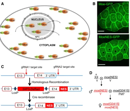
Generation of themoeNESmutant
To obtain more insight into the nuclear activities of Drosophila Moe and to determine the biological rele- vance of this nuclear localization, we created mutant animals with decreased level of nuclear Moe. We fused the protein with a NES which ensures constant removal of Moe from the nucleus (Fig.1A). We chose the evolutionarily highly conserved, chromosomal region maintenance 1 (CRM1)-recognized NES sequence motif of the cAMP-dependent protein kinase inhibitor alpha (PKIA) [17] and tested the effect of this NES-tag on the intracellular distribution of Moe
in a pilot experiment. The nuclei of Drosophila larval salivary gland cells expressing GFP-labeled MoeNES protein from a transgene showed unambiguous decrease in the nuclear Moe level (Fig.1B).
We then tagged the Moe protein in situ with the NES sequence using CRISPR-Cas9 technology[18]. In Drosophila, themoe gene is located on the X chromo- some and with the use of alternative START sites and introns it encodes 13 different transcripts that give rise to seven different protein isoforms. We inserted the NES sequence in front of the STOP codon between the 14th exon and the 30UTR which are present in all 13 moe transcripts. For the selection of the mutants, the fluorescent dsRed expressing marker gene (3xP3 >dsRed) flanked by two loxP recombination sites was inserted into the last intron (Fig.1C). From the embryos co-injected with the guide RNAs (gRNAs) and the donor construct, four independent mutant lines, hereafter referred to as moeNES, were generated. Next, the dsRed marker gene was removed with the help of Cre recombinase, leaving a single loxP sequence in the last intron of themoegene. Sequencing of the mRNA expressed from themoeNESgene verified successful insertion of the NES sequence, the lack of the marker gene, and the normal excision of the last intron (data not shown).
We noticed that all the progenies of the females car- rying at least one copy of themoeNESallele were ster- ile. The sterility was specific to mothers only; the progeny of hemizygous mutant males (moeNES/Y) were fertile, indicating that the phenotype is due to a domi- nant maternal effect. Therefore, to maintain the moeNES allele for future experiments, mutant males were crossed to females carrying attached X chromo- somes (X^X/Y) to establish the stable moeNES stock.
The attached X chromosome consists of two full- length X chromosomes sharing a common centromere so that they are always inherited together. As a result, in crosses of moeNES males to females carrying an attached X, male progenies inherit theirmoeNES bear- ing X from their father and their Y from their mother, while the females never carry themoeNESallele. In this way, the problem of maternal female sterility is elimi- nated. The moeNES/Y mutant males were viable and fertile; however, to analyze females carrying at least one copy of the moeNES allele, moeNES males were crossed in every experiment to females carrying a wild- type (WT) X chromosome (w1118) or a null allele (moeG0415) of moe. The resulting female progeny (moeNES/moeG0415, referred to asmoeNESmutant in the text) expressing only the MoeNESprotein were used in the assays (Fig.1D) and compared throughout the experiments to control animals carrying either themoe
null (moeG0415/w1118) or the moeNES allele (moeNES/ w1118) in heterozygous form.
The cytoplasmic activity of the MoeNES protein is unaffected
The complete loss of Moe functions is lethal to the fly.
Null mutant animals die at the third larval stage [19,20]. In contrast, the moeNES mutant females and males were viable, suggesting that at least the main cytoplasmic functions of Moe are unaffected. In the next experiments, we aimed to confirm this notion.
In order to verify that the NES-tag does not inhibit the activation and activity of the Moe protein, salivary glands ofmoeNESmutant males and ovaries ofmoeNES mutant mothers were stained with an anti-phospho- ERM antibody specifically recognizing the activated form of Moe [21]. The immunostaining revealed that the MoeNES protein is activated through phosphory- lation and, similarly to the WT protein, it primarily localizes to the cell cortex (Fig.2A). This demonstrates the F-actin-binding ability of MoeNES and suggests that its molecular cross-linking function is intact. The
quantitation of the immunostainings showed that, in addition to localization, the nucleo-cytoplasmic distri- bution of P-Moe is normal in the moeNES mutant ovary (Fig. 2A’). The measurement of the levels of P- Moe and Moe in the ovary by western blotting (Fig. 2A”) suggested that the amount of P-Moe rela- tive to total Moe amount in the mutant differs only by 10% from that of the control (1.38 and 1.52 Moe/P- Moe ratio in the mutant and the control, respectively).
The quantitation also revealed that the total amount of Moe is reduced by 30% in the moeNES mutant (142 837 vs. 99 815 intensity values in the control and the mutant, respectively).
The female fly possesses two ovaries joined with a common oviduct, and around 16 ovarioles per ovary.
The ovarioles are parallel strings of 6–7 progressively older egg chambers. The egg chamber is covered with a monolayer of somatic epithelial cells, the follicle cells. In the egg chambers, 15 polyploid nurse cells are connected to the oocyte and deposit a large amount of RNA and protein into it. During stage 10 of oogene- sis, nurse cells produce unipolar, filopodia-like actin cables between their cell membrane and nucleus to
′
′
′
A
C
D B
Fig. 1.Rationale of the experimental system. (A) NES ensures that Moe is constantly exported from the nucleus while its cytoplasmic functions remain intact. NPC: Nuclear pore complex. (B) Live images of larval salivary glands expressing Moe-GFP or MoeNES-GFP (green) demonstrate that MoeNES-GFP is not present in the nucleus. Representative images of salivary glands of eight larvae examined each genotype in one experiment. Scale bars, 50lm. (C)In vivotagging of themoegene with NES signal using the CRISPR-Cas9 system. The NES sequence was inserted between the protein-coding sequence and the 30UTR. (D) ThemoeNESallele is maintained with the help of attached X females. To generate moeNES mutant females (highlighted in red), the moeNES/Y mutant males (red) are crossed in all experiments with females carrying amoenull allele (moeG0415).
moe[NES]
oocyte oocyte
B
F
0.8 1.0 1.2 1.4 1.6 1.8 2.0
Nucl./Cytopl. Fluor. Ratio of Med-GFP
n= 46 n= 40 WT moe[NES]
P< 0.05
*
WT moe[NES]
Actin Actin
WGA WGA
+DAPI +DAPI
nurse cells nurse cells D
Staufen-GFPOskar-GFPVasa-GFP
WT
C E WT
moe[NES]
P-Moe DAPIPhalloidin merge
WTmoe[NES]WTmoe[NES]
A
A′′
A′
WT moe[NES]
moe[NES]
WT
Med-GFP Med-GFP
w 2.0 2.5 3.0 3.5
Length/widthratio
moe[NES]
WT n.s.
0.0 0.2 0.4 0.6 0.8
w[1118]
moe[G0415]
moe[NES]
moe[G0415]
Nucl/cytofluor. intensity ratio of P-Moe
n = 128 n = 128 n.s.
42 Act
72 Moe
95
55 140
α- Moe α- P-Moe
α- Act
kDa
prevent the nuclei from moving and to block the squeezing of cytoplasm into the oocyte [22]. Moe is required for the formation of these tethering actin cables in the ovary and anchors the cortical actin net- work to the cell membrane in the developing oocyte.
Therefore, the impairment of Moe function leads to the disorganization of the microfilament network [21]
and the detachment of cortical actin from the cell membrane[19]. Visualization of the actin cytoskeleton and the membranes at stage 10 of oogenesis in ovaries of control (moeG0415/w1118) and moeNES mutant females demonstrated that the intracellular position of nurse cell nuclei and the actin network are normal in the mutant (Fig.2B). The perturbation of nurse cell filamentous actin cable organization or their integra- tion with the cortical actin network blocks nurse cell dumping which in turn results in reduced embryonic size [23]. The regular size and shape of the embryos laid bymoeNESmutant mothers confirmed further that the cytoskeletal functions of Moe are unaffected (Fig.2C).
The intact cytoskeletal network of the oocyte also ensures the correct localization of maternal factors within the cytoplasm, which is necessary for the for- mation of germ cell precursors at the posterior pole of the developing embryo. Moe plays essential role in this process, as demonstrated previously by the mislocaliza- tion of maternal factors Oskar, Staufen, and Vasa, and significantly reduced number of pole cells [19,21]
in the embryos of moeNES mutant mothers. However, we found that in the eggs laid by moeNES mutant females, the maternal factors Oskar, Staufen, and Vasa
all localize properly at the posterior pole of the oocyte or the early embryo (Fig. 2D), and at the cellulariza- tion phase, the number of embryonic pole cells is nor- mal also (Fig. 2E), providing additional evidence that the cytoplasmic functions of MoeNES are normal.
The MoeNES protein is exported from the nucleus by the CRM1 (XPO1) export factor which recognizes the NES motif. In the next set of experiments, we investigated whether the expression of the MoeNES protein alters the normal activity of CRM1, for exam- ple, by overloading the CRM1-mediated nuclear export pathway. We thus compared the ratio of nuclear and cytoplasmic fluorescent intensity values of the GFP-tagged Medea (SMAD4) protein, a known cargo of the CRM1 exportin [24,25], in the nurse cells of WT and mutant ovaries (Fig.2F). We expected the nuclear accumulation of Med-GFP if the export is overloaded, but the protein exhibited very similar nuclear-cytoplasmic distribution in normal and mutant nurse cells. This confirms that the CRM1 nuclear export pathway functions normally in the moeNES mutant and that reduced CRM1 pathway activity can- not be responsible for the observed phenotypes.
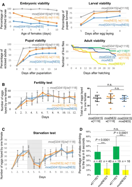
Lack of nuclear Moesin causes diverse phenotypes
The moeNES mutant males and females were viable and fertile; however, they exhibited numerous pheno- types (Fig. 3A). These defects, such as slow develop- ment, decreased lifespan, egg production and climbing ability, as well as male genitalia rotation, were of
Fig. 2.The MoeNES protein functions normally in the cytoplasm. (A) Immunostaining for phospho-Moe (P-Moe, white) in the larval salivary gland (upper rows) and the ovary (bottom rows) reveals that the MoeNES protein is phosphorylated and localizes in the cytoplasm normally.
Representative images of three independent experiments. w1118flies were used as WT. Scale bars, 50lm. (A’) Nuclear/cytoplasmic fluorescence intensity ratios in moeNES mutant and control ovaries of three independent experiments. Horizontal line represents mean fluorescent ratioSD, ‘n’is the number of cells examined, n.s.—not significant (unpairedt-test). (A”) Quantitation of the amount of Moe and P-Moe proteins in moeNES mutant (moeNES/moeG0415) and control (moeG0415/w1118) ovaries with western blotting. Representative pictures of one of three independent experiments. Actin was used as loading control. Act—Actin. (B) The organization of the actin network and the position of the nuclei are intact in stage 10B egg chambers ofmoeNES(moeNES/moeG0415) females. Phalloidin was used to stain for actin: green, WGA for membrane staining: magenta, DAPI: blue.w1118flies were used as WT. Representative images of at least 35 egg chambers examined in two independent experiments. Scale bars, 50lm. (C) The eggs laid bymoeNESmutant mothers have WT size and shape.w1118embryos were used as WT. Scale bars, 500lm. Dot plot data represent the meanSD of length/width ratio of eggs. n.s.— not significant (unpaired t-test). 100 embryos were measured each genotype. (D) Immunostaining reveals that the localization of the maternal factors Staufen and Oskar (green) are normal in the oocytes of stage 9 egg chambers and that Vasa (green) localizes properly in the embryos ofmoeNESmutant females. Representative images of one of three independent experiments. Scale bars, 10lm (Staufen and Oskar) and 100lm (Vasa). (E) The number of pole cells at the posterior pole of the embryo laid bymoeNESmothers is normal as reflected by the immunostaining for Vasa (green) and DAPI (blue).w1118embryos were used as WT. Scale bars, 10lm. Representative images of two independent experiments. (F) Medea localizes properly in moeNES mutant ovaries. Representative images of two independent experiments. The nuclear/cytoplasmic fluorescence ratio of the Medea-GFP signal reveals that the nuclear level of Medea (green) is unaffected in the ovaries ofmoeNESfemales.w1118flies were used as WT. Stage 8 egg chambers are shown. 20 egg chambers of 10 females were analyzed of each genotype. ‘n’indicates the total number of nurse cells measured. Error bars=SD, *P<0.05, unpaired t-test. Scale bars, 50lm.
zygotic origin. The ratio ofmoeNESmutant males with genitalia rotation (Fig.3B) was 32.4% (out of 145 ani- mals), and the frequency of this phenotype decreased to 12.3% (out of 171 animals) when the males were bearing a Y chromosome that contained one copy of the WTmoe gene (Dp(1:Y)619). This suggests that an extra copy of the moe gene can rescue genitalia rota- tion by about 60% and that the phenotype is due to the loss of Moe function.
In contrast to the zygotic phenotypes, maternal-ef- fect phenotypes appear in the F1 descendants of moeNES females, independently from the genotype of the progeny. The maternal phenotypes primarily affected the posterior part of the animals and caused rudimentary genitalia and, as a consequence, sterility (Fig.3A). The maternal-effect sterility of the F1
descendants was due to the undeveloped genitalia both in males and in females (Fig.3C). The visible pheno- types in the F1 progeny of moeNES mutant mothers were defects in tergite development and pigmentation, extra hairs, wing malformations, and dark and rough eye (Fig.3D).
The moeNES mutant stock was weak, and thus, we analyzed the viability of the animals at every main developmental stage. Out of 100 eggs laid by the moeNES mutant mothers, only 30% hatched, in con- trast to the control (moeG0415/w1118) where 98% of the embryos developed to larval stage. In addition, embry- onic viability further decreased with the aging of the mutant mothers (0% in the mutant vs. 92% in the control). The phenotype, especially at younger age, was partially rescued by a WT moe allele (moeNES/ A
B
C
C Fig. 3.Phenotypes ofmoeNESmutant
animals. (A) Both sexes ofmoeNESmutant parents (P) exhibited slow development, decreased lifespan, heat stress sensitivity, and lethality. The decreased climbing ability and genitalia rotation are manifested only in moeNESmales, whilemoeNESmutant females show grandchildless phenotype.
The F1 offsprings ofmoeNESmothers are 100% sterile and have various
developmental problems. (B) Rotated male genitalia inmoeNES/Y parents (32.4% of 145 males examined). (C) All the F1 offsprings ofmoeNESmutant mothers have rudimentary and nonfunctional genitalia (100% of 82 flies). (D) Developmental problems of F1 progeny include tergit development and pigmentation problems (10.7%), extra bristles (1.2%), absence of genitalia (5.8%), notched wing margin and reduced or missing cross-veins (4.9%), darker and rough eye (100%). Three hundred and fifty flies were analyzed.
Defects are circled or marked by red arrows. WT (w1118), A5-A6: abdominal segments 5 and 6, ACV: anterior cross-vein, PCV: posterior cross-vein.
w1118) (Fig.4A). DAPI staining of early embryos demonstrated that, similarly to the progeny of control females, 100% of the eggs laid by moeNES mutant mothers (n =100) pass the syncytial blastoderm stage, indicating that lack of fertilization does not contribute to embryonic lethality (not shown). After the embry- onic stage, 30% lethality was observed at both larval and pupal stages ofmoeNES mutants. The viability at these stages was fully rescued by one copy of the WT moe gene (moeNES/w1118), indicating that the lethality during development is caused by the loss of Moe func- tion. These data also demonstrated that the develop- ment of mutant animals is slower compared with the control. The viability experiments revealed a shortened
adult life span only in the case of moeNES/Y mutant males. In these males, life span was found to be decreased by~50% compared with mutant females or control males. Since these flies displayed a reduced hatching rate, we analyzed the fecundity of moeNES mutant females. They exhibited normal egg produc- tion, the daily egg yield as well as the total number of eggs laid by one female during the examined 12-day period were almost identical to those of the control females (Fig.4B).
In the next set of experiments, we inspected the pos- sibility that low energy homeostasis is contributing to the phenotype. We measured the egg production of mutant and control females during starvation. Two
0 25 50 75 100
5 6 7 8
Days after egg laying
0 10 20 30
0 4 8 12 16 20 24 28 32 36 0
25 50 75 100
3 4 5 6 7 10
Percentageof hatchedlarvae
Embryonic viability
Percentageof pupaeformed
Larval viability
Age of females (days)
0 25 50 75 100
8 9 10 11 12 13 14
Days after pupariation Pupal viabilty
Percentageof eclosedadults
moe[G0415]/w[1118]
moe[G0415]/w[1118]
moe[NES]/w[1118]
moe[G0415]/moe[NES]
moe[G0415]
moeNES moe[NES]/Y moe[G0415]/moe[NES]
moe[G0415]/moe[NES]
moe[NES]/w[1118]
moe[G0415]/w[1118]
moe[NES]/w[1118]
moe[G0415]/w[1118]
moe[NES]
w[1118]
w[1118]/Y
0 20 40 60
1. 2. 3. 4. 5. 6. 7. 8. 9. 10. 11. 12.
Days Fertility test
Numberof eggs layedbyonefemale
moe[G0415]/moe[NES]
moe[G0415]/w[1118]
moe[NES]/w[1118]
0 10 20 30 40 50 60 70
1. 2. 3. 4. 5. 6. 7. 8. 9. 10. 11. 12.
Days Starvation test
Numberof eggslayedbyonefemale
moe[G0415]/moe[NES]
moe[G0415]/w[1118]
moe[NES]/w[1118]
Adult viability
Numberof liveflies
Days after hatching
0 20 40 60
moe[G0415]
w[1118] moe[G0415]
moe[NES]
moe[NES]
w[1118]
Total no. of eggslayed byonefemale
n.s.
n.s.
0%
10%
20%
30%
40%
50%
60%
70%
80%
90%
Percentageof malesclimbing tothetop in 8 seconds
n.s.
P< 0.0001
***
P< 0.0001
7 DAYS 1 DAY
n = 40n = 40n = 19n = 16
***
A
B
C D
Fig. 4.moeNESmutants have physiological problems. (A)moeNESmutant animals show increased lethality at every developmental stage. Decreased lifespan at 25°C is observed only inmoeNES/Y males. (B) The fertility test shows that there is no difference between themoeNES/moeG0415 and control females in the number of laid eggs. The egg yield is normal during a 12- day period as shown in the graph (n=16, error bars=SD). The total number of eggs laid by one female is also normal (Dot plot).
Error bars=SD,n=11, n.s.—not significant (unpairedt-test). (C) The response ofmoeNESfemales to starvation is normal. Data represent the meanSD of the number of eggs laid by one female. 30 females were tested of each genotype. (D) moeNES/Y mutant males perform poorly in the climbing test: only 13% of them is able to climb the full distance in 8 s, in contrast to 48% of the WT. The ability of climbing did not decrease significantly with age. Data show quantitative evaluation (meanSD) of three independent experiments. n.s.— not significant,***P<0.0001 (unpairedt- test), ‘n’is the number of flies examined. In all panels, genotypes are indicated by the following colors: gray—moeG0415/w1118, orange—moeNES/w1118, blue—moeG0415/ moeNES, green—w1118/Y, yellow—moeNES/Y.
days of food deprivation resulted in a dramatic decrease in egg production in both groups, followed by a rapid return to normal levels. The kinetics of egg yield changes were the same in both groups (Fig.4C), indicating that moeNES mutants show normal physio- logical reactions to temporary food deprivation and suggesting that altered energy homeostasis is not responsible for reduced life span.
During the experiments, we also noticed that mutant flies move more slowly than the control animals; there- fore, we tested the basic locomotor behavior of the mutant (Fig.4D). For the investigation of locomotor activity, we performed a negative geotaxis assay and found that mutant males move significantly more slowly than WT males, with only 13% of them climb- ing to the top of the vial during 8 s, compared to 48%
in case of the control animals. In contrast, the locomo- tor activity of mutant females was normal (not shown). We also conducted the test on 1-week-old males, but the difference between the mutant and the control flies was about the same (59% vs. 10% in WT and mutant flies, respectively) (Fig.4D).
Transcriptome analysis reveals altered gene expression inmoeNESmutants
Because the progeny of mutant females exhibit various maternal-effect phenotypes (reduced viability, develop- mental defects, sterility), and the ovary is the place of very extensive gene expression, we performed mRNA- Seq analysis on ovaries of moeNES/moeG0415 mutant females, and compared their transcriptomes with moeNES/+ and moeG0415/+ controls (Fig.5). Principal component analysis (PCA) of the mRNA-Seq datasets showed 24% total variance in the first principal com- ponent (PC1) and clearly separated the mutant animals from the two controls (Fig.5A), indicating significant differences between the transcriptomes of WT (moeG0415/w1118) and mutant samples. Compared to
WT (moeG0415/w1118), approximately the same number of differentially expressed genes were upregulated (n =371) or downregulated (n=315) in the mutant (Fig.5B).
To gain more biologic insight, we performed Gene Ontology (GO) term analysis on statistically significant genes [twofold (log2(FC)≥1 and adjusted P-value of
<0.05 for upregulated genes, and log2(FC)≤-1 and adjusted P-value of <0.05 for downregulated genes]
differentially expressed between the genotypes moeG0415/w1118, moeNES/w1118, and moeNES/moeG0415 by subjecting the data (summarized in TableS1) to the GO database of Flybase (https://flybase.org/). We observed that major transcriptional changes in upregu- lated genes related to cellular component terms involve the cell membrane and extracellular matrix, while in the case of downregulated genes, nuclear and nuclear+cytoplasmic terms were affected (Fig. 5B).
The top biological process terms in both up- and downregulated gene groups are related to ‘develop- mental process’, ‘response to stimuli’, and ‘transcrip- tion’ (Fig.5C). In addition, we found the GO terms
‘chromosome organization’ and ‘cell cycle’ in downreg- ulated genes and ‘signal transduction’ and ‘protein metabolic process’ in the case of upregulated genes.
These GO terms cover 68.3% of the downregulated genes and 51.5% of the upregulated genes.
Among the upregulated genes, three key regulators of development,vasa, Notch, and dpp, showed signifi- cant changes in gene expression activity (Fig.5D), which may contribute to the developmental defects and sterility of moeNES mutants. Genes that might account for normal locomotor activity were also iden- tified in the upregulated group (Fig.5E), suggesting that abnormal gene expression may be responsible for the phenotype in adult males. These results show that the biological processes behind the phenotypes are impaired by gene programs that are directly affected in moeNESmutants.
Fig. 5.Altered gene expression activity inmoeNESmutants. (A) PCA of the transcriptome of two independent replicates of the two control and the mutant ovaries. X and Y axes show principal component 1 and principal component 2 variance that explain 24% and 16% of the total variance, respectively. (B) From the data analysis, 12 965 genes were identified; 371 genes had higher and 315 genes had lower transcript levels than the control groups. Based on the grouping of cellular components, the downregulated genes are predominantly nuclear (39% only nuclear and 13% nuclear and cytoplasmic), while the upregulated genes have the same nuclear and cytoplasmic ratio as the total gene group. (C) Biological function analysis. Three biological functions—developmental process, response to stimuli, and transcription—involve most of the genes that show increased or decreased transcript level. (D) Posterior developmental malformations in the mutants could be due to the change in the transcription level of maternal developmental factors. The transcript levels of thedppand Notchgenes, which play key roles in the Decapentaplegic (Dpp) signaling pathway, were significantly increased, while a small increase in the transcript level ofvasawas detected. Error bar represents minimum and maximum values. Horizontal line=mean of normalized gene expression values. (E) Six genes (bt,Mhc,Mlc1,Mlc2,Neurochondrin, andup) were identified with increased expression in the mutant (green) which can account for decreased climbing activity. Error bar represents minimum and maximum values. Horizontal line=mean of normalized gene expression values.
315 GENES DOWN-REGULATED 12,965 GENES ANALYZED Without group
44%
Nuclear 13%
Membrane associated 9%
Extracellular space 5%
Cytoplasm 22%
Nuclear and cytoplasm 7%
Signal transduction Protein
metabolic process Developmental
process
Response to stimuli
Transcription
UPREGULATED GENES
DOWNREGULATED GENES
Transcription
Chromosome organization
Cell cycle Response
to stimuli Developmental
process
Mlc2
9.5 9.0 8.5 8.0
Neurochondrin
7.5
6.0 6.5 7.0
Up
9.6
8.4 8.8 9.2 6.0
6.5 7.0 7.5
dpp Vasa
13.0 13.25 13.5 13.75 14.0
11.0 11.2 11.4 11.6
Notch
8.50 8.75 9.0 9.25 9.50
9.75 bt Mhc
9.2 9.6 10.0 10.4
6.5 7.0 7.5 8.0
Mlc1 Membrane
associated 4%
Extracellular space 2%
Cell division 2%
Without goup 17%
Nuclear 39%
Nuclear and
cytoplasm 13% Cytoplasm 23%
moe[G0415]
moe[NES]
moe[G0415]
w[1118]
moe[NES]
w[1118]
60
0
-60 30
-30
-60 -30 0 30 60
PCA Plot
PC1: 24% variance
PC2: 16% variance
371 GENES UP-REGULATED
Cell division
1%
Nuclear 11%
Cytoplasm 19%
Nuclear and cytoplasm 7%
Membrane associated 16%
Extracellular space 10%
Without goup 36%
Expression
ExpressionExpression ExpressionExpressionExpression Expression
Expression Expression
A B
C D
E
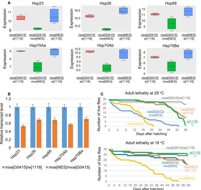
Nuclear Moesin is required for the transcription of heat-shock genes
We noticed during the transcriptomic experiments that among the best hits in genes with downregulated expression in moeNES mutant, six genes encoded heat shock-inducible protein chaperons (hsp70Aa,hsp70Ab, hsp70Ba, hsp68, hsp26, and hsp23) (Fig. 6A). The
decrease in hsp gene activity was rescued with one copy of WT moe allele, strongly suggesting that the nuclear activity of Moe is necessary for the normal expression of these genes. RT–qPCR on heat-shocked mutant animals reinforced the idea that nuclear Moe participates in the transcription ofhspgenes (Fig.6B).
In addition to thermotolerance [26,27], proper
0 5 10 15 20 25 30
0. 4. 8. 12. 16. 20. 24. 28. 32. 36. 40. 44. 48. 52. 56. 60.
0 5 10 15 20 25 30
0 4 8 12 16 20 24 28 32 36
0 0.2 0.4 0.6 0.8 1 1.2
Relative transcript level
moe[G0415]/w[1118] moe[NES]/moe[G0415]
Adult lethality at 29oC
Numberof liveflies
Days after hatching moe[G0415]
moe[NES]
moe[NES]/Y
moe[G0415]/w[1118]
moe[NES]
w[1118]
w[1118]
Y
Adult lethality at 18oC
Numberof liveflies
Days after hatching
moe[NES]/Y moe[G0415]/w[1118]
moe[NES]
w[1118]
w[1118]
Y moe[G0415]
moe[NES]
Hsp23
9 Expression 10
11
moe[G0415]
w[1118]
moe[G0415]
moe[NES]
moe[NES]
w[1118]
Hsp26
Expression
15.0 16.5 16.0
moe[G0415]
w[1118]
moe[G0415]
moe[NES]
moe[NES]
w[1118]
Expression
Hsp68
9 10 11 12
moe[G0415]
w[1118]
moe[G0415]
moe[NES]
moe[NES]
w[1118]
Hsp70Aa
Expression
7 8 9
moe[G0415]
w[1118] moe[G0415]
moe[NES] moe[NES]
w[1118]
Hsp70Ab
Expression 9.0
8.0 8.5 9.5 10.0 10.5
moe[G0415]
w[1118] moe[G0415]
moe[NES] moe[NES]
w[1118]
Expression
Hsp70Ba
4 5 6 7 8
moe[G0415]
w[1118] moe[G0415]
moe[NES] moe[NES]
w[1118]
A
B C
Fig. 6.Nuclear Moe is required for heat-shock gene expression. (A) The expression of sixhspgenes is decreased in the moeNESmutant flies (green). Error bar represents minimum and maximum values. Horizontal line=mean of normalized gene expression values. (B) RT– qPCR experiment on heat-shocked animals further demonstrates that nuclear Moe is required for hsptranscription. Data represent the meanSD of relative transcript levels measured in ovaries from five females of each genotype. (C) Both sexes ofmoeNESmutant show decreased viability at 29°C. Only mutant males exhibit shortened life span at 18°C. gray—moeG0415/w1118, orange—moeNES/w1118, blue— moeG0415/moeNES, green—w1118/Y, yellow—moeNES/Y.
functioning of Drosophila hsp genes is necessary for normal life span [28], fertility and germ cell mainte- nance [29,30], locomotor activity [31,32], and develop- mental timing [27]. Since all these processes are affected in moeNES animals, we performed additional experiments to confirm the involvement of Moe inhsp gene activity.
The most obvious sign for hsp malfunctioning is heat intolerance; therefore, we first analyzed the viabil- ity of the mutants at elevated temperature. At 25°C, only mutant males showed shortened life span (Fig.4A), but at 29°C, both sexes displayed a signifi- cant decrease in life span (Fig.6C). The phenotype was fully rescued by one copy of the WT moe gene, indicating that loss of Moe function is responsible for the decreased heat tolerance. To further demonstrate that heat intolerance causes reduced viability at high temperature in males and females, we also performed the experiment at 18°C. The result was similar to the data obtained at 25°C, only mutant males exhibited shortened life span; however, at this temperature, the mutant males lived much longer (51 vs. 21 days) than at 29°C (Fig. 6C). We also investigated whether the observed heat stress intolerance affects the negative geotaxis behavior of the mutants. The 35% decrease in the climbing ability of moeNES animals at 25 °C (Fig.4D) did not change significantly at higher tem- perature (data not shown), indicating that heat stress does not affect climbing activity. This in turn suggests that impaired hsp expression does not contribute to locomotor problems of the mutant.
In our previous work, we discovered that upon heat shock the amount of Moe significantly increases in the nucleus [16]. Therefore, we tested if the NES-tagged Moe isoform can also localize to the nucleus at high levels, by ectopically expressing GFP-labeled Moe and Moe NES proteins in the larval salivary gland cells (Fig.7A). In contrast to WT Moe, MoeNES-GFP did not accumulate in the nuclei after heat shock, demon- strating that the NES-tag inhibits the increase in Moe level in the nucleus. The immunostaining of WT and moeNES larvae with an anti-Moe antibody revealed that, although the endogenous MoeNES protein is not completely eliminated from the nucleus, upon heat shock it fails to accumulate in the nucleus (Fig.7B).
Moe has been shown previously to localize to the heat-shock puffs at cytological locations 87A and 87C [16]. To further confirm that Moe is responsible for the regulation of heat-shock gene transcription, we tested whether WT Moe localizes to the actively tran- scribing heat-shock genes identified in the transcrip- tome experiment. The activation of these genes by heat stress leads to the formation of chromosome puffs,
which are special euchromatic regions of the polytene chromosomes with extremely high levels of transcrip- tion. We found that Moe accumulated in high levels to the heat-shock puff sites, as demonstrated by the immunostaining of chromosome preparations from salivary gland cells (Fig.7C), providing additional evi- dence that Moe is involved in the transcription of hsp genes. Because in the cytoplasm Moe is organizing cytoskeletal structures, we wanted to exclude the possi- bility that Moe is needed for the formation of the puff structure. For this aim, we induced transcription with heat shock at a specific chromosomal location without the formation of the puff structure and stained for Moe. The animals used in this experiment carried a heat shock-inducible transgene at cytological location 86E in heterozygous form (Fig. 7D). The homologous chromosomes in the giant polytene chromosomes are paired in interphase and often separate from each other during the preparation at the nonhomologous sites. Without induction, no Moe staining can be observed in the corresponding region, but after heat shock, we found an extra Moe-positive band at this chromosomal location though only on one of the homologues (Fig.7D). This suggests that Moe partici- pates in hsp gene transcription and not the construc- tion of the puff structure. To confirm this finding, we tested whether heat-shock puffs are formed in the moeNESmutant. Staining for polymerase II CTD phos- pho-serine 5 (Pol2-PS5) on heat-shocked polytenic chromosomes prepared from moeNES mutant salivary glands showed that puffs can develop after heat shock (Fig. 7E). The results of the chromosome staining experiments together with the mRNA-seq results prove that Moe participates in heat-shock gene transcription.
Discussion
To attain deeper knowledge about the nuclear func- tions of the Drosophila ERM protein, Moe, we tagged the moe gene in situ with an NES sequence and ana- lyzed the consequences of the lack of nuclear Moe.
The moeNESmutants exhibited diverse phenotypes that were of both zygotic and maternal origin. Maternal-ef- fect phenotypes included posterior developmental mal- formations, dominant sterility, and lethality at embryonic and larval stages. Decreased life span, male genitalia rotation, reduced climbing ability, lethality at embryonic and larval stages, and decreased heat stress tolerance are zygotic phenotypes. Interestingly, decrease in viability at 18 and 25 °C and in climbing activity was observed in moeNES mutant males only.
Considering that the moe gene is on the first chromo- some, these sex-specific phenotypic differences are
+ HEAT SHOCK
Wi ld t y p e
No heat shock Heat shock No heat shock Heat shock
Mo e -G F P
Moe Moe
mo e [N ES]
M o e N ES -GFP
Moe Moe
63C
Pol2-PS5 DAPI
No heat shock Heat shock
+ HEAT SHOCK P< 0.0001
***
P< 0.0001 P< 0.0001 ***
P< 0.0001 ***
***
P< 0.0001 P< 0.0001 ***
***
P< 0.0001
***
P< 0.0393
*
Nucl./Cytopl. fluorescenceratio Nucl./Cytopl. fluorescenceratio
Moe- GFP MoeNES-
GFP Moe-
GFP
MoeNES-
GFP Moe MoeNES Moe MoeNES
n = 7/48n = 5/39 n = 6/55 n = 9/57
n = 3/41 n = 4/61
n = 3/56 n = 6/58
Transcription without puff formation at 86E Inactive, heat shock
inducible transgene at 86E
0 50 100 150 200 250
1 15 29 43 57 71 85 99 113 127 141 155 169 183 197 211
0 50 100 150
1 13 25 37 49 61 73 85 97 109 121 133 145 157 169 181
86E
86E
86D
86 D
86E 86D86E 86D
Moe Moe
Fluor.intensityFluor. intensity
distance distance paternal
maternal 67B 95D
Moesin + DAPI
67B 95D
High
Fluorescenceintensity Low
A B
C
D
E
most likely the consequence of dosage compensation.
The X chromosome of hemizygous males presumably produces more MoeNES protein than that of the female, which therefore suggests a dominant-negative effect for the protein in the development of these phe- notypes.
The quantitation of Moe and P-Moe levels in the moeNES mutant ovary revealed that the phosphoryla- tion, cytoplasmic localization, and intracellular distri- bution of the MoeNES protein are normal. The experiment also showed that the total Moe protein amount is reduced by about 30% in the mutant. This, in theory, could contribute to the phenotypes observed in the mutant. However, as shown earlier, the hypo- morphic moe allele, EP1652, has a reduced Moe pro- tein level [19] but moe[EP1652] homozygous animals are fertile and viable with no apparent visible pheno- type, strongly suggesting that the decreased amount of Moe cannot be responsible for the phenotypes The subsequent control experiments demonstrated that the cytoplasmic functions of Moe are unaffected; there- fore, the observed phenotypes are due to the perturba- tion of nuclear Moe function.
The mRNA-seq experiment revealed that the exclu- sion of Moe from the nucleus causes altered transcrip- tion of nearly 690 genes, from which 371 are significantly upregulated and 315 are downregulated in the mutant. Nuclear Moe was shown to participate in mRNA export[16] but it is conceivable that, similarly to the cytoplasm, Moe has multiple functions in the nucleus. In the case of the genes with decreased expres- sion, Moe most likely plays a positive role in their transcription regulation, while in the case of upregu- lated genes, the protein is involved in their repression.
Alternatively, the block of mRNA export, as a conse- quence of the lack of nuclear Moe, can also increase the activity of the genes affected, as suggested earlier [16], thereby accounting for the elevation of transcript levels.
The transcriptome analysis was performed on the ovary, but it is a reasonable assumption that part of
these gene expression changes is not specific to the ovary. Moe is almost ubiquitously expressed in the animal at all developmental stages; therefore, the lack of nuclear Moesin should affect gene expression in other tissues as well. On the other hand, most of the genes with altered expression in the moeNES mutant are active in many other tissues throughout develop- ment. In fact, for example, the analysis of the heat- shock genes provides supporting evidence for this assumption, because decreased heat stress tolerance was observed in the adults.
The mRNA-seq data showed that inmoeNESmutant animals, the transcription of three key regulators of development,Notch, dpp, andvasa, is disturbed. Notch is a transmembrane receptor of a highly conserved cell signaling system which plays major role in the regula- tion of development. Notch mutants in Drosophila have characteristic wing margin ‘notches’, a phenotype also manifested inmoeNESmutants (Fig.3B). Dpp is a secreted morphogen necessary for the correct pattern- ing and development of the early embryo. It has been shown recently that it is also essential for the mainte- nance of primordial germ cell identity [33]. Vasa is an evolutionarily conserved ATP-dependent RNA helicase that plays central role in germ cell determination and function [34]. Our microscopy experiments demon- strated that despite altered vasa expression in moeNES mothers and 100% sterility in their progenies, the Vasa protein localizes properly at the posterior pole of the developing embryo laid by moeNES mothers (Fig.2D).
Interestingly, we also found that the Vasa signal van- ishes in the embryonic germ cells, and as a conse- quence, all germ cells disappear after cellularization in the embryos of moeNES mothers (C.B. unpublished result). Future experiments should shed light on the role of Dpp and Vasa in the molecular mechanism responsible for germ cell loss inmoeNESprogenies.
As a consequence of the changes in transcript levels, different molecular mechanisms can be responsible for the development of the observed phenotypes. For instance, the altered activity of the apoptotic
Fig. 7.Moe is not required for the formation of the chromosome heat-shock puff structure. (A-B) The relative level of the MoeNES protein is significantly lower in the nuclei of larval salivary gland cells both at 25°C and under heat shock (37°C) as revealed by immunostaining for transgenically expressed MoeNES-GFP (GFP antibody—green) (A) and endogenous MoeNES (Moe antiserum—green) (B) proteins.
Representative images of two independent experiments.n–is the total number of salivary glands and cells examined, respectively.w1118 flies were used as WT in (B). Error bars=SD,*P<0.05,***P<0.0001 (unpairedt-test). Scale bars, 50lm. (C) Moe accumulates in the heat-shock puff sites (indicated by white arrows) at cytological locations 67B2:hsp26, 67B3:hsp23, and 95D:hsp68. Representative picture of one of four independent experiments. (D) Heat scale images demonstrating that Moesin localizes to thehspgene also in the absence of the puff structure. The heat shock-inducible transgene (hs>FLP) is inserted at cytological position 86E (thick arrow) but only in one of the homologues. Upon heat shock, the transgene becomes transcriptionally active without puff formation and induces the accumulation of Moe at the site (Moe-HA—yellow). Representative images of five independent experiments. (E) Heat-shock puffs are formed in themoeNES mutant as revealed by polymerase II CTD phospho-serine5 (Pol2-PS5) staining (green). 63C: hsp82. Representative image of one experiment.
machinery or sex-specific transcription factors can lead to genitalia rotation in males [35]. The complete loss of pole cells observed in the F1 progeny can be due to uncontrolled transposon activity[36], impaired sumoy- lation[37], or compromised BMP signaling[38]).
The exploration of the causes behind the various phenotypes was beyond the scope of this study but our mRNA-seq data uncovered that the transcription of a group of heat shock-induciblehspgenes is signifi- cantly reduced in the moeNES mutants. Drosophila hsp genes encode protein chaperones with versatile func- tions such as thermotolerance, normal life span, fertil- ity and germline stem cell maintenance, locomotor activity, and developmental timing. Therefore, it is tempting to speculate that the low level of hsp gene transcription alone can contribute to most of the phe- notypes observed in themoeNESmutant. Although the lack of nuclear Moe does not fully eliminate hspgene expression, the combination of reduced expression of multiple hspgenes can account for the severity of the phenotypes observed. Given this possibility, as well as the findings that during heat stress Moe accumulates in the nucleus at thehsploci and thatmoeNESmutants exhibit reduced thermotolerance, we propose that in the nucleus Moe is required for the normal expression of heat-shock genes. The mechanism through which Moe contributes to hsp transcription is still an open question. No DNA-binding ability of Moe has been described; therefore, the association of Moe with the hspgenes is most likely indirect. An obvious candidate for a binding partner of Moe is nuclear actin, which plays important role in transcription [1,39]. However, the exact activity Moe performs in the nucleus during hspgene transcription has to be explored in the future.
We believe that our results open up a new avenue to understanding the ERM protein family, their exact functions in growth and differentiation, and contribute to a better understanding of the transcription regula- tion of heat-shock genes.
Materials and methods
Fly stocks
Fly strains were maintained, and crosses were carried out on standard cornmeal, yeast, and sucrose Drosophila med- ium at 25°C. Stocks number 51324 (w1118; PBac{y[+mDin- t2]=vas-Cas9}VK00027), 42278 (w1118; PBac{Med- GFP.FLAG}VK00037), 12015 (w67c23 P{w+ mC=lacW}
moeG0415/FM7c) and 279 (w1118; MKRS; P{hsFLP}86E/
TM6B, Tb) were obtained from the Bloomington Droso- phila Stock Center (Indiana University, Bloomington, IN, USA). The Dp(1;Y)619, y[+] BS/w1 oc9/C(1)DX, y1 f1
(No. 108358) stock was obtained from Kyoto Stock Center (https://kyotofly.kit.jp; Institute of Technology, Kyoto, Japan). The attached X and vasa:AID:EGFP lines were provided by Miklos Erdelyi (BRC, Szeged, Hungary), and the oskMS2-MS-GFP andw,P{w+,matTub4:GFP-Staufen};
stauD3/CyO stocks were kind gifts from Daniel St Johnston (University of Cambridge, UK). The transgenic lines expressing Moe-GFP and Moe-HA have been described previously[16].
To generate flies expressing NES and GFP (MoeNES- GFP)-tagged full-length Moe, the coding region was PCR amplified from the cDNA SD10366 (DGC Gold collection, BDGP) using the primer pair Moe_cDNSFw and Moe- NES_cDNSRev with gateway recombination sites. The resulting PCR product was sequence-verified and recom- bined into pDONR221 plasmid, then subcloned into the pPWG gateway vector (Drosophila Gateway Vector Collec- tion) which enables P-element-based random integration into the fly genome. The construct was sequence-verified, and standard Drosophila methods were used to generate transgenic flies. Moe_cDNSFw: 50-GGG GAC AAG TTT GTA CAA AAA AGC AGG CTT CAC CAT GTC TCC AAA AGC GCT A-30; MoeNES_cDNSRev (NES-tag underlined): 50-GGGG AC CAC TTT GTA CAA GAA AGC TGG GTC TGT CTT GTT GAT ATC AAG ACC TGC TAA TTT CAA GGC TAA TTC ATT GCT GGA CAT GTT CTC AAA C-30.
Drosophilaovary dissection andin vivo microscopy
Flies were dissected in PBS (137 mM NaCl, 2.7 mM KCl, 10 mM Na2HPO4, 1.8 mM KH2PO4), and the ovaries were transferred to a microscope slide with a drop of Drosophila Schneider’s medium (Lonza, Basel, Switzerland, Schneider’s Drosophila Medium, Modified, 04-351Q), then covered with a glass coverslip. The samples were imaged with an Olympus confocal microscope (Olympus Europa SE & Co.
KG, Hamburg, Germany, Olympus Fluoview FV1000 Con- focal Microscope, 409oil immersion objective, 1.3 NA).
Moesin antisera
GST-tagged FERM domain of the Drosophila Moe protein was expressed by the tac promoter-IPTG system using the pGEX-6P-1 vector and the BL21E. colistrain. The protein was purified on Glutathione Sepharose 4B beads (GE Healthcare, Chicago, IL, USA, 17-0756-01); then, high level of soluble FERM-Moe protein was eluted from the beads by the PreScission protease (GE Healthcare, 27-0843-01) and used to immunize three rabbits, yielding three poly- clonal sera (aFERM-Moe1, aFERM-Moe2, aFERM- Moe3). All animal experimentation protocols used in this study were performed in accordance with the European animal experimentation and ethics guidelines and have been