University of Szeged
Advanced
Microbiology Practicals
for MSc students
Z. Hamari, I. Pfeiffer, M. Takó and C. Vágvölgyi
2019
Table of content
1. How To Work In A Microbiological Laboratory:
Instructions And Rules 7
1.1. The regulations of the microbiological laboratory 9
1.2. Materials and laboratory tools 10
2. Basic Microbiological Practices Applied During The
Course 12
2.1. Inoculation – Practice 1 13
2.2. Dilution – Practice 2 15
2.3. Cell count – Practice 3 17
2.4. Staining methods – Practice 4 21
3. Advanced Microbiological Practices 25 3.1. Studies On Microbiological Genetics 25
3.1.1. Sexual cycle of S. cerevisiae 26
3.1.1.1. Conjugation – Practice 5 29
3.1.1.2. Diploid isolation – Practice 6 30 3.1.1.3.Induction of the sporulation – Practice 7 31 3.1.1.4. Ascospore isolation – Practice 8 32 3.1.1.5. Random spore analysis – Practice 9 33 3.1.1.6. Genetic analysis – Practice 10 34 3.1.2. Study of the killer system of Saccharomyces
cerevisiae 37
3.1.2.1. Killer activity – Practice 11 39 3.1.2.2. RNase protection assay – Practice 12 40
3.1.3. General introduction to the asexual, sexual and parasexual life cycles of the filamentous fungus model organism, Aspergillus
nidulans 42
3.1.2.1. Asexual life cycle of A. nidulans – Practice 13 49
3.1.2.2. Formation and maintenance of heterokaryons
3.2. Basic Techniques On The Field Of Microbial
Ecology 119
3.2.1. Determination of bacterial and fungal CFU in
soil samples – Practice 18 120
3.2.2. Isolation of psychrophilic, mesophilic and thermo-
philic bacteria from water sample – Practice 19 127 3.2.3. Isolation of Pseudomonas and Bacillus strains from
soil samples – Practice 20 134
3.2.4. Isolation of Trichoderma strains from soil sample
– Practice 21 142
3.2.5. Isolation of laccase producing fungi – Practice 22 148 3.2.6. Analysis of in vitro antagonism of bacterium isolates
against filamentous fungi – Practice 23 153 3.2.7. Fungicide tolerance of fungi – Practice 24 162
3.3. Studies On Microbial Exoenzymes 169 3.3.1. Lipase production of microorganisms 170 3.3.1.1. Detection of extracellular lipase activity
– Practice 25 176
3.3.1.2. Measurement of lipase hydrolytic activity
– Practice 26 178
3.3.1.3. Measurement of lipase synthetic activity
– Practice 27 180
3.3.2. Chitinase production of microorganisms 183 3.3.2.1. Detection of extracellular chitinase activity
– Practice 28 188
3.3.1.2. Measurement of chitinase activity – Practice 29 191
Description of the subject Master level
(1.) Title of the subject: Advanced microbiology practicals 1 and
2
Credits: 6/6Type of the subject: compulsory
The ratio of the theoretical and practical character of the subject: 10-90 (credit%) The type of the course: laboratory practice
The total number of the contact hours: 4 per week Language: English
Type of the evaluation: written test
Other methods for evaluation of the student’s competence:
preparation of a note- book, skills during the practice
The term of the course: II-III. semester
Prerequisite of the subject: Advanced microbiology I.
Description of the subject The aim of the subject:
The courses are directed to students who have some skills in basic microbial techniques.
It provides an overview of techniques generally used in environmental and molecular microbiology; methods used in microbial ecology are also presented. Participants will acquire techniques generally used to manipulate microorganisms and they will study the genetic background of the life cycle of two important eukariotic modell systems
Aspergillus nidulans and Saccharomyces cerevisiae.Course description:
-
The sexual life cycle of Aspergillus nidulans (heterokaryon formation, diploid isolation, ascospore formation and genetic analysis of the progenies)
-
The parasexual life cycle in filamentous fungi (heterokaryon formation by anastomosis and protoplast fusion, diploid isolation, haploidization);
-
Study of the sexual life cycle of Saccharomyces cerevisiae (conjugation, diploid isolation, spore forming, random spore analysis);
-
The killer phenomenon in yeasts;
-
The presence of dsRNA viruses in Saccharomyces cerevisiae;
-
Counting of viable bacterial and fungal cells in soil samples;
-
Counting of psychro-, meso- and thermophilic bacteria in water samples;
-
Selective isolation of Pseudomonas and Bacillus strains from soil;
-
Selective isolation of Trichoderma strains from environmental samples;
-
In vitro testing of the microbial antagonism between bacteria and filamentous fungi;
-
Study of the extracellular lipase and esterase secretion of bacterial strains;
Detection and measurement of the cellulase, xylanase and phosphatase activity of
-
Determination of the susceptibility of Trichoderma strains to fungicides.
Selected bibliography (2-5) (author, title, edition, ISBN)
Baltz, R. H., Davies, J. E., Demian, A. L. (2010): Manual of Industrial Microbiology
and Biotechnology. Third Edition. ASM Press, Washington DC.Lammert J.M.: Techniques in Microbiology: A Student Handbook 1st Edition, ISBN- 13: 978-0132240116
HERSKOWITZ I.: Life cycle of the Budding Yeast Saccharomyces cerevisiae.
MICROBIOLOGICAL REVIEWS, 1988, 536-553, 0146-0749/88/04536-18$02.00/0
Pontecorvo G (1953) The genetics of Aspergillus nidulans. Adv Genet 5: 141–238.Wickner R.B., Fujimura T., Esteban R.: Viruses and prions of Saccharomyces
cerevisiae. Adv Virus Res. 2013; 86: 1–36. doi: 10.1016/B978-0-12-394315-6.00001-5
Hamari Zs., Pfeiffer I., Takó M., Vágvölgyi Cs.: Advanced microbiology practicals (typescript)
General competence (knowledge, skills, etc., KKK 8.) promoted by the subject a) knowledge
-
Familiar with the tools and methods applied in modern biology.- Know the terminology of microbiology and apply it in the correct way.
- Understand the social problems with biological relevance.
- Understand the significance of the interdisciplinary approach.
- Know the coherency among the area of biology.
b) skills
- Able to participate in biological research project and to create new scientific results under competent supervision.
- Able to plan research projects in the field of biology
- Able to recognize the coherency among the different area of biology.
- Able to apply new tools and techniques independently.
- Able for the interpretation and the presentation of the results.
c) attitude
- open to cooperate with other research groups
- ready to understand the evolution, structure and function of the living organisms - interested in new results, techniques and methods; contribute to new scientific results
and methods
- keep the ethical rules of the biological research d) autonomy and responsibility
- able to organize the work of small research teams independently - provide and require safety work conditions in the laboratory - help his collegues in the completion of the research projects - build his/her scientific career consciously
Special competence promoted by the subject:
experiments to study the sexual and parasexual life cycles of eukaryotic
microorganisms
microbiological
laboratory. sterile work in the microbiology lab and always
keep the
laboratory rules working with living materials.
Open to study new techniques and methods.
Ready to prepare a correct and up- to-date notebook.
Ready to
cooperate with his/her
colleagues in the completion of the experiments.
Apply the acquired methods independently.
Evaluate the results independently.
Know the method for genetic analysis of the progeny
Students can apply protoplast fusion and sexual cross to obtain heterokaryon.
Know the
methods to isolate microbes from different
environmental samples.
Able to apply the
media and
methods for isolation of microbes from the environment.
Know the
counting methods for evaluation the microbial cell
numbers in
environmental samples.
Plan experiments to count the cell
number in
environmental samples.
Acquire
techniques to
detect and
measure extracellular enzyme activity of microorganisms.
Present that microbes
produce different
kind of
extracellular enzymes.
Know methods to detect antagonistic relationships among microbes.
Demonstrate the antagonistic relationship between the microbes.
Carry out
susceptibility tests.
Able to plan experiments to reveal the drug susceptibility of microbes.
List the types of killer toxins and their chemical
Able to
demonstrate that
killer toxins have
killer toxin production.
Instructor of the course (name, position, scientific degree):
Papp Tamás associate professor PhD Teachers (name, position, scientific degree):
Hamari Zsuzsanna associate professor PhD Pfeiffer Ilona associate professor PhD Takó Miklós senior lecturer PhD
1. How to work in a
microbiological laboratory:
instructions and rules
The purpose of this practical handbook is to support the preparation for microbiological practices, furthermore, with the detailed description of the experiments it serves as a guide for students in the laboratory. Experiments could be performed independently; at the same time, they are didactically intertwined, and they relate to the knowledge acquired in theoretical courses.
At the beginning of the handbook, the microbiological laboratory safety regulations are briefly described followed by a general description of the basic materials and equipment used in a microbiological laboratory.
Each exercise is started with a summary of the theoretical knowledge, then the material and equipment need of the experiment are described, followed by a description of the experimental work. It is specified what questions should be answered in relation to the experiment performed and suggest how to evaluate the experiment.
Successful implementation of the experiments requires great accuracy and clarity. Generally, it is true for all type of laboratory work but it has special importance when we have to handle microorganisms safely.
Some experiments could be started at the same time and performed side by side so that they can be fully executed within the time available. Due to the nature of the microbiological work, the results can be evaluated in 24-48 hours after the experiments or even later, if it requires a prolonged incubation.
1. General Introduction
1.1. The regulations of the microbiological laboratory
The safety rules of general laboratory activities have to be respected in microbiological laboratory to avoid unwanted situations from working with electricity, gas, acids, poisonous and other dangerous chemicals. In addition, special precautions must be taken when using microbial cultures because of the risk of infection.
1.1. Any microbial culture used in the laboratory should always be handled as a pathogenic one!
1.2. It is strictly forbidden to eat or drink in the laboratory!
1.3. Do not use laboratory equipment for other purposes!
1.4. Laboratory tools and glassware have to be in perfect condition; do not use unsafe or broken items!
1.5. After completion of the experiments, the laboratory bench must be cleaned with a proper disinfectant.
1.6. Wash your hands before leaving the laboratory.
1.7. It is strictly forbidden to dispose microbial cultures in the drain. All such wastes have to be destroyed by heat or chemical treatment.
1.8. Do not pour melted agar medium into the drain!
1.9. Before washing, the used laboratory glassware should be disinfected by autoclaving or by chemical treatment.
1.10. You have to wear lab coat performing experiments in the laboratory. This coat should preferably not be used for other purposes.
1.2. Materials and laboratory tools
1.2.1. Media
Lots of different culture media are used for the cultivation of microorganisms in laboratory practice. Though the nutritional requirements are extremely variable between microorganisms, these media basically must contain the essential nutrients: carbon, nitrogen, mineral salts and water.
The classification of culture media can be based on various aspects.
1.2.1.1. According to the absence or presence of a solidifying agent 1.2.1.1.1. Liquid media (broth, nutrient solutions)
1.2.1.1.2. Solid media: complement the liquid medium with a solidifying agent.
Solidifying agents may include:
A) Agar-agar: a polysaccharide purified from seaweed; used in 1-3% concentration;
hydrolyzed by very few microorganisms.
B) Gelatine: a polypeptide; used in 10-30% concentration; its melting point is a function of concentration; hydrolyzed by several microbes.
C) Silica gel: its advantage is that it does not contain organic components;
preparation of the medium is time-consuming; rarely used.
1.2.1.2. According to the materials used
1.2.1.2.1. Undefined (Natural) media: based on organic materials whose exact composition is unknown, e.g., meat extract, yeast extract, malt, molasses, juices
1.2.1.2.2. Defined (Synthetic) media: their composition is well known, exactly defined.
1.2.1.2.3. Semi-synthetic media: mixtures of natural and synthetic components.
1.2.1.3. According to the nutrient components used
1.2.1.3.1. Basal media: except for the ingredients needed for growth, they do not contain any special additives.
1.2.1.3.2. Selective media: basal medium + selective inhibitor (e.g., antibiotic). Suppress the growth of certain microorganisms.
1.2.1.3.3. Differential or indicator media: basal medium + indicator compound that indicates specific metabolic reactions (e.g., lactose utilization, acid formation).
1.2.1.3.4. Special media: media supporting microbial growth for various special purposes.
Preparation of the culture medium: first weigh the solid constituents and then dissolve them in a
solidifying agent. Agar-containing media can be sterilized under pressure, those, which contains gelatin flowing steam must be used (without pressure).
The heat-sensitive media components must be added to the autoclaved and properly cooled basal medium after sterilization by filtration.
1.2.2. Laboratory Glassware
All laboratory vessels used for microbial cultivation must be fitted with cotton swabs or other safe closure.
1.2.2.1. Test tubes: generally, 5 ml of liquid medium, 10 ml of solid medium for sloped agar, and 10 ml of solid medium for high agar, is required.
1.2.2.2. Flasks: for liquid microbial cultures most frequently Erlenmeyer flasks or flat bottom spherical flasks are used.
1.2.2.3. Special breeding vessels: Roux bottle and the Kolle flask; these provide large surfaces for the microbial culture.
The abovementioned glassware types are suitable for shaking and for standing liquid cultures.
The culture vessels are sealed with a cotton/paper swab after loading the medium and then sterilized by autoclaving. It is advantageous to cover the cotton swab with e.g., aluminium foil before sterilization to prevent its moistening in the autoclave.
1.2.2.4. Petri dish: the most commonly used laboratory glassware. Sterilize the medium before pouring into a sterile petri dish. When pouring the medium, raise the lid only to the extent necessary. In a 9-10 cm diameter dish, 20-25 ml of medium is required.
1.2.2.5. The fermenter is a special laboratory equipment frequently with a large glass reaction vessel to cultivate microorganisms in large volume. The parameters (conditions) of cultivation can be ensured by appropriate control systems with data recording. Generally, it has a stirring system to ensure homogeneity of the microbial culture in the medium and auxiliary parts for controlled supplement of nutrients, gases, and other necessary additives.
In the laboratory, the culture vessels are marked as follows: 1.) the name of the microorganism, 2.) the date of inoculation, 3.) the type of medium, 4.) the number of the experiment, 5.) the name of the researcher.
Nowadays, practically all laboratory glassware has its disposable variant made of plastic. When performing experiments, special attention must be paid to the limit of their heat resistance.
2. Basic microbial techniques
applied during the course
I. Introduction
Under laboratory condition inoculation means the transfer of a pure culture into liquid or solid culture medium. Solid media immobilize cells and allow them to grow and form a visible mass of cells called colonies.
Loops are simple tools widely used for inuculation. It can be used for inoculation of liquid or solid media as well in different types of cultivation dishes. Working with deep-agar, needles are used to inoculate the media.
During the proccess of inoculation a series of steps will prevent contamination during the transfer of the cells: flaming the loops or needles and the surface of the cultivation tube sterilize them.
During inoculation, work always close to the flame because the warm rising air prevent contamination.
Microorganisms: Saccharomyces cerevisiae culture on slant agar Required materials
Learning Objectives
Students will know
what inoculation means
simple tools for inoculation
different inoculation techniques
2.1. Inoculation
/- Practice 1 -/
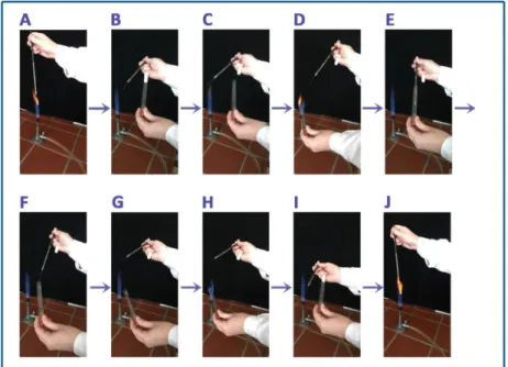
1. Flame the loop (Figure 1.A)
2. Remove the cap of the tube contaning the culture (Figure 1.B-C) 3. Flame the tube tip (Figure 1.D)
4. Take out the inoculum (Figure 1.E-F) 5. Flame the tube tip again (Figure 1.G-H) 6. Recap the tube (Figure 1.I)
Remove the the cap of the sterile tube containing the slant agar. Flame the tube tip. Transfer the inoculum onto the surface of the slant agar. Flame the tip of the tube and recap it. Flame the loop (Figure 1.J).
Write the name of the strain onto the tube. Incubate the culture for 48 hours at 30 °C.
Figure 1. Taking out the inoculum of the culture
Check your progress
Prepare agar slants and inoculate coloured
bacterial strains to them! Check the quality of the cultures after 2 days of incubation!
What does inoculation refer to?
What kind of tool is suitable for inoculation of
deep-agar?
I. Introduction
When the cell number of a liquid culture or a suspension is too high it is necessary to dilute it.
This practice provides an example for preparing tenfold dilution seies.
Microorganisms: Saccharomyces cerevisiae 24-hour-old liquid culture Materials: pipette, pipette tips, sterile glass tubes, sterile distilled water
Prepare a serial dilution as shown in Figure 1. Fill 6 glass tubes with 4.5 ml sterile distilled water.
Add 500 µl liquid culture to the first tube. Vortex it, take out 500 µl suspension and add it to the next tube. Follow this steps until the 6th tube. The cell number of the original culture decreases
Learning Objectives Students will know
how to prepare a dilution series of a suspension
how the cell number of the population will change during the dilution
2.2. Dilution
/- Practice 2 -/
Required materials
Method
Figure 1. Preparation of dilutions
Check your progress
A 10-fold dilution series in 6 steps was prepared from a Saccharomyces cerevisiae suspension.
The total cell number of the original suspension was estimated as 6.3x10
7cells/ml.
Establish the cell number in the 5th step of the
dilution series!
I. Introduction
There are direct and indirect methods to determine the cell number of a microbial population.
Direct methods count directly the cells in the population therefore refer to the total cell number (dead and living cells together) indirect methods assess a property of the population what is proportional to the cell number.
This practice demonstrates a direct cell counting method to determine the total cell number of a yeast population.
Counting chambers are generally used to establish the number of the suspended particles (bacterial or yeast cells, fungal spores) in a given volume. Counting chambers are microscopic slides with grooved cross-channels. The middle part of the slide is thinner with standard size, so the height of the liquid column between the slide and the cover slip is known. As the size of the grid is also known, the volume between the slide and cover slip can also be defined.
The height of the more widely used Bürker-chambers (Fig. 1.) is 0.1 mm. The cross-channels
2.3. Total cell count of a yeast population /- Practice 3 -/
Learning Objectives
Students will know
how to use the Bürker chamber
a simple method to determine the total cell number of
a yeast population by a direct cell counting method
As the height of the chamber is 0.1 mm the volume of the big square is 1/250 mm3,
the small square is 1/4000 mm3, the rectangle is 1/1000 mm3.
Figure 1. Bürker-chamber
(A) Parts of the chamber 1. clamp 2. cover glass 3. counting chamber (B) Enlarged grid (C) Enlarged grid with yeast cells.
Microorganisms: Saccharomyces cerevisiae 24-hour-old liquid culture
Materials: pipette, pipette tips, sterile glass tubes, sterile distilled water, Bürker chamber, cover slip, light microscope
Required materials
Method
slip due to the capillary action. Focus the microscope on the area of the chamber and count the cells in one unit (either small or big squares, rectangles)! Repeat the process for 10 units!
Calculate the average!
The total cell count should be given for 1 ml (= 1 cm3)!
Potential pitfalls:
Use always the same type of unit for the counting!
If the suspension contains countless cells dilute the yeast suspension! In this case you have to take the rate of the dilution under consideration calculating the cell number of the original suspension!
Example 1:
You obtained the following counts in big squares: 8, 3, 4, 2, 7, 7, 5, 11, 3, 9 Calculate the average (x):
The volume of the big squares is 1/250 mm3.
Estimate the mean cell number of the suspension using the following formula:
In an abbreviated form: 1.475 106 cells/ml Example 2:
The original suspension contained countless number of cells therefore you have made 50 fold dilution, and counted the cell number in the diluted sample.
The volume of the rectangles is 1/1000 mm3.
Estimate the mean cell number of the original suspension using the following formula:
In an abbreviated form: 3.2 108 cells/ml
Check your progress
Compare the direct an the indirect cell counting methods!
What do direct cell counting methods refer to?
What can counting chambers is used for?
Estimate the total cell number of a yeast
population! The cell counts in rectangles were as follows: 12, 4, 6, 3, 5, 13, 9, 5, 8, 14.
Estimate the total cell number of the yeast
population! The cell counts in small squares were as follows: 2, 2, 3, 0, 5, 4, 0, 3, 2, 1.
As the original suspension was too dense, these
counts were made in 100-fold dilution.
I. Introduction
To observe microbial cells one needs a microscope. Bacteria, fungi, yeasts, unicellular algae and parasites can be examined under optical microscope. This type of microscope works with lower magnification therefore it is suitable for visualization of larger objects only (viruses cannot be observed under this type of microscope!). The cells are visible under optical microscope because they absorb and scatter the light differently from the surroundings. However, the contrast between the cells and the background can be elevated by using specific dyes what stains the cells or cell compartments but not the surroundings.
To stain microbes, generally positive staining methods are used. During these methods the
2.4. Staining method for cell and ascospore differentiation in a Saccharomyces cerevisiae population /- Practice 4 -/
Learning Objectives
Students will know
what differential staining means
what aggressive staining method means and what it is good for
a simple method for staining S. cerevisiae cells and
ascospores
During differential staining methods two dyes with different color are used to distinguish between the cell types. One dye is used as a primary stain and the other one is applied for counterstaining.
Spores (endospores of bacteria or different spore types of fungi) are usually resistant to simple staining techniques. Therefore, so called “aggressive” method is used to stain them. During this technique the dye (often phenol-containing) is forced to penetrate into the spores by heating.
Experienced users can observe vegetative cells and ascospores in a S. cerevisiae population under bright field microscope. For students, to use differential staining is more ubiquitous. During this practice the Schaeffer-Fulton method will be used to differentiate between the vegetative cells and ascospores of S. cerevisiae. Malachite green will be used as primary dye and safranin as a counterstain. The sample will be visualized under bright field microscope.
Microorganisms: Saccharomyces cerevisiae seven-day-old culture on sporulation medium
Materials: malachite green solution, safranin solution, slides, cover plates, ethanol-ether (1:1) mix, distilled water, filter paper, staining tub, water bath, light microscope
Malachite green solution:
1 g malachite green 1 g phenol
100 ml distilled water
Safranin solution:
6 ml 5% safranin O 10 ml 3% KOH 5 ml 87% glycerol 79 ml distilled water
Clean the slide with a detergent and rinse it in distilled water. After drying, place a drop of distilled water onto the slide and prepare the smear: Spread the culture in a thin film. Let it air
Required materials
Method
Saturate the blotting paper with malachite green and place the slide over a boiling water bath for 10-15 minutes. Keep the paper moist by adding more dye as required. Remove the filter paper and decolorize the sample with water then counter-stain it with safranin: flood the smear with safranin solution for 30 seconds. Wash it with water and after drying it can be visualized under the microscope. Ascospores have to be green, vegetative cells have to be red.
Figure 1. Saccharomyces cerevisiae cells and ascopores.
Safranin-stained vegetative cells (red) and malachite green-stained ascospores (green).
Check your progress
Briefly characterize each positive staining method!
Why can basic dyes be applied to stain microbial cells?
What kind of technique is suitable for staining the spore?
Outline the Schaeffer-Fulton staining method, how
it is used?
3. Advanced Microbiological Practices
3.1. Studies On
Microbiological Genetics
I. Introduction
The haploid Saccharomyces cerevisiae has two different mating types ’a’ and ’α’. Separately the cells of the different mating types can be divided by budding (Figure 1). Budding is a mitotic division, during this process the number of the chromosomes does not change.
Learning Objectives
Students will know
what is homothallism and heterothallism
what does haplo-diploid life cycle refer to
what kind of methods are available to study the segregation of the genetic markers in the progenies
3.1.1. Sexual cycle of Saccharomyces cerevisiae
/ Practice 5-10 /
The ‘a’ cells produce and secrete ‘a’ factor, ‘α‘ cells produce ‘α‘ factor. Both ‘a’ and ‘α‘
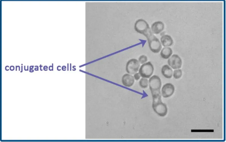
factors are pheromones and both are able to bind to the cell surface receptors of the opposite mating type. If the cells of the two opposite mating types are mixed, the pheromones induce morphological changes in the cells finally they will fuse (this process is called conjugation) creating diploid zygotes (Figure 2). Diploid cells can divide by mitotic division (budding) establishing a diploid cell population. As vegetative cells can be haploid or diploid and both haploid and diploid cells are able to undergo mitotic division the life cycle of Saccharomyces cerevisiae is called haploid-diploid life cycle.
Figure 2. Conjugated cells
Diploid cells can become a zygote and the zygote can undergo meiotic division under starvation. The result of the meiotic division is an ascus with 4 haploid ascospores in it (tetrad).
The distribution of the mating type alleles in the ascus is: 2 a: 2 α. When the wall of the ascus disrupts the ascospores deliberate and will be haploid vegetative cells those start to propagate by budding.
The genetic analysis of ascospores in one ascus is called tetrad analysis. One needs micromanipulator to carry out this study. The other way of the genetic analysis is called random spore analysis. In this case a population of ascopores is under the study independently of their position in the ascus.
Haploid S. cerevisiae cells are able to change their mating type the process is called mating type switching. The mating type of the cell is determined by the allele located in the MAT
switch their mating type are called heterothallic and the pure culture derived from single heterothallic cell will never sporulate.
Heterothallic strains are useful during genetic crossing experiments because conjugation of the strains can be controlled more easily comparing to homothallic strains.
Microorganisms: Fourteen-hour-old shaken culture of Saccharomyces cerevisiae strains 0666 (a, ura3, ade1) and GRF-18 (α, leu2, his3) in 20 ml YPD. Twenty-four-hour-old mixed (1:1) culture of the two strains.
Media:
YPD: 0.5% yeast extract, 1% pepton, 1% D-glucose, 2% agar
Minimal medium: 0.5% (NH4)2SO4, 0.1% KH2PO4, 0.05% MgSO4, 1% glucose, 0.1% Wickerham vitamin solution, 2% agar
Materials: pipette, pipette tips, sterile glass tubes, sterile distilled water, Bürker chamber, cover slip, light microscope, microcentrifuge, Petri dishes, ethanol,
A. Count the cells of the fourteen-hour-old shaken culture of strains 0666 and GRF-18 in Burker chamber. Pipette 107 cells from each culture in a sterile Eppendorf tube. Pellet the cells by centrifugation (5 min, 5000 rpm) after that re-suspend the cells in 1 ml YPD and incubate them for 2 hours at 30 ˚C. Follow the conjugation process under the microscope!
B. Take out 2x10⁷ cells from the twenty-four-hour-old mixed (1:1) culture in a sterile Eppendorf tube. Pellet the cells by centrifugation (5 min, 5000 rpm) after that re-suspend the cells in 1 ml sterile water and pellet the cells again (5 min, 5000 rpm).
Re-suspend the cells again in 1 ml sterile water and prepare a tenfold dilution series in four steps.
The dilution can be made in sterile water.
Spread 50 µl aliquots from the original suspension and the 10x and 100x dilutions to the surface of minimal medium (2 paralels) to select diploid cells.
Required materials
Method
3.1.1.1. Conjugation
/- Practice 5 -/
Microorganisms: colonies grown on minimal medium Materials: 4 slant agar made of YPD medium, loop
Isolate 4 colonies from the minimal medium (these are diploid cells!) inoculate them onto slant agar and incubate them for 6 days at 30 ˚C.
Required materials
Method
3.1.1.2. Diploid isolation
/- Practice 6 -/
Microorganisms: cultures on slant agar inoculated on the previous practice Media
PSM (presporulation medium): 0.8% yeast extract, 0.3% pepton, 10% D-glucose
SM (sporulation medium): 0.5% Na-acetate, 0.5% KCl, 0.1% yeast extract, 0.05% D-glucose, 2%
agar
Materials: sterile centrifuge tube, microcentrifuge, pipette, tips
One day before the practice inoculate the selected diploid strains into PSM medium and incubate them for 24 hours at 30 ˚C.
Pellet the cells by centrifugation (5 min, 5000 rpm) after that re-suspend the cells in 1 ml sterile water and pellet the cells again (5 min, 5000 rpm).
Re-suspend the cells in 1 ml sterile water and inoculate 25 µl onto the surface of SM medium. Dry the surface under sterile box. Incubate the plates for 1 week at 27 ˚C.
Required materials
Method
3.1.1.3. Induction of the sporulation
/- Practice 7 -/
Microorganism: cultures on SM medium inoculated in the previous practice Medium: 2xYPD: 1% yeast extract, 2% pepton, 2% D-glucose, 2% agar
Materials: sterile distilled water, loop, microcentrifuge tubes, water bath, snail gut enzyme solution (5% in sterile distilled water), Triton X-100 (0.1%), sterile glass beads (0.5 mm diameter), ethanol, slides, cover slips, microscope
Check the ascus formation under the microscope. Select the strain with several ascus and make a dense suspension in 0.5 ml sterile water in a sterile test tube. Place the suspension to a water- bath at 55 °C for 20 min. During this step the vegetative cells will die.
Add 1.5 ml snail gut enzyme to the suspension and incubate it at 30 ˚C until the lysis of the ascus wall. Check the process under the microscope!
Add 1/5 volume of sterile glass beads to the suspension and vortex it to separate the ascospores from each other.
Pellet the beads and pipette 1 ml of the supernatant into a sterile microcentrifuge tube.
Centrifuge it (5 min, 5000 rpm) and wash the cells with 0.1% Triton X-100 solution, centrifuge again (5 min, 5000 rpm).
Suspend the cells in 1 ml sterile water and prepare a tenfold dilution series in three steps (10x, 100x, 1000x). Spread 50 µl from each dilution onto the surface of 2xYPD medium in two replicates. Incubate the plates for 72 hours at 30 ˚C.
Required materials
Method
3.1.1.4. Ascospore isolation
/- Practice 8 -/
Media
A. Minimal medium: 0.5% (NH4)2SO4, 0.1% KH2PO4 , 0.05% MgSO4, 1% glükóz, 0.1% Wickerham vitamin solution, 2% agar supplemented with uracil, leucine, histidine and adenine.
B. Minimal medium supplemented with uracil, leucine and histidine C. Minimal medium supplemented with uracil, leucine and adenine D. Minimal medium supplemented with adenine, leucine and histidine E. Minimal medium supplemented with uracil, adenine and histidine
The concentration of leucine, histidine and uracil: 30 µg/ml, adenine: 5 µg/ml.
Materials: sterile velvets
Choose the plates with countable number of colonies and transfer the colonies to the surface of the supplemented minimal medium with sterile velvets (replica plating).
Incubate the plates for 72 hours at 30 ˚C.
Required materials
Method
3.1.1.5. Random spore analysis
/- Practice 9 -/
Replica plates from Practice 5.
Establish the distribution of the genetic markers among the progenies (pr)!
Cross: GRF-18 X 0666 Media
Strains
MM uracil Ade Leu His
MM Ade Leu His
MM uracil Leu His
MM uracil Ade His
MM uracil Ade Leu
PHENO
-TYPE GENOTYPE
GRF-18 0666 pr 1 pr 2 pr 3 pr 4 pr 5 pr 6 pr 7 pr 8 pr 9 pr 10 pr 11 pr 12 pr 13 pr 14 pr 15
Required materials
Method
3.1.1.6. Genetic analysis
/- Practice 10 -/
pr 17 pr 18 pr 19 pr 20 pr 21 pr 22 pr 23 pr 24 pr 25 pr 26 pr 27 pr 28 pr 29 pr 30 pr 31 pr 32 pr 33 pr 34 pr 35 pr 36 pr 37 pr 38 pr 39 pr 40 pr 41 pr 42 pr 43 pr 44 pr 45 pr 46
Check your progress
What does homothallism/heterothallism mean?
Why do we use heterothallic strains during the practice?
How the diploids can be selected?
What is the principle of the diploid selection?
How can the sporulation be induced?
What method is suitable to transfer several colonies in the same time to a plate?
What are the methods suitable for genetic analysis of the progenies?
What is the phenotype of the ade1 or ade2 mutants in S. cerevisiae?
Could you establish linkage between the analysed
genetic markers?
I. Introduction
The killer yeast system was first described in 1963. Killer yeast such as Saccharomyces cerevisiae is able to secrete a toxic protein, which is lethal to receptive cells. The killer yeast cells are immune to the toxic effects of the protein due to an intrinsic immunity.
In S. cerevisiae the toxins encoded by a double-stranded RNA virus, translated to a precursor preproprotein. After maturation, the preprotoxin is secreted outside of the cells, where it may affect susceptible cells.
The virus, L-A, is an icosahedral virus of S. cerevisiae comprising a 4.6 kb genomic dsRNA segment. L-A virus has several satellite double-stranded RNA viruses, called M particles (M1, M2, M28). The genomic segment of L-A encodes for the viral coat protein and a protein which replicates the viral genomes (dsRNA-dependent RNA polymerase). The M dsRNAs encode the toxin, what has at least three variants in S. cerevisiae (K1, K2, K28).
The initial protein product resulted by translation of the M dsRNA is the so-called preprotoxin, which is targeted to the yeast secretory pathway. The preprotoxin is processed and
Learning Objectives
Students will know
what are the killer toxins
what is the mode action of the killer toxins
what is the genetic background of the toxin production
3.1.2. Study of the killer system of Saccharomyces
cerevisiae /- Practice -11-12 /
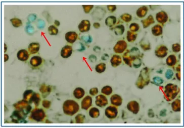
leaky cells can be stained by methylene blue (Figure 1.) because this dye with small molecular weight can enter to the dead cells across the pores opened by the toxin.
K28 uses the cell wall α-1,6-mannoprotein receptors to enter to the cell. From the ER, K28 moves into the cytoplasm and shuts down DNA synthesis in the nucleus, triggering apoptosis.
Figure 1. Killer activity of S. cerevisiae
Sensitive cells were inoculated into the background. The toxin diffused into the medium and caused a wide inhibition zone around the killer strain. The methylene blue supplemented in the medium stained the dead sensitive cells (red arrow).
Microorganisms: Four-day-old shaken culture of Saccharomyces cerevisiae strain T158C (K1 toxin producing strain) in YPD medium and one-day-old S. cerevisae S6 (K1 toxin sensitive) strain on slant agar.
Media:
T-28A: 0.5% yeast extract, 1% pepton, 1% D-glucose, 2% agar Citrate/phosphate buffer (pH 4.2)
methylene blue solution (0.3 mg/ml)
Materials: Microtubes, pipettes, tips, proteinase K solution (10 mg/ml), water bath, sterile tube, sterile water, Petri dish, sterile borer, loop, microcentrifuge
Melt the T-28A media in a pressure cooker. Adjust the pH of the medium to 4.2 with citrate buffer (T-28B). Add some drops of methylene blue (T-28C) to the medium and pour one plate.
Prepare a dense suspension of S. cerevisiae S6 (killer sensitive strain) in 3 ml sterile distilled water. Make a massive inoculation on the plate. Make 4 wells with a sterile borer in the inoculated plate.
Centrifuge 1 ml suspension of S. cerevisiae T158C (K1 toxin producing strain) in a sterile Eppendorf tube (5 min, 5000 rpm). Pipette 200 µl supernatants into 3 sterile Eppendorf tubes.
One will be the control it will be loaded to one well. Add 2 µl Proteinase K to the second Eppendorf tube and incubate it at 37 ˚C for 30 min. After the incubation load the sample to the second well. Place the third Eppendorf tube to a hot water bath for 30 min. Load the sample to the third well after 30 min. Pipette cells of the killer strain to the 4th well.
Required materials
Method
3.1.2.1. Killer activity
/- Practice 11 -/
Microorganisms: Four-day-old shaken culture of Saccharomyces cerevisiae strain T158C (K1 toxin producing strain) in YPD medium
Materials: Microtubes, pipettes, tips, RNase solution (10 mg/ml), water bath, centrifuge tubes, sterile phosphate buffer (0.1 M), phenol:chloroform:iso-amyl-alcohol 25:24:1, loading buffer, agarose, TBE buffer, ethidium-bromid, sterile distilled water, cylinder, scale, gel electrophoresis unit, microcentrifuge
Take 50 ml three-old-day culture of S. cerevisiae T158C. Pellet the cells by centrifugation (10 min, 3000 rpm, 4°C). Suspend the cells in 5 ml ice-cold phosphate buffer (0.1 M, pH: 7.4) and disrupt them in French pressure at 20000 Ψ. Pellet the cells and cell debris by centrifugation (30 min, 15000 rpm, 4°C).
Pipette 200 µl supernatant in 3 Eppendorf tubes.
Tube 1.: Add equal amount of PCI (phenol:chloroform:iso-amyl-alcohol 25:24:1), centrifuge it (10 min, 10000 rpm, room temperature). Pipette 50 µl from the upper phase to a new Eppendorf tube. Add 10 µl bromo-phenol blue to the sample and load it to a well of an agarose gel.
Tube 2.: Add equal amount of PCI (phenol:chloroform:iso-amyl-alcohol 25:24:1), centrifuge it (10 min, 10000 rpm, room temperature). Pipette 50 µl from the upper phase to a new Eppendorf tube. Add 1 µl RNase to the sample and incubate it at 37°C for 30 min. After the incubation time add 10 µl bromo-phenol blue to the sample and load it to a well of an agarose gel.
Tube 3.: Add 2 µl RNase to the sample and incubate it at 37°C for 30 min. After the incubation time add equal amount of PCI (phenol:chloroform:iso-amyl-alcohol 25:24:1), centrifuge it (10 min, 10000 rpm, room temperature). Pipette 50 µl from the upper phase to a new Eppendorf tube. Add 10 µl bromo-phenol blue to the sample and load it to a well of an agarose gel.
Gel electrophoresis Required materials
Method
3.1.2.2. RNase protection assay
/- Practice 12 -/
Check your progress
What is the mode of action of K1 toxin?
What are the optimal conditions for toxin activity?
What is the role of the methylene blue in the experiment?
How can the chemical nature of the toxin be determined?
What is the genetic background of killer toxin production in S. cerevisiae?
What „RNase protection assay” is good for?
What method is suitable to open the yeast cells?
What proteins are coded on L-A virus?
Introduction of the model organism Aspergillus nidulans
The filamentous fungus A. nidulans has many advantageous characteristics, which make this fungus an excellent candidate for studying life cycle, genetic interactions, various molecular biological processes (sensing and signal transduction of environmental factors and response to them; regulation of conidiogenesis and -ascosporogenesis; regulation and biosynthesis of primary- and secondary metabolites; DNA metabolism; organisation of actin and cytoskeleton;
regulation and molecular events of polarized growth; transport mechanisms; mitochondrial functions; role of remodeling complexes, activators and repressors in chromatin functions and many more...), physiological functioning and evolution. These characteristics are the following.
It has minimal nutritional requirements and in that one source of organic carbon (glucose, lactose, galactose, saccharose, mannose, ethanol, certain amino acids and many more...), one source of nitrogen (nitrate, ammonium, acetamide, hypoxanthine, xanthine, uric acid, allantoin, urea and many more...), inorganic salts and a few trace elements are needed. This simplicity supports the studies on the genetics of biosyntheses.
It grows rapidly, with which the fungus produces great amount of biomass in a few days that could be subjected to experimental processing.
It has filamentous growth.
It can be cultured in liquid medium.
It performs a compact growth with easily accessible asexual and sexual reproductive structures. The sexual structures (cleistothecia) are closed and remain closed even when the ascospores are ripe. Therefore cleistothecia with viable ascospores can be preserved for years.
Metabolic versatility with wide range of primary- and secondary metabolic pathways.
It performs complete sexual-, asexual and parasexual life cycles.
It is self-fertile due to homothallism.
Its uninucleate conidiospores and the clonal reproduction make possible the rapid purification of mutants or transformants obtained by genetic crosses, mutagenesis or
3.1.3. General introduction to the asexual, sexual and
parasexual life cycles of the filamentous fungus model
organism, Aspergillus nidulans
Its haploid genome makes mutants easily selectable and execution of Mendelian analysis within one generation.
Heterokaryons and diploids can be easily isolated for complementation tests, dominance tests and study of somatic crossing overs.
The availability of dozens of auxotroph mutants for genetic and molecular biological studies.
Its transformation and genetic manipulation is easy that support the application of molecular biological methods.
Its whole genome is sequenced and annotated (http://www.aspergillusgenome.org).
Life cycles of A. nidulans
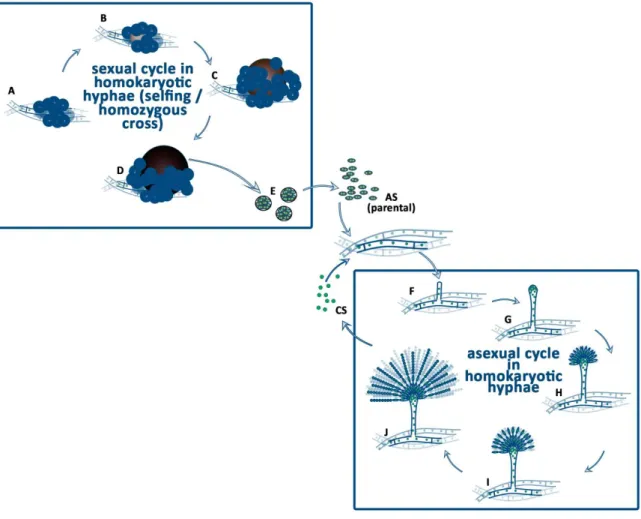
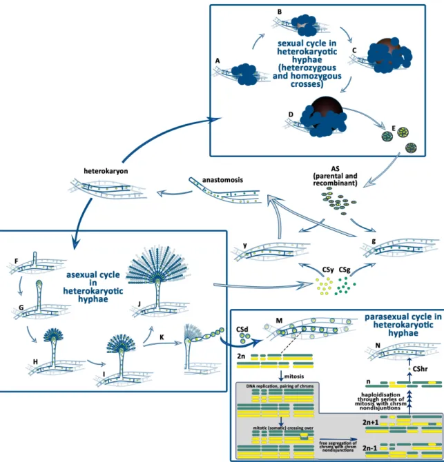
The A. nidulans is a homothallic filamentous fungus. Its genome carries both the MAT1-1 and MAT1-2 mating type genes, with orthologues corresponding to the two oposite mating type genes in the heterothallic closely-related species, such as Podospora anserina. Since both mating type genes are present and expressed in the genome, A. nidulans is able to accomplish the full sexual cycle itself, without the need of a mating partner. When this happens, we call the process selfing, inbreeding, or more accurately, homozygous cross (Figure 1). As a result of the selfing the nuclei of the reproductive structures, the ascospores, are identical with that were found in the parental hyphae. The heterozygous sexual cycle (outcross) starts with a heterokaryon formation by anastomosis between the hyphae of two, genetically different mycelia. The result of the heterozygous sexual cycle is the production of both recombinant and parental types of ascospores (Figure 2).
A. nidulans colonies can be formed by the germination of either the asexually produced conidiospores or the sexually produced ascospores (Figure 1). Ascospores are binucleate (each contains two identical nuclei) but the conidiospores are uninucleate. The colony, which develops from the germination of a single ascospore or a conidiospore, is homokaryotic. This means that all the nuclei in the hyphae are identical.
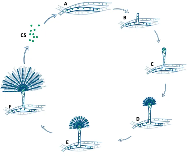
Asexual reproduction in homokaryotic mycelium
formed, which is full with nuclei (Figure 1). Metula cells forming a cell layer on the surface of vesicle are generated by yeast like mitotic division-, budding of the nuclei in the vesicle (Figure 1).
Figure 1. Life cycle of A. nidulans homokaryotic hyphae.
The homozygous sexual cycle of A. nidulans (A-E) begins with the production of Hülle cells when the environmental conditions are supportive (A). Surrounded by the Hülle cells, ascogenous hyphae are differentiated (B). These ascogenous hyphae form a ball shape nest, which is called primordium (B).
Around the ball of ascogenous hyphae flattened longitudional hyphae are differentiated to the pericarp of the cleistothecium. The primordium grows and forms the µ-cleistothecium, in which the ascogenous hyphae start to develop ascus mother cells and asci, where meiotic events occur (C). Upon maturation, the µ-cleistothecium hardens its pericarp that is accompanied by a dark pigment accumulation of the pericarp cells, while hundred thousands of asci are formed inside the cleistothecium (D). Each ascus contains eight binucleate, parental type ascospores with dark red color. The asci and the free ascospores (AS) are released to the environment. Germination of an ascospore will result in a homokaryotic mycelium formation. The homozygous asexual cycle (the conidiogenesis) starts with the differentiation of a hyphal compartment into a foot cell, which starts to grow upward (forms the stalk), high above the mycelial mat (F). At the tip of the stalk a vesicle is formed (G). Through budding of the nuclei in the vesicle metula cells are formed (H). The metula cells are uninucleate and by covering the surface of vesicle, they form the layer of the primary sterigmata (H). Through the budding of the metula cells the secondary sterigmata layer composed of uninucleate phialide cells is formed (I). Each of the phialide cells starts to produce uninucleate conidiospores, by subsequent repetition of budding. As a result, chains of conidiospores (CS) are formed by the phialide cells (J). Germination of a conidiospore will result in a homokaryotic mycelium formation.
uninucleate phialide cells is formed (Figure 1). Each of the phialide cells starts to produce uninucleate conidiospores, by subsequent repetition of budding. As a result, chains of conidiospores are formed from the phialide cells (Figure 1). Germination of a conidiospore will result in a homokaryotic mycelium formation.
Sexual reproduction in homokaryotic mycelium
The homozygous sexual cycle of A. nidulans begins in 4 days old colonies when the environmental conditions support the sexual development (darkness, decrease of oxygen tension, availability of ammonium nitrogen source). The first event of sexual development is the formation of unicellular Hülle cells (Figure 1). Hülle cells are thick walled multinucleate cells, which protect and nurse the developing asci. Surrounded by the Hülle cells, dikaryotic ascogenous hyphae are differentiated (Figure 1). These ascogenous hyphae form a ball shape nest, which is called primordium (Figure 1). Around the ball of ascogenous hyphae flattened longitudional hyphae are differentiated to the pericarp (outer coat/wall) of the immature closed fruiting body (cleistothecium). The primordium grows and forms the µ-cleistothecium, in which the ascogenous hyphae start to develop ascus mother cells and asci, where meiotic events occur (Figure 1). Upon maturation, the µ-cleistothecium hardens its pericarp that is accompanied by a dark pigment accumulation in the pericarp cells, while hundred thousands of asci are formed inside the cleistothecium (Figure 1).
Each ascus contains eight binucleate, parental type ascospores with dark red color (Figure 1).
When the free ascospores are released to the environment they germinate and form a homokaryotic mycelium.
Sexual, asexual and parasexual reproduction in heterokaryotic mycelium
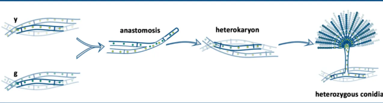
When the mycelia of two neighbouring colonies are mixed, hypha anastomosis (plasmogamy) between the hyphae of different partners will occur from time to time (Figure 2).
Following the anastomosis, the hyphal compartments are heterokaryotic, because they carry both types of nuclei originated from the two parental hyphae. The heterokaryotic compartments form the heterokaryotic mycelium. In nature as well as in the laboratory, the heterokaryotic mycelia are maintained when the environmental conditions are changed and the new conditions do not support the growth of the parental types of mycelia. If the strains carry complementary
riboflavin and pyridoxine. In the absence of the required vitamins, only those compartments can grow and maintained, which are heterokaryotic, thus they contain both types of parental nuclei. In the heterokaryons the auxotrophies of the individual nuclei are complemented by the other parental nuclei. In such a selective environment, the homokaryotic mycelia will die, while the heterokaryotic mycelia thrive.
In the heterokaryotic mycelium sexual, asexual and parasexual life cycles may take place (Figure 2). Technically the the series of events are the same that happen during the homokaryotic conidiogenesis or sexual development, however the outcomes of these processes are different (Figure 2). In the heterokaryotic mycelium the dikaryotic ascogenous hyphae may contain two identical nuclei (homozygous dikaryon) of either parent or two different nuclei of the two parents
Figure 2. Life cycle of A. nidulans heterokaryotic hyphae.
The heterozygous sexual cycle of A. nidulans (A-E) begins with the production of Hülle cells and
genetically identical with either the one or the other parent. In case the ascogenous hypha in the primordium was heterokaryotic, the whole cleistothecia will be of recombinant type (D). Germination of an ascospore will result in a homokaryotic mycelium formation. The heterozygous asexual cycle (the conidiogenesis) starts with the differentiation of a hyphal compartment into a foot cell and continues with the stalk (F), vesicle (G), metula layer (H) and the phialide layer (I) formation as it is decribed in details in the homokaryotic life cycle in Figure 1. Since the vesicle contains the nuclei of both parents and the metula cells formed by the mitosis of an individual nucleus of the vesicle are uninucleate, henceforth the metula cells may possess either one or the other parental nucleus. The uninucleate phialide inherits the nucleus of the underneath metula cell and the nucleus of the conidiospore will be identical with that of the phialid cell. Since one phialide cell produces many conidiospores that stick together forming a chain of spores, all the conidiospores belonging to the same chain will carry identical nuclei. Sometimes diploid nuclei are formed in the heterokaryon by the fusion of two haploid nuclei (K). When a heterozygous diploid nucleus plays role in metula cell formation, the metula cell and the subsequently formed phialide cell and all the conidiospores (CSd) in the cognate spore chain will carry a single diploid nucleus Germination of a diploid conidiospore will result in the formation of a diploid mycelium (M).
When heterozygous diploid nuclei undergo mitosis, the failure of the segregation of sister chromatids, the so called chromosome non-disjunction, will result in the loss of one or more of the chromosomes in the progeny (grey boxed area). In these progeny the chromosome set is different from 2n and n (grey boxed area). They are called aneuploids. Over several mitoses the repeated events of chromosome non- disjunction will result in the haploidization of the diploid nuclei. The random losses of the parental chromosomes due to the chromosome non-disjunctions and the rarely occurring mitotic crossing overs will result in haploid genomes (n) that carry recombinant genetic material. When such a haploid and recombinant nucleus participates in conidiogenesis, the haploid and recombinant nucleus will be inherited in the conidiospores (CShr) and will be propagated in the environment. From these haploid and recombinant conidiospores a new, recombinant type of mycelia will be developed.
(heterozygous dikaryon). When the ascogenous hypha is homozygous, the ascospores in the whole cleistothecium will have the same parental nuclei (Figure 2). In case the ascogenous hypha is heterozygous, then the ascospores in the whole cleistothecium will have recombinant nuclei (Figure 2). Regarding the asexual reproduction, it will result in conidiosores with nuclei of either the one or the other parent. Nuclei of conidiospores belonging to the same chain will be always identical with each other, since they derive from the same uninucleate phialide cell. In the heterokaryotic mycelium heterozygous diploid nuclei may be formed by the fusion of two non- identical nuclei. Diploid formation is a relatively rare event (with 10-5-10-8 frequency). The diploid nuclei will undergo mitosis and can take part in conidiogenesis that results in the formation of diploid conidiospores (Figure 2). The diploid conidiospores can germinate and form diploid mycelia. Sometimes the sister chromatides cannot separate from each other (called as chromosome non-disjunction) during the mitosis of the diploid nuclei and one nucleus will inherit both chromatides (it will possess three of that type of chromosome, the chromosome set will be 2n+1), while the other nucleus will not inherit that chromatid at all (it will posses only one from
haploid genomes derived from the haploidization of a diploid genome will be of recombinant- type (differs from both parents). In the laboratory, the failure of the segregation of the sister chromatides, the chromosome non-disjunction can be mediated by the usage of the inhibitor of the microtubules, benomyl.
Literature:
Pontecorvo G. (1956) The parasexual cycle in fungi. Annu. Rev. Microbiol. 10, 393-100.
Pontecorvo G., Roper J. A., Hemmons L. M., Macdonald K. D., and Bufton A. W. J. (1953) The genetics of Aspergillus nidulans. Adv. Genet. 5, 141-238.
Kafer E. (1977) Meiotic and Mitotic Recombination in Aspergillus and Its Chromosomal Aberrations. In Advances in Genetics. Volume 19, Pages 33-131
3.1.3.1. Asexual life cycle of A. nidulans - Practice 13 -
I. Introduction
Asexual life cycle of A. nidulans
II. Practice 13
Study of the asexual reproductive structures of A. nidulans
Learning Objectives
After the completion of the practical course, you should be able to:
Explain the different stages of conidiogenesis and name the structural units of a conidia.
Define the following terms: uninucleate,
conidiogenesis, metula, phialide, conidiospore
Acquire the following laboratory skills: usage of
the microscope, making cellux-tape preparates
from conidia, calculate the necessay amounts of
components of a laboratory used buffer.
I. Introduction
Asexual life cycle of A. nidulans
When a uninucleate haploid conidiospore germinates, a colony will be formed by the mycelia of colorless septate hyphae (Figure 1). The compartments bordered by two septa are multinucleate and homokaryotic (Figure 1). 40-48 hour after the germination of a conidiospore, the young colony starts to develop foot cells by the differentiation of certain compartments (Figure 1). A foot cell gain a brownish thick cuticle and produces an up-growing stalk (conidiophore) with length of 100 µm and diameter of 6 µm (Figure 1). At the end of the stalk a vesicle is formed, 10 µm in diameter (Figure 1). The foot cell, the stalk and vesicle are multinucleate, aseptate and covered with a thick brownish cuticle. From the surface of the vesicle uninucleate elongated buds with 5 µm in length are developed synchronously (Figure 1) by budding. These daughter cells are called metula cells and they form the primary sterigmata layer of the conidia. The metula cells will undergo a budding process synchronously, giving origin to a layer of uninucleate phialide cells (Figure 1). This second layer of cells is called secondary sterigmata layer. Each phialide cell begins a series of budding, by which a chain of uninucleate conidiospores will emerge from the end of the phialide cell (Figure 1). The youngest conidiospore is always the nearest one to the phialide cell. The older ones are further away, gradually attaining full size and full green color upon maturation. All the conidiospores of a certain chain derive their nucleus from the cognate single nucleus of the sterigma cells (phialide and metula). The conidiospores are green in wild type A.
nidulans.
Figure 1. Conidiogenesis.
Conidiogenesis starts with the differentiation of a vegetative hyphal compartment (A, B) by the development of the stalk high above the surface of the colony (B). At the end of the stalk a vesicle containing many nuclei is formed (C). Nuclei in the vesicle undergo mitosis and by budding (same as budding in yeasts) a layer of uninucleate cells (primary sterigmata layer) bud out of the vesicle (D). These daughter cells are called metula cells. After that the metula cells will synchronously undergo budding and as a result, a secondary sterigmata layer of uninucleate phialide cells are formed (E). After that the phialide cells begin a series of budding, by which chains of conidiospores are formed at the end of the phialide cells. During maturation, the conidiospores are kept together in the chain. Upon maturing, the conidiospores are disbanded and will be spread as free individual conidiospores by the air or water to new habitat.
Literature:
Pontecorvo G., Roper J. A., Hemmons L. M., Macdonald K. D., and Bufton A. W. J. (1953) The genetics of Aspergillus nidulans. Adv. Genet. 5, 141-238.