Novel mechanisms of GPCR functions: AT1 angiotensin receptor acts as a signaling hub and focal point of receptor cross-talk
András D. Tóth1, Gábor Turu1,2, László Hunyady1,2,3, András Balla1,2
1Department of Physiology, Faculty of Medicine, Semmelweis University, Budapest, Hungary, 2MTA-SE Laboratory of Molecular Physiology, Hungarian Academy of Sciences and Semmelweis University, Budapest, Hungary,
3To whom correspondence should be addressed: László Hunyady, Address: Department of Physiology, Faculty of Medicine, Semmelweis University, H-1428 Budapest, P.O. Box 2, Hungary. Tel.: +36 1 266 9180; fax: +36 1 266 6504. E-mail:
hunyady.laszlo@med.semmelweis-univ.hu Abstract
AT1 angiotensin receptor (AT1R), a prototypical G protein-coupled receptor (GPCR), is the main receptor, which mediates the effects of the renin-angiotensin system (RAS). AT1R plays a crucial role in the regulation of blood pressure and salt-water homeostasis, and in the development of pathological conditions, such as hypertension, heart failure, cardiovascular remodeling, renal fibrosis, inflammation, and metabolic disorders. Stimulation of AT1R leads to pleiotropic signal transduction pathways generating arrays of complex cellular responses.
Growing amount of evidence shows that AT1R is a versatile GPCR, which has multiple unique faces with distinct conformations and signaling properties providing new opportunities for functionally selective pharmacological targeting of the receptor. Biased ligands of AT1R have been developed to selectively activate the β-arrestin pathway, which may have therapeutic benefits compared to the conventional angiotensin converting enzyme inhibitors and angiotensin receptor blockers. In this review, we provide a summary about the most recent findings and novel aspects of the AT1R function, signaling, regulation, dimerization or oligomerization and its cross-talk with other receptors, including epidermal growth factor (EGF) receptor, adrenergic receptors and CB1 cannabinoid receptor. Better understanding of the mechanisms and structural aspects of AT1R activation and cross-talk can lead to the development of novel type of drugs for the treatment of cardiovascular and other diseases.
Keywords
GPCR, angiotensin II, receptor cross-talk, bias, dimerization, Abbreviations
α2CAR, α2C adrenergic receptor; AngII, angiotensin II; AT1R, AT1 angiotensin receptor;
β2AR, β2-adrenergic receptor; EGFR, epidermal growth factor receptor; GPCR, G protein- coupled receptors; HB‒EGF, heparin-binding epidermal growth factor-like growth factor;
RAS, renin‒angiotensin system; VSMC, vascular smooth muscle cell
Introduction
The octapeptide (Asp‒Arg‒Val‒Tyr‒Ile‒His‒Pro‒Phe) hormone angiotensin II plays a crucial role in the maintenance of blood pressure and fluid homeostasis. It is produced by a two-step cleavage process from the precursor angiotensinogen by protease enzymes, namely renin and angiotensin convertase enzyme (ACE). The AngII effects are mediated by two distinct G protein-coupled receptors (GPCRs), the AT1 and AT2 angiotensin receptors, but there is a substantial difference in their importance in favor of the former. The AT1R is expressed in numerous tissues and mediates the "classical" physiological actions of circulating AngII on mechanisms including blood pressure regulation, salt-water balance, aldosterone secretion and effects on the central nervous system, such as thirst sensation and regulation of sympathetic outflow [1,2]. In addition, increased AT1R activity has been associated with the development of several pathological conditions, including hypertension, heart failure, vascular remodeling, diabetic nephropathy, atherosclerosis and inflammation [2]. Therefore, dampening of AT1R activity has enormous therapeutic benefits and has been successfully exploited in the last decades using ACE inhibitors and AT1R blockers. However, application of these drugs also hinders the beneficial functions of AT1R. In recent years, novel drug compounds with different pharmacodynamic properties were discovered, which are able to selectively activate specific signaling pathways of AT1R. These compounds may reduce side effects and/or have advantageous actions in treatment of diseases and could open a new era in AT1R-targeted therapies.
In addition, a substantial knowledge has been gained about the main properties of AT1R, such as ligand preference, signaling, regulation, and trafficking. However, it is less known how AT1R and other plasma membrane receptors affect each other’s function, and how these crosstalk mechanisms can be utilized in the clinical practice. The new results in the field of receptor crosstalk can reveal not just new drug targets, but can also explain certain interactions between pharmaceutical compounds. In this review, we highlight the traditional and novel features of AT1 angiotensin receptor (AT1R), which is a prototypical GPCR, and can be considered as paradigm for other GPCRs not only in its pleiotropic functions and action of mechanisms but also in its clinical importance and druggability.
Structural aspects of AT1R functions
AT1R is a member of the rhodopsin family of GPCRs, and shares their common structural architecture [1]. It possesses an extracellular N terminus, an intracellular C terminus and seven highly hydrophobic transmembrane α-helices (H1-7), connected by three extra- and intracellular loops (ECL1-3 and ICL1-3, respectively). Ligands of GPCRs can be classified as agonists or antagonists. Agonists are capable to induce structural rearrangements in the receptor, achieving an active conformation of the GPCR, whereas antagonists stabilize the inactive state. Our understanding of the structural aspects of GPCR activation has improved strikingly in recent years thanks to the growing numbers of high-resolution GPCR structures.
Surprisingly, despite the relatively low sequence homology between GPCRs, the conformational features of the transmembrane helices are considerably conserved [3]. In contrast, the composition of ligand binding pockets is diverse among GPCRs, which ensures the specific recognition of receptor ligands. Accordingly, our knowledge of the interaction between AT1R and its specific antagonists (or more precisely: inverse agonists) has hugely improved by the first crystal structures of AT1R in complex with the AT1R antagonists ZD7155 and olmesartan [4,5]. In good agreement with the previous results of site-directed mutagenesis studies, the ligand binding pocket of AT1R is formed by several key residues of ECL2 and the transmembrane helices H1, H2, H3 and H7. With the help of docking
simulations, these structures could explain the different binding properties of various receptor blockers. For instance, losartan, an antagonist with a relatively low binding affinity, forms only one salt bridge with the ligand binding pocket, whereas candesartan, an antagonist with insurmountable binding, is speculated to engage AT1R with two additional salt bridges [4].
The knowledge of the antagonist-stabilized state may also help to predict the conformational changes of the agonist-stimulated AT1R.
Signal transducers of AT1R
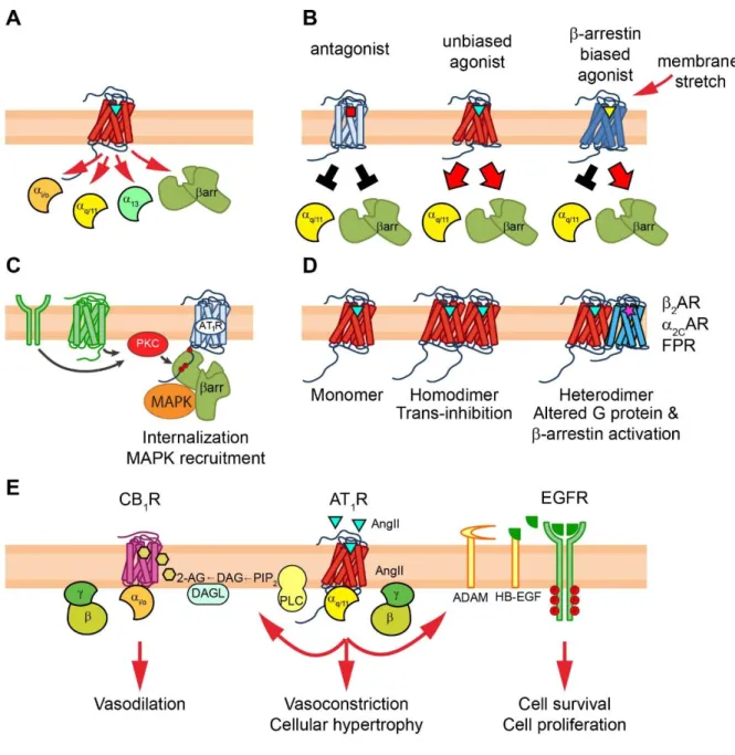
Heterotrimeric G proteins initiate the first wave of GPCR signaling (Fig. 1) [6]. Upon agonist activation, the Gα subunit binds to the core region of active GPCRs via the opened cytosolic cavity. This interaction triggers nucleotide exchange of Gα from GDP to GTP and dissociation of Gα from Gβγ. The separated subunits modulate the activity of downstream effector proteins to induce robust signaling cascades. These signaling mechanisms have wide spectra, including second messenger generation, activation of small G proteins and cytoplasmic tyrosine kinases, regulation of ion channels or transactivation of growth factor receptors. Likewise, G protein-mediated signaling pathways are responsible for the vast majority of AT1R-evoked cellular responses [1]. AngII stimulation of AT1R is able to activate not just one but various G proteins, such as Gq/11, Gi/o, or G12/13 [7]. Gq/11 protein induces the hydrolysis of phosphatidylinositol 4,5-bisphosphate (PIP2) into the second messenger inositol trisphosphate (IP3) and diacylglycerol (DAG) by activation of phospholipase Cβ, IP3 triggers intracellular Ca2+ mobilization via binding and opening its calcium channel receptor. Ca2+ is a central regulator of many intracellular proteins and, in co-operation with DAG, also leads to the activation of various protein kinase C (PKC) isoforms [1,2]. Meanwhile, the signal transduction via Gi/o and G12/13 proteins lead to inhibition of adenylyl cyclase, regulation of L- and T-type Ca2+ channels and activation of phospholipase D, Rho GTPases or Rho kinase. It seems that the Gq/11-mediated signal transduction mechanisms of AT1R prevail in the major physiological target tissues, including kidney, adrenal cortex, vascular smooth muscle and cardiac cells [2].
The first wave of signaling is terminated by the elimination of the GTP bound to Gα and by distinct mechanisms of desensitization [8]. In the course of homologous desensitization, GRK enzymes recognize and phosphorylate agonist-bound GPCRs on serine/threonine residues of the receptor C-tail and/or ICL loops. The phosphorylation contributes to receptor desensitization through promotion of high affinity binding of arrestin proteins [9]. Moreover, since GRKs have a large number of non-receptor targets, they may also act as effectors of GPCR signaling [10]. Although the phosphorylation target sites of the seven GRK isoforms show substantial overlap, there are marked differences as well. It was demonstrated that distinct ligands of the β2AR initiate different phosphorylation patterns in the cytoplasmic tail of the receptor, “barcode”, due to alternative interaction with GRK2 and/or GRK6 [11]. Similarly, different phosphorylation barcode by GRK2/3 and GRK5/6 were suggested in the case of AT1R [12,13]. In addition, the C-tail of AT1R contains several consensus PKC phosphorylation sites [14]. Phosphorylation by PKC can be induced by activation other plasma membrane receptors and occurs not only in the active but the inactive state of the receptor. This mechanism of regulation of AT1R sensitivity is termed as heterologous desensitization.
GRK-phosphorylation of agonist-bound AT1R is followed by the recruitment of β- arrestins. β-arrestins are multi-functional adaptor proteins of GPCRs. The two β-arrestins (β- arrestin1 and β-arrestin2) are expressed ubiquitously in mammalian tissues, and show high sequence and structural homology [9]. Their major roles are identical, but several isoform- specific functions were also reported (reviewed in [15]). They bind receptors in a two-step process [16]. First, β-arrestins interact with receptor-attached phosphates, then dock to the
intrahelical cavity of the activated GPCR. The binding of β-arrestin to the GPCR core sterically prevents the further activation of G proteins. Moreover, activated β-arrestins can interact with numerous adaptor proteins involved in the endocytic cargo transport [8].
Association of β-arrestin with the β2-appendage of adaptor protein 2 induces translocation of the GPCR-β-arrestin complex to clathrin-coated pits, which step is followed by internalization mediated by clathrin-coated vesicles. AT1R interacts with β-arrestins in sustained manner, due to strong interaction of β-arrestins with phosphorylated C-terminal serine/threonine clusters [17,18]. Because of the stable interaction, AT1R and β-arrestins internalize together as a complex. This allows β-arrestins to govern the intracellular trafficking of the receptor [8].
Basically, receptors can have two fates after endocytosis (Fig. 2). They can be degraded in lysosomes or be recycled to the plasma membrane via fast and/or slow recycling endosomes.
During recycling, the receptor ligands detach from the receptor due to lower pH in endosomes, and the receptor is dephosphorylated by protein phosphatases. These processes induce resensitization of the receptor, i.e. they are able to respond to agonists again. Sustained β-arrestin binding of AT1R favors late endosomal and lysosomal trafficking, inducing its down-regulation [8]. Furthermore, β-arrestin-independent and caveolae-mediated internalization routes of AT1R have also been described [19].
Initially, β-arrestins were considered only as negative regulators of GPCR functions by mediating receptor desensitization and internalization. It is now widely-accepted that they have much broader roles, as they are central organizers of distinct signaling cascades [16]. As scaffolds, they orchestrate a vast array of signaling proteins, such as various mitogen- activated protein kinases (MAPK), phosphoinositide 3-kinase, Akt or protein phosphatase 2A [16,20,21]. In that function, β-arrestins fine tune a second wave of GPCR signaling. This signaling is substantially different from the first wave regarding its temporal, spatial and mechanistic features [6]. One of the most known β-arrestin-mediated function is the modulation of MAPK activation. Various MAPK cascade members are regulated by β- arrestins upon AT1R activation, including Raf1, MEK, ERK, p38 MAPK and JNK [16]. The formation of AT1R–β-arrestin–MAPK complexes alter the localization of MAPK activation.
These complexes keep activated MAPKs away from the nucleus, thereby preventing the induction of their transcriptional response [20]. On the other hand, the β-arrestin-bound MAPK complexes can phosphorylate and regulate other target proteins, which are brought in proximity as well by β-arrestins. β-arrestins were shown to interact with hundreds of signaling proteins, indicating that β-arrestins are central regulators of complex signaling networks [22,23]. Among a wide range of functions, β-arrestins regulate cytoskeletal rearrangements, chemotaxis, or protein synthesis. In the light of these observations, it is not surprising that β- arrestins were demonstrated to mediate a plethora of AT1R effects, such as positive inotropy in the heart, cardiac hypertophy or proliferation of cardiomyocytes [24–26].
Interestingly, β-arrestins show a remarkable degree of conformational plasticity [9].
They can adopt multiple active conformations with distinct signaling properties. An important determinant of the β-arrestin conformation is the receptor C-terminal phosphorylation barcode [16]. Different phosphorylation motifs induce distinct β-arrestin conformational changes, which provides the possibility to evoke distinct AT1R induced cellular responses [12]. In addition, activated β-arrestins are ubiquitinated by ubiquitin ligases, such as Mdm2 [27].
Interestingly, the ubiquitination patterns of β-arrestin may differ in the distinct active conformations, which may contribute to the fine regulation of β-arrestin-mediated response [27].
Recent data suggest that the β-arrestin-mediated second signaling wave is dependent on G proteins, suggesting that β-arrestins, in fact, broaden the signaling options of G proteins by governing their signaling in time and space [28]. Interestingly, several GPCRs have been reported to generate a third, sustained signaling wave from endosomes after internalization
[29,30]. Interestingly, GPCR–G protein–β-arrestin supercomplexes have been identified to be responsible for the sustained endosomal signaling [31]. In contrast to the generally believed competition between G proteins and β-arrestins, in this complex Gα binds to the receptor core and β-arrestin interacts with the phosphorylated C-tail of the same receptor. These results were obtained with Gs protein-coupled GPCRs, and it would be curious to investigate whether the mainly Gq-coupled AT1R could form such complexes as well. These data shed light on the complex interplay between the GPCR transducers and suggest that spatiotemporal features of signaling are more prominent than they were previously supposed.
In addition to the above-mentioned effectors, GPCRs also have numerous other interacting partners. These proteins can be either plasma membrane proteins such as ion channels, transporters, various serine/threonine-specific protein kinases, cytoskeletal proteins, Src homology and PDZ domain-containing proteins, small G proteins or extracellularly located adhesion molecules [32]. Accordingly, AT1R was shown to interact with wide spectra of other proteins beside G proteins and β-arrestins, such as AT1R-associated protein (ATRAP), phospholipase Cγ, JAK2 or other GPCRs, just to name a few, which may also take important parts in the complex pleiotropic effects of AT1R in the target tissues of the renin- angiotensin system (RAS) [1].
Biased agonism of AT1R
β-arrestin binding and internalization of AT1R and other GPCRs does not require G protein activation [28,33–35]. Although recent data suggest that β-arrestin-mediated signaling of GPCRs requires G protein activation [28], β-arrestin binding and its effect on receptor regulation is G protein independent, therefore compounds that selectively activate G proteins and β-arrestin binding (biased or functional selective compounds) can modify the intracellular fate of receptors, which may have therapeutic relevance. It was speculated that AT1R may adopt multiple active conformations with distinct signaling properties, and conformation- specific targeting of AT1R could offer the intriguing possibility of pathway-selective intervention. Evidence for this concept is the successful development of β-arrestin-biased peptide agonists [36]. These peptides induced no or partial activation of G proteins, while they triggered efficient receptor phosphorylation and β-arrestin recruitment (Fig. 3A-B) [7,35]. Using AT1R conformational biosensors, it was demonstrated that the conformations stabilized by β-arrestin-biased peptides are indeed distinct from that of the AngII-induced conformation [37]. These biased peptides lack the aromatic amino acid in position 8, the indispensable residue for adoption of the G protein-activating conformation of AT1R. The first such a peptide was [Sar1,Ile4,Ile8]-AngII, which was then followed by a series of higher- affinity ligands including, TRV120023 or TRV120027 [38]. These peptides revolutionized AT1R pharmacology and unveiled new aspects of AT1R functions [39]. In addition, they show not only selective activation of the β-arrestin-mediated signaling pathway but change the intracellular trafficking of AT1R [40,41]. The altered intracellular fate upon treatment with β- arrestin-biased compounds may be explained by the interesting finding that they also induce a different active conformation of the β-arrestin [42,43]. In addition, lack of Gq-dependent hydrolysis of PIP2, a known determinant of endocytosis [44], accelerates internalization of the receptor, which may have profound effects on the spatiotemporal features of signaling (Fig. 2) [40,45]. Moreover, these results offer the intriguing possibility that biased agonists could be applied in diseases where the intracellular receptor processing should be changed. The unique pharmacodynamic properties of biased agonists were also demonstrated in vivo, as they possessed beneficial effects on cardiac contractility and performance [38,46]. Unfortunately, although promising results were obtained with TRV120027 for the treatment of acute heart failure in animal studies [39,47], it failed to deliver the expected results in a Phase II clinical trial [48]. However, long-term treatment with another β-arrestin-biased peptide, TRV120067
was shown to have benefits compared to losartan treatment in a mouse model of dilated cardiomyopathy [49]. This suggests that chronic treatment with biased agonists could still be useful in the therapeutic strategies of some cardiovascular diseases. Although β-arrestin- mediated signaling pathways were mostly suggested to be beneficial, β-arrestin activation could also have adverse side effects. In the treatment of certain conditions, such as aldosterone overproduction, the preferable medicines will still probably be the full antagonists of AT1R, such as candesartan or valsartan, since the use of β-arrestin-biased compounds may lead to aldosterone escape [50], due to the fact that the aldosterone production is partly β- arrestin mediated in response to AngII [51].
AT1R as a stretch mechano-sensor
There is a growing number of evidence that AT1R not only behaves as a hormone receptor, but also serves as a sensor of membrane stretch [52–55]. In that function, AT1R is activated in the absence of ligand binding and shows biased signaling properties. Upon osmotic stretch, AT1R binds β-arrestin in a Gi/o activity dependent manner, but does not activate Gq/11 proteins [55]. These processes are followed by the transactivation of epidermal growth factor receptor (EGFR) and activation of ERK [55]. This mechanism was suggested to mediate the Frank-Starling law of the heart, i.e. the enhanced contraction response upon increased ventricular filling [56].
AT1R as a signaling hub
It was generally believed that the signal transduction of AT1R requires ligand binding and/or adoption of its active conformation. As reviewed above, activated AT1R induces a plethora of signaling pathways, in contrast to inactive AT1R, which was thought to be silent in terms of signaling. It has been demonstrated that pharmacological activation of PKC causes β- arrestin2 recruitment to AT1R even in the absence of receptor agonists [57]. Moreover, stimulation of either epidermal growth factor receptor or a distinct Gq/11-coupled GPCR, such as α1A-adrenergic receptor or endogenous purinergic receptors could exert the same effect, proving that the interaction can be triggered at physiological levels of PKC activation (Fig.
3C). It was also found that this heterologous mechanism of β-arrestin recruitment to AT1R was not sensitive to treatment with the inverse agonist candesartan, showing that this process does not require the active state of the receptor. However, it depends on stable association between the PKC-phosphorylated serine/threonine clusters in the receptor’s C-terminus and two conserved phosphate-binding lysines of β-arrestin2. Using β-arrestin2 conformational biosensors it was demonstrated that β-arrestin2 binds to PKC-phosphorylated AT1R in a distinct conformation. Moreover, this conformation is also active functionally, since it triggers MAPK recruitment and receptor internalization with altered intracellular receptor trafficking.
Taken together, the unliganded, but phosphorylated AT1R is able to recruit β-arrestins and consequently, the inactive AT1R may also participate in signaling as a scaffold protein [57].
This mechanism could be particularly important in the cases of receptors, like α1A-adrenergic receptor, which do not interact with β-arrestins, since the presence of AT1R could aid them to initiate β-arrestin dependent signaling. Since phosphorylation of AT1R is induced by a variety of other receptors, the β-arrestin activation by heterologously-phosphorylated AT1R may represent a central cross-talk mechanism for regulation of signal transduction.
Transactivation mechanisms driven by AT1R Cannabinoid receptor regulation by AT1R
Cannabinoid receptors (CB1 and CB2 receptors) were first recognized as the targets of the phytocannabinoid tetrahydrocannabinol, the active compound of marijuana [58,59]. Later
on, anandamide and 2-arachidonoyl glycerol (2-AG) were described as the main endocannabinoids produced in brain and other tissues [60]. The role of 2-AG as a cannabinoid receptor ligand was an intriguing finding, since it is produced from DAG by DAG lipase (DAGL) [61]. Since DAG is produced after Gq protein activation, and DAGL is expressed widely in many tissues, we have hypothesized that AT1R stimulation may lead to production of 2-AG. Indeed, 2-AG is produced in cell culture models after stimulation of the AT1R with AngII, leading to activation of the CB1 cannabinoid receptor (CB1R) in both autocrine and paracrine manner (Fig. 3E) [62,63]. Moreover, it has been shown later, that in different arteries [64–67] and central nervous system AT1R function is altered by CB1R activation, which effects are dependent on DAGL activity [68]. AngII itself induces contraction in rat and mouse aorta and coronary, renal, skeletal, muscle and pulmonary arteries, and this effect is attenuated through the parallel induction of 2-AG production and activation of CB1R [64–
67]. This mechanism may serve as a fine-tuning negative feedback regulation in the vasomotor system, dampening the effects of many Gq-coupled receptors in vascular smooth muscle cells (VSMCs), which may serve as a protection mechanism from overactivation.
Although the interaction between the RAS and the cannabinoid system seems to be present in the arteries, identification of the precise cellular location of the individual elements requires additional studies.
AT1R‒CB1R interaction is not limited to the regulation of endocannabinoid release. In rat astrocytes, AngII-induced PKC-mediated phosphorylation and heterologous desensitization of the cannabinoid receptors [69]. In these cells, CB1R activity inhibits the MAP kinase pathway, and desensitization may regulate the balance between active and inactive receptors after activation through endocannabinoid release. Alternatively, CB1R receptor signaling bias might be regulated through PKC-mediated phosphorylation, although these aspects of the interaction have not been yet investigated. On the other hand, AT1R‒
CB1R heterodimerization has also been reported in Neuro2A cells, where AT1R-induced full ERK1/2 required expression of the CB1R [70]. Together, these data show that there may be multiple levels of interaction between RAS and the cannabinoid system, which may be distinct in different tissues and cell types. They also suggest that autocrine and paracrine activations of CB1R might be not just spatially, but also functionally different.
EGFR transactivation by AT1R
It is very characteristic for the AT1R that several cellular responses upon AngII stimulation are mediated by receptor tyrosine kinases, among which the EGFR plays the most important role in the cardiovascular system [71] and in other tissues, such as hepatocytes [72].
The EGFR transactivation is mediated via calcium signal and matrix metalloprotease (ADAM) activation, which causes the cleavage of heparin-binding epidermal growth factor- like growth factor (HB‒EGF) resulting in agonist release for the EGFR stimulation (Fig. 3E) [73]. It turned out that this mechanism is crucial for several pathological effects of AngII, including cardiac and vascular remodeling, and the pharmacological inhibition of ADAM17 can be promising new possibility in treatment of hypertension [74]. Although EGFR transactivation seems to be the most important among growth factor receptor transactivation pathways, other growth factor receptors including insulin-like growth factor I receptor, and platelet-derived growth factor receptor transactivation mechanisms in physiological target cells, such as VSMCs, were demonstrated in response to AngII stimulation [75].
Dimer formation of AT1R with other GPCRs
It is now widely accepted that AT1R is capable to form higher order complexes, i.e homodimers/oligomers and heterodimers/oligomers with other GPCRs. Several GPCR‒AT1R heterodimers were published, including adiponectin receptor [76], α2C-adrenergic receptor
(α2CAR) [77], apelin receptor [78], β2AR [79], bradykinin B2 receptor [80], CB1 cannabinoid receptor [70], chemokine (C-C Motif) receptor 2 [81], prostaglandin F2α receptor [82] and purinergic P2Y6 receptor [83]. Interestingly, several dimers have been associated with altered ability to activate G protein and/or β-arrestins (Fig. 3D). For instance, several studies have demonstrated that homodimerization has negative allosteric effect on AT1R function [84–86], and recently the structural requirements of homodimer formation were proposed [87]. In addition, heterodimerization between the mainly Gi/o-coupled α2CAR and Gq-coupled AT1R was shown to change their G protein preference, and switches to Gs proteins and cAMP signaling [77]. Furthermore, altered pharmacological profile of the heterodimerized AT1R and β2AR has been demonstrated. Antagonist binding of either receptor was found to induce trans- inhibition of the other protomer, i.e. one antagonist could block the G protein activation of both receptors [79]. The AT1R‒β2AR heterodimer also influences the β-arrestin binding properties [88]. Dual agonist occupancy potentiates the β2AR‒β-arrestin recruitment without affecting the β-arrestin binding of AT1R. β-arrestin biased AT1R agonists, in contrast to the conventional AT1R antagonists, could also evoke this phenomenon [88]. These results suggest that some pharmacological effects of β-arrestin-biased AT1R agonists may be transmitted by the regulation of β2AR leading to unexpected side effects of these drugs. However, it must be noted that physical interaction between receptor dimer partners, was mostly demonstrated using methods prone to inherent errors, and several parallel independent experimental approaches, as well as careful experimental design [89] is needed to verify many of these findings.
AT1R in diseases
The overactivity of AT1R is detrimental, induces pathophysiological conditions, and frequently associated with various diseases especially in the cardiovascular system and in the kidney. Due to the complexity of the AT1R signaling, the various cell/tissue/organ dysfunctions could be promoted by several parallel mechanisms. The growth factor transactivation (mainly EGFR) is accounted as the key player in AngII-induced maleficent cardiac and vascular hyperplasia and hypertrophy [90]. In addition, excessive AngII-induced reactive oxygen species (ROS) production can lead to oxidative stress within the cells by promoting lipid, protein, and nucleic acid oxidations. Depending on the type of cells the oxidative stress itself can result in endothelial dysfunction, cardiovascular remodeling, hypertension, cardiac and vascular smooth muscle cell hypertrophy, diabetes, atherosclerosis [91]. AngII also induces inflammatory signals [92], and the proinflammatory actions of AT1R were implicated in the development of several diseases including hypertension, myocardial and renal fibrosis [93,94]. On top of the cardiovascular and renal symptoms, the deleterious AngII signaling is also implicated in metabolic diseases and diabetes based on the results of clinical studies using various AT1R blockers [95,96]. The mechanisms which lead to those conditions are not fully understood, although it is well established that insulin resistance can be caused by excessive AngII action and the blockade of RAS improves the insulin sensitivity [97].
Concluding remarks
In recent years, the high-resolution crystal structures of antagonist-bound AT1R greatly improved our understanding of the molecular aspects of AT1R functions. However, there is still an urgent need for the structures of both unbiased and biased agonist-bound AT1Rs, which could aid the development of biased compounds with better pharmacodynamic profile.
Although TRV120027 failed in a trial of acute heart failure, investigation of biased drugs in other diseases is highly desirable to answer whether these compounds be could be applied in
clinical practice. The new insights of receptor cross-talk mechanisms showed that AT1R is much more complex than previously appreciated. Further studies are needed to answer whether the cross-talk mechanisms of AT1R could be successfully targeted in the treatment of diseases.
Practice points:
Recent results of biased agonism help to understand how different drugs acting on the same GPCR can have different pharmacological effects.
The wide array of therapeutic effects of ACE inhibitors and AT1R blockers, including blood pressure control, renoprotection, amelioration of peripheral inflammation, and beneficial effects in metabolic disorders, are mediated not only by dampening RAS activity but most likely also by hindering the endocannabinoid and growth factor receptor tyrosine kinase pathways.
Drug interactions can be caused by receptor cross-talk mechanisms.
Exploitations of receptor interactions and biased agonism offer the possibility of new therapeutic strategies in the near future
Research agenda:
Determination of high-resolution structures of unbiased and biased agonist-bound AT1R
Elucidation of the spatiotemporal properties of AT1R actions and the role of internalized receptors in G protein activation and signaling
Development of novel biased AT1R agonists with better pharmacokinetic and dynamic profiles
Investigation of the effects of biased compounds in different disease models
Conflict of interest
The authors declare that there is no conflict of interest.
Acknowledgements
This work was supported by the Hungarian National Research, Development and Innovation Fund (NKFI K116954, and NVKP_16-1-2016-0039), and the ÚNKP-17-3-III-SE-23 New National Excellence Program of the Ministry of Human Capacities (ADT).
References:
[1] Hunyady L, Catt KJ. Pleiotropic AT1 receptor signaling pathways mediating
physiological and pathogenic actions of angiotensin II. Mol Endocrinol 2006;20:953–
70. doi:10.1210/me.2004-0536.
[2] Karnik SS, Unal H, Kemp JR, Tirupula KC, Eguchi S, Vanderheyden PML, et al.
International Union of Basic and Clinical Pharmacology. XCIX. Angiotensin
Receptors: Interpreters of Pathophysiological Angiotensinergic Stimuli. Pharmacol Rev 2015;67:754–819. doi:10.1124/pr.114.010454.
[3] Carpenter B, Tate CG. Active state structures of G protein-coupled receptors highlight the similarities and differences in the G protein and arrestin coupling interfaces. Curr Opin Struct Biol 2017;45:124–32. doi:10.1016/j.sbi.2017.04.010.
*[4] Zhang H, Unal H, Gati C, Han GW, Liu W, Zatsepin NA, et al. Structure of the Angiotensin receptor revealed by serial femtosecond crystallography. Cell 2015;161:833–44. doi:10.1016/j.cell.2015.04.011.
[5] Zhang H, Unal H, Desnoyer R, Han GW, Patel N, Katritch V, et al. Structural Basis for Ligand Recognition and Functional Selectivity at Angiotensin Receptor. J Biol Chem 2015;290:29127–39. doi:10.1074/jbc.M115.689000.
[6] Grundmann M, Kostenis E. Temporal Bias: Time-Encoded Dynamic GPCR Signaling.
Trends Pharmacol Sci 2017;38:1110–24. doi:10.1016/j.tips.2017.09.004.
*[7] Saulière A, Bellot M, Paris H, Denis C, Finana F, Hansen JT, et al. Deciphering biased- agonism complexity reveals a new active AT1 receptor entity. Nat Chem Biol
2012;8:622–30. doi:10.1038/nchembio.961.
[8] Moore CAC, Milano SK, Benovic JL. Regulation of receptor trafficking by GRKs and arrestins. Annu Rev Physiol 2007;69:451–82.
doi:10.1146/annurev.physiol.69.022405.154712.
[9] Gurevich EV., Gurevich VV. Arrestins: ubiquitous regulators of cellular signaling pathways. Genome Biol 2006;7:236. doi:10.1186/gb-2006-7-9-236.
[10] Gurevich EV., Tesmer JJG, Mushegian A, Gurevich VV. G protein-coupled receptor kinases: more than just kinases and not only for GPCRs. Pharmacol Ther 2012;133:40–
69. doi:10.1016/j.pharmthera.2011.08.001.
[11] Nobles KN, Xiao K, Ahn S, Shukla AK, Lam CM, Rajagopal S, et al. Distinct
phosphorylation sites on the β(2)-adrenergic receptor establish a barcode that encodes differential functions of β-arrestin. Sci Signal 2011;4:ra51.
doi:10.1126/scisignal.2001707.
[12] Kim J, Ahn S, Ren X-R, Whalen EJ, Reiter E, Wei H, et al. Functional antagonism of different G protein-coupled receptor kinases for beta-arrestin-mediated angiotensin II receptor signaling. Proc Natl Acad Sci U S A 2005;102:1442–7.
doi:10.1073/pnas.0409532102.
[13] Zimmerman B, Beautrait A, Aguila B, Charles R, Escher E, Claing A, et al.
Differential β-arrestin-dependent conformational signaling and cellular responses revealed by angiotensin analogs. Sci Signal 2012;5:ra33.
doi:10.1126/scisignal.2002522.
[14] Hunyady L, Bor M, Balla T, Catt KJ. Identification of a cytoplasmic Ser-Thr-Leu motif that determines agonist-induced internalization of the AT1 angiotensin receptor. J Biol Chem 1994;269:31378–82.
[15] Srivastava A, Gupta B, Gupta C, Shukla AK. Emerging Functional Divergence of β- Arrestin Isoforms in GPCR Function. Trends Endocrinol Metab 2015;26:628–42.
doi:10.1016/j.tem.2015.09.001.
[16] Peterson YK, Luttrell LM. The Diverse Roles of Arrestin Scaffolds in G Protein- Coupled Receptor Signaling. Pharmacol Rev 2017;69:256–97.
doi:10.1124/pr.116.013367.
[17] Oakley RH, Laporte SA, Holt JA, Caron MG, Barak LS. Differential affinities of visual arrestin, beta arrestin1, and beta arrestin2 for G protein-coupled receptors delineate two major classes of receptors. J Biol Chem 2000;275:17201–10.
doi:10.1074/jbc.M910348199.
[18] Wei H, Ahn S, Barnes WG, Lefkowitz RJ. Stable interaction between beta-arrestin 2 and angiotensin type 1A receptor is required for beta-arrestin 2-mediated activation of extracellular signal-regulated kinases 1 and 2. J Biol Chem 2004;279:48255–61.
doi:10.1074/jbc.M406205200.
[19] Hunyady L, Catt KJ, Clark AJL, Gáborik Z. Mechanisms and functions of AT(1) angiotensin receptor internalization. Regul Pept 2000;91:29–44.
*[20] Luttrell LM, Roudabush FL, Choy EW, Miller WE, Field ME, Pierce KL, et al.
Activation and targeting of extracellular signal-regulated kinases by beta-arrestin scaffolds. Proc Natl Acad Sci U S A 2001;98:2449–54. doi:10.1073/pnas.041604898.
[21] Beaulieu J-M, Sotnikova TD, Marion S, Lefkowitz RJ, Gainetdinov RR, Caron MG.
An Akt/beta-arrestin 2/PP2A signaling complex mediates dopaminergic neurotransmission and behavior. Cell 2005;122:261–73.
doi:10.1016/j.cell.2005.05.012.
[22] Xiao K, McClatchy DB, Shukla AK, Zhao Y, Chen M, Shenoy SK, et al. Functional specialization of beta-arrestin interactions revealed by proteomic analysis. Proc Natl Acad Sci U S A 2007;104:12011–6. doi:10.1073/pnas.0704849104.
[23] Xiao K, Sun J-P, Kim J, Rajagopal S, Zhai B, Villén J, et al. Global phosphorylation analysis of beta-arrestin-mediated signaling downstream of a seven transmembrane receptor (7TMR). Proc Natl Acad Sci U S A 2010;107:15299–304.
doi:10.1073/pnas.1008461107.
[24] Zhai P, Yamamoto M, Galeotti J, Liu J, Masurekar M, Thaisz J, et al. Cardiac-specific overexpression of AT1 receptor mutant lacking G alpha q/G alpha i coupling causes hypertrophy and bradycardia in transgenic mice. J Clin Invest 2005;115:3045–56.
doi:10.1172/JCI25330.
[25] Aplin M, Christensen GL, Schneider M, Heydorn A, Gammeltoft S, Kjølbye AL, et al.
The angiotensin type 1 receptor activates extracellular signal-regulated kinases 1 and 2 by G protein-dependent and -independent pathways in cardiac myocytes and
langendorff-perfused hearts. Basic Clin Pharmacol Toxicol 2007;100:289–95.
doi:10.1111/j.1742-7843.2007.00063.x.
[26] Aplin M, Christensen GL, Schneider M, Heydorn A, Gammeltoft S, Kjølbye AL, et al.
Differential extracellular signal-regulated kinases 1 and 2 activation by the angiotensin type 1 receptor supports distinct phenotypes of cardiac myocytes. Basic Clin
Pharmacol Toxicol 2007;100:296–301. doi:10.1111/j.1742-7843.2007.00064.x.
[27] Shenoy SK, Lefkowitz RJ. β-Arrestin-mediated receptor trafficking and signal transduction. Trends Pharmacol Sci 2011;32:521–33. doi:10.1016/j.tips.2011.05.002.
*[28] Grundmann M, Merten N, Malfacini D, Inoue A, Preis P, Simon K, et al. Lack of beta-
arrestin signaling in the absence of active G proteins. Nat Commun 2018;9:341.
doi:10.1038/s41467-017-02661-3.
[29] Feinstein TN, Wehbi VL, Ardura JA, Wheeler DS, Ferrandon S, Gardella TJ, et al.
Retromer terminates the generation of cAMP by internalized PTH receptors. Nat Chem Biol 2011;7:278–84. doi:10.1038/nchembio.545.
[30] Wehbi VL, Stevenson HP, Feinstein TN, Calero G, Romero G, Vilardaga J-P.
Noncanonical GPCR signaling arising from a PTH receptor-arrestin-Gβγ complex.
Proc Natl Acad Sci U S A 2013;110:1530–5. doi:10.1073/pnas.1205756110.
*[31] Thomsen ARB, Plouffe B, Cahill TJ, Shukla AK, Tarrasch JT, Dosey AM, et al.
GPCR-G Protein-β-Arrestin Super-Complex Mediates Sustained G Protein Signaling.
Cell 2016;166:907–19. doi:10.1016/j.cell.2016.07.004.
[32] Sokolina K, Kittanakom S, Snider J, Kotlyar M, Maurice P, Gandía J, et al. Systematic protein-protein interaction mapping for clinically relevant human GPCRs. Mol Syst Biol 2017;13:918.
*[33] Hunyady L, Baukal AJ, Balla T, Catt KJ. Independence of type I angiotensin II receptor endocytosis from G protein coupling and signal transduction. J Biol Chem 1994;269:24798–804.
[34] Gáborik Z, Jagadeesh G, Zhang M, Spät A, Catt KJ, Hunyady L. The role of a conserved region of the second intracellular loop in AT1 angiotensin receptor
activation and signaling. Endocrinology 2003;144:2220–8. doi:10.1210/en.2002-0135.
*[35] Wei H, Ahn S, Shenoy SK, Karnik SS, Hunyady L, Luttrell LM, et al. Independent beta-arrestin 2 and G protein-mediated pathways for angiotensin II activation of extracellular signal-regulated kinases 1 and 2. Proc Natl Acad Sci U S A
2003;100:10782–7. doi:10.1073/pnas.1834556100.
[36] Holloway AC, Qian H, Pipolo L, Ziogas J, Miura S, Karnik SS, et al. Side-chain substitutions within angiotensin II reveal different requirements for signaling,
internalization, and phosphorylation of type 1A angiotensin receptors. Mol Pharmacol 2002;61:768–77.
*[37] Devost D, Sleno R, Pétrin D, Zhang A, Shinjo Y, Okde R, et al. Conformational Profiling of the AT1 Angiotensin II Receptor Reflects Biased Agonism, G Protein Coupling, and Cellular Context. J Biol Chem 2017;292:5443–56.
doi:10.1074/jbc.M116.763854.
[38] Violin JD, DeWire SM, Yamashita D, Rominger DH, Nguyen L, Schiller K, et al.
Selectively engaging β-arrestins at the angiotensin II type 1 receptor reduces blood pressure and increases cardiac performance. J Pharmacol Exp Ther 2010;335:572–9.
doi:10.1124/jpet.110.173005.
[39] Violin JD, Crombie AL, Soergel DG, Lark MW. Biased ligands at G-protein-coupled receptors: promise and progress. Trends Pharmacol Sci 2014;35:308–16.
doi:10.1016/j.tips.2014.04.007.
[40] Szakadáti G, Tóth AD, Oláh I, Erdélyi LS, Balla T, Várnai P, et al. Investigation of the fate of type I angiotensin receptor after biased activation. Mol Pharmacol 2015;87:972–
81. doi:10.1124/mol.114.097030.
[41] Namkung Y, Le Gouill C, Lukashova V, Kobayashi H, Hogue M, Khoury E, et al.
Monitoring G protein-coupled receptor and β-arrestin trafficking in live cells using enhanced bystander BRET. Nat Commun 2016;7:12178. doi:10.1038/ncomms12178.
[42] Shukla AK, Violin JD, Whalen EJ, Gesty-Palmer D, Shenoy SK, Lefkowitz RJ.
Distinct conformational changes in beta-arrestin report biased agonism at seven- transmembrane receptors. Proc Natl Acad Sci U S A 2008;105:9988–93.
doi:10.1073/pnas.0804246105.
*[43] Lee M-H, Appleton KM, Strungs EG, Kwon JY, Morinelli TA, Peterson YK, et al. The
conformational signature of β-arrestin2 predicts its trafficking and signalling functions.
Nature 2016;531:665–8. doi:10.1038/nature17154.
[44] Tóth DJ, Tóth JT, Gulyás G, Balla A, Balla T, Hunyady L, et al. Acute depletion of plasma membrane phosphatidylinositol 4,5-bisphosphate impairs specific steps in endocytosis of the G-protein-coupled receptor. J Cell Sci 2012;125:2185–97.
doi:10.1242/jcs.097279.
[45] Balla A, Tóth DJ, Soltész-Katona E, Szakadáti G, Erdélyi LS, Várnai P, et al. Mapping of the localization of type 1 angiotensin receptor in membrane microdomains using bioluminescence resonance energy transfer-based sensors. J Biol Chem
2012;287:9090–9. doi:10.1074/jbc.M111.293944.
[46] Kim K-S, Abraham D, Williams B, Violin JD, Mao L, Rockman HA. β-Arrestin-biased AT1R stimulation promotes cell survival during acute cardiac injury. Am J Physiol Heart Circ Physiol 2012;303:H1001-10. doi:10.1152/ajpheart.00475.2012.
[47] Boerrigter G, Lark MW, Whalen EJ, Soergel DG, Violin JD, Burnett JC. Cardiorenal actions of TRV120027, a novel ß-arrestin-biased ligand at the angiotensin II type I receptor, in healthy and heart failure canines: a novel therapeutic strategy for acute heart failure. Circ Heart Fail 2011;4:770–8.
doi:10.1161/CIRCHEARTFAILURE.111.962571.
[48] Pang PS, Butler J, Collins SP, Cotter G, Davison BA, Ezekowitz JA, et al. Biased ligand of the angiotensin II type 1 receptor in patients with acute heart failure: a randomized, double-blind, placebo-controlled, phase IIB, dose ranging trial (BLAST- AHF). Eur Heart J 2017;38:2364–73. doi:10.1093/eurheartj/ehx196.
[49] Ryba DM, Li J, Cowan CL, Russell B, Wolska BM, Solaro RJ. Long-Term Biased β- Arrestin Signaling Improves Cardiac Structure and Function in Dilated
Cardiomyopathy. Circulation 2017;135:1056–70.
doi:10.1161/CIRCULATIONAHA.116.024482.
[50] Lymperopoulos A, Aukszi B. Angiotensin receptor blocker drugs and inhibition of adrenal beta-arrestin-1-dependent aldosterone production: Implications for heart failure therapy. World J Cardiol 2017;9:200–6. doi:10.4330/wjc.v9.i3.200.
[51] Lymperopoulos A, Rengo G, Zincarelli C, Kim J, Soltys S, Koch WJ. An adrenal beta- arrestin 1-mediated signaling pathway underlies angiotensin II-induced aldosterone production in vitro and in vivo. Proc Natl Acad Sci U S A 2009;106:5825–30.
doi:10.1073/pnas.0811706106.
[52] Zou Y, Akazawa H, Qin Y, Sano M, Takano H, Minamino T, et al. Mechanical stress activates angiotensin II type 1 receptor without the involvement of angiotensin II. Nat Cell Biol 2004;6:499–506. doi:10.1038/ncb1137.
[53] Hunyady L, Turu G. The role of the AT1 angiotensin receptor in cardiac hypertrophy:
angiotensin II receptor or stretch sensor? Trends Endocrinol Metab 2004;15:405–8.
doi:10.1016/j.tem.2004.09.003.
[54] Rakesh K, Yoo B, Kim I-M, Salazar N, Kim K-S, Rockman HA. beta-Arrestin-biased agonism of the angiotensin receptor induced by mechanical stress. Sci Signal
2010;3:ra46. doi:10.1126/scisignal.2000769.
[55] Wang J, Hanada K, Gareri C, Rockman HA. Mechanoactivation of the angiotensin II type 1 receptor induces β-arrestin-biased signaling through Gαi coupling. J Cell Biochem 2017. doi:10.1002/jcb.26552.
[56] Abraham DM, Davis RT, Warren CM, Mao L, Wolska BM, Solaro RJ, et al. β-Arrestin mediates the Frank-Starling mechanism of cardiac contractility. Proc Natl Acad Sci U S A 2016;113:14426–31. doi:10.1073/pnas.1609308113.
*[57] Tóth AD, Prokop S, Gyombolai P, Várnai P, Balla A, Gurevich VV., et al.
Heterologous phosphorylation-induced formation of a stability lock permits regulation
of inactive receptors by β-arrestins. J Biol Chem 2018;293:876–92.
doi:10.1074/jbc.M117.813139.
[58] Munro S, Thomas KL, Abu-Shaar M. Molecular characterization of a peripheral receptor for cannabinoids. Nature 1993;365:61–5. doi:10.1038/365061a0.
[59] Matsuda LA, Lolait SJ, Brownstein MJ, Young AC, Bonner TI. Structure of a cannabinoid receptor and functional expression of the cloned cDNA. Nature 1990;346:561–4. doi:10.1038/346561a0.
[60] Mechoulam R, Ben-Shabat S, Hanus L, Ligumsky M, Kaminski NE, Schatz AR, et al.
Identification of an endogenous 2-monoglyceride, present in canine gut, that binds to cannabinoid receptors. Biochem Pharmacol 1995;50:83–90.
[61] Bisogno T, Howell F, Williams G, Minassi A, Cascio MG, Ligresti A, et al. Cloning of the first sn1-DAG lipases points to the spatial and temporal regulation of
endocannabinoid signaling in the brain. J Cell Biol 2003;163:463–8.
doi:10.1083/jcb.200305129.
[62] Turu G, Simon A, Gyombolai P, Szidonya L, Bagdy G, Lenkei Z, et al. The role of diacylglycerol lipase in constitutive and angiotensin AT1 receptor-stimulated cannabinoid CB1 receptor activity. J Biol Chem 2007;282:7753–7.
doi:10.1074/jbc.C600318200.
[63] Turu G, Várnai P, Gyombolai P, Szidonya L, Offertaler L, Bagdy G, et al. Paracrine transactivation of the CB1 cannabinoid receptor by AT1 angiotensin and other Gq/11 protein-coupled receptors. J Biol Chem 2009;284:16914–21.
doi:10.1074/jbc.M109.003681.
[64] Szekeres M, Nádasy GL, Turu G, Soltész-Katona E, Tóth ZE, Balla A, et al.
Angiotensin II induces vascular endocannabinoid release, which attenuates its vasoconstrictor effect via CB1 cannabinoid receptors. J Biol Chem 2012;287:31540–
50. doi:10.1074/jbc.M112.346296.
[65] Szekeres M, Nádasy GL, Turu G, Soltész-Katona E, Benyó Z, Offermanns S, et al.
Endocannabinoid-mediated modulation of Gq/11 protein-coupled receptor signaling- induced vasoconstriction and hypertension. Mol Cell Endocrinol 2015;403:46–56.
doi:10.1016/j.mce.2015.01.012.
[66] Karpińska O, Baranowska-Kuczko M, Kloza M, Ambroz Ewicz E, Kozłowski T, Kasacka I, et al. Activation of CB1 receptors by 2-arachidonoylglycerol attenuates vasoconstriction induced by U46619 and angiotensin II in human and rat pulmonary arteries. Am J Physiol Regul Integr Comp Physiol 2017;312:R883–93.
doi:10.1152/ajpregu.00324.2016.
[67] Szekeres M, Nádasy GL, Soltész-Katona E, Hunyady L. Control of myogenic tone and agonist induced contraction of intramural coronary resistance arterioles by cannabinoid type 1 receptors and endocannabinoids. Prostaglandins Other Lipid Mediat
2018;134:77–83. doi:10.1016/j.prostaglandins.2017.10.001.
[68] Gyires K, Rónai AZ, Zádori ZS, Tóth VE, Németh J, Szekeres M, et al. Angiotensin II- induced activation of central AT1 receptors exerts endocannabinoid-mediated
gastroprotective effect in rats. Mol Cell Endocrinol 2014;382:971–8.
doi:10.1016/j.mce.2013.10.002.
[69] Haspula D, Clark MA. MAPK activation patterns of AT1R and CB1R in SHR versus Wistar astrocytes: Evidence of CB1R hypofunction and crosstalk between AT1R and CB1R. Cell Signal 2017;40:81–90. doi:10.1016/j.cellsig.2017.09.002.
[70] Rozenfeld R, Gupta A, Gagnidze K, Lim MP, Gomes I, Lee-Ramos D, et al. AT1R- CB₁R heteromerization reveals a new mechanism for the pathogenic properties of angiotensin II. EMBO J 2011;30:2350–63. doi:10.1038/emboj.2011.139.
[71] Eguchi S, Numaguchi K, Iwasaki H, Matsumoto T, Yamakawa T, Utsunomiya H, et al.
Calcium-dependent epidermal growth factor receptor transactivation mediates the angiotensin II-induced mitogen-activated protein kinase activation in vascular smooth muscle cells. J Biol Chem 1998;273:8890–6.
[72] Olivares-Reyes JA, Shah BH, Hernández-Aranda J, García-Caballero A, Farshori M. P, García-Sáinz JA, et al. Agonist-induced interactions between angiotensin AT1 and epidermal growth factor receptors. Mol Pharmacol 2005;68:356–64.
doi:10.1124/mol.104.010637.
[73] Mifune M, Ohtsu H, Suzuki H, Nakashima H, Brailoiu E, Dun NJ, et al. G protein coupling and second messenger generation are indispensable for metalloprotease- dependent, heparin-binding epidermal growth factor shedding through angiotensin II type-1 receptor. J Biol Chem 2005;280:26592–9. doi:10.1074/jbc.M502906200.
[74] Takayanagi T, Forrester SJ, Kawai T, Obama T, Tsuji T, Elliott KJ, et al. Vascular ADAM17 as a Novel Therapeutic Target in Mediating Cardiovascular Hypertrophy and Perivascular Fibrosis Induced by Angiotensin II. Hypertens (Dallas, Tex 1979) 2016;68:949–55. doi:10.1161/HYPERTENSIONAHA.116.07620.
[75] Du J, Sperling LS, Marrero MB, Phillips L, Delafontaine P. G-protein and tyrosine kinase receptor cross-talk in rat aortic smooth muscle cells: thrombin- and angiotensin II-induced tyrosine phosphorylation of insulin receptor substrate-1 and insulin-like growth factor 1 receptor. Biochem Biophys Res Commun 1996;218:934–9.
doi:10.1006/bbrc.1996.0165.
[76] Zha D, Cheng H, Li W, Wu Y, Li X, Zhang L, et al. High glucose instigates tubulointerstitial injury by stimulating hetero-dimerization of adiponectin and angiotensin II receptors. Biochem Biophys Res Commun 2017;493:840–6.
doi:10.1016/j.bbrc.2017.08.047.
[77] Bellot M, Galandrin S, Boularan C, Matthies HJ, Despas F, Denis C, et al. Dual agonist occupancy of AT1-R-α2C-AR heterodimers results in atypical Gs-PKA signaling. Nat Chem Biol 2015;11:271–9. doi:10.1038/nchembio.1766.
[78] Siddiquee K, Hampton J, McAnally D, May L, Smith L. The apelin receptor inhibits the angiotensin II type 1 receptor via allosteric trans-inhibition. Br J Pharmacol 2013;168:1104–17. doi:10.1111/j.1476-5381.2012.02192.x.
[79] Barki-Harrington L, Luttrell LM, Rockman HA. Dual inhibition of beta-adrenergic and angiotensin II receptors by a single antagonist: a functional role for receptor-receptor interaction in vivo. Circulation 2003;108:1611–8.
doi:10.1161/01.CIR.0000092166.30360.78.
[80] AbdAlla S, Lother H, Quitterer U. AT1-receptor heterodimers show enhanced G- protein activation and altered receptor sequestration. Nature 2000;407:94–8.
doi:10.1038/35024095.
[81] Ayoub MA, Zhang Y, Kelly RS, See HB, Johnstone EKM, McCall EA, et al.
Functional interaction between angiotensin II receptor type 1 and chemokine (C-C motif) receptor 2 with implications for chronic kidney disease. PLoS One
2015;10:e0119803. doi:10.1371/journal.pone.0119803.
[82] Goupil E, Fillion D, Clément S, Luo X, Devost D, Sleno R, et al. Angiotensin II type I and prostaglandin F2α receptors cooperatively modulate signaling in vascular smooth muscle cells. J Biol Chem 2015;290:3137–48. doi:10.1074/jbc.M114.631119.
[83] Nishimura A, Sunggip C, Tozaki-Saitoh H, Shimauchi T, Numaga-Tomita T, Hirano K, et al. Purinergic P2Y6 receptors heterodimerize with angiotensin AT1 receptors to promote angiotensin II-induced hypertension. Sci Signal 2016;9:ra7.
doi:10.1126/scisignal.aac9187.
[84] Hansen JL, Theilade J, Haunsø S, Sheikh SP. Oligomerization of wild type and nonfunctional mutant angiotensin II type I receptors inhibits galphaq protein signaling
but not ERK activation. J Biol Chem 2004;279:24108–15.
doi:10.1074/jbc.M400092200.
[85] Karip E, Turu G, Süpeki K, Szidonya L, Hunyady L. Cross-inhibition of angiotensin AT1 receptors supports the concept of receptor oligomerization. Neurochem Int 2007;51:261–7. doi:10.1016/j.neuint.2007.05.018.
[86] Szalai B, Barkai L, Turu G, Szidonya L, Várnai P, Hunyady L. Allosteric interactions within the AT₁ angiotensin receptor homodimer: role of the conserved DRY motif.
Biochem Pharmacol 2012;84:477–85. doi:10.1016/j.bcp.2012.04.014.
[87] Young BM, Nguyen E, Chedrawe MAJ, Rainey JK, Dupré DJ. Differential
Contribution of Transmembrane Domains IV, V, VI, and VII to Human Angiotensin II Type 1 Receptor Homomer Formation. J Biol Chem 2017;292:3341–50.
doi:10.1074/jbc.M116.750380.
[88] Tóth AD, Gyombolai P, Szalai B, Várnai P, Turu G, Hunyady L. Angiotensin type 1A receptor regulates β-arrestin binding of the β2-adrenergic receptor via
heterodimerization. Mol Cell Endocrinol 2017;442:113–24.
doi:10.1016/j.mce.2016.11.027.
[89] Szalai B, Hoffmann P, Prokop S, Erdélyi LS, Várnai P, Hunyady L. Improved
Methodical Approach for Quantitative BRET Analysis of G Protein Coupled Receptor Dimerization. PLoS One 2014;9:e109503. doi:10.1371/journal.pone.0109503.
[90] Forrester SJ, Kawai T, O’Brien S, Thomas W, Harris RC, Eguchi S. Epidermal Growth Factor Receptor Transactivation: Mechanisms, Pathophysiology, and Potential
Therapies in the Cardiovascular System. Annu Rev Pharmacol Toxicol 2016;56:627–
53. doi:10.1146/annurev-pharmtox-070115-095427.
[91] Cai H, Griendling KK, Harrison DG. The vascular NAD(P)H oxidases as therapeutic targets in cardiovascular diseases. Trends Pharmacol Sci 2003;24:471–8.
doi:10.1016/S0165-6147(03)00233-5.
[92] Jurewicz M, McDermott DH, Sechler JM, Tinckam K, Takakura A, Carpenter CB, et al. Human T and natural killer cells possess a functional renin-angiotensin system:
further mechanisms of angiotensin II-induced inflammation. J Am Soc Nephrol 2007;18:1093–102. doi:10.1681/ASN.2006070707.
[93] Crowley SD, Song Y-S, Sprung G, Griffiths R, Sparks M, Yan M, et al. A role for angiotensin II type 1 receptors on bone marrow-derived cells in the pathogenesis of angiotensin II-dependent hypertension. Hypertens (Dallas, Tex 1979) 2010;55:99–108.
doi:10.1161/HYPERTENSIONAHA.109.144964.
[94] Zhang J, Patel MB, Griffiths R, Dolber PC, Ruiz P, Sparks MA, et al. Type 1 angiotensin receptors on macrophages ameliorate IL-1 receptor-mediated kidney fibrosis. J Clin Invest 2014;124:2198–203. doi:10.1172/JCI61368.
[95] NAVIGATOR Study Group, McMurray JJ, Holman RR, Haffner SM, Bethel MA, Holzhauer B, et al. Effect of valsartan on the incidence of diabetes and cardiovascular events. N Engl J Med 2010;362:1477–90. doi:10.1056/NEJMoa1001121.
[96] Putnam K, Shoemaker R, Yiannikouris F, Cassis LA. The renin-angiotensin system: a target of and contributor to dyslipidemias, altered glucose homeostasis, and
hypertension of the metabolic syndrome. Am J Physiol Heart Circ Physiol 2012;302:H1219-30. doi:10.1152/ajpheart.00796.2011.
[97] Nawano M, Anai M, Funaki M, Kobayashi H, Kanda A, Fukushima Y, et al. Imidapril, an angiotensin-converting enzyme inhibitor, improves insulin sensitivity by enhancing signal transduction via insulin receptor substrate proteins and improving vascular resistance in the Zucker fatty rat. Metabolism 1999;48:1248–55.
Figures:
Figure 1. Signal generation of ligand stimulated GPCR
The activation of a GPCR usually initiates multiple and complex signaling pathways in the target cells. The first wave of signaling depends on the G protein coupling and second messenger production. The β-arrestin binding not just decouple the receptor from G protein but serving as a signaling scaffold initiates the slightly delayed second, β-arrestin-mediated wave of receptor signaling. Recent evidences revealed that the internalized receptors are also capable to organize a third wave of signaling, which results in sustained cell response.
Figure 2. Intracellular trafficking of AT1R
Agonist stimulation of AT1R leads to endocytosis predominantly via a β-arrestin- and dynamin-dependent mechanism. The internalized receptors can either be degraded through the late endosomal/lysosomal route or be resensitized and recycled to the cell surface by recycling endosomes. The β-arrestin-biased AT1R peptides promote distinct and accelerated trafficking of the receptor due to the lack of PIP2 depletion in the plasma membrane and the different β- arrestin binding properties.
Figure 3. Pleiotropic functions of AT1R signaling
AT1R acts as multifaceted organizer of signal transduction processes. (A) AT1R can interact with various effector proteins, including Gq/11, Gi/o, G12/13 proteins and β-arrestin molecules.
(B) Binding of ligands with different pharmacodynamic properties or membrane stretch can promote distinct conformational rearrangements in the receptor, leading to alternative signaling outcomes: blockade or activation of distinct downstream effectors. (C) The inactive AT1R acts as a focal point of signaling of other plasma membrane receptors via β-arrestin recruitment. (D) AT1R can function in distinct molecular compositions. Homodimerization of AT1R induces allosteric trans-inhibition in the dimer partners, whereas the heterodimerization with other receptors can alter the G protein preference and/or the β-arrestin binding properties. (E) Transactivation mechanisms driven by AT1R. In many cell types, AT1R acts also via transactivation of other receptors, including growth factor receptor transactivation (i.e. EGFR) and GPCR transactivation (i.e. CB1R).