Novel mechanisms of peptidergic signaling in reproductive regulation
Ph.D. Thesis
Csilla Molnár
Semmelweis University
János Szentágothai Doctoral School of Neuroscience
Supervisor: Dr. Erik Hrabovszky, Ph.D., scientific advisor
Opponents: Dr. Árpád Párducz, D.Sc., professor emeritus Dr. Árpád Dobolyi, Ph.D., senior research fellow
Chairman of committee: Dr. András Csillag, D.Sc., professor Members of committee: Dr. István Ábrahám, Ph.D., associate
professor and scientific advisor Dr. Katalin Köves, D.Sc., professor
Budapest
2014
2
TABLE OF CONTENTS
1. LIST OF ABBREVIATIONS ... 5
2. INTRODUCTION ... 7
2.1. Central regulation of the gonadal axis ... 7
2.2. Peptidergic systems and mechanisms acting upstream from the GnRH neuronal systems ... 9
2.3. Role of kisspeptin, neurokinin B, RF-amide related peptides in sex steroid feedback ... 11
2.4. Sex differences in steroid feedback mechanisms ... 13
2.5. Aging related alterations in the central regulation of the reproductive axis ... 14
3. SPECIFIC AIMS ... 16
4. MATERIALS AND METHODS ... 17
4.1. Animals ... 17
4.1.1. Surgery (Gonadectomy and hormone replacement of female mice) ... 17
4.1.2. Transcardiac perfusion ... 17
4.2. Human tissues ... 18
4.2.1. Collection of human hypothalamic tissues ... 18
4.3. Section preparation and pretreatments for immunohistochemistry and in situ hybridization ... 18
4.3.1. Preparation of hypothalamic sections for immunohistochemistry in studies using mouse tissues ... 18
4.3.2. Preparation of frozen tissue sections for in situ hybridization experiments on mouse tissues ... 19
4.3.3. Preparation of human tissue sections for immunohistochemistry ... 19
4.4. Immunohistochemical methods ... 20
4.4.1. Peroxidase-based immunohistochemical single-labeling ... 20
4.4.2. Silver-gold intensification of the Ni-DAB chromogen ... 20
4.4.3. Innervation and colocalization studies with the combined use of silver-gold- intensified Ni-DAB and DAB chromogens ... 21
4.4.4. Dual-immunofluorescent investigations to study the colocalization of KP and NKB or KP and DYN immunoreactivities in the Inf ... 21
4.4.5. Triple-immunofluorescent studies to analyze the colocalization of KP and NKB in neuronal afferents to human GnRH neurons ... 22
3
4.5. In situ hybridization ... 23
4.5.1. Preparation of hybridization probes ... 23
4.5.2. In situ hybridization studies with 35S-labeled cRNA hybridization probes ... 24
4.5.3. Autoradiography ... 24
4.6. Microscopy and data analysis ... 25
4.6.1. Light- and fluorescent microscopy ... 25
4.6.2. Confocal laser microscopy ... 25
4.6.3. Data analysis and statistics ... 25
4.6.3.1. Quantitative analysis of ISH signal ... 25
4.6.3.2. Analysis of quantitative immunohistochemical experiments ... 26
4.6.3.2.1. Perikaryon size (mean immunoreactive profile area) ... 27
4.6.3.2.2. Regional incidence of cell bodies ... 27
4.6.3.2.3. Regional density of labeled fibers ... 27
4.6.3.2.4. Neuropeptide colocalization in cell bodies ... 28
4.6.3.2.5. Studies of neuropeptides in afferents to GnRH neurons ... 28
5. RESULTS ... 30
5.1. Estrogenic down-regulation of RF-amide related peptide expression via estrogen receptor-α ... 30
5.1.1. RFRP mRNA levels of OVX mice decrease in response to E2 treatment ... 30
5.1.2. Nuclear estrogen receptor-α occurs in a small subset of RFRP-synthesizing neurons, whereas estrogen receptor-β is absent ... 32
5.2. Immunohistochemical evidence for the absence of ‘KNDy neurons’ in young human males ... 33
5.2.1. Incidence of KP-, NKB- and DYN-immunoreactive perikarya in the Inf ... 35
5.2.2. Abundance of KP- and NKB-immunoreactive fibers in the Inf ... 37
5.2.3. Abundance of KP- and NKB-immunoreactive fibers in the InfS ... 38
5.2.4. Frequency of KP-IR and NKB-IR appositions onto GnRH-IR neurons ... 41
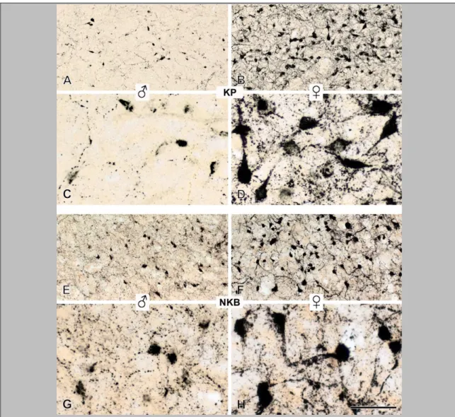
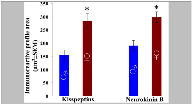
5.3. Sexual dimorphism of kisspeptin and neurokinin B systems in aged human individuals ... 42
5.3.1. Perikaryon size of KP-IR and NKB-IR neurons ... 42
5.3.2. Incidence of KP-IR and NKB-IR cell bodies in the Inf ... 44
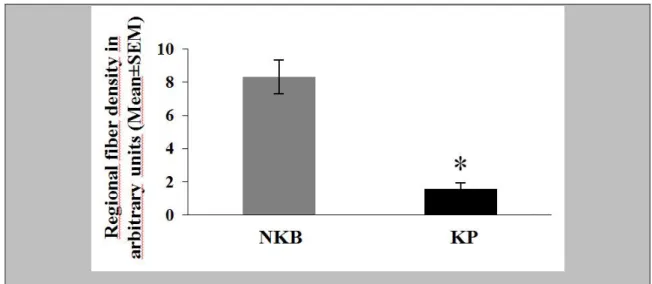
5.3.3. Regional density of KP-IR and NKB-IR fibers ... 45
5.3.4. Frequency of KP-IR and NKB-IR appositions onto GnRH-IR neurons ... 46
5.3.5. Colocalization of KP and NKB in neuronal afferents to GnRH neurons ... 49
5.4. Aging related changes of the human hypothalamic kisspeptin and neurokinin B neuronal systems ... 51
5.4.1. Incidence of KP-IR and NKB-IR perikarya in the Inf ... 51
5.4.2. Perikaryon size of NKB-IR neurons ... 53
5.4.3. Regional density of KP-IR and NKB-IR fibers ... 53
5.4.4. Colocalization of KP and NKB in neuronal perikarya of the Inf ... 54
5.4.5. Incidence of KP-IR and NKB-IR appositions onto GnRH-IR neurons ... 56
5.4.6. Colocalization of KP and NKB in neuronal afferents to GnRH neurons ... 59
4
6. DISCUSSION ... 61
6.1. Possible contribution of the estrogenic regulation of RF-amide related peptide to estrogen feedback mechanisms ... 61
6.1.1. The expression of RFRP mRNA is negatively regulated by estrogen ... 61
6.1.2. A relatively small subset of RFRP neurons expresses ER-α immunoreactivity 61 6.1.3. Putative sites of action of RFRP on the reproductive axis ... 62
6.2. Species differences in the neuropeptide compliment of hypothalamic kisspeptin neurons ... 63
6.2.1. Species differences in the colocalization of KNDy peptides... 63
6.2.2. Role of NKB in the regulation of the human reproductive axis ... 64
6.2.3. Role of KP in the regulation of the human reproductive axis ... 66
6.2.4. Absence of DYN immunoreactivity from most KP elements ... 66
6.3. Importance of sex differences in the central regulation of human reproduction ... 68
6.3.1. Sex difference in perikaryon size of KP and NKB neurons ... 68
6.3.2. Higher relative levels of NKB vs. KP immunolabeling ... 69
6.3.3. Sex differences in KP immunoreactivity ... 71
6.3.4. Sex differences in NKB immunoreactivity ... 72
6.4. Role of enhanced kisspeptin and neurokinin B signaling in aging men, as a cause or a consequence of reproductive decline ... 74
6.4.1. Aging-dependent variations in KP and NKB immunoreactivities and their colocalization pattern ... 74
6.4.2. Aging-dependent changes in the central regulation of male reproduction ... 75
7. CONCLUSIONS ... 78
8. SUMMARY ... 80
9. ÖSSZEFOGLALÁS ... 81
10. REFERENCES ... 82
11. LIST OF PUBLICATIONS ... 100
11.1. List of publications underlying the thesis ... 100
11.2. List of other publications ... 101
12. ACKNOWLEDGEMENTS ... 103
5
1. List of abbreviations
ARC - arcuate nucleus
AVPV - anteroventral periventricular nucleus
BV - blood vessel
DAB - diaminobenzidine
DMN - dorsomedial nucleus
DYN - dynorphin
E2 - 17β-estradiol
ER-α - estrogen receptor alpha
ER-β - estrogen receptor beta
FSH - follicle-stimulating hormone
GABA - gamma-aminobutyric acid
GnIH - gonadotropin-inhibiting hormone GnRH - gonadotropin-releasing hormone
icv - intracerebroventricular
Inf - Infundibular nucleus
InfS - Infundibular stalk
i.p. - intraperitoneal
IR - immunoreactive
ISH - in situ hybridization
KISS1R - kisspeptin receptor
KNDy - Kisspeptin/Neurokinin B/Dynorphin
KP - kisspeptin
KOR - dynorphin receptor
LH - luteinizing hormone
ME - median eminence
MPOA - medial preoptic area
Ni-DAB - nickel-intensified diaminobenzidine
NK3 - neurokinin B receptor
NKB - neurokinin B
OVLT - organum vasculosum of the lamina terminalis
6
OVX - ovariectomized
PBS - phosphate buffered saline
PFA - paraformaldehyde
PVN - paraventricular nucleus
RFRP - RFamide-related peptide
RP3V - rostral periventricular area of the third ventricle
sc. - subcutan
SSC - standard saline citrate
T - testosterone
TBS - Tris buffered saline
VMN - ventromedial nucleus
7
2. Introduction
2.1. Central regulation of the gonadal axis
Gonadotropin-releasing hormone (GnRH) synthesizing neurons of the hypothalamus play a crucial role in the central regulation of reproduction in all mammals [1]. The hypophysiotropic axons of GnRH neurons secrete the GnRH decapeptide into the fenestrated capillaries of the hypophysial portal circulation at the hypothalamic median eminence (ME). From here, long portal veins carry GnRH to the anterior pituitary gland. GnRH binds to type-1 GnRH receptor on the surface of gonadotroph cells to regulate the synthesis and secretion of the gonadotropins, follicle-stimulating hormone (FSH) and luteinizing hormone (LH).
FSH and LH released into the systemic circulation, in turn, act on the gonads to stimulate gametogenesis and gonadal steroid secretion, respectively. Gonadal hormones act on both the hypothalamus and the pituitary in a classical negative feedback loop, inhibiting thereby the synthesis of GnRH and the two gonadotropin [2, 3].
GnRH neurons are derived from the nasal placod and only migrate into the brain during fetal development [4, 5]. Defects of this migratory process can cause reproductive deficiencies due to the absence of GnRH neurons in the forebrain.
Hypogonadotropic hypogonadism characterizing this status often coincides with anosmia (Kallmann syndrome).
The number of GnRH neurons regulating reproduction is surprisingly low (1000- 2000). By the time of birth, these cells have already settled in the hypothalamus. They are distributed in a relatively large area, without forming solid groups of neurons. In rats, GnRH cells are located in diagonal band of Broca, medial septum, medial preoptic area (MPOA), organum vasculosum of the lamina terminalis (OVLT) and medial preoptic nucleus [6]. In humans, GnRH cells are even more scattered. A relatively large subset of them can be found in the infundibular nucleus (Inf) [7], which is analogous to the arcuate nucleus (ARC) of rodents.
The cell body of GnRH neurons is relatively small (10-20µm) and fusiform. GnRH neurons have one or two dendrites at the poles of the elongated perikaryon and one axon
8
emanating from either the cell body or one of the main dendrites [8]. According to a widely accepted view, GnRH cells form a network via establishing axo-somatic and axo-dendritic connections with one another [9], making the coordinate firing and GnRH secretion possible. GnRH cells project to two circumventricular organs, the external zone of the ME and the OVLT [4, 5].
At the level of the ME, GnRH terminals release GnRH into the fenestrated capillaries of the portal vasculature in the form of secretory pulses which occur once every 30-90 minutes [2]. The episodic secretion of GnRH has been shown to correlate with the pulsatile secretion of LH from the adenohypophysis in many species [10, 11].
Coordinated function of the cells, to allow regular pulses to be generated in terms of secretory events, could be achieved through dendro-dendritic connections [12-14]. The neuronal network accounting for the establishment of the GnRH neurosecretory pulses is commonly referred to as the GnRH/LH ‘pulse generator’. Similarly to observations on various laboratory animal species, the pattern of LH secretion was also found to be pulsatile in primates [15].
It has been shown in the sheep that a strong temporal correlation exists between the GnRH secretory episodes measured in the portal blood and the pulses of LH in the peripheral blood [16]. In contrast, the relationship between GnRH and FSH secretion is more complex as FSH secretion exhibits a random pattern, without any strict correlation with the GnRH/LH secretory episodes [17]. The pattern of GnRH secretion carries relevant information for adenohypophysial gonadotroph cells. The control of FSH and LH synthesis is closely linked to the transcription of the distinct β-subunits, Fshb and Lhb respectively. Both FSH and LH contain a common α-subunit (CGA); therefore, it is FSHβ and LHβ that confer the specific actions of the gonadotropins [18]. Notably, in follicular phase, low frequency GnRH secretion preferentially stimulates FSHβ gene expression and protein synthesis. To the opposite, during the preovulatory hypersecretion (‘surge’) of GnRH and LH, the frequent GnRH pulses preferentially stimulate LHβ gene expression and LH secretion from the gonadotrophs [19-23].
The functions of GnRH cells are strongly modulated by neuronal afferents. Classical neurotransmitters as well as neuropeptides released from these afferents play important roles in the neuronal regulation of GnRH cells.
9
One of the most important neurotransmitters in the neuronal regulation of the GnRH neuron is gamma-aminobutyric acid (GABA). A significant proportion of synaptic contacts on GnRH neurons is GABAergic [24]. Both GABAA and GABAB [25]
receptors have been described on GnRH cells. The direct action of GABA on GnRH neurons is mediated by postsynaptic GABAA receptors, which are ligand-gated chloride-ion channels composed of five subunits [26]. Functional GABAB receptors have also been detected in GnRH neurons [27, 28]. GABAergic afferents play important roles in the mediation of metabolic [29], circadian [30] and estrogen signals to the GnRH system [30]. Our recent work indicates that GABAergic neurons also provide a highly abundant input to GnRH neurons in the human [31].
L-glutamate is a critically important excitatory neurotransmitter in the afferent control of GnRH cells [32]. Activation of ionotropic glutamate receptors contributes to both the pulsatile [33] and the surge [34] release of GnRH. Our recent immunohistochemical work has revealed an abundant glutamatergic input to GnRH neurons of the human. While GABAergic afferents dominates over glutamatergic afferents on the perikaryon of GnRH cells, the combined incidence of two different subtypes of glutamatergic inputs exceeds the incidence of GABAergic inputs on the dendritic compartment of GnRH neurons in human [31].
2.2. Peptidergic systems and mechanisms acting upstream from the GnRH neuronal systems
A large number of neuropeptides can regulate GnRH secretion from the hypophysiotropic GnRH axon terminals by acting on either the somato-dendritic or the axonal compartment of the GnRH neurons.
Kisspeptin (KP), a neuropeptide encoded by the metastasis suppressor gene KiSS1, plays a particularly important role in the central regulation of reproduction.
Administration of KP, KP antibodies [35-37], KP antagonists [38-40] or the use of transgenic animal models deficient for KP or its receptor (KISS1R) [41] have provided evidence that KP regulates the pulsatile [37] and surge [36, 42] release of GnRH.
KP synthesizing neurons in various mammals have been localized to two major anatomical sites, the preoptic region and the ARC. The KP cell population of the
10
preoptic region exhibits evident anatomical variations among species. In rodents, these KP cells form a compact nucleus in the rostral periventricular area of the third ventricle (RP3V) [43-45] (also cited often as the kisspeptin cell population of the anteroventral periventricular nucleus; AVPV). Kisspeptin neurons are much more scattered and present in lower numbers in the preoptic region of the sheep [46, 47], the monkey [48]
and the human [49]. The second (and largest) kisspeptin synthesizing cell group can be consistently detected within the ARC in all mammalian species studied so far [35, 36, 43, 44, 46-48, 50-73] and in the analogous Inf of primates [49, 58].
KP axons innervate the perikaryon and dendrites of GnRH neurons [53]. The majority of GnRH neurons express KISS1R [50, 51, 74] and respond to KP with increased neuronal activity [75].
Neurokinin B (NKB) is another critically important neuropeptide which interacts with KP in the neuroendocrine regulation of GnRH/LH secretion. NKB belongs to the family of tachykinins. From the three tachykinin receptors, NKB primarily activates the NK3 receptor form [76], but it has been shown recently that the reproductive effects of NKB in mice also involve the other two types of tachykinin receptors, NK1 and NK2 [77].
NKB is encoded by the TAC3 gene in humans, and the Tac2 gene in rodents, whereas NK3 is encoded by the TACR3/Tacr3 gene [76]. Inactivating mutations of the neuropeptide- or the neuropeptide receptor gene are associated with hypogonadotropic hypogonadism in humans [78, 79].
NKB may increase GnRH secretion from the hypothalamic ME where GnRH axons are apposed to NKB axons [80, 81] and express NK3 immunoreactivity [80] in the rat.
In addition, NKB neurons also influence reproduction by acting on other NKB neurons in the ARC where they establish frequent contacts with one another [82, 83] and express NK3 autoreceptors.
An inhibitory neuropeptide named gonadotropin-inhibiting hormone (GnIH) has been identified in the quail hypothalamus; GnIH inhibits gonadotropin release from the pituitary in a dose-dependent manner [84]. In addition to acting as a release-inhibiting hormone on gonadotrophs, GnIH also regulates fertility via influencing the neurosecretory output of hypophysiotropic GnRH neurons. Accordingly, GnIH- immunoreactive (IR) neuronal contacts [85] and GnIH receptors [86] are present on
11
avian GnRH neurons. Putative GnIH homologues, RF-amide related peptides (RFRP-1, RFRP-2, RFRP-3), have also been identified in mammals [87]. With some anatomical species differences, the majority of neurons that synthesize preproRFRP mRNA and RFRP peptides have been localized to the dorsomedial nucleus (DMN) of the hypothalamus in hamsters, rats, mice and sheep [88] and to the intermediate periventricular nucleus in monkeys [89]. Unlike in birds where GnIH-IR axons have access to the hypophysial portal vasculature [84, 90], RFRP neurons in rodents do not project to the external zone of the ME [88] and do not accumulate Fluoro-Gold (retrograde neuronal tracer) from the systemic circulation [91], suggesting that RFRP peptides regulate fertility primarily via central mechanisms. These mechanisms include direct inhibitory actions exerted upon GnRH neurons, as indicated by RFRP-IR neuronal contacts on GnRH neurons [62, 92] and by the RFRP-3-induced hyperpolarization [93] and reduced electric activity of a large subset of GnRH neurons [94] in slice preparations of GnRH-GFP transgenic mice.
2.3. Role of kisspeptin, neurokinin B, RF-amide related peptides in sex steroid feedback
The GnRH neuronal system which represents the final common pathway in the neuroendocrine control of reproduction responds to feedback actions of circulating 17β- estradiol (E2). The feedback effect of sex steroids takes place at the level of the hypothalamus to regulate GnRH neurons as well as at the level of pituitary gonadotrophs. The fact that the maintenance of plasma level of gonadal steroids acts as a negative brake on the GnRH- gonadotropin axis is most simply demonstrated by gonadectomy, which causes GnRH and gonadotropin levels to rise. There is a difference between males and females regarding the feedback effect of sex steroids on brain, most notably the presence of ‘positive feedback’ mechanism in females and the absence of the same in males. This positive feedback drives the preovulatory surge in GnRH and LH secretion in female. While direct estrogen actions upon GnRH neurons can be exerted via estrogen receptor-β (ER-β) [95-99], interneurons expressing the classical estrogen receptor (ER-α) play a critically important role in sensing and conveying indirect information on circulating estrogens to the GnRH neuronal system [100].
12
KP neurons of the ARC also synthesize NKB in the sheep [47, 70], the goat [73], the mouse [101], monkey [72] and the human [49]. The recently introduced terminology of KNDy (Kisspeptin/Neurokinin B/Dynorphin) neurons [70] refers to the co-synthesis of dynorphins (DYN) by the majority of these KP/NKB cells at least in the sheep [47, 102], the rat [82], the mouse [101, 103, 104] and the goat [73]. Evidence from studies of sheep suggests that KNDy neurons of the ARC play an important role in conveying the negative feedback effects of sexual steroids onto GnRH neurons [59], and possibly, also the positive feedback effects of estrogens [105], at least in this species. In addition, KNDy neurons also appear to constitute an important component of the GnRH pulse generator [73, 101]. Recent models of the GnRH pulse generator [73, 101, 103] suggest that KNDy neurons communicate with one another via NKB and its receptor, NK3, and possibly, also DYN and its receptor, KOR. In ovariectomized goats, central NKB increases and DYN decreases the frequencies of multiunit activity volleys and LH secretory pulses [73]. Pulse generator cells, in turn, appear to communicate with GnRH neurons primarily via KP/KISS1R signaling. GnRH neurons express KISS1R [50, 51, 106] and the majority of GnRH neurosecretory pulses show temporal association with KP pulses in the ME of monkeys [107].
The closer analysis of anatomical reports describing the colocalization of
‘KNDy’ neuropeptides, in retrospect, reveals that neuropeptide and receptor colocalizations are often only partial and also variable in the different studies, species, sexes and age groups. In particular, NKB-IR neurons and their fibers are partly distinct from the KP-IR elements in various human models [49], thus challenging the universal validity of the KNDy neuron concept. Based on our preliminary data indicating that the number of KP neurons is very low in young human males subjects [49], we predicted that the degree of overlap between the KNDy neuropeptides is much lower in young male humans than suggested earlier for sheep [47, 70], goats [73] or mice [101]. In one of our studies (5.2) underlying this thesis, we investigated the universal validity of the KNDy neuron concept via the parallel immunohistochemical analysis of KP-, NKB- and DYN immunoreactivities in the Inf and the infundibular stalk (InfS) of young men.
Evidence that the RFRP neuronal system may be involved in estrogen feedback signaling to GnRH neurons has emerged from studies of hamsters. RFRP neurons in this rodent species contain ER-α and respond with c-Fos expression to an acute
13
administration of E2 [88]. RFRP axons provide inputs to GnRH neurons, suggesting direct regulation. Intracerebroventricular injection of RFRP-3 reduced plasma LH secretion in ovariectomized (OVX) hamsters [88] and gonadally intact rats [92, 108]. In one study (5.1) underlying this thesis we investigated the putative estrogen responsiveness of the RFRP neuronal system in mice, by addressing the estrogenic regulation of RFRP gene expression and the presence of the two estrogen receptor isoforms in RFRP neurons.
2.4. Sex differences in steroid feedback mechanisms
The pulsatile pattern of GnRH secretion into the hypophysial portal circulation is shaped by a sex steroid-sensitive neuronal circuitry that acts upstream from GnRH cells [109]. In both males [110] and females [111], gonadal steroid hormones exert homeostatic negative feedback on GnRH release via this upstream neuronal circuitry. In females, elevated estradiol in the late follicular phase of the reproductive cycle contributes to a switch from negative to positive feedback to induce a surge of GnRH from the hypothalamus. The subsequent surge of LH from the adenohypophysis triggers ovulation [109].
KP cells of the rodent RP3V exhibit a robust sexual dimorphism. They occur in higher numbers [35, 53, 69, 112] and provide input to a higher percentage of GnRH neurons in females compared with males [53]. There is strong evidence suggesting that in rodents, the kisspeptin cell population of the RP3V is critically involved in positive estrogen feedback to GnRH neurons [35, 113]. In contrast, positive feedback appears to be attributable mainly to the KP cells of the ARC in sheep [114] and primates [2, 115, 116].
The ARC also exhibits sexual dimorphism in several species. In the sheep, this nucleus contains higher NKB [117] and KP [70] cell numbers in females than in males.
In rats, sex differences were reported in the projection field of NKB-IR axons in the infundibular area [81].
The sex-specific spatial and temporal patterns of hypothalamic KP expression strongly depend on the activational effects of sexual steroid hormones. Large subsets of KP neurons in both the preoptic region and the ARC contain receptors for estradiol,
14
testosterone (T) and progesterone in various species [35, 41, 44-46, 59, 70, 118]. In rodents, androgens and estrogens upregulate KP expression in the RP3V [35, 44, 112, 118, 119] during the phase of positive estrogen feedback. In contrast, KP expression in the ARC/Inf is regulated negatively by sex steroid hormones in rodents and other species [35, 44, 112, 118-120] and so is NKB expression at this site [120-124].
The sex-dependent pattern of hypothalamic KP expression also depends on the organizational effects of T exposure in males, in addition to the circulating levels of other sex steroid hormones. There is a neonatally determined robust sexual dimorphism of the RP3V in adult rats [112] and mice [53], with higher cell numbers in females compared with males. Prenatal T exposure accounts for a similar sexual dimorphism of KP neurons in the preoptic area of the sheep [70].
In contrast, no apparent organizational effects of the perinatal T exposure on the abundance of KP neurons have been observed in the rodent ARC which contains similar KP cell numbers in intact males and in diestrous females, or in gonadectomized males and females receiving the E2 or T regimen [112]. No sex difference appears to exist in the number of NKB neurons either. However, in the rat a sexual dimorphism that develops under the organizational effects of sex steroids has been reported in the projection fields of NKB-IR axons [81]. Unlike in rodents, the ARC of the female sheep contains higher NKB [117] and KP [70] cell numbers, compared with males. Moreover, previous work from our laboratory identified higher KP neuron and fiber densities in the Inf of women vs. men [49]; it requires clarification to what extent this sexual dimorphism reflects the organizational effects of sex steroids during development or the difference in the adult hormonal status between men and women.
In one of the studies underlying this thesis (5.3), we have addressed several immunohistochemical correlates of the functional differences that characterize the KP and NKB neuronal systems of the Inf in men and women. Quantitative immunohistochemistry was used to study various aspects of the putative morphological sex differences.
2.5. Aging related alterations in the central regulation of the reproductive axis
Reproductive aging is accompanied by sex-specific neuromorphological alterations of the Inf. While gonadal functions in aging men can be quite well-preserved throughout
15
life [125], the negative feedback response of the reproductive axis to T shows a declining trend [126]. This reduced feedback may be correlated with a mild neuronal hypertrophy in the Inf of aged men [127]. Reproductive aging is more dramatic in postmenopausal women after the depletion of ovarian follicles, leading to the loss of circulating estrogen and causing the reduction of negative estrogen feedback [128].
Previous comparison of histological samples from pre- and postmenopausal women revealed profound anatomical changes in the Inf where negative feedback is thought to take place [128]. Accordingly, in situ hybridization studies identified the postmenopausal hypertrophy of neurons that express ER-α [129], substance P [121], NKB [121], KP [58] and prodynorphin [130]. These morphometric alterations were also associated with increased NKB [121] and KP [58] and decreased prodynorphin mRNA expression [130] at this site.
Reproductive aging in men during midlife transition is characterized by decreased serum levels of free T and increased levels of LH, FSH and sex hormone binding globulin, among other endocrine alterations [131, 132]. The aging-related hypogonadism coincides with functional disturbances occurring at different levels of the reproductive axis, which include reduced androgen receptor-mediated negative feedback to the hypothalamus [126]. In view of the proposed involvement of KP/NKB neurons in negative feedback [44, 47, 118], in one study (5.4) included in this thesis we addressed the hypothesis that the weakening of the inhibitory T feedback in elderly men coincides with morphological signs of enhanced KP and NKB signaling in the Inf.
16
3. Specific aims
1) To investigate the putative estrogen responsiveness of the RFRP neuronal system in mice
2) To characterize the 'KNDy' neuronal system in adult human males
3) To investigate the sexual dimorphism of the human hypothalamic kisspeptin and neurokinin B neuronal systems
4) To address aging-related anatomical changes of human kisspeptin and neurokinin B neurons
17
4. Materials and Methods
4.1. Animals
The experiments were performed on CD1 mice that were purchased from a local colony bred at the Medical Gene Technology Unit of the Institute of Experimental Medicine (IEM). The animals were housed in light- (12:12 light-dark cycle, lights on at 07:00h) and temperature (22 2°C) controlled environment, with free access to standard food and tap water. All studies were carried out with permission from the Animal Welfare Committee of the Institute of Experimental Medicine of the Hungarian Academy of Sciences (No.: A5769-01) and in accordance with legal requirements of the European Community (Decree 86/609/EEC).
4.1.1. Surgery (Gonadectomy and hormone replacement of female mice)
In a study to analyze the estrogenic regulation of RFRP mRNA expression in mice (5.1), we used gonadectomy, followed by hormone replacement. The ovariectomy was carried out under deep anesthesia with an intraperitoneal (i.p.) cocktail of ketamine (25 mg/kg), xylavet (5 mg/kg) and pipolphen (2.5 mg/kg) in saline. On post-ovariectomy day 9, the mice were re-anesthetized and implanted subcutaneously (sc.) with a single silastic capsule (Sanitech; Havant, UK; l=10 mm; ID=1.57 mm; OD=3.08 mm) containing either sunflower oil (OVX, n=5) or 100µg/ml E2 (Sigma Chemical Company, St Louis, MO) in sunflower oil (OVX+E2, n=4).
4.1.2. Transcardiac perfusion
Four days later, the mice were anesthetized and killed by transcardiac perfusion with 40 ml 4% paraformaldehyde (PFA) in phosphate buffered saline solution (PBS; 0.1M;
pH 7.4). The brains were removed, postfixed in 4% PFA solution for 1h at 4°C, infiltrated with 20% sucrose (in PBS) overnight, and then snap-frozen on powdered dry ice. These mice were used in colocalization studies of ER-α and RFRP-1 immunoreactivities (5.1). Another six mice were OVX and treated similarly to generate OVX (n=3) and OVX+E2 (n=3) groups and perfused with a mixture of 2%
paraformaldehyde and 4% acrolein. These mice perfused with the acrolein-containing
18
fixative were used in colocalization studies of ER-β and RFRP-1 immunoreactivities (5.1).
4.2. Human tissues
4.2.1. Collection of human hypothalamic tissues
Human hypothalamic tissue samples were obtained from autopsies at the Forensic Medicine Department of the University of Debrecen with permission from the Regional Committee of Science and Research Ethics of the University of Debrecen (DEOEC RKEB/IKEB: 3183-2010). Selection criteria included sudden causes of death, lack of history of neurological and endocrine disorders and post mortem delay below 48h.
In studies to characterize the 'KNDy' neuronal system of adult humans (5.2), we used tissues of six young male individuals (age 21-37 years). To investigate the sexual dimorphism of the human hypothalamic KP and NKB neuronal systems (5.3), we used tissue samples from nine male subjects above 50 years of age (50-67 years) and from seven postmenopausal female subjects above 55 years of age (57- 70 years). To address the aging-related anatomical changes of human KP and NKB neurons, arbitrarily defined ‘young’ (21-49 years; N=11) and ‘aged’ (50-67 years; N=9) male groups were formed (5.4).
Following dissection, the human hypothalamic tissue blocks were rinsed briefly with running tap water and then, immersion-fixed with 4% paraformaldehyde in 0.1M PBS (pH 7.4) for 7-14 days at 4°C. Following fixation, the hypothalami were trimmed further to include the optic chiasma rostrally, the mammillary bodies caudally and the anterior commissure dorsally [99].
4.3. Section preparation and pretreatments for immunohistochemistry and in situ hybridization
4.3.1. Preparation of hypothalamic sections for immunohistochemistry in studies using mouse tissues
Serial 20μm (5.1) coronal sections were cut from the hypothalami with a Leica SM 2000R freezing microtome (Leica Microsystems, Nussloch Gmbh, Germany). The
19
sections were stored at -20˚C in 24-well tissue culture plates containing anti-freeze solution (30% ethylene glycol; 25% glycerol; 0.05 M phosphate buffer; pH 7.4).
In studies to investigate the putative colocalization of ER-β with RFRP (5.1), the perfusion solution contained acrolein. In these cases the sections were treated with 1%
sodium borohydridefor 30 min and washed in PBS until the sections sank to the bottom of washing dish. Prior to immunohistochemistry, the sections were treated with 0.2%
Triton X-100 and 0.5% H2O2 in PBS for 30 min. Between treatments, the sections were washed in PBS for 30 min (3 x 10 min).
4.3.2. Preparation of frozen tissue sections for in situ hybridization experiments on mouse tissues
For in situ hybridization experiments serial 20μm coronal sections were cut and stored as described above.
In studies to analyze RFRP mRNA levels in OVX and OVX+E2 mice (5.1), every 6th section from each paraformaldehyde-fixed mouse hypothalamus was mounted on silanized microscope slides from sterile Tris buffered saline (TBS; 50 mM; pH 7.8) with an RNase-free paint brush and air-dried. Then, the sections were processed through the following prehybridization steps: 2 min rinse in 2X standard saline citrate (SSC) solution; 10 min acetylation in 0.25% acetic anhydride (Sigma)/0.9% NaCl/0.1 M triethanolamine (pH 8.0; Sigma) [133]; a brief rinse in 2XSSC solution; dehydration in 70%, 80%, 95% and 100% ethanol (2 min each); delipidation in chloroform (5 min), then partial rehydration in 100% followed by 95% ethanol (2 min each). The slides were finally air-dried.
4.3.3. Preparation of human tissue sections for immunohistochemistry
Sagittal cuts were made 2cm lateral from midsagittal plane on both sides and then, the blocks were cut in halves and infiltrated with 20% sucrose for 5 days at 4°C. The right hemihypothalami were placed in a freezing mold, surrounded with Jung tissue freezing medium (Leica Microsystems, Nussloch Gmbh, Germany; diluted 1:1 with 0.9% sodium chloride solution), snap-frozen on powdered dry ice, and sectioned serially at 30μm with a freezing microtome parallel to the plane of the lamina terminalis. The
20
sections were stored similarly to mouse sections. All experiments (5.2, 5.3, 5.4) were performed on every 24th hemihypothalamic section from each subject.
Prior to immunohistochemistry, human tissues were permeabilized and endogenous peroxidase activity reduced using a mixture of 0.2% Triton X-100 and 0.5% H2O2 in PBS for 30 min. Subsequently, antigen retrieval was carried out by using a 0.1M citrate buffer (pH 6.0) wash at 80°C for 30 min. In immunofluorescent experiments, the sections were also pretreated with Sudan black to reduce tissue autofluorescence from lipofuscin deposits [134] [49].
4.4. Immunohistochemical methods
4.4.1. Peroxidase-based immunohistochemical single-labeling
Sections processed for peroxidase-based immunohistochemistry were first incubated in the working dilution of primary antibodies (24-72h; 4ºC). The primary antibodies were reacted with biotinylated secondary antibody (1:500; 1h) and then, with the ABC Elite reagent (Vector, Burlingame, CA, 1:1000; 1h). The peroxidase signal was developed with diaminobenzidine (DAB) or with nickel-intensified diaminobenzidine (Ni-DAB) chromogen. The immunostained sections were mounted on microscope slides from Elvanol, air-dried, dehydrated with 95% (5 min), followed by 100% (2X5 min) ethanol, cleared with xylene (2X5 min) and coverslipped with DPX mounting medium (Sigma, St. Louis, USA).
4.4.2. Silver-gold intensification of the Ni-DAB chromogen
In some dual-peroxidase-based experiments we studied the localization of two different antigens (one nuclear and one cytoplasmic) in the same neuron (5.1, 5.2). In others, the afferent connectivity between two different neuronal phenotypes was investigated (5.3, 5.4). In these studies the Ni-DAB chromogen was post-intensified with silver-gold [135].
21
4.4.3. Innervation and colocalization studies with the combined use of silver-gold- intensified Ni-DAB and DAB chromogens
In studies to examine the KP-IR and NKB-IR afferent inputs to GnRH neurons (5.2, 5.3, 5.4), two series of sections were processed for the detection of KP or NKB immunoreactivities. Subsequently, GnRH neurons were detected with a new guinea pig antiserum (#1018; see in Table 1, 1:10,000). The primary antibodies were reacted with biotinylated anti-guinea pig IgG (Jackson ImmunoResearch; 1:500; 60 min) and the ABC reagent (1:1000; 60 min) and then, the peroxidase signal was developed with DAB chromogen.
In studies to reveal the presence of the estrogen receptor isoforms in RFRP neurons (5.1) ERs were detected with an antiserum raised in rabbit (see in Table 1), followed by biotinylated secondary antibodies (Jackson ImmunoResearch Europe Ltd., Soham, Cambridgeshire, UK; 1:500) and the ABC Elite reagent for 60 min each. The signal was visualized with Ni-DAB and then post-intensified with silver-gold. Subsequently, RFRP-1 immunoreactivity was detected with mouse monoclonal antibodies (IF3;
Takeda Pharmaceutical Co. Ltd, Japan; 1:20,000) against the C-terminus of rat RFRP-1 [136], using the biotinylated secondary antibody-ABC technique and non-intensified DAB as the chromogen.
4.4.4. Dual-immunofluorescent investigations to study the colocalization of KP and NKB or KP and DYN immunoreactivities in the Inf
In studies to examine the colocalization between KP and NKB or KP and DYN (5.2, 5.3, 5.4), dual-immunofluorescent labeling was used. Incubation in a cocktail of primary antibodies (rabbit anti-NKB, 1:1000 and sheep anti-KP, 1:1000; or rabbit anti-DYN, 1:1000 and sheep anti-KP, 1:1000; see in Table 1; 48h; 4ºC) was followed by a cocktail of fluorochrom-conjugated secondary antibodies (Jackson ImmunoResearch; anti- rabbit-FITC, 1:250; anti-sheep-Cy3, 1:1000) for 5h at room temperature.
To maximize sensitivity, in some dual-immunofluorescent studies (5.2, 5.3, 5.4) tyramide signal amplification was used. In these experiments KP was detected first using sequential incubations in sheep KP antibodies (1:30,000; 48h; 4ºC), biotinylated anti-sheep IgG (Jackson ImmunoResearch Laboratories; 1:500; 1h), the ABC Elite reagent (Vector; 1:1000; 1h), biotin tyramide working solution (1:1000, in 0.05M Tris-
22
HCl buffer, pH 7.6, containing 0.003% H2O2; 30 min) [137] and finally, avidin-Cy3 (Jackson ImmunoResearch; 1:1000; 1h). Then, the sections were treated for 30 min with 0.5% H2O2 and 0.1% sodium azide in PBS, to inactivate horseradish peroxidase. To detect NKB or DYN, the rabbit primary antibodies were used at 1:50,000 (48h; 4ºC) and reacted with anti-rabbit-peroxidase (Jackson ImmunoResearch; 1:500; 1h). Then, FITC-tyramide [137] (diluted 1:500 with 0.05M Tris-HCl buffer, pH 7.6, containing 0.003% H2O2; 30 min) was deposited on the peroxidase sites. Control experiments included the omission of the NKB and DYN primary antibodies. Lack of FITC labeling in these control sections indicated that no FITC-tyramide deposition is caused by residual peroxidase activity on KP-IR sites.
4.4.5. Triple-immunofluorescent studies to analyze the colocalization of KP and NKB in neuronal afferents to human GnRH neurons
Incubation in a cocktail of primary antibodies (rabbit anti-NKB, 1:1000; sheep anti- KP, 1:1000; guinea pig anti-GnRH, 1:3000) for 48h at 4ºC was followed by a cocktail of fluorochrom-conjugated secondary antibodies (all raised in donkey; anti-rabbit-FITC, 1:250; anti-sheep-Cy3, 1:1000; anti-guinea pig-AMCA, 1:100; Jackson ImmunoResearch) for 5h (5.3, 5.4).
Sections processed for immunofluorescent experiments were mounted from 0.1M Tris–HCl buffer (pH 7.6) and coverslipped with the aqueous mounting medium Mowiol.
Table 1. Summary of the antibodies and reagent used in different experiments
Primary antibodies Dilution Chromogen
5.1. Dual-label immunohistochemical experiments to colocalize estrogen receptors and RFRP-1 immunoreactivities
rabbit ER-α antiserum
(C1355; Millipore, Temecula, CA, USA)
1:10,000 Ni-DAB rabbit ER-β antiserum
(Z8P; Zymed Laboratories; Lot 01162852)
150ng/ml Ni-DAB mouse RFRP-1 monoclonal antibody
(IF3; Takeda Pharmaceutical Co. Ltd, Japan)
1:20,000 DAB
23
5.2. 'KNDy' neuronal system in adult human males sheep polyclonal antiserum to human KP-54
(GQ2; gift from Dr. S.R. Bloom)
1:200,000/
1:1,000
Ni-DAB/
Cy3 rabbit polyclonal antiserum to human NKB
(gift from Dr. Philippe Ciofi)
1:100,000/
1:1,000
Ni-DAB/
FITC rabbit polyclonal antiserum to DYN
(T-4268; Peninsula Laboratories; San Carlos, CA.)
1:100,000/
1:1,000
Ni-DAB/
FITC guinea-pig GnRH antiserum
(#1018; Hrabovszky et al., 2011)
1:50,000 DAB 5.3. Sexual dimorphism of the human hypothalamic kisspeptin and neurokinin B neuronal systems
sheep polyclonal antiserum to human KP-54
(GQ2; gift from Dr. S.R. Bloom)
1:100,000/
1:1,000
Ni-DAB/
Cy3 rabbit polyclonal antiserum to NKB
(gift from Dr. Philippe Ciofi)
1:100,000/
1:1,000
Ni-DAB/
FITC guinea-pig GnRH antiserum
(#1018; Hrabovszky et al., 2011)
1:50,000/
1:3,000
DAB/
AMCA 5.4. Aging-related anatomical changes of human kisspeptin and neurokinin B neurons
sheep polyclonal antiserum to human KP-54
(GQ2; gift from Dr. S.R. Bloom)
1:100,000/
1:1,000
Ni-DAB/
Cy3 rabbit polyclonal antiserum to NKB
(gift from Dr. Philippe Ciofi)
1:100,000/
1:500
Ni-DAB/
FITC guinea-pig GnRH antiserum
(#1018; Hrabovszky et al., 2011)
1:10,000/
1:5,000
DAB/
AMCA
4.5. In situ hybridization
4.5.1. Preparation of hybridization probes
To prepare a probe to preproRFRP mRNA (5.1), a 424-bp cDNA fragment (corresponding to bases 136-559 of the rat preproRFRP mRNA; AB040288, respectively) was amplified with PCR from rat hypothalamic cDNA. The amplicons
24
were inserted into a plasmid vector using the pGEM-T Easy Vector System from Promega (Madison, WI). The plasmids were grown in DH5α cells (Invitrogen, Carlsbad, CA, USA), isolated with the QIAGEN Plasmid Maxi kit (Qiagen; Valencia, CA, USA), linearized with Sal1 and purified with phenol/chloroform/isoamyl alcohol (PCI), followed by chloroform/isoamyl alcohol extractions and then precipitation with NaCl and ethanol. The linearized RFRP template was transcribed with T7 RNA polymerase in the presence of 35S-UTP (NEN Life Science Products, Boston, MA, USA).
4.5.2. In situ hybridization studies with 35S-labeled cRNA hybridization probes
In studies to detect E2 dependent changes of RFRP mRNA level (5.1) in the mouse hypothalamus, after prehybridization the sections were placed in humidity chamber. 50 μl of hybridization solution was pipetted onto each section, covered with a glass coverslip and compassed with DPX. The standard hybridization reaction was carried out at 56°C for 16h (overnight). Following post-hybridization treatments including the RNase A digestion (20 μg/ml, 60 min at 37°C) of probe excess and a 30-min stringent treatment in 0.1X SSC at 60°C, the sections were rinsed briefly in 70% ethanol and air dried.
4.5.3. Autoradiography
The sections labeled with the RFRP probe (5.1) were first exposed to Kodak BioMax MR autoradiography films for 3 days and signals developed with standard film processing procedures.
To visualize the isotopic signal for RFRP (5.1), the slides were dipped into Kodak NTB autoradiographic emulsion (Kodak; Rochester, NY) and exposed for 1-2 weeks.
The emulsion autoradiographs were developed using standard procedures and Kodak processing chemicals. The sections were air-dried, dehydrated with 95%, followed by 100% ethanol (5 min each), cleared with xylene (2X5 min), and coverslipped with DPX mounting medium.
25 4.6. Microscopy and data analysis
4.6.1. Light- and fluorescent microscopy
The light- (5.1, 5.2, 5.3, 5.4) and fluorescent (5.2) microscopic images were captured with an AxioCam MRc 5 digital camera mounted on a Zeiss AxioImager M1 microscope using the AxioVision 4.6 software (Carl Zeiss, Göttingen, Germany).
4.6.2. Confocal laser microscopy
To analyze the double- and triple-immunofluorescent labeling (5.3, 5.4) we used a Radiance 2100 confocal microscope (Bio-Rad Laboratories, Hemel Hempstead, UK).
Multiple stacks of optical slices (512x512 pixels, z-steps 0.6 µm) were obtained using a 60x oil immersion objective. The fluorochromes were detected with the following laser lines and filters: 488 nm for FITC, 543 nm for Cy3 and Alexa594, 405nm for AMCA, with dichroic/emission filters 560 nm/500–540 nm for FITC, 650 nm/560-610 nm for Cy3 and Alexa594, 500 nm/420-480 nm for AMCA. The separately recorded green, red and blue channels were merged and displayed with the Laser Vox software (Bio-Rad) running on an IBM-compatible personal computer.
Furthermore, confocal images (5.2, 5.4) were prepared with an inverted Nikon Eclipse Ti-E microscope equipped with an A1R confocal system (Nikon, Japan) too.
4.6.3. Data analysis and statistics
4.6.3.1. Quantitative analysis of ISH signal
X-ray film images were used for the quantitative analysis of RFRP mRNA levels in OVX and OVX+E2 mice (5.1). The films were scanned using a HP ScanJet 4600 Flatbed Scanner equipped with a transparent material adapter. For consistency, the autoradiographic images of the most heavily labeled four sections in each animal were selected for the quantitative analysis of RFRP mRNA expression. The digital image files were saved with TIF extension and opened for analysis with the Image J software (public domain at http://rsb.info.nih.gov/ij/download/src/). During measurements, a
26
threshold was set and held constant across all sections to highlight the entire positive hybridization signal area. The autoradiographic signal in each animal was characterized with the mean of the four bilateral ‘integrated density’ measurements (sum of pixel density values in the highlighted signal area; mean gray value X area). The OVX and the OVX+E2 groups were compared using one-way ANOVA.
The integrated density of X-ray film autoradiographs depends both on the number of labeled neurons in the signal area and the single-cell levels of RFRP mRNA expression in individual RFRP neurons. These two parameters were analyzed further using the computerized image analysis of emulsion autoradiographs from the same sections that had been selected for film analysis. Each animal was characterized with eight digital photomicrographs from the dorsomedial nuclei of these four sections. We used a consistent sampling method to reach the densest concentration of RFRP neurons in each microscope field. The eight microscopic images of each animal were scanned with an AxioCam MRc 5 digital camera, using a 20X objective lens. The TIF files were analyzed by an investigator blind to treatments. The files were opened with Image J and the threshold was set to only highlight the silver grains in the sections. Then, all neurons found bilaterally in the eight image files of each animal were identified and selected individually, using the lasso tool of the Image J software. The integrated density of highlighted pixels covered by silver grains (Mean gray value X Area) was determined for each neuron. Each animal was finally characterized with the mean integrated density over individual RFRP neurons, as determined from all cells found in the eight photomicrographs. The number of silver grain clusters identified as RFRP neurons in the OVX and the OVX+E2 groups as well as the mean integrated density of RFRP neurons in the two treatment groups were compared with one-way ANOVA.
4.6.3.2. Analysis of quantitative immunohistochemical experiments
The digital images were processed with the Adobe Photoshop CS software (Adobe Systems, San José, CA, USA) at a 300 dpi resolution. Quantitative data were expressed as mean±SEM and statistical comparisons were carried out one-way ANOVA followed by Newman-Keuls post-hoc test using the Statistica 8.0 software package (StatSoft, Inc, Tulsa, USA).
27
For quantitative immunohistochemical studies on humans (5.2, 5.3, 5.4), the immunostained microscopic specimens as well as the digital photographs were randomized, coded and analyzed by investigators blind to the origin of samples.
4.6.3.2.1. Perikaryon size (mean immunoreactive profile area)
To determine the average size of KP-IR and NKB-IR cell bodies (5.3, 5.4), 10-30 solitary neurons, which showed no overlap with one another, were identified in digital images of the Inf from each individual. To exclude immunoreactive neuronal processes from the area analyzed, the tissue area surrounding each immunolabeled neuron was erased using the Adobe Photoshop CS software. The digital images of selected cell bodies were compiled into TIF files and opened for area/cell body analysis with the Image J software. A threshold was determined and set to only highlight the labeled cell bodies in all specimens. The signal areas were measured this way and then, converted to µm2 using appropriate calibration. For each human subject the mean profile area of labeled perikarya was derived from an average of 10-30 labeled cells.
4.6.3.2.2. Regional incidence of cell bodies
The number of immunoreactive cell bodies was counted (5.2, 5.3, 5.4) at 100X magnification within a 0.25 mm2 counting area with the aid of a 5X5 ocular grid, as described previously [49]. Each subject was characterized by the maximal number of immunoreactive perikarya in this counting area (determined from 2-6 sections).
4.6.3.2.3. Regional density of labeled fibers
Digital images were taken (5.2, 5.3, 5.4) from the bulk of KP-IR and NKB-IR neurons in the Inf. The files were opened with the Adobe Photoshop CS software. The immunolabeled cell bodies and their proximal dendrites were erased (‘eraser tool’) from the photomicrographs. The remaining images were compiled into TIF files and opened with the Image J software. The regional fiber density in each photograph was defined as the area occupied by immunoreactive fibers/total area. For each subject, the mean fiber density was derived from 1-3 digital images. The overlap between NKB-IR and KP-IR
28
axons or DYN-IR and KP-IR axons (5.2) was also studied qualitatively in confocal images of dual-immunofluorescent specimens.
Projections of NKB-, KP-, DYN- and GnRH-IR axons around the portal blood vessels of the InfS were analyzed in the 5.2 experiment. Based on previous immunohistochemical results in the median eminence of different species [72] [81], we assumed that fibers containing KNDy peptides around the portal vasculature arise mostly from the ARC/Inf. First, sections labeled with peroxidase-based immunohistochemistry were used to study the relationship of fibers with the superficial and deep capillary plexuses of the human postinfundibular eminence [138]. Then, the extents of overlap between NKB and KP immunoreactivities and between DYN and KP immunoreactivities were assessed from dual-immunofluorescent specimens.
4.6.3.2.4. Neuropeptide colocalization in cell bodies
In studies to colocalize KP and NKB in the Inf the incidence of double-labeled KP- IR and NKB-IR perikarya (5.2, 5.4) were determined quantitatively from the dual- immunofluorescent specimens in which the tyramide signal amplification was used.
This analysis included 1-3 representative confocal images per subject.
4.6.3.2.5. Studies of neuropeptides in afferents to GnRH neurons
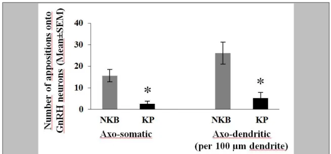
Dual-immunoperoxidase labeled sections were selected (1-2 from each individual) to determine the number of KP/NKB axonal contacts along the outlines of GnRH-IR cell bodies and dendrites (5.2, 5.3, 5.4). Counting of the appositions was carried out using a 63X oil-immersion objective and contacts defined using stringent criteria. The axon and the GnRH profile had to be in the same focus plane without any visible intervening gap, and uncertain instances of partial overlap were not considered. For each subject, the mean number of contacts per GnRH soma and 100µm GnRH dendrite was calculated.
In studies to colocalize KP and NKB in neuronal afferents to GnRH neurons of the Inf (5.3, 5.4), one section from the triple-immunofluorescent specimens of the Inf was selected from each individual to analyze single- and double-labeled KP-IR and NKB-IR neuronal appositions onto GnRH neurons. Multiple stacks of optical slices (512x512
29
pixels, z-steps 0.6 µm) were obtained by scanning GnRH neurons in the Inf and their KP-IR and NKB-IR contacts using a 60x oil immersion objective and a Radiance 2100 confocal microscope. Appositions were validated if no gap was visible between the juxtaposed profiles in at least one optical slice.
30
5. Results
5.1. Estrogenic down-regulation of RF-amide related peptide expression via estrogen receptor-α
5.1.1. RFRP mRNA levels of OVX mice decrease in response to E2 treatment
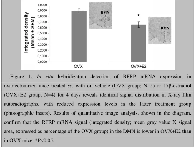
Radioisotopic in situ hybridization studies revealed a restricted regional distribution of RFRP mRNA synthesizing neurons in the mouse hypothalamus, in accordance with the results of earlier studies [88]. The majority of labeled neurons were observed in the dorsomedial nucleus (DMN), an area ventral to it and in the periventricular nucleus of the caudal hypothalamus. The distribution patterns were identical in the OVX and OVX+E2 groups, but the signal was much weaker in the latter (photographic insets in Figure 1). Quantitative analysis established that a 4-day E2 treatment of OVX mice significantly decreased the integrated density of X-ray film images, used as a signal measure (P=0.012; Fig. 1).
Figure 1. In situ hybridization detection of RFRP mRNA expression in ovariectomized mice treated sc. with oil vehicle (OVX group; N=5) or 17β-estradiol (OVX+E2 group; N=4) for 4 days reveals identical signal distribution in X-ray film autoradiographs, with reduced expression levels in the latter treatment group (photographic insets). Results of quantitative image analysis, shown in the diagram, confirm that the RFRP mRNA signal (integrated density; mean gray value X signal area, expressed as percentage of the OVX group) in the DMN is lower in OVX+E2 than in OVX mice. *P<0.05.
31
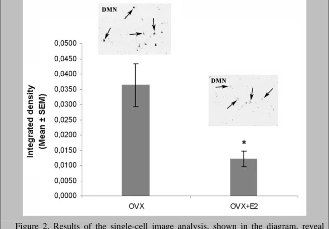
Silver grain clusters in the emulsion autoradiographs were analyzed to determine if E2 treatment decreased the number of detectable RFRP neurons, the single-cell levels of RFRP mRNA, or both. The integrated density analysis of silver grains over individual neurons (Fig. 2) revealed lower single-cell levels of RFRP mRNA expression in the OVX+E2 vs. the OVX group (P=0.021). In retrospect, this unbiased analysis identified significantly fewer (P=0.026) silver grain clusters (RFRP neurons) in OVX+E2 mice (26.4±2.6 neurons/animal; Mean±SEM), compared with OVX controls (39.0±3.6 neurons/animal).
Figure 2. Results of the single-cell image analysis, shown in the diagram, reveal reduced single-cell levels of RFRP mRNA expression (integrated density; mean gray value X area of silver grains above individual neurons), in ovariectomized mice treated sc. with 17β-estradiol for 4 days (OVX+E2 group; N=4) vs. their oil-treated controls (OVX group; N=5). Integrated density values are expressed as percentage of the OVX group. *P<0.05. Individual RFRP neurons are indicated by arrows in representative emulsion autoradiographs.
32
5.1.2. Nuclear estrogen receptor-α occurs in a small subset of RFRP-synthesizing neurons, whereas estrogen receptor-β is absent
The use of silver-gold-intensified Ni-DAB chromogen enabled the sensitive visualization of ER-α (Fig. 3, A-G). A heavy ER-α immunolabeling was present in the ARC and ventromedial nuclei (VMN) (Fig. 3A) and in scattered cell nuclei within the dorsomedial and periventricular nuclei where most RFRP-1-IR cells occurred (Fig. 3, A and B). A pale nuclear ER-α signal was detected in 18.7±3.8% of RFRP-1-IR cells (Fig.
3, C-E, H), whereas the majority of RFRP neurons did not contain ER-α signal (Fig. 3, C, F, G). Using the Z8P antiserum against ER-β, many ER-β-IR cell nuclei were detectable in the hypothalamic paraventricular nucleus (PVN) (Fig. 3I), but only scattered cell nuclei were labeled for ER-β in the dorsomedial nucleus (Fig. 3J). ER-β- positive RFRP neurons were not revealed (Fig. 3J).
33
Figure 3. Use of dual-label immunohistochemistry provides evidence for the presence of ER-α within a small subset (18.7%) of RFRP-1-IR neurons and the absence of ER-β in ovariectomized mice. Use of the silver-gold-intensified Ni-DAB chromogen reveals a high density of darkly labeled ER-α-IR cell nuclei (black signal) in the ARC and VMN (A). Heavily labeled cell nuclei are also present in the dorsomedial nucleus (DMN) where the majority of RFRP-1-IR neuronal cell bodies (brown cytoplasmic labeling) occurs (B). As shown in the high-power images (C-G) of framed areas in B, typical RFRP neurons are either devoid of the nuclear ER-α signal (white arrows; C, F, G) or exhibit only weak ER-α labeling (black arrows; C, D, E). Very few RFRP neurons show medium (H) and none show high labeling intensity. Arrowheads in C point to ER- α-IR cell nuclei in non-RFRP neurons. The application of silver-gold-intensified Ni- DAB to detect ER-β immunoreactivity reveals a large number of labeled nuclei in the hypothalamic paraventricular nucleus (PVN; I) but only a few labeled cells in the DMN (arrowhead in J). RFRP-1-IR cells in the DMN (arrows) do not contain ER-β signal (arrowhead) in the high-power inset. Scale bars=50μm in A, B, I, J and 10μm in C-H and in the high-power inset in J.
5.2. Immunohistochemical evidence for the absence of ‘KNDy neurons’ in young human males
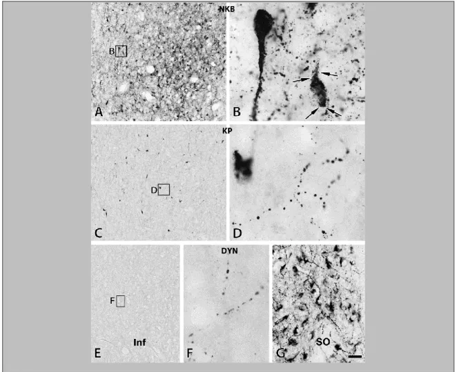
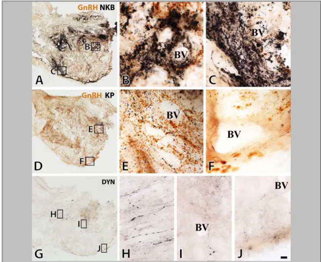
The comparative analysis of NKB, KP and DYN immunoreactivities in immunoperoxidase-labeled sections of the Inf (Fig. 4) and the InfS (Fig. 6) revealed strikingly different labeling intensities for KNDy neuropeptides. In general, NKB-IR elements showed much higher abundance than KP-IR elements. DYN immunoreactivity, both in perikarya and fibers, was relatively sparse and weak.
34
Figure 4. The silver-gold intensified Ni-DAB chromogen was used to visualize NKB (A, B), KP (C, D) and DYN (E-G) immunoreactivities in adjacent sections of the Inf from a 31-year-old male subject. NKB-IR perikarya as well as nerve fibers (A, B) occur in much higher numbers than do KP-IR elements (C, D). For results of the quantitative analyses which reveal about five-fold differences for both perikaryon and fiber densities, see Figure 5 and 7, respectively. Arrowheads point to NKB-IR cell bodies in B and a KP-IR cell body in D. Note that NKB-IR neurons receive numerous afferent contacts (arrows in B) from NKB-IR varicose axons; analogous juxtapositions in other species were proposed to underlie the main peptidergic signaling mechanism among the putative pulse-generating KNDy neurons. Few of any DYN-IR cell bodies are detectable in the Inf (none visible in this specific case) and only scattered DYN-IR fibers occur (E, F). This low level of the DYN signal does not appear to reflect a technical limitation of the immunohistochemical approach, given that IR perikarya and fibers are abundant elsewhere in the hypothalamus, including the supraoptic nucleus (SO; G). Scale bar=100μm for A, C, E and 9μm elsewhere.
35
5.2.1. Incidence of KP-, NKB- and DYN-immunoreactive perikarya in the Inf
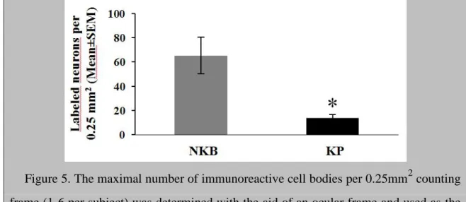
In peroxidase-based immunohistochemistry, many NKB-IR perikarya were identified in the Inf (Fig. 4, A and B). KP-IR cell bodies occurred in much lower numbers in neighboring sections (Fig. 4, C and D). Quantitative analysis showed that the density of KP neurons was about 5 times lower than that of NKB-IR perikarya (Fig. 5; P=0.01 by ANOVA). DYN-IR perikarya were either entirely absent in some subjects (Fig. 4, E and F) or extremely rare in the Inf of others, preventing quantitative studies. In contrast, the supraoptic nucleus contained many DYN labeled perikarya (Fig. 4G), making it unlikely that the low DYN signal in the Inf reflects technical limitations.
Figure 5. The maximal number of immunoreactive cell bodies per 0.25mm2 counting frame (1-6 per subject) was determined with the aid of an ocular frame and used as the index of the density of labeled perikarya. Results show that in young male human individuals, the mean incidence of NKB-IR cell bodies is about 5 times higher than the incidence of KP-IR cell bodies. *P<0.05 by ANOVA.
A surprising segregation of NKB-IR and KP-IR perikarya was revealed in dual- immunofluorescent specimens (Fig. 6, A and B). Tyramide signal amplification was crucial for sensitive detection of NKB/KP dual-labeled cell bodies which represented only 32.9±4.7% of the NKB-IR and 75.2±6.6% of the KP-IR perikarya (Fig. 6, A and B). Tyramide signal amplification was capable of visualizing only a few DYN-IR perikarya (not shown).
36
37
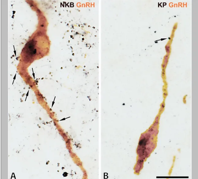
Figure 6. Results of immunofluorescent studies reveal a significant degree of mismatch between KP and NKB immunoreactivities (A-E) and KP and DYN (or dynorphin B) immunoreactivities (F-K) in the Inf (A, B, F, G) and the InfS (C-E, H) of the human. The dual-immunofluorescent visualization of NKB (green) and KP (red) immunoreactivities in the Inf (A, B) not only confirms the dominance of NKB-IR (green) over KP-IR (red) cell bodies (arrowheads) and axons (arrows) in the Inf, but also reveals a considerable degree of segregation between the two different perikaryon (arrowheads)- and fiber (arrows) populations. Yellow double-arrows and arrowheads point to dual-labeled fibers and cell bodies, respectively. Single-labeled NKB-IR axons are also typical around the portal blood vessels (BV) of the postinfundibular eminence in the InfS (C-E), in addition to single-labeled KP-IR fibers (red) and NKB/KP-dual- labeled (yellow) axons. The dual-immunofluorescent visualization of KP and DYN (F- H) illustrates the absence of DYN immunoreactivity from KP-IR cell bodies in the Inf (red arrowheads in F). With the exceptions of a few scattered dual-labeled fibers (yellow double-arrows) in the Inf (F, G) and the InfS (H), the majority of KP-IR (red) and DYN-IR (green) axons are separate. Negative colocalization results are reproducible in the Inf (I) and the InfS (J) using an antiserum against a different prodynorphin cleavage product, dynorphin B. Note the high density of dynorphin B-IR fibers in the neighboring ventromedial nucleus (VMH) in K. High-power micrographs (A, B, D-K) represent single optical slices (0.7µm). Scale bar=100μm for C, 5μm for D and 10μm for A, B, E-K.
5.2.2. Abundance of KP- and NKB-immunoreactive fibers in the Inf
The incidence of immunolabeled fibers in the Inf followed a similar trend as labeled perikarya. The most frequently encountered phenotype was, again, IR for NKB. These axons established many appositions (Fig. 4B) to NKB-IR cell bodies and their dendritic processes. Quantitative analysis of the area covered by immunohistochemical signal established that the mean incidence of NKB-IR fibers was about 5 times higher than that of KP-IR fibers (Figure 7; P=0.0001 by ANOVA). DYN-IR fibers were also detectable in the Inf, although less frequently than either NKB-IR or KP-IR axons (Fig. 4, E and F).
38
Figure 7. The area covered by immunoreactive fibers (divided by the total area analyzed) was determined with the ImageJ software in digital photographs of the Inf and used as a fiber density measure. Areas that were occupied by labeled cell bodies and their thick proximal dendrites were deleted from the photographs with the eraser tool of the Adobe Photoshop software and thus, excluded from the analysis. The density of NKB-IR fibers (expressed in arbitrary units) is 5.4 times higher than the density of KP- IR fibers. *P<0.0005 by ANOVA.
In immunofluorescent specimens, many NKB-IR fibers without KP immunolabeling as well as KP-IR fibers without NKB labeling could be seen in the Inf, in addition to dual-labeled axons (Fig. 6, A and B). DYN-IR fibers showed a high intensity of labeling only if the tyramide signal amplification approach was also used. Most of them were distinct from KP-IR axons, although dual-labeled KP/DYN-IR fibers occasionally occurred (Fig. 6, F and G). Similarly, the majority of KP-IR fibers were also devoid of dynorphin B immunoreactivity in the Inf and the InfS (Fig. 6, I and J), whereas this second antiserum performed well in regions rich in DYN fibers, including the ventromedial nucleus (Fig. 6K).
5.2.3. Abundance of KP- and NKB-immunoreactive fibers in the InfS
The InfS was associated with the superficial and the deep capillary plexuses of the postinfundibular eminence [138]. Both were abundantly innervated by GnRH-IR axons (brown color in Fig. 8, A-F), suggesting they contribute to the GnRH supply of adenohypophysial gonadotrophs. The relative abundance of the different types of