Helicobacter pylori infection is associated with anemia, weight loss or both conditions among Bulgarian children
LYUDMILA BOYANOVA
1pand PETYO HADZHIYSKI
21Department of Medical Microbiology, Medical University of Sofia, 2 Zdrave str., 1431, Sofia, Bulgaria
2Specialized Hospital for Active Pediatric Treatment, Medical University of Sofia,“Acad. Ivan Evstatiev Geshov”Blvd, 1606, Sofia, Bulgaria
Received: February 10, 2020 • Accepted: May 5, 2020 Published online: December 1, 2020
ABSTRACT
Some studies suggested an association betweenHelicobacter pyloriinfection and iron-deficiency anemia, however, the link between weight loss and the infection in childhood remains non-established. In a retrospective cohort study, we comparedH. pyloripositivity rates of Bulgarian children without or with anemia (47 children in each group) or weight loss (45 children in each group) and both conditions (17 children in each group).H. pyloriinfection was associated with the presence of anemia (in 76.6% of the anemic vs. 21.3% of the non-anemic patients,P< 0.0001) and weight loss (in 82.2% of the patients vs.
17.8% of the control children,P< 0.0001). All 17 patients with both conditions wereH. pyloripositive.
Relative risk of anemia, weight loss and both conditions was 3.6 (95% CI, 2.0–6.4), 4.6 (95% CI, 2.4–8.8) and 5.7 (95% CI, 2.0–15.8), respectively, in the children withH. pyloriinfection. In conclusion,H. pylori infection was significantly associated with iron-deficiency anemia or/and weight loss in Bulgarian pe- diatric patients. Therefore, diagnostics and treatment of the infection as well as a proper control of the eradication success can be beneficial and thus, can be recommended for children with those conditions.
KEYWORDS
Helicobacter pylori, infection, children, anemia, weight loss
INTRODUCTION
In 1993, Bruel et al. [1] reported a case of 11-year old patient with severe anemia, Heli- cobacter pylorigastritis and upper gastrointestinal bleeding and suggested that the infection and anemia were linked. Other authors also detected an association between H. pylori infection and iron-deficiency anemia [2–4]. Anemia was refractory to iron treatment, whereas patients recovered after both iron therapy and eradication ofH. pyloriinfection [5–
7]. BothH. pyloriinfection and anemia, in particular iron-deficiency anemia, are predomi- nant in populations with low socio-economic status [8]. However, results of some studies did not show any association betweenH. pylori status and iron deficiency anemia [9].
In Maastricht V/Florence Consensus Report, the need of gastro-duodenoscopy of patients with anemia has been stressed [10]. However, unlike the probable link ofH. pyloriinfection to the iron-deficiency anemia, the association between weight loss/obesity and the infection in childhood remains unclear [11]. Therefore, the aim of the present study was to assess a possible association betweenH. pylori status and the presence of anemia or weight loss, or both conditions in Bulgarian pediatric patients.
MATERIAL AND METHODS
We carried out a retrospective cohort study onH. pyloripositivity rates with 47 children with anemia 45 children with weight loss and 17 children and both conditions and compared the
Acta Microbiologica et Immunologica Hungarica
67 (2020) 4, 239–242 DOI:
10.1556/030.2020.01158
© 2020 Akademiai Kiado, Budapest
RESEARCH ARTICLE
*Corresponding author.
Tel.:þ3592 91 72 730.
E-mail:l.boyanova@hotmail.com
results with groups of children with the same number in each group and similar characteristics (Table 1).
Anemia was documented by laboratory tests such as hemoglobin (Hb) level <120 g/L (<12 g/dL), hematocrit (<40%), erythrocytes, Mean Corpuscular Volume, MCV and Mean Corpuscular Hemoglobin, MCH). Presence of weight loss (≥2 BMI-for-age percentiles) during the last year most often occurred during the last 3–5 months. Ten boys and seven girls had both anemia and weight loss.
Two gastric biopsy specimens were taken from each patient and were used for direct Gram staining with carbol fuchsine, a rapid urease test with 10% urea and culture on both non-selective and selective media as described in our prior publications [12, 13]. Positivity of either culture or two of the three diagnostic tests was considered as H. pylori positive status. Written informed consent was taken from the children’parents. The study was approved by the Ethics Committee of the Medical University of Sofia, Sofia, Bulgaria.
Statistical analysis.c2with Fisher’s exact test was used to compare the groups. Odd ratios and relative risks were calculated as well (https://www.medcalc.org/calc/odds_ratio.
php;https://www.medcalc.org/calc/relative_risk.php).
RESULTS
We found that the children who had anemia were signifi- cantly more oftenH. pyloripositive (76.6%, 36/47 children) compared with those without anemia (21.3%, 10/47, P <
0.0001), (Table 1). Children who had weight loss were much more frequentlyH. pyloripositive (82.2%, 37/45) compared with the other patients (17.8%, 8/45,P< 0.0001).
All 17 children with both anemia and weight loss wereH.
pylori positive vs. only 17.7% (3/17, P < 0.0001) in the control group. The calculated relative risk of anemia was 3.6 (95% CI, 2.0–6.4), that of weight loss was 4.6 (95% CI, 2.4–
8.8) and that of both conditions was 5.7 (95% CI, 2.0–15.8) in theH. pyloripositive children compared with those in the control groups (Table 2).
DISCUSSION
H. pylorigastritis is a common, although not the only reason for anemia refractory to treatment, mostly in adolescence [5]. The high incidence ofH. pyloriinfection in patients with anemia suggests that the bacteria may be involved in the pathogenesis of the condition trough different mechanisms such as H. pylori competition with the host for available iron, H. pylori proteins for iron transport or storage, diminished iron absorption owing to hypochlorhydria and bleeding erosions or microerosions [4].
Decreased iron absorption has been linked to the increased pH of gastric juice and transient hypochlorhydria in the early phase of infection and atrophic changes in the stomach later in life [14]. Moreover, H. pylori virulence factors were related to the acquisition of iron by bacteria.
CagA (cytotoxin-associated gene A) protein and VacA (vacuolating cytotoxin A) are conjointly involved in bacterial iron acquisition through the cholotransferin and in enhanced colonization of gastric cells, NapA (neutrophil-activating protein A) can increase bacterial Fe ion uptake and SabA (sialic acid–binding adhesin) can be of importance as well [2, 4, 15]. In our previous study, we detected a high frequency (>85%) of virulent (vacA s1 and cagA positive) H. pylori strains in symptomatic Bulgarian patients [16].
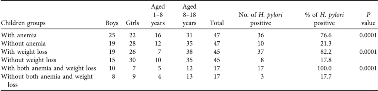
Table 1. H. pyloripositivity in Bulgarian pediatric patients according to the presence or absence of anemia, weight loss or both conditions
Children groups Boys Girls
Aged 1–8 years
Aged 8–18
years Total
No. ofH. pylori positive
% ofH. pylori positive
P value
With anemia 25 22 16 31 47 36 76.6 0.0001
Without anemia 19 28 12 35 47 10 21.3
With weight loss 19 26 7 38 45 37 82.2 0.0001
Without weight loss 15 30 10 35 45 8 17.8
With both anemia and weight loss 10 7 5 12 17 17 100.0 0.0001
Without both anemia and weight loss
8 9 4 13 17 3 17.7
Table 2.Risk factors for anemia and weight loss amongH. pyloripositive children
Outcome variable
Exposure
(independent) variable OR
95% CI for OR Z
statistic Pvalue RR
95% CI for RR Z
statistic Pvalue
Lower Upper Lower Upper
Anemia H. pylori 12.1 4.6 32.0 5.0 <0.0001 3.6 2.0 6.4 4.4 <0.0001
Weight loss H. pylori 21.4 7.3 63.0 5.6 <0.0001 4.6 2.4 8.8 4.7 <0.0001
Both anemia and weight loss
H. pylori 175.0 8.4 3,645 3.3 0.0009 5.7 2.0 15.8 3.3 0.0009
OR–odd ratio (https://www.medcalc.org/calc/odds_ratio.php); RR-relative risk (https://www.medcalc.org/calc/relative_risk.php).
95% CI–95% confidence interval.
240
Acta Microbiologica et Immunologica Hungarica67 (2020) 4, 239–242Unlike the anemia, weight loss association withH. pylori positivity remains unclear. The weight loss was found to be neither an indication for gastroduodenoscopy in the USA [17], nor as a risk factor forH. pyloriinfection in China [18].
An important result of our study was the significant association of H. pylori infection with weight loss. This result indirectly corresponds to that of Choi et al. [11], who found that, although there was no difference in H.
pylori positivity rates between obese and non-obese Korean children, a significant gain of weight was observed two months after H. pylori eradication of the treated children. Some authors have suggested an association be- tween H. pylori infection and gastric or serum concen- trations of appetite-regulating peptides such as leptin, ghrelin and obestatin, however, more studies are required to evaluate the association [11, 19, 20]. Nevertheless, Talley et al. [21] have reported the presence of dyspepsia, involving reduced or lost appetite in 65% of the infected children vs. only 15% in those with H. pylori negative status. The results of our study show that both anemia and weight loss, alone and in combination with each other, are much more common inH. pyloripositive children than in the noninfected pediatric patients.
CONCLUSION
In conclusion,H. pyloriinfection was closely linked to iron- deficiency anemia and weight loss in Bulgarian children. The infected pediatric patients were at much higher risk of anemia and weight loss compared with H. pylori negative children. The results imply that in children with anemia and weight loss, the diagnosis of infection and the proper control of eradication success are of utmost importance.
Conflicts of interest: The authors declare that there are no conflicts of interest.
Funding information:The authors received no specific grant from any funding agency.
ACKNOWLEDGEMENTS
We express our gratitude to our Statistical analysis consul- tant Assoc. Prof. Mircho Vukov.
REFERENCES
[1] Bruel H, Dabadie A, Pouedras P, Gambert C, Le Gall E, Jezequel C. Anemie aigu€e revelatrice d’une gastrite aHelicobacter pylori [Helicobacter pylori gastritis manifested by acute anemia]. Ann Pediatr (Paris) 1993; 40(6): 364–7.
[2] Yokota S, Toita N, Yamamoto S, Fujii N, Konno M. Positive relationship between a polymorphism in Helicobacter pylori
neutrophil-activating protein a gene and iron-deficiency anemia.
Helicobacter 2013; 18(2): 112–16.
[3] Hudak L, Jaraisy A, Haj S, Muhsen K. An updated systematic review and meta-analysis on the association betweenHelicobacter pylori infection and iron deficiency anemia. Helicobacter 2017;
22: 1.
[4] Kato S, Osaki T, Kamiya S, Zhang XS, Blaser MJ. Helicobacter pylori sabA gene is associated with iron deficiency anemia in childhood and adolescence. PLoS One 2017; 12(8): e0184046.
[5] Duclaux-Loras R, Lachaux A. Anemie ferriprive de l’adolescent liee a une infection a Helicobacter pylori, a propos d’un cas [Helicobacter pylori infection, a classic but often unrecognized cause of iron deficiency anemia in teenagers]. Arch Pediatr 2013;
20(4): 395–7.
[6] Hacibekiroglu T, Basturk A, Akinci S, Bakanay SM, Ulas T, Guney T, et al. Evaluation of serum levels of zinc, copper, and Heli- cobacter pyloriIgG and IgA in iron deficiency anemia cases. Eur Rev Med Pharmacol Sci 2015; 19(24): 4835–40.
[7] Gheibi SH, Farrokh-Eslamlou HR, Noroozi M, Pakniyat A. Re- fractory iron deficiency anemia andHelicobacter pyloriinfection in pediatrics: a review. Iran J Ped Hematol Oncol 2015; 5(1):
50–64.
[8] Wenzhen Y, Yumin L, Kehu Y, Bin M, Quanlin G, Donghai W, et al. Iron deficiency anemia inHelicobacter pyloriinfection: meta- analysis of randomized controlled trials. Scand J Gastroenterol 2010; 45(6): 665–76.
[9] John S, Baltodano JD, Mehta N, Mark K, Murthy U. Unexplained iron deficiency anemia: does Helicobacter pylorihave a role to play? Gastroenterol Rep (Oxf) 2018; 6(3): 215–20.
[10] Malfertheiner P, Megraud F, O’Morain CA, Gisbert JP, Kuipers EJ, Axon AT. Management of Helicobacter pylori infection-the Maastricht V/Florence consensus report. Gut 2017; 66(1): 6–30.
[11] Choi JS, Ko KO, Lim JW, Cheon EJ, Lee GM, Yoon JM. The as- sociation betweenHelicobacter pyloriinfection and body weight among children. Pediatr Gastroenterol Hepatol Nutr 2016; 19(2):
110–15.
[12] Boyanova L, Hadzhiyski P, Markovska R, Yaneva P, Yordanov D, Gergova G, et al. Prevalence of Helicobacter pyloriis still high among symptomatic Bulgarian children. Acta Microbiol Immunol Hung 2019; 66(2): 255–60.
[13] Boyanova L, Gergova G, Nikolov R, Davidkov L, Kamburov V, Jelev C, et al. Prevalence and evolution of Helicobacter pylori resistance to 6 antibacterial agents over 12 years and correlation between susceptibility testing methods. Diagn Microbiol Infect Dis 2008; 60(4): 409–15.
[14] Windle HJ, Kelleher D, Crabtree JE. ChildhoodHelicobacter pylori infection and growth impairment in developing countries: a vi- cious cycle? Pediatrics 2007; 119(3): e754–9.
[15] Boyanova L. Role ofHelicobacter pylorivirulence factors for iron acquisition from gastric epithelial cells of the host and impact on bacterial colonization. Future Microbiol 2011; 6(8): 843–6.
[16] Boyanova L, Markovska R, Yordanov D, Marina M, Ivanova K, Panayotov S, et al. High prevalence of virulentHelicobacter pylori strains in symptomatic Bulgarian patients. Diagn Microbiol Infect Dis 2009; 64(4): 374–80.
[17] Raj P, Thompson JF, Pan DH.Helicobacter pyloriserology testing is a useful diagnostic screening tool for symptomatic inner city children. Acta Paediatr 2017; 106(3): 470–7.
Acta Microbiologica et Immunologica Hungarica67 (2020) 4, 239–242
241
[18] Shu X, Ping M, Yin G, Jiang M. Investigation ofHelicobacter pyloriinfection among symptomatic children in Hangzhou from 2007 to 2014: a retrospective study with 12,796 cases. Peer J 2017; 5: e2937.
[19] Franceschi F, Annalisa T, Teresa DR, Giovanna D, Ianiro G, Franco S, et al. Role ofHelicobacter pyloriinfection on nutrition and metabolism. World J Gastroenterol 2014; 20(36): 12809–17.
[20] Romo-Gonzalez C, Mendoza E, Mera RM, Coria-Jimenez R, Chico-Aldama P, Gomez-Diaz R, et al.Helicobacter pyloriinfec- tion and serum leptin, obestatin, and ghrelin levels in Mexican schoolchildren. Pediatr Res 2017; 82(4): 607–13.
[21] Talley NJ, Stanghellini V, Heading RC, Koch KL, Malagelada JR, Tytgat GN. Functional gastroduodenal disorders. Gut 1999;
45(Suppl. 2): II37–42.