Cite this article as: Blomstro¨m-Lundqvist C, Traykov V, Erba PA, Burri H, Nielsen JC, Bongiorni MGet al. European Heart Rhythm Association (EHRA) international consensus document on how to prevent, diagnose, and treat cardiac implantable electronic device infections—endorsed by the Heart Rhythm Society (HRS), the Asia Pacific Heart Rhythm Society (APHRS), the Latin American Heart Rhythm Society (LAHRS), International Society for Cardiovascular Infectious Diseases (ISCVID) and the European Society of Clinical Microbiology and Infectious Diseases (ESCMID) in collaboration with the European Association for Cardio-Thoracic Surgery (EACTS).
Eur J Cardiothorac Surg 2020;57:e1–31.
European Heart Rhythm Association (EHRA) international consensus document on how to prevent, diagnose, and treat cardiac
implantable electronic device infections—endorsed by the Heart Rhythm Society (HRS), the Asia Pacific Heart Rhythm Society
(APHRS), the Latin American Heart Rhythm Society (LAHRS), International Society for Cardiovascular Infectious Diseases (ISCVID)
and the European Society of Clinical Microbiology and Infectious Diseases (ESCMID) in collaboration with the European
Association for Cardio-Thoracic Surgery (EACTS)
Carina Blomstro¨m-Lundqvist (Chair)
a*, Vassil Traykov (Co-Chair)
b, Paola Anna Erba
c, Haran Burri
d, Jens Cosedis Nielsen
e, Maria Grazia Bongiorni
f, Jeanne Poole (HRS representative)
g, Giuseppe Boriani
h, Roberto Costa (LAHRS representative)
i, Jean-Claude Deharo
j, Laurence M. Epstein (HRS representative)
k,
Laszlo Saghy
l, Ulrika Snygg-Martin (ESCMID and ISCVID representative)
m, Christoph Starck (EACTS representative)
n, Carlo Tascini (ESCMID representative)
o, and
Neil Strathmore (APHRS representative)
pa Department of Medical Science and Cardiology, Uppsala University, Uppsala, Sweden
b Department of Invasive Electrophysiology and Cardiac Pacing, Acibadem City Clinic Tokuda Hospital, Sofia, Bulgaria
c Nuclear Medicine, Department of Translational Research and New Technology in Medicine, University of Pisa, Pisa, Italy, and University of Groningen, University Medical Center Groningen, Medical Imaging Center, Groningen, Netherlands
d Department of Cardiology, University Hospital of Geneva, Geneva, Switzerland
e Department of Cardiology, Aarhus University Hospital, Aarhus, Denmark
f Division of Cardiology and Arrhythmology, CardioThoracic and Vascular Department, University Hospital of Pisa, Pisa, Italy
g Division of Cardiology, University of Washington, Seattle, WA, USA
h Division of Cardiology, Department of Biomedical, Metabolic and Neural Sciences, University of Modena and Reggio Emilia, Policlinico di Modena, Modena, Italy
i Department of Cardiovascular Surgery, Heart Institute (InCor) of the University of S~ao Paulo, S~ao Paulo, Brazil
EHRA international consensus document on how to prevent, diagnose and treat cardiac implantable electronic device infections,EP Europace2019, doi:10.1093/
europace/euz246. Reprinted by permission of Oxford University Press on behalf of the European Society of Cardiology.
This publication comprises a reprint of Journal item “EHRA international consensus document on how to prevent, diagnose and treat cardiac implantable electronic device infections” originally published in the English language inEP Europaceby Oxford University Press under licence from the European Society of Cardiology (“ESC”).
VCEuropean Heart Rhythm Association 2019.
All rights reserved; no part of this publication may be reproduced, stored in a retrieval system, or transmitted in any form or by any means, electronic, mechanical, photocopying, recording, or otherwise, without prior written permission of the Publishers.
For Permissions, please email: journals.permissions@oup.com
The opinions expressed in the Journal item reproduced as this reprint are those of the authors and contributors, and do not necessarily reflect those of the European Society of Cardiology, the editors, the editorial board, Oxford University Press or the organization to which the authors are affiliated.
The mention of trade names, commercial products or organizations, and the inclusion of advertisements in this reprint do not imply endorsement by the Journal, the editors, the editorial board, Oxford University Press or the organization to which the authors are affiliated. The editors and publishers have taken all reasonable pre- cautions to verify drug names and doses, the results of experimental work and clinical findings published in the Journal. The ultimate responsibility for the use and dosage of drugs mentioned in this reprint and in interpretation of published material lies with the medical practitioner, and the editors and publisher cannot accept liability for damages arising from any error or omissions in the Journal or in this reprint. Please inform the editors of any errors.
EHRACONSENSUSPAPER
European Journal of Cardio-Thoracic Surgery 57 (2020) e1–e31
EHRA CONSENSUS PAPER
doi:10.1093/ejcts/ezz296 Advance Access publication 14 November 2019
Downloaded from https://academic.oup.com/ejcts/article/57/1/e1/5625578 by 81728827 user on 19 January 2022
j Department of Cardiology, Aix Marseille Universite´, CHU la Timone, Marseille, France
k Electrophysiology, Northwell Health, Hofstra/Northwell School of Medicine, Manhasset, NY, USA
l Division of Electrophysiology, 2nd Department of Medicine and Cardiology Centre, University of Szeged, Szeged, Hungary
mDepartment of Infectious Diseases, Institute of Biomedicine, Sahlgrenska Academy, University of Gothenburg, Gothenburg, Sweden
n Department of Cardiothoracic and Vascular Surgery, German Heart Center Berlin, Berlin, Germany
o First Division of Infectious Diseases, Cotugno Hospital, Azienda ospedaliera dei Colli, Naples, Italy
p Department of Cardiology, Royal Melbourne Hospital, Melbourne, Australia
* Corresponding author. Tel: +46 18 611 3113. E-mail address: carina.blomstrom.lundqvist@akademiska.se Received 1 August 2019; editorial decision 11 August 2019; accepted 19 August 2019
Abstract
Pacemakers, implantable cardiac defibrillators, and cardiac resynchronization therapy devices are potentially life-saving treatments for a number of cardiac conditions, but are not without risk. Most concerning is the risk of a cardiac implantable electronic device (CIED) infec- tion, which is associated with significant morbidity, increased hospitalizations, reduced survival, and increased healthcare costs.
Recommended preventive strategies such as administration of intravenous antibiotics before implantation are well recognized.
Uncertainties have remained about the role of various preventive, diagnostic, and treatment measures such as skin antiseptics, pocket anti- biotic solutions, anti-bacterial envelopes, prolonged antibiotics post-implantation, and others. Guidance on whether to use novel device alternatives expected to be less prone to infections and novel oral anticoagulants is also limited, as are definitions on minimum quality requirements for centres and operators and volumes. Moreover, an international consensus document on management of CIED infections is lacking. The recognition of these issues, the dissemination of results from important randomized trials focusing on prevention of CIED infections, and observed divergences in managing device-related infections as found in an European Heart Rhythm Association worldwide survey, provided a strong incentive for a 2019 International State-of-the-art Consensus document on risk assessment, prevention, diagno- sis, and treatment of CIED infections.
Keywords: Infection • Endocarditis • Microbiology • Cardiac implantable electronic devices • Implantable cardioverter-defibrillators • Pacemakers • Cardiac resynchronization therapy • Leads • Extraction • Re-implantation • EHRA consensus document
TABLE OF CONTENTS
Introduction . . . e3 Scope of the consensus document . . . e3 Methodology . . . e3 Background and epidemiology . . . e3 Pathogenesis and microbiology of cardiac implantable
electronic device infections . . . e4 Risk factors for cardiac implantable electronic device infection . e5 Risk stratification . . . e6 Prevention . . . e6 Pre-procedural measures . . . e6 Patient selection . . . e6 Lead management . . . e6 Patient factors . . . e6 Anticoagulation and antiplatelet drugs . . . e6 Appropriate environment . . . e8 Staff training . . . e8 Nasal swabs/S. aureusdecolonization of patients . . . e8 Pre-procedure skin preparation . . . e8 Pre-procedure antibiotic therapy . . . e8 Peri-procedural measures . . . e8 Patient surgical preparation . . . e8 Good surgical technique . . . e9 Antibiotic envelope . . . e9 Local instillation of antibiotics or antiseptics . . . e9 Capsulectomy . . . e9 Closure . . . e9 Post-procedural measures . . . e9 Post-procedure antibiotic therapy . . . e9 Wound care . . . e9
Re-intervention . . . e9 Diagnosis of cardiac implantable electronic device infections and related complications . . . e10
Clinical findings . . . e10 Identification of the causative microorganisms . . . e11 Imaging . . . e11 Echocardiography . . . e11 Radiolabelled leucocyte scintigraphy, positron emission tomography, and computerized tomography . . . e11 Management of cardiac implantable electronic device infections:
when, how, and where . . . e14 Cardiac implantable electronic device removal . . . e14 Antimicrobial therapy including long-term suppressive
therapy . . . e15 Preventive strategies after cardiac implantable electronic
device implantations, new re-implantations, and
alternative novel devices . . . e16 Preventive strategies after cardiac implantable electronic
device implantations . . . e16 Re-implantations . . . e18 Alternative novel devices . . . e18 Prognosis, outcomes, and complications of cardiac
implantable electronic device infections . . . e19 Special considerations to prevent device-related
infections (elderly, paediatrics, adult with congenital
heart disease) . . . e19 Minimum quality requirements concerning centres and
operator experience and volume . . . e20 Health economics for cardiac implantable electronic
devices infections and strategies to reduce costs . . . e22 Divergent recommendations from different societies . . . e22
Downloaded from https://academic.oup.com/ejcts/article/57/1/e1/5625578 by 81728827 user on 19 January 2022
General definitions and minimal requirements of
variables in scientific studies and registries . . . e24 Gaps of evidence . . . e25 Summary of emerging messages and call for scientific
evidence . . . e25 References . . . e26
INTRODUCTION
Scope of the consensus document
Pacemakers (PM), implantable cardiac defibrillators (ICDs), and cardiac resynchronization therapy (CRT) devices are life-saving treatments for a number of cardiac conditions. Device-related in- fection is, however one of the most serious complications of car- diac implantable electronic device (CIED) therapy associated with significant morbidity, mortality, and financial healthcare burden.
Although many preventive strategies such as administration of intravenous (i.v.) antibiotic therapy before implantation are well recognized, uncertainties still exist about other regimens.
Questions still remain such as the use of CIED alternatives expected to be less prone to infections and how to manage medication, such as anticoagulants during CIED surgery, and the role of minimum quality and volume requirements for centres and operators. The recognition of these gaps in knowledge, reports of new important randomized trials, observed divergen- ces in managing device-related infections [1], and the lack of international consensus documents specifically focusing on CIED infections provided a strong incentive for a 2019 State-of-the-art Consensus document on risk assessment, prevention, diagnosis, and management of CIED infections. The aim of this document is to describe the current knowledge on the risks for device-related infections and to assist healthcare professionals in their clinical decision making regarding its prevention, diagnosis, and man- agement by providing the latest update of the most effective strategies.
Methodology
This consensus document is an international collaboration among seven professional societies/associations, including the European Heart Rhythm Association (EHRA), the Heart Rhythm Society (HRS), the Asia Pacific Heart Rhythm Society (APHRS), the Latin American Heart Rhythm Society (LAHRS), the European Association for Cardio-Thoracic Surgery (EACTS), the European Society of Clinical Microbiology and Infectious Diseases (ESCMID), and the International Society for Cardiovascular Infectious Diseases (ISCVID). The writing group consisting of 16 Task Force Members, were selected based on their expertise and medical specialty (12 cardiologists with vary- ing subspecialties, 2 infectious disease specialists, 1 imaging specialist, and 1 thoracic surgeon), from 11 countries in 4 continents.
All experts undertook a detailed comprehensive literature search until May 2019 (human research published in English and indexed in major databases such as MEDLINE, EMBASE, the Cochrane Library, and others as required) related to studied pa- tient cohort and CIED infection topics using relevant search terms related to the field and prior guidelines. Systematic reviews of published evidence for management of given conditions and
clinical problems were performed. Members were asked to weigh the strength of evidence for or against a particular diagnostic in- strument, procedure, or treatment, include estimates of expected health outcomes and assess risk–benefit ratios where data existed. Patient-, device-, and procedure-specific modifiers were considered, as were the results of the international survey on CIED infections conducted for this purpose [1] and of previous registries [2]. Consensus statements were evidence-based, derived primarily from published data and by consensus opinion after thorough deliberations, requiring at least 80% predefined con- sensus delivered via email by chairs to all expert members for their approval/rejection.
The EHRA user-friendly ranking system, for consensus docu- ments, with ‘coloured hearts’ providing the current status of the evidence and consequent guidance was used for the coding of the scientific evidence for statements made (Table1). The grading does not have separate levels of evidence, which instead are defined in each of the coloured heart grades. A letter coding
‘ROME’ defining existing scientific evidence was applied: R for randomized trials, O for observational studies, M for meta- analyses, and E for expert opinion (Table1).
The document was peer-reviewed by official external reviewers representing EHRA, the participating societies, and ESC Committee for Practice Guidelines (CPG). All members of the writing group as well as reviewers have disclosed potential con- flicts of interest, at the end of this document.
Since this consensus document includes evidence and expert opinions from various countries and healthcare systems, the medical approaches discussed may include drugs or devices that are not approved by governmental regulatory agencies in all countries. Moreover, the ultimate decision on management must be made by the healthcare provider and the patient in light of in- dividual factors presented.
BACKGROUND AND EPIDEMIOLOGY
Over the last decades, there has been a substantial increase in the number and complexity of CIED implantations as a result of expanded indications and progressive aging of the population.
Although these devices improve cardiovascular outcomes, they also expose patients to a risk for potential complications.
Infection is one of the most serious complications of CIED therapy and is associated with significant mortality, morbidity, and financial healthcare burden. It is difficult to give a precise rate of CIED infections because of divergent definitions, varied populations, and the range of rates in retrospective and pro- spective studies. In the Danish registry including 46 299 consecu- tive patients who underwent pacemaker implantation between 1982 and 2007, the incidence of infection was 4.82/1000 device- years after a primary implantation, and 12.12/1000 device-years after replacement [3]. Greensponet al. found that the incidence of CIED infection in the USA increased from 1.53% in 2004 to 2.41% in 2008 [4] and a National Inpatient Sample database study showed an increase from 1.45% to 3.41% (P< 0.001) from 2000 through 2012, particularly for CRT devices [5]. Infection rates in prospective observational studies [6,7], registries [8] and more re- cent cross-over cluster PADIT- and randomized WRAP-IT trials [9,10], were only 0.6–1.3%, as compared to retrospective studies [11,12], reporting significantly higher rates (2.3–3.4%) in the first year after implantation.
EHRACONSENSUSPAPERDownloaded from https://academic.oup.com/ejcts/article/57/1/e1/5625578 by 81728827 user on 19 January 2022
PATHOGENESIS AND MICROBIOLOGY OF CARDIAC IMPLANTABLE ELECTRONIC DEVICE INFECTIONS
Cardiac implantable electronic device infections occur via two major mechanisms. The most common is contamination of leads and/or pulse generator during implantation or subsequent ma- nipulation [13]. Device erosion late after interventions may either be due to, or result in pocket infection. In either case, contamin- ation and subsequent bacterial colonization result in pocket in- fection which can spread along the intravascular parts of the leads and progress to systemic infection. The second mechanism is a bloodstream infection [14]. Direct lead seeding can occur during bacteraemia caused by a distant infectious focus, such as a local septic thrombophlebitis, osteomyelitis, pneumonia, surgi- cal site infection, contaminated vascular catheters or bacterial entry via the skin, mouth, gastrointestinal, or urinary tract.
Factors, which play a role in the pathogenesis of CIED infections, can be related to the host, the device, or the micro- organism. The patient’s own skin flora can be introduced into the wound at the time of skin incision and thereby contaminate the device. Contamination may also occur before implantation via the air in the operating room (both host and staff) or via the hands of anyone handling the device. From a pathophysiological standpoint, device-related factors are those affecting bacterial adherence to the generator or lead and the biofilm formation on these surfaces. Bacterial adherence is facilitated by irregular and hydrophobic surfaces [15]. Of the commonly used polymers, pol- yvinylchloride and silicone allow better adherence than poly- tetrafluoroethylene, while polyurethane allows less adherence than polyethylene. Metals also differ in their propensity for bac- terial adherence—e.g. titanium has less propensity for bacterial adherence than steel. Normally non-pathogenic microorganisms such as Coagulase-negative Staphylococci (CoNS) may adhere to the CIED and establish a focus of infection. The microorganisms most frequently isolated have been Gram-positive bacteria (70–
90%), especially CoNS (37.6% of the isolates) andStaphylococcus (S.)aureus(30.8%), which are far more prone to adhere to non-
biological material than others (Table 2) [16, 17, 19].
Staphylococcus aureusis the most common cause of bacteraemia and early pocket infections. Altogether, methicillin-resistant staphylococci were isolated in 33.8% of CIED infections (49.4% of all staphylococcal infections) [16], their frequency varied by country, and even hospital [18, 20]. Over the past decade the rates of methicillin resistance seem to be greater than those reported earlier [16]. Gram-negative bacteria were isolated in 8.9% while other microbes such as streptococci, anaerobes, and fungi were less often isolated (Table2). Enterobacteriaceae, other Gram-negative rods and fungi were rare (Table2).
Table 1: Scientific rationale of recommendations Consensus statement
related to a treatment or procedure
Definitions of consensus statement Statement class Scientific evidence coding (SEC)
Ref.
Recommended/indicated or ‘should do this’
Scientific evidence that a treatment or proced- ure is beneficial and effective. Requires at least one randomized trial, or is supported by large observational studies and authors’ consensus
R
May be used or recommended
General agreement and/or scientific evidence fa- vour the usefulness/efficacy of a treatment or procedure. May be supported by randomized trials based on small number of patients or not widely applicable
O
Should NOT be used or recommended
Scientific evidence or general agreement not to use or recommend a treatment or procedure
E
This categorization for the consensus document should not be considered as being directly similar to that used for official society guideline recommendations which apply a classification (I–III) and level of evidence (A, B, and C) to recommendations.
The ‘ROME’ coding was applied for each consensus statement, defining existing scientific evidence.
E: expert opinion; M: meta-analyses; O: observational studies; R: randomized trials.
Table 2: Pathogens isolated in patients undergoing interven- tions for device infection from three large patient cohorts in North America, Europe, and Asia
Percentage of isolates
Pathogen North
America [16]
Europe [17]
Asia [18]
Coagulase-negative staphylococci 69 45.2
Methicillin-resistant 18.8 Methicillin-sensitive 18.8
S. aureus 13.8 4.1
Methicillin-sensitive 15.8 Methicillin-resistant 15.0
Streptococcusspp. 2.5
Enterococcusspp.
Vancomycin-sensitive 2.8
Vancomycin-resistant 1.4
Cutibacteriumspp. (previously Propionibacteriumspp.)
2.5
Corynebacterium 5
Gram-negative bacteria 8.9 6.1 9.1
Enterobacteriaceae 3 3.2
Non-fermentative bacilli, incl.
Pseudomonasspp.
1.5 5.9
Anaerobes 1.6
Fungi 0.9 1 0.9
Mycobacteria 0.2
Downloaded from https://academic.oup.com/ejcts/article/57/1/e1/5625578 by 81728827 user on 19 January 2022
RISK FACTORS FOR CARDIAC IMPLANTABLE ELECTRONIC DEVICE INFECTION
Risk factors for CIED infection may be divided into patient- related, procedure-related, and device-related factors. These risk factors may be modifiable or non-modifiable. Identification of modifiable risk factors is important because they may allow for preventive measures to reduce the risk. In patients with non- modifiable risks, alternative approaches may be an option to lower the overall risk. For example, renal dialysis is a non- modifiable patient risk factor. By changing the procedure and/or device and selecting an epicardial or subcutaneous system the risk may be reduced. Several studies have examined large data- bases for the most common risk factors. A meta-analysis [21] of pooled data including 206 176 patients in 60 studies (of which 21 were prospective and 39 retrospective) is presented in Table3. Other large studies analysing risk factors include device registry data matched with Medicare fee-for-service claims data [22], the National Inpatient Sample database study with 85 203 device-related infections [5], and the recent Danish device-cohort study, including 97 750 patients [23].
A summary of the most important risk factors identified in these trials are listed in Table 3 (adapted from Polyzos et al. [21]) Unfortunately, the importance of risk factors varied from study to study and in some cases findings were contradictory (age as an example).
Of thepatient-related factors, end-stage renal disease was con- sistently associated with the highest risk, underscoring the im- portance of a careful clinical evaluation in these patients. In the meta-analysis risk factors included: end-stage renal disease, renal insufficiency, diabetes mellitus, chronic obstructive pulmonary disease, corticosteroid use, history of previous device infection, malignancy, heart failure, pre-procedural fever, anticoagulant drug use, and skin disorders, but not age or gender [21].
However younger age, along with prior device infection were identified as significant risks in the Danish device-cohort study [23]. Others identified malnutrition (OR 2.44, P< 0.001) as a strong risk factor [5].
Regarding procedure-related factors, antibiotic prophylaxis was associated with a 70% relative risk reduction in infection and is now the standard of care [21]. The presence of a haematoma was associated with an approximately nine-fold increased risk of Table 3: Pooled effect estimates for potential risk factors predisposing to cardiac implantable electronic device infection
Prospective + retrospective studies Prospective studies only
Factor Studies
(n)
Total (n)
Pooled estimate
P-value Studies (n)
Total (n)
Pooled estimate
P-value
Patient-related factors
ESRDa 8 3045 8.73 [3.42, 22.31] 0.00001 NA
History of device infection 4 463 7.84 [1.94, 31.60] 0.004 NA
Fever prior to implantation 3 6652 4.27 [1.13, 16.12] 0.03 2 6580 5.34 [1.002, 28.43] 0.05
Corticosteroid use 10 3432 3.44 [1.62, 7.32] 0.001 3 1349 2.10 [0.47, 9.32] 0.33
Renal insufficiencyb 5 2033 3.02 [1.38, 6.64] 0.006 NA
COPD 6 2810 2.95 [1.78, 4.90] 0.00003 2 2393 2.30 [0.97, 5.48] 0.06
NYHA class >_ 2 3 2447 2.47 [1.24, 4.91] 0.01 2 2393 2.77 [1.26, 6.05] 0.01
Skin disorders 4 6810 2.46 [1.04, 5.80] 0.04 2 6519 2.60 [0.88, 7.70] 0.08
Malignancy 6 1555 2.23 [1.26, 3.95] 0.006 NA
Diabetes mellitus 18 11839 2.08 [1.62, 2.67] <0.000001 7 9815 1.88 [1.19, 2.98] 0.007
Heparin bridging 2 6373 1.87 [1.03, 3.41] 0.04 NA
CHF 6 1277 1.65 [1.14, 2.39] 0.008 NA
Oral anticoagulants 9 8527 1.59 [1.01, 2.48] 0.04 3 7271 1.18 [0.44, 3.11] 0.75
Procedure-related factors
Procedure duration 9 4850 9.89 [0.52, 19.25] 0.04 6 4508 13.04 [-0.64, 26.73] 0.06
Haematoma 12 14228 8.46 [4.01, 17.86] <0.000001 6 9715 9.33 [2.84, 30.69] 0.0002
Lead repositioning 5 1755 6.37 [2.93, 13.82] 0.000003 4 1659 7.03 [2.49, 19.85] 0.0002
Inexperienced operatorc 2 1715 2.85 [1.23, 6.58] 0.01 2 1715 2.85 [1.23, 6.58] 0.01
Temporary pacing 10 10683 2.31 [1.36, 3.92] 0.002 4 8683 3.29 [1.87, 5.80] 0.00004
Device replacement/revision/upgrade 26 21214 1.98 [1.46, 2.70] 0.00001 8 8793 0.95 [0.49, 1.87] 0.89
Generator change 20 12134 1.74 [1.22, 2.49] 0.002 6 2139 0.91 [0.37, 2.22] 0.83
Antibiotic prophylaxis 16 14166 0.32 [0.18, 0.55]d 0.00005 11 10864 0.29 [0.13, 0.63] 0.002
Device-related factors
Epicardial leads 3 623 8.09 [3.46, 18.92] 0.000001 NA
Abdominal pocket 7 4017 4.01 [2.48, 6.49] <0.000001 2 2268 5.03 [1.96, 12.91] 0.0008
>_2 leads 6 1146 2.02 [1.11, 3.69] 0.02 NA
Dual-chamber device 14 45224 1.45 [1.02, 2.05] 0.04 7 12102 1.28 [0.73, 2.25] 0.38
Risk parameters which were statistically significant for retrospective and prospective data are shown. Analyses restricted to prospective data only for the same parameters (if available) are also shown. Adapted from Polyzoset al.[21]
CHF: congestive heart failure; COPD: chronic obstructive pulmonary disease; ESRD: end-stage renal disease; NA: not available; NYHA: New York Heart Association.
aGFR <_15 mL/min or haemodialysis or peritoneal dialysis.
bGlomerular filtration rate (GFR) <60 mL/min or creatinine clearance (CrCL) <60 mL/min.
c<100 previous procedures.
dThe pooled effect estimate from randomized studies was 0.26 [0.13, 0.52].
EHRACONSENSUSPAPERDownloaded from https://academic.oup.com/ejcts/article/57/1/e1/5625578 by 81728827 user on 19 January 2022
infection. These findings were later confirmed by the prospective BRUISE-CONTROL study, which reported data from 659 patients in whom there was a hazard ratio of infection of 7.7 (95% CI 2.9–20.5;
P< 0.0001) in case of clinically significant haematoma (requiring surgery and/or resulting in prolonged hospitalization >_24 h, and/or requiring interruption of anticoagulation), with as many as 11% of these patients developing this complication over 1-year follow-up [24]. Early reoperation for haematoma or lead dislodgement were identified as the strongest risk factors for CIED infection in a device registry data matched with Medicare fee-for-service claims data [22]. Haematoma was also one of the strongest risk factors (OR 2.66,P< 0.001) in a National Inpatient Sample database study [5].
Procedure duration was associated with a multifold increased risk of infection, although there was significant heterogeneity in the studies [21]. Data from the Danish device registry [23] showed that compared to procedures lasting <30 min, the relative risk [95% CI]
of infection for procedures lasting 60–90, 90–120, or >120 min were 1.54 [1.24–1.91], 1.85 [1.36–2.49], and 2.42 [1.77–3.33], re- spectively. The same registry identified implantation of CRT and reoperations as high and significant risks [23]. Another study con- firmed early lead repositioning as a strong predictor of infection al- though it is as yet unknown whether delaying the re-intervention would reduce risk [21]. Temporary pacing has also been shown to increase the risk of infection [21] (and carries a risk of perforation/
tamponade). This may be due to deviations in sterility measures due to urgent placement, need for lead re-manipulation and sim- ply as a chronic portal of entry to the bloodstream. Indication for temporary transvenous pacing should therefore be carefully con- sidered, and alternative measures such as backup transthoracic pacing or infusion of rate-accelerating drugs evaluated. Device gen- erator replacement roughly doubles the risk of infection, possibly due to activation of pre-existing bacterial colonization or reduced penetration of antibiotics into the encapsulated generator pocket [21]. As with any procedure, experience has an impact on outcome [25], and risk of infection may be increased by allocating generator changes to inexperienced operators.
There are fewerdevice-related factors for CIED infection. After restricting analysis to prospective studies, an abdominal pocket was the only significant risk factor [21], although factors such as patient profile and type of intervention may have confounded the results.
Data from the Danish registry [23] showed that device complexity and the numbers of leads were factors significantly associated with increased infection risk on multivariate analysis with a HR of 1.26, 1.67, and 2.22 for ICD, CRT-P, and CRT-D systems, respectively as compared to PMs (P<_0.002 for all comparisons).
Risk stratification
Considering that CIED infections occur in the presence of mul- tiple host and procedure-related factors, risk scores have been developed to identify patients at low and high risk. Scoring sys- tems could play a role in better identifying patients at risk than individual factors, especially considering the inconsistency of the reported factors in various studies.
A single centre study of 2891 ICD or CRT-D recipients identi- fied a novel composite score of 7 independent risk factors for in- fection and defined patients as low (1% risk), medium (3.4%), and high (11.1%) risk for infection [26]. Related to its moderate pre- dictive range, the model has not been adopted for risk stratifica- tion. Another study identified 10 preoperative risk factors associated with CIED infection for a risk score system that
defined score <1 as low risk (1%) and >_3 as high risk (infection rate 2.4%) [27]. Despite the potential practical use of such risk scores, they can currently not be recommended because the evi- dence behind them remains weak.
PREVENTION
A summary of recommended preventive measures is shown in Table 4. A flowchart that indicates how modifiable risk factors can be minimized on various levels is shown in Fig.1.
Pre-procedural measures
Patient selection. The best treatment of device-related infec- tions is prevention. Careful consideration should be given to whether the risks of device implantation, in any individual patient, outweighs the benefit. If there is a significant risk of infection delay of implantation for a period of observation or longer-term antibiot- ic treatment might be of value. For patients undergoing device re- moval for infection, one-third to one-half may not require device re-implantation [38]. If the decision is to proceed with an implant- ation, it is important to ‘think before you choose’. Avoiding a trans- venous system, and implanting an epicardial system, may be preferential in high-risk patients [39]. There is hope that ‘leadless’
pacemakers will be less prone to infection and can be used in a similar manner in high-risk patients [40, 41]. Subcutaneous ICDs (S-ICD) are an option in patients requiring sudden death protection without requiring pacing. Decisions must be made on an individual basis, weighing all known risks and benefits.
Lead management. The number of leads and the presence of abandoned leads are associated with increased risk for compli- cations, including infection. The decision to abandon or extract a lead can be complex and must be made on an individual basis weighing all known risks and benefits. The increased risk of infec- tion, and increased risk of extraction if an infection occurs, must be considered in this decision [42,43].
Patient factors. In patients who have fever or signs of active infection, a procedure should be delayed until a patient has been afebrile for at least 24 h [28]. The need for temporary pacing wires increases the risk of infection and should be avoided if pos- sible [28]. Temporary pacing via a jugular route may provide a lower risk of infection than groin access, although this remains to be proven. Studies have demonstrated that better glycaemic con- trol in the peri-procedural period may reduce infections in surgi- cal patients [44].
Anticoagulation and antiplatelet drugs. The development of a pocket haematoma increases the risk for infection [24].
Studies have demonstrated that a ‘bridging’ approach with anti- coagulation increases the risk of haematoma and is no longer recommended [30]. In patients who are not at high risk for thrombo-embolic events (e.g. CHA2DS2VASc score <4), holding anticoagulation for the procedure and restarting when the bleed- ing risk is reduced seems prudent. In higher-risk patients, such as those with prior embolic event or mechanical valve, continuing anticoagulation with Warfarin is recommended. Preliminary data from the BRUISE-Control 2 study suggests the same may be true for non-vitamin K antagonist oral anticoagulants [29].
Downloaded from https://academic.oup.com/ejcts/article/57/1/e1/5625578 by 81728827 user on 19 January 2022
Table 4: List of recommended preventive measures for CIED infections
Consensus statement Statement class Scientific evidence
coding
References
Pre-procedural measures
Confirm indication for CIED E
Delay CIED implantation in patients with infection E [28]
Avoid temporary transvenous pacing and central venous lines, which should ideally be removed prior to introducing new hardware, whenever possible
O, M [21]
Measures to avoid pocket haematoma are recommended (avoid heparin bridg- ing, discontinue antiplatelets if possible)
R [21,29–31]
Periprocedural use of therapeutic low-molecular-weight-heparin R, M, O [30,32,33]
Perform the CIED procedure in an operating room/suite with complete sterile environment as required for other surgical implant procedures
E [34]
Procedure should be performed or supervised by an operator with sufficient training and experience (Table12)
O [45]
TopicalS. aureusdecolonization may be performed E
Pre-procedural skin wash may be performed E
Hair removal with electric clippers (not razors) is recommended O [35]
Antibiotic prophylaxis is recommended within 1 h of incision for cefazolin and flucloxacilline, within 90-120 min for vancomycin
R, M [21]
A continuous surveillance program of infection rates and associated microbiol- ogy should be set-up at the level of each implanting centre
E –
Peri-procedural measures
Surgical preparation with alcoholic chlorhexidine should be used rather than povidone-iodine
R [36,37]
Allow sufficient time for the antiseptic preparation to dry E
Adhesive iodophor-impregnated incise drapes may be used E
Perform the procedure with adequate surgical technique—minimize tissue damage, haemostasis, adequate wound closure
E
Antibiotic envelope in high-risk situations is recommendeda R [10]
If the operator performs the prepping and draping, glove change/re-scrub or remove outer glove of a double-glove before incision
E
Using local instillation of antiseptic and antibiotics in the pocket R, E [9]
Use of braided sutures for final skin closure E
Post-procedural measures
Use of postoperative antibiotic therapy R [9]
Adequate dressing for 2–10 days is recommended E
Patient instructions on wound care should be provided E
Delay or reconsider indication for re-intervention if possible E
Haematoma drainage or evacuation (unless tense, wound dehiscence is present or pain is severe)
O [24,28]
aCandidates are those as defined in the WRAP-IT study population [10] (patients undergoing pocket or lead revision, generator replacement, system upgrade, or an initial CRT-D implantation) and patients with other high risk factors as outlined in Table3, considering also the local incidence of CIED infections.
CIED: cardiac implantable electronic device; E: expert opinion; M: meta-analysis; O: observational studies; R: randomized trials.
EHRACONSENSUSPAPERDownloaded from https://academic.oup.com/ejcts/article/57/1/e1/5625578 by 81728827 user on 19 January 2022
Therapeutic low-molecular-weight-heparin (LMWH) should be avoided [30,32,33]. Antiplatelet agents, especially P2Y12 inhibi- tors (clopidogrel, prasugrel, ticagrelor) significantly increase the risk for bleeding and should (unless clearly indicated) preferably be discontinued for 5–10 days before the intervention, especially if they are combined with oral anticoagulation [31].
Appropriate environment. Both in operating rooms and Electrophysiology/Catheterization laboratories, the standards for sterile procedures (e.g. cleaning, room design, ventilation, limitation of area traffic, etc.) must be met as for other surgical procedures associated with implants. Minimum standards for the environment for CIED procedures have been published [41]. It is recommended that each centre set up a continuous surveillance program of their infection rates and flora involved. Data must be correlated with pa- tient, procedure, staff, and device information (Table4).
Staff training. All staff involved in CIED implantation must be trained in appropriate strict sterile techniques and behaviour in an operating room setting (scrubbing, set up of tables, patient preparation, and strict limitation to room traffic). Operators should be adequately trained [45] and supervised.
Nasal swabs/S. aureus decolonization of patients. For elective procedures, S. aureuscolonization can be detected by nasal swabs. Nasal treatment with mupirocin and chlorhexidine skin washing can reduce colonization and has been shown in some surgical studies to reduce the risk for infection [46], but there are no studies relating specifically to CIED interventions.
Pre-procedure skin preparation. In many hospitals, pre- surgical washing with an anti-microbial agent is employed. The
data on this practice for general surgical procedures are diverse and a recommendation for its routine use therefore can not be strongly supported [47]. If chest hair needs to be removed, elec- tric clippers with a single-use head (and not razors) should be used on the day of the procedure [35].
Pre-procedure antibiotic therapy. The use of prophylactic systemic antibiotics has been proven to lower infection rates of CIED and is the standard of care [48,49]. It significantly reduces the incidence of device infection, compared with no antibiotic therapy, with a 40–95% relative risk reduction [21]. Antibiotics must be completed within 1 h of incision to ensure adequate tis- sue levels.Staphylococcus aureusis the most common organism involved in acute CIED infections. The degree of methicillin re- sistance varies. Antibiotics should at least coverS. aureusspecies.
Currently, there are no significant data to support routine Methicillin-Resistant S. Aureus (MRSA) coverage and its usage should be guided by the prevalence of MRSA in the implanting institution and patient risk. Randomized trials have used i.v. flu- cloxacillin (1–2 g) and first-generation cephalosporins such as cefazolin (1–2 g) [9,48,49]. Vancomycin (15 mg/kg) may be used in case of allergy to cephalosporins and since it should be admin- istered slowly (approximately over 1 h) it needs to be started 90- 120 min prior to the incision.
Peri-procedural measures
Patient surgical preparation. Randomized studies have demonstrated alcoholic 2% chlorhexidine to be superior to povidone-iodine (with or without alcohol) for skin preparation prior to surgery [36] or intra-vascular catheter insertion [37] but no randomized data exist regarding CIED implantation. The anti- septic should be allowed to dry completely before incision, in
Device type: CRT or ICD More than 2 leads Abandoned / complex route leads Dual chamber device Presence of epicardial leads
Evaluaon of risk factors for CIED infecon Evaluaon of risk factors for CIED infecon Modifiable
Modifiable Non-modifiable Non-modifiable
Paent-related factors
Paent-related factors Procedure-related factorsProcedure-related factors Device-lead-related factorsDevice-lead-related factors
Device replacement/upgrade Paent-related factors
Paent-related factors Procedure-related factorsProcedure-related factors Device-lead-related factorsDevice-lead-related factors
Lead reposioning
Postpone procedure if
fever or infecon
Treat any comorbidity
OAC uninterrupted
Anplatelets paused 1 w prior surgery if possible
Experienced operator (shortens procedure duraon &
reduces lead dislodgement risk)
Limit number of persons in operang room
Follow outlined surgical field preparaon /techniques
Limit number of
IV lines Consider epicardial pacing, leadless
pacing, subcutaneous ICD Fever prior to implantaon
Skin disorders Heparin bridging Oral ancoagulants Fever prior to implantaon Skin disorders Heparin bridging Oral ancoagulants
Prolonged procedure Hematoma Prior procedure(s) Inexperienced implanter Temporary pacing wire
Abdominal pocket
Reassess indicaons for primary implantaon, reoperaon or re-implantaon of a new device following lead extracon Reassess indicaons for primary implantaon, reoperaon or re-implantaon of a new device following lead extracon
Replace temporary
pacing by external pacing
or drugs in non-dependent
paent
Evaluate need to use anbacterial envelopes
Administer preprocedural anbioc prophylaxis as
recommended Reduce risk by taking acon on modifiable risk factors
Reduce risk by taking acon on modifiable risk factors
End-stage renal disease Corcosteroid use Renal failure History of device infecon COPD
Heart Failure NYHA > II Malignancy Diabetes mellitus
Figure 1:A flowchart indicating how device-related infections can be minimized by targeting modifiable risk factors on various levels. Risk factors ranked in order of strength from top to bottom. CIED: cardiac implantable electronic device; COPD: chronic obstructive pulmonary disease; CRT: cardiac resynchronization therapy;
ICD: implantable cardiac defibrillator; NYHA: New York Heart Association; OAC: oral anticoagulation; w: week.
Downloaded from https://academic.oup.com/ejcts/article/57/1/e1/5625578 by 81728827 user on 19 January 2022
order to provide sufficient time for it to be effective. In addition, alcoholic antiseptic agents may carry a fire hazard with electro- cautery, especially if there is pooling. Many operators use adhe- sive incise drapes, but there is no evidence that it reduces infection rates (and may even increase risk of infection when non-iodophor incise drapes are used [50]).
Good surgical technique. Minimizing tissue damage, strict attention to haemostasis, and adequate wound closure are all im- portant measures to reduce infection. Many operators change gloves (e.g. by double-gloving) when draping the patient and also before handling the generator. Non-powdered gloves may reduce the risk of infection by reducing local inflammation [51].
Pocket haematoma is associated with an increased risk of infec- tion [24]. There are no data supporting the routine use of topical haemostatic agents, although, they may be useful in selected patients. Vigorous pocket irrigation is important to remove devi- talized tissue as well as dilute any contaminants [52]. Diagnostic or therapeutic aspiration of a haematoma is contraindicated given the risk of ‘inoculating’ the pocket and causing an infection [24, 28]. Haematoma evacuation should only be undertaken if pain is unmanageable or wound closure is threatened, and should ideally be performed in an operating room [24].
Antibiotic envelope. An antibacterial mesh envelope [TYRXTM, Medtronic, MN, USA] has been developed, which locally releases minocycline and rifampin for a minimum of 7 days to prevent infections and biofilm formation and is fully absorbed in 9 weeks. The WRAP-IT trial [10] has shown that the envelope significantly reduces the incidence of CIED infection in high-risk patients (undergoing pocket or lead revision, generator replace- ment, system upgrade, or an initial CRT-D implantation) without a higher incidence of complications. A total of 6983 patients were randomized to receive the envelope or not, with a lower in- cidence of primary endpoints (infection resulting in system ex- traction or revision, long-term antibiotic therapy, or death) within 12 months after the CIED implantation in patients who received the envelope vs. controls: 0.7% and 1.2%, respectively (hazard ratio 0.60; 95% confidence interval 0.36–0.98; P= 0.04) [10]. While the population treated showed benefit, the number of patients needed to treat to prevent one infection was high. The exclusion of higher-risk patients (those treated with immunosup- pressive treatments, with vascular access, or on dialysis) may have contributed to a lower-than-expected rate of infections (1.2%) also observed in other prospective studies [6, 7, 9]. A heightened awareness of infection prevention when participating in prospective trials may also explain such low rates. Higher in- fection rates (2.3–3.4%), as observed in less-selected retrospective studies [11,12], would improve the overall cost-effectiveness of the envelope. Recommendation for the use of the antibacterial envelope is outlined in Table4. The use should be individualized based upon presence of risk factors (Table3) and the local inci- dence of CIED infections.
The use of other ‘envelopes’ (bioscaffold or pericardium patches) for stabilization, antibiotic soaked gauze, etc. has not been rigorously studied and cannot be supported.
Local instillation of antibiotics or antiseptics. While vigor- ous pocket irrigation is recommended the use of local installation of an antibiotic or antiseptic is not. The recent PADIT trial dem- onstrated no benefit (see below) [9].
Capsulectomy. Even in the absence of signs of clinical infec- tions, cultures taken at the time of generator change demonstrate a significant incidence of colonization [53]. In addition, the fi- brous capsule inhibits the body’s normal defence mechanisms and antibiotic penetration. Theoretically, ‘capsulectomy’ mitigates these issues but could also result in more pocket bleeding/
haematoma, and therefore cannot be recommended as routine practice [54].
Closure. Wound dehiscence or superficial infection can lead to a frank pocket infection. Closure in layers minimize wound tension and reduces the risk of dehiscence and infection [55].
Skin closure can be with a subcuticular absorbable suture, non- absorbable suture, surgical staples, or surgical adhesive. If non- absorbable material is used, it must be removed in a timely man- ner when clinically appropriate (usually 7–14 days). Absorbable sutures must be placed with care to allow for absorption and avoidance of a ‘stitch abscess’ especially at the site of the knot.
Although there are no data indicating that the type of suture ma- terial impacts the risk of infection, many operators prefer non- braided monofilament sutures for skin closure as they may avoid bacterial adhesion (see Pathogenesis and microbiology of cardiac implantable electronic device infections section). Some sutures are impregnated with antibiotics, but since there is no evidence that it reduces infection, it cannot be recommended over stand- ard sutures.
Post-procedural measures
Post-procedure antibiotic therapy. Some physicians ad- minister post-implant antibiotics from a single dose to a week i.v.
and oral administration [1]. The recent PADIT trial [9], with its cluster cross-over design, tested the clinical effectiveness of incre- mental perioperative antibiotics to reduce device infection. The conventional treatment was a single-dose preoperative cefazolin infusion vs. a combination of pre-procedural cefazolin plus vancomycin, intra-procedural bacitracin pocket wash, and 2-day postoperative oral cephalexin in almost 20 000 patients under- going CIED implantation. The primary outcome of 1-year hospi- talization for device infection in the high-risk group was not statistically significant (non-significant 20% reduction of infec- tion). The device infection rates were low. As there are no data supporting this practice, it is not recommended to administer postoperative antibiotic therapy.
Wound care. An appropriate dressing should cover the incision at the end of the operation (except in the case of surgical adhesive).
Clinical practice varies with the dressing being left on for 2–10 days.
Pressure dressing may be used for the first 24 h to avoid haema- toma. It is not necessary to change the dressing, unless it becomes impregnated. Some dressings are waterproof and allow the patient to shower. Patients should be advised to avoid soaking the wound (e.g. by swimming) until it is entirely healed (which usually takes ap- proximately a month). They should also be instructed to seek med- ical attention in case of signs of local infection.
Re-intervention. It is well known that early re-intervention dramatically increases the risk of infection [19,21,28], so all meas- ures must be taken to avoid this need (i.e. avoid haematoma, lead dislodgment, etc.). Some operators delay re-intervention by weeks
EHRACONSENSUSPAPERDownloaded from https://academic.oup.com/ejcts/article/57/1/e1/5625578 by 81728827 user on 19 January 2022
(e.g. for lead repositioning) in an attempt to reduce this risk. This strategy may also alleviate the pain associated with early re- intervention, but further research is needed to determine whether this effectively reduces the risk of infection.
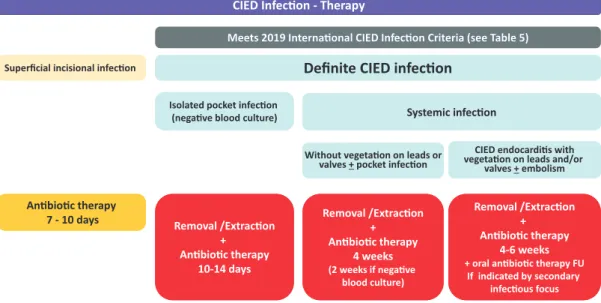
DIAGNOSIS OF CARDIAC IMPLANTABLE
ELECTRONIC DEVICE INFECTIONS AND RELATED COMPLICATIONS
Clinical findings
A superficial incisional infection should be differentiated from a pocket infection, as it involves only the skin and the subcutane- ous tissue without communication with the pocket (and hence does not require CIED system extraction) [56,57]. Close monitor- ing of the patient must be pursued in order to recognize early re- currence that may be a sign of a significant pocket infection.
Pocket infectionis defined as an infection limited to the gener- ator pocket. It is clinically associated with local signs of inflam- mation that may be mild and characterized by erythema, warmth, and fluctuation [14,57]. Deformation of the pocket, ad- herence or threatened erosion are often signs of low grade, indo- lent infection. Symptoms and signs of an infected surgical wound may fluctuate and although it can be difficult to recognize initial- ly it is not recommended to take a sample of pocket material.
Once a wound dehiscence occurs, a purulent drainage or a sinus is established, and a pocket infection is clearly present. If the gen- erator or proximal leads are exposed, the device should be con- sidered infected, irrespective of the results of the microbiology.
Material from the pocket may be used for culture, recognizing the potential for contamination. Pocket infections may be associ- ated with lead infections and CIED systemic infections and/or in- fective endocarditis. The actual rates depend on the definitions used in different studies [58].
The diagnosis ofCIED systemic infection and infective endocar- ditis without local infection may be more challenging (Table 5).
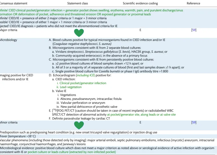
Table 5: Recommendations for diagnosis of CIED infections and/or infective endocarditis: the Novel 2019 International CIED Infection Criteria
Consensus statement Statement class Scientific evidence coding Reference
‘Definite’ CIED clinical pocket/generator infection = generator pocket shows swelling, erythema, warmth, pain, and purulent discharge/sinus formation OR deformation of pocket, adherence and threatened erosion OR exposed generator or proximal leads
‘Definite’ CIED/IE = presence of either 2 major criteria or 1 major + 3 minor criteria
‘Possible’ CIED/IE = presence of either 1 major + 1 minor criteria or 3 minor criteria
‘Rejected’ CIED/IE diagnosis = patients who did not meet the aforementioned criteria for IE
Major criteria E [59]
Microbiology A. Blood cultures positive for typical microorganisms found in CIED infection and/or IE (Coagulase-negative staphylococci,S. aureus)
B. Microorganisms consistent with IE from 2 separate blood cultures:
a. Viridans streptococci,Streptococcus gallolyticus(S. bovis), HACEK group,S. aureus; or b. Community-acquired enterococci, in the absence of a primary focus
C. Microorganisms consistent with IE from persistently positive blood cultures:
a. >_2 positive blood cultures of blood samples drawn >12 h apart; or
b. All of 3 or a majority of >_4 separate cultures of blood (first and last samples drawn >_1 h apart); or c. Single positive blood culture forCoxiella burnetiior phase I IgG antibody titre >1:800
Imaging positive for CIED infections and/or IE
D. Echocardiogram (including ICE)positive for:
a. CIED infection:
i. Clinical pocket/generator infection ii. Lead-vegetation
b. Valve IE i. Vegetations
ii. Abscess, pseudoaneurysm, intracardiac fistula iii. Valvular perforation or aneurysm
iv. New partial dehiscence of prosthetic valve
E. [18F]FDG PET/CT (caution should be taken in case of recent implants) or radiolabelled WBC SPECT/CT detection of abnormal activityat pocket/generator site, along leads or at valve site F. Definite paravalvular leakage by cardiac CT
Minor criteria E [59]
a. Predisposition such as predisposing heart condition (e.g. new onset tricuspid valve regurgitation) or injection drug use b. Fever (temperature >38C)
c. Vascular phenomena (including those detected only by imaging): major arterial emboli, septic pulmonary embolisms, infectious (mycotic) aneurysm, intracranial haemorrhage, conjunctival haemorrhages, and Janeway’s lesions
d. Microbiological evidence: positive blood culture which does not meet a major criterion as noted above or serological evidence of active infection with organism consistent with IE orpocket culture or leads culture (extracted by non-infected pocket)
Based on merging of the modified Duke and ESC 2015 Guidelines criteria, see text [59,60]. Green text refers to CIED-related infection criteria.
CIED: cardiac implantable electronic device; CT: computerized tomography; E: expert opinion; ICE: intracardiac echocardiography; IE: infective endocarditis; M:
meta-analysis; O: observational studies; R: randomized trials; SPECT: single-photon emission tomography; WBC: white blood cell.
Downloaded from https://academic.oup.com/ejcts/article/57/1/e1/5625578 by 81728827 user on 19 January 2022


![Table 7: Recommendations for diagnosis of CIED infections by imaging [59]](https://thumb-eu.123doks.com/thumbv2/9dokorg/998580.61823/13.918.83.841.130.760/table-recommendations-diagnosis-cied-infections-imaging.webp)
![6 weeks is reasonable (Table 9, Fig. 3). If oral suppressive therapy is planned, antibiotic therapy should be chosen according to cul-ture results but evidence-based recommendations cannot be made [103, 118]](https://thumb-eu.123doks.com/thumbv2/9dokorg/998580.61823/16.918.82.837.132.707/reasonable-suppressive-therapy-planned-antibiotic-according-evidence-recommendations.webp)