Cite this article as: Kéri, O., Kócs, L., Hórvölgyi, Z., Baji, Zs., László, K., Takáts, V., Erdélyi, Z., Szilágyi, I. M. "Photocatalytically Active Amorphous and Crystalline TiO2 Prepared by Atomic Layer Deposition", Periodica Polytechnica Chemical Engineering, 63(3), pp. 378–387, 2019. https://doi.org/10.3311/PPch.13873
Photocatalytically Active Amorphous and Crystalline TiO
2Prepared by Atomic Layer Deposition
Orsolya Kéri1*, Lenke Kócs2, Zoltán Hórvölgyi2, Zsófia Baji3, Krisztina László2, Viktor Takáts4, Zoltán Erdélyi5, Imre Miklós Szilágyi1
1 Department of Inorganic and Analytical Chemistry, Faculty of Chemical Technology and Biotechnology, Budapest University of Technology and Economics, H-1111 Budapest, Szt. Gellért tér 4., Hungary
2 Department of Physical Chemistry and Materials Science, Faculty of Chemical Technology and Biotechnology, Budapest University of Technology and Economics, H-1111 Budapest, Budafoki út 6-8., Hungary
3 Hungarian Academy of Sciences, Centre for Energy Research, Institute for Technical Physics, H–1121 Budapest, Konkoly–Thege út 29–33., Hungary
4 Hungarian Academy of Sciences, Institute for Nuclear Research (Atomki), H–4026 Debrecen, Bem tér 18/c., Hungary
5 Department of Solid State Physics, Faculty of Sciences and Technology, University of Debrecen, H–4026 Debrecen, Bem tér 18/b., Hungary
* Corresponding author, e-mail: keri.orsolya@mail.bme.hu
Received: 12 February 2019, Accepted: 03 March 2019, Published online: 26 March 2019
Abstract
In this work, the photocatalytic properties of amorphous and crystalline TiO2 deposited on oxide and polymer nanoparticles by atomic layer deposition (ALD) were studied. Beside TiO2, as reference, both ALD grown amorphous Al2O3 and crystalline ZnO layers were also examined. When choosing the carrier, the priority was that it had no effect on the photocatalytic activity of TiO2; therefore the oxide layers were deposited on SiO2 and poly(methyl-methacrylate) (PMMA) nanoparticles. The amorphous SiO2 particles were synthetized by the Stöber method, while the PMMA particles were prepared by emulsion polymerization. Both the bare and core/shell composite nanoparticles were investigated by SEM-EDX, TEM, FT-IR, and XRD. Finally, the photocatalytic activity of PMMA, SiO2 and the core/shell nanoparticles was measured by the decomposition of methylene orange, monitored by UV-Vis spectroscopy. Based on the results, the SiO2 was uniformly coated with the deposited oxide films, while in the case of the PMMA a contiguous web-like polymer/TiO2 matrix was formed. During the photocatalytic reactions, the amorphous Al2O3 was not active, while the crystalline ZnO and TiO2 showed good photocatalytic activity. The amorphous TiO2 deposited by ALD on the SiO2 and PMMA nanoparticles had smaller, but a clearly detectable photocatalytic effect on the photodegradation of methylene orange.
Keywords
titanium-dioxide, amorphous, crystalline, atomic layer deposition, photocatalysis
1 Introduction
It has long been an important issue how the energy of the sun- light can be effectively converted into chemical energy [1], and in recent years the interest in semiconductor oxide pho- tocatalysts such as WO3, ZnO, and TiO2 has grown expo- nentially [2]. These photocatalysts are widely used in water purification [3, 4], decomposition of organic contaminants [5–7] or in the preparation of self-cleaning surfaces [8, 9].
TiO2 is the most widespread out of the semiconductor oxides in these fields, due to its low cost, good physical and chemi- cal stability and being environmentally friendly.
TiO2 has three crystalline forms: anatase, rutile, and brookite. Since the 1960s all forms of crystalline TiO2
have been thoroughly investigated in photocatalytic reac- tions, e.g. in the splitting of water and in many other reac- tions [10, 11]. It was revealed that generally anatase TiO2 was more active than rutile [12], while brookite was not investigated thoroughly due to the difficulties in obtaining it as a pure phase. In some cases it was proved that brook- ite could have even higher photocatalytic activity than anatase [13]; however, its application is hindered, thanks to the complicated and relatively expensive preparations methods [14]. It has also been found that the photocatalytic activity of crystalline TiO2 can be increased and shifted to visible light by doping it with heteroatoms (e.g. C, N,
S) [15, 16] and other metal atoms [17, 18] or by using it in composites with different semiconductor oxides [19–21].
Amorphous TiO2 in itself is considered not to have any photocatalytic property, because the defects in the crys- tal structure can serve as recombination centers [22].
Nevertheless, it was observed that if magnetron sputtered amorphous TiO2 was coupled with other semiconductor oxides, the amorphous material alone was inactive, but the composites showed photo-reactivity and the response could be expanded to the visible region [23]. Recently it was also discovered that amorphous TiO2 grown by atomic layer deposition (ALD) on lotus leaves could show some photo- catalytic activity [24]. ALD is a self-limiting, sequential thin film deposition method; owing to the alternating sur- face-controlled reactions in the ALD reactor the growth of thin films from the gas phase is possible within nanometer range precision [25–27]. This technique allows the coating of flat surfaces, nanoparticles and even nanofibers [28, 29]
with amorphous or crystalline oxide layers depending on the deposition temperature and among other applications these core/shell nanocomposites can be used as photocatalysts.
Hence, we investigated ALD grown amorphous and crystalline TiO2, deposited onto SiO2 and PMMA nanoparticles and compared the photocatalytic properties.
SiO2 and PMMA were chosen as carriers, because they did not affect the course of the photocatalytic reactions, and hence the efficiency was only dependent on the TiO2 shell. First, the carrier particles were prepared, i.e. the SiO2 by the Stöber method, while the PMMA nanoparti- cles by emulsion polymerization. After that, the TiO2 was deposited onto them, and as a reference, amorphous Al2O3 and crystalline ZnO layers were also grown on the SiO2 nanoparticles by ALD. All samples were investigated by SEM-EDX, TEM, FT-IR, and XRD. Finally, the photocat- alytic activities were measured by the decomposition of methylene orange, monitored by UV-Vis spectroscopy.
2 Experimental
The SiO2 nanoparticles were prepared by the Stöber method [30,31] from tetraethyl orthosilicate (TEOS).
8 cm3 TEOS was added to a mixture of 200 cm3 ethanol and 12 cm3 25 % ammonium hydroxide. The solution was stirred for 24 h at room temperature, during which in var- ious hydrolysis and polycondensation steps SiO2 nanopar- ticles were formed.
The poly(methyl methacrylate) (PMMA) nanopar- ticles were prepared by emulsion polymerization [32]
from methyl methacrylate monomer, using potassium
peroxodisulfate as the initiator and sodium lauryl sulfate as the emulsifier. From the methyl methacrylate 25.81 g was mixed with 180 g distilled water, 0.18 g sodium lauryl sulfate, and 0.18 g potassium peroxodisulfate.
The reaction mixture was continuously stirred at 80 °C under nitrogen atmosphere. The completion of the reac- tion was indicated by the disappearance of the character- istic odor of the monomer. After cooling the mixture to room temperature the polymer nanoparticles were precip- itated with the addition of 400 cm3 1 M HCl solution. The SiO2 and PMMA particles were separated from the liquid phases by centrifuging (3000 rpm, 10 min), then those were washed 3 times with distilled water and dried in air at room temperature.
The deposition of ALD thin films on the nanoparti- cles was done in a Picosun SUNALE R-100 reactor. The overall pressure in the reactor chamber was around 10 mbar during all the reactions. The TiO2 films were pre- pared using titanium tetraisopropoxide (TTIP) and H2O as precursors. Before coating the nanoparticles, it had to be determined from what deposition temperature the pro- duced TiO2 layer was crystalline. For this several deposi- tions were done onto Si wafers at different temperatures from 80 °C to 300 °C (80 °C, 160 °C, 200 °C, 250 °C, 300 °C). In this case, the precursor pulse time was 0.1 s for both TTIP and H2O with a 3 s purge after the TTIP and a 4 s purge after the H2O pulses. The TiO2 was deposited in 1000 cycles. After this, the preparation of the TiO2 lay- ers was done at 80 °C and 300 °C on the SiO2 and only at 80 °C on the PMMA particles. Beside the TiO2 coatings, as reference, Al2O3 and ZnO were also grown at 80 °C and 250 °C. The Al2O3 films were synthetized from trimeth- ylaluminium (TMA) and H2O as precursors, while ZnO from diethylzinc (DEZ) and H2O. In the preparation of the nanocomposites, the TTIP, DEZ, TMA and H2O pulse times were all 0.5 s. The purge times were uniformly 15 s after the precursors. The TiO2 was deposited in 250, while the Al2O3 and ZnO in 100 cycles.
The composition of the samples was studied with energy dispersive X-ray (EDX) analysis in a JEOL JSM- 5500LV scanning electron microscope. The measure- ments were done at 20 kV voltage. Before the measure- ment, the nanocomposites were coated with a thin Au/Pd layer in a sputter coater.
X-ray photoelectron spectra (XPS) were taken on a PHOIBOS HSA3500 100 R6 device. Before the measure- ment the powders were pressed to obtain pellets, the sur- face of the pellets were argon sputter cleaned.
The XRD patterns were recorded with a PANalytical X’pert Pro MPD X-ray diffractometer using Cu Kα irradiation.
The FTIR spectra of the composites were measured by a Biorad Excalibur Series FTS 3000 FTIR spectrophotom- eter between 400 and 4000 cm-1 in KBr pellets.
TEM images were taken by an FEI Morgagni 268 TEM microscope.
Nitrogen adsorption/desorption isotherms were mea- sured at -196 °C with a Nova2000e (Quantachrome) com- puter-controlled apparatus. The apparent surface area (SBET) was calculated using the Brunauer–Emmett–
Teller (BET) model [33].
The photocatalytic properties of the samples were stud- ied by decomposing methyl orange (MO) dye. 1 mg of the nanocomposites was put into 3 cm3 aqueous 0.04 mM MO solution, kept in the dark for 1 h for adsorbing the dye, then illuminated by two parallel UV backlights (Osram black- light UV-A, 18 W) that were placed 5 cm from the sam- ples on either side. The measured wavelength of the lamp is between 350-390 nm (maximum intensity at 375 nm) (Fig. 1), and the estimated power at the samples is 0.5 W.
The UV-Vis spectra of the MO solutions were measured every 30 minutes during the photocatalytic reactions by a Jasco V-550 UV-VIS spectrophotometer. The relative absorbance values, which were plotted versus time, were determined at the 464 nm absorption peak. As a reference the samples were compared to P25 TiO2 (Aeroxide) mea- sured under the same conditions.
3 Result and Discussion
Before the preparation of the composites, the ALD depo- sition temperature was determined in order to grow both amorphous and crystalline TiO2. The layers were
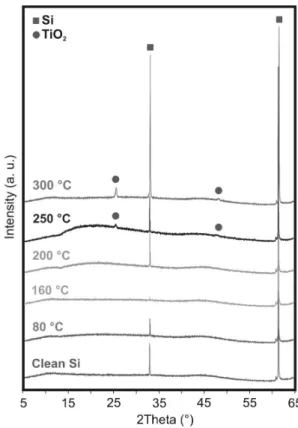
synthetized onto Si wafers and the crystallinity was checked by XRD. On Fig. 2 it is visible that till 200 °C only the peaks referring to crystalline Si are visible (33° and 62°, ICDD 00-040-0932), therefore the TiO2 layer is amor- phous, but from 250 °C, the pattern of anatase TiO2 (25°
and 48°, ICDD 01-083-2243) appears on the diffractogram.
Based on these findings, later the deposition of TiO2 onto the nanoparticle substrates was done at 80 °C and 300 °C in order to be able to compare the properties of the amorphous layers with that of the crystalline ones. The reference Al2O3 and ZnO layers were grown at 80 °C and 250 °C.
The preparation of SiO2 nanoparticles by sol-gel method and the synthesis of the PMMA particles by emul- sion polymerization were successful. It can be seen in the TEM images (Fig. 3a-b and Fig. 4a-b) that in both cases the particles were spherical; the SiO2 nanoparticles had a diameter of 100-150 nm (Fig. 3 a-b), while the size of the PMMA particles was 50-100 nm (Fig. 4 a-b).
After the deposition, the composites were investigated with TEM again (Fig. 3 and Fig. 4). In the case of the SiO2 substrate (Fig. 3 c-h), it was visible that core/shell struc- tured nanoparticles were successfully prepared, the thick- ness of the deposited oxide layers was about 10-20 nm in all samples. This showed that the growth-per-cycle (GPC) [34]
value for these oxides vary greatly, i.e. achieving the same
Fig. 2 XRD results of TiO2 deposited at different temperatures onto Si wafers
Fig. 1 Emittance of the UV blacklight source
thickness for the TiO2 layer as the Al2O3 and ZnO required 2.5 times as many cycles. The TiO2 and Al2O3 formed uni- form and even coatings, while the ZnO layer consisted of smaller, aggregated particles. It was visible in the TEM images that the PMMA particles (Fig. 4 c) softened during the preparation of the composites, and a contiguous matrix was formed from the polymer and the oxide, which could have been caused by the exoterm nature of the ALD reac- tions. Therefore not core/shell PMMA/TiO2 particles, but a PMMA/TiO2 web-like nanostructure was obtained.
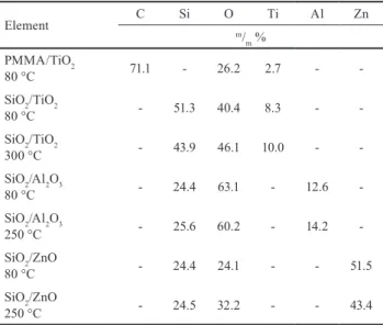
The EDX results (Table 1) also proved that the ALD depositions were successful; Ti, Al, and Zn could be found in the samples. The amount of Al2O3 grown at different temperatures was the same, but at 250 °C smaller amount of ZnO was deposited than at 80 °C. This can be explained by that at 250 °C the upper limit of the ZnO ALD window
was reached [35]. The SiO2/TiO2 nanocomposite sample prepared at higher temperature contained more TiO2 than the 80 °C composite, while when the PMMA was used as carrier, the amount of deposited TiO2 was the lowest. This can be explained by the presence of less functional groups on the surface of PMMA, than on the SiO2.
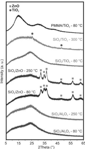
From the XRD patterns (Fig. 5) it can be seen that after the deposition the TiO2 is amorphous when deposited at 80 °C and crystalline at 300 °C, but the Al2O3 is amor- phous and the ZnO is crystalline (zincite, ICDD 00-036- 1451) at both temperatures (80 °C and 250 °C). Based on the Scherrer-equation the crystallite size of the ZnO was about 20 nm, which was in accordance with the ZnO par- ticle size on the TEM images. This was determined for the crystalline TiO2 layer as well, but the calculated value of about 20 nm is rather an estimate for the thickness of the
Fig. 3 TEM images of a-b) the SiO2 nanoparticles and the SiO2/oxide nanocomposites; c) SiO2/TiO2 (80 °C), d) SiO2/TiO2 (300 °C), e) SiO2/ Al2O3 (80 °C), f) SiO2/ Al2O3 (250 °C), g) SiO2/ZnO (80 °C), h) SiO2/
ZnO (250 °C)
Table 1 Composition of the core/shell nanoparticles measured by EDX
Element C Si O Ti Al Zn
m/m % PMMA/TiO2
80 °C 71.1 - 26.2 2.7 - -
SiO2/TiO2
80 °C - 51.3 40.4 8.3 - -
SiO2/TiO2
300 °C - 43.9 46.1 10.0 - -
SiO2/Al2O3
80 °C - 24.4 63.1 - 12.6 -
SiO2/Al2O3
250 °C - 25.6 60.2 - 14.2 -
SiO2/ZnO
80 °C - 24.4 24.1 - - 51.5
SiO2/ZnO
250 °C - 24.5 32.2 - - 43.4
Fig. 4 TEM images of a-b) the PMMA nanoparticles and c) the PMMA/
TiO2 nanocomposites
TiO2 film, since it was a continuous layer, and not made up by individual particles like the ZnO shell. In the case of amorphous TiO2 during the TEM measurements the electron diffraction was investigated as well to confirm that the samples did not contain nanocrystalline domains.
There were no observable electron diffraction patterns, thus these samples were completely amorphous.
In the FTIR spectra (Fig. 6) of the samples containing SiO2, the peaks at 3410 cm-1 and 1634 cm-1 correspond to the vibrations of the surface O-H groups and the absorbed water. The 949 cm-1 is the stretching vibration of the Si-OH bond and the peaks at 1099 cm-1, 799 cm-1, 468 cm-1 are the stretching, bending and out of plane rocking of Si-O and Si-O-Si bonds [36, 37]. In the spectra of the PMMA, the 3441 cm-1 peak refers again to the adsorbed water. The peaks at 2997 cm-1 and 2951 cm-1 can be assigned to the stretching, the 1449 cm-1 peak to the bending vibrations of C-H bonds.
The 1730 cm-1 peak can indicate the presence of the acrylate carboxyl group, while the 750 cm-1 one is referring to the α-methyl groups. The 1242 cm-1 and 1150 cm-1 peaks can be attributed to the C-O-C stretching vibrations. The bands
at 989 cm-1 and 841 cm-1 are characteristic PMMA absorp- tion vibrations [38, 39]. The TiO2, ZnO and Al2O3 lattice vibrations are expected below 1000 cm-1. On the spectra, the slight shift in the position of the peaks between 1000- 400 cm-1 and the changes in the ratio of intensity can refer to the presence of the ALD deposited oxide.
For the samples containing TiO2 some further measure- ments were carried out. The specific surface area of the samples was determined based on nitrogen adsorption/
Fig. 5 XRD measurement of the core/shell nanocomposites
Fig. 6 FTIR measurement of the pure nanoparticles and core/shell nanocomposites
desorption isotherms (Table 2). After the deposition, for the PMMA/TiO2 composite the surface area decreased slightly, which can be attributed to the change in the shape of the PMMA substrate. In the case of the SiO2/TiO2 composites, at 80 °C the surface area did not differ much from the pure SiO2 substrate, while after the 300 °C deposition there was a con- siderable decrease. This was possibly due to the sintering of the SiO2/TiO2 particles at the higher deposition temperature.
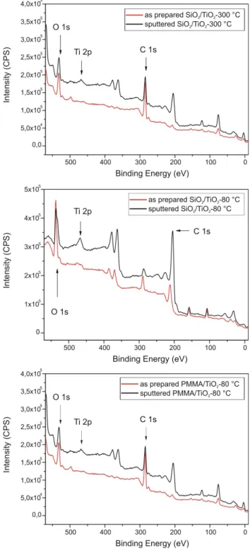
Also the chemical composition of the ALD TiO2 layer was further investigate by XPS measurements (Fig. 7), for this the Ti 2p peaks were decomposed by a standard non- linear least-squares fitting method. Based on the results, next to TiO2, Ti was present in the form of Ti2O3 suboxide phase as well on the surface of the sample. The formation of this phase on ALD layers and other TiO2 samples was already described in the literature, and in some cases it was found to be thermodynamically favored [40, 41].
The result of the photocatalytic measurements (Fig. 8) of the composites containing Al2O3 and ZnO have met the expectations. The amorphous Al2O3 layer had no photo- catalytic property (Fig. 8a), i. e. the SiO2/Al2O3 prepared both at 80°C and 250 °C had the same effect as the sub- strate SiO2, while the crystalline ZnO layer had good pho- tocatalytic properties (Fig. 6b). The SiO2/ZnO compos- ite that was synthetized at 80 °C had a higher efficiency, because it contained more crystalline ZnO. The SiO2/crys- talline TiO2 sample (Fig. 8c) had similar activity as the SiO2/ZnO samples. Interestingly this sample had a very low specific surface area, but a good photocatalytic effi- ciency. The SiO2/amorphous TiO2 core/shell nanoparticles had low activity; however, it clearly had higher decompo- sition rate than the SiO2 substrate, pointing to a small, but observable photocatalytic effect of amorphous TiO2. In the case of PMMA/amorphous TiO2 nanocomposite (Fig. 8d), the small, but detectable photocatalytic property of amor- phous TiO2 was even more visible. This was a surprising
effect, since previously it was only observed when charge separation was possible between the substrate and the coating [24, 42]. The photocatalytic properties of the two samples that had the highest efficiency (SiO2/ZnO - 80 °C, SiO2/TiO2 - 300 °C) were compared to P25 TiO2 (Fig. 9a).
Both samples that were synthetized by ALD had a higher photocatalytic activity than the P25 TiO2 measured under the same conditions. The apparent reaction rate constants,
Table 2 Specific surface area of the pure nanoparticles and the samples containing TiO2
Sample s(BET)
m2/g
PMMA 56
PMMA/TiO2
80 °C 50
SiO2 14
SiO2/TiO2
80 °C 17
SiO2/TiO2
300 °C 1
Fig. 7 XPS results of the samples containing TiO2
which were indicated by the slope of the fitted lines on Fig. 9b, were determined assuming pseudo first order reac- tion kinetics. The reaction rate constants in the case of the SiO2/ZnO - 80 °C, SiO2/TiO2 - 300 °C samples were more than two times higher than that of the P25 TiO2.
4 Conclusion
The photocatalytic properties of amorphous and crystalline TiO2 thin films deposited by atomic layer deposition (ALD) were studied. With ALD the amorphous and crystalline lay- ers could be prepared with the same morphology and their
Fig. 8 Photocatalytic efficiency of the prepared nanocomposites
Fig. 9 a) Photocatalytic efficiency of the SiO2/ZnO – 80 °C and SiO2/TiO2 – 300 °C compared to P25 TiO2; b) kinetics of the photocatalytic reactions
References
[1] Grätzel, M. "Photoelectrochemical Cells", Nature, 414(6861), pp. 338–344, 2001.
https://doi.org/10.1038/35104607
[2] Hoffmann, M. R., Martin, S. T., Choi, W., Bahnemann, D. W.
"Environmental Applications of Semiconductor Photocatalysis", Chemical Reviews, 95(1), pp. 69–96, 1995.
https://doi.org/10.1021/cr00033a004
[3] Baruah, S., Najam, M., Joydeep, K. "Perspectives and Applications of Nanotechnology in Water Treatment", Environonmental Chemistry Letters, 14(1), pp. 1–14, 2016.
https://doi.org/10.1007/s10311-015-0542-2
[4] Sreeja, S., Shetty, K. V. "Photocatalytic Water Disinfection under Solar Irradiation by Ag@TiO2 Core-Shell Structured Nanoparticles", Solar Energy, 157(15), pp. 236–243, 2017.
https://doi.org/10.1016/j.solener.2017.07.057
[5] Fujishima, A., Zhang, X., Tryk, D. "TiO2 Photocatalysis and Related Surface Phenomena", Surface Science Reports, 63(12), pp. 515–582, 2008.
https://doi.org/10.1016/j.surfrep.2008.10.001
[6] Jallouli, N., Pastrana-martínez, L. M., Ribeiro, A. R., Moreira, N. F.
F., Faria, J. L., Hentati, O., Silva, A. M. T., Ksibi, M. "Heterogeneous Photocatalytic Degradation of Ibuprofen in Ultrapure Water, Municipal and Pharmaceutical Industry Wastewaters Using a TiO2/UV-LED System", Chemical Engineering Journal, 334(15), pp. 976–984, 2017.
https://doi.org/10.1016/j.cej.2017.10.045
[7] Réti, B., Kiss, G. I., Gyulavári, T., Baan, K., Magyari, K., Hernadi, K. "Carbon Sphere Templates for TiO2 Hollow Structures:
Preparation, Characterization and Photocatalytic Activity", Catalysis Today, 284(15), pp. 160–168, 2017.
https://doi.org/10.1016/j.cattod.2016.11.038
[8] Krishna, M. G., Vinjanampati, M., Purkayastha, D. D. "Metal Oxide Thin Films and Nanostructures for Self-Cleaning Applications : Current Status and Future Prospects", The European Physical Journal, Applied Physics, 62(3), pp. 1–12, 2013.
https://doi.org/10.1051/epjap/2013130048
[9] Gholami, A., Alemrajabi, A. A.; Saboonchi, A. "Experimental Study of Self-Cleaning Property of Titanium Dioxide and Nanospray Coatings in Solar Applications", Solar Energy, 157(15), pp. 559–565, 2017.
https://doi.org/10.1016/j.solener.2017.08.075
[10] Schneider, J., Matsuoka, M., Takeuchi, M., Zhang, J., Horiuchi, Y., Anpo, M., Bahnemann, D. W. "Understanding TiO2 Photocatalysis : Mechanisms and Materials", Chemical Reviews, 114(19), pp.
9919–9986, 2014.
https://doi.org/10.1021/cr5001892
[11] Hashimoto, K., Irie, H., Fujishima, A. "TiO2 Photocatalysis: A Historical Overview and Future Prospects", Japanese Journal of Applied Physics, 44(12), pp. 8269–8285, 2005.
https://doi.org/10.1143/JJAP.44.8269
photocatalytic properties could be investigated in compar- ison with each other. As carrier SiO2 and PMMA nanopar- ticles were chosen, because those did not interact with the TiO2 during photocatalytic reactions. About 20 nm thick TiO2 layers were grown at different deposition temperatures (80 °C, 300 °C); and as reference, the SiO2 nanoparticles were covered with amorphous Al2O3 and crystalline ZnO as well at 80 °C and 250 °C. The TEM images revealed that the SiO2 nanoparticles were uniformly coated with the depos- ited oxide films and the PMMA formed a web-like struc- ture with the TiO2. During the photocatalytic reactions, the amorphous Al2O3 was not active, while the crystalline ZnO and TiO2 showed good photocatalytic activity. The amor- phous TiO2 deposited by ALD on the SiO2 and PMMA had smaller, but clearly detectable photocatalytic effect. This was an unexpected and previously not described phenom- enon. For this behavior we assume three possible reasons:
(1) the coating interacts with the substrate during the photo- catalytic reactions, (2) the samples contain nanocrystalline domains or (3) there is a small amount of dopant C atoms left over from the ALD precursor (TTIP) in the TiO2 layers.
In our case, between the TiO2 and chosen carriers (amor- phous SiO2 and PMMA) there is no possibility for charge separation, and it was also confirmed by XRD and elec- tron diffraction measurements that the samples were com- pletely amorphous. A detailed computational chemistry
investigation could provide useful answers to these ques- tions; however, that is beyond the scope of the present work.
According to the results, the synthesis of amorphous TiO2 layers does not require high deposition temperature, so the films might be grown by ALD on heat sensitive, highly structured substrates and used for e.g. in the preparation of self-cleaning surfaces.
Acknowledgements
I. M. Szilágyi thanks for a János Bolyai Research Fellowship of the Hungarian Academy of Sciences and an ÚNKP-18-4- IV-BME-238 grant supported by the ÚNKP-17-4-IV New National Excellence Program of the Ministry of Human Capacities, Hungary. The research was financed by the Higher Education Institutional Excellence Programme of the Ministry of Human Capacities in Hungary, within the framework of the Energetics thematic programme of the University of Debrecen. An OTKA PD-109129 grant and a K 124212 grant are acknowledged. The research within project No. VEKOP-2.3.2-16-2017-00013 was supported by the European Union and the State of Hungary, co-fi- nanced by the European Regional Development Fund.
József Hári and Levente Kárpáti (Budapest University of Technology and Economics, Department of Physical Chemistry and Materials Science) are acknowledged for their support in PMMA nanoparticle preparation.
[12] Luttrell, T., Halpegamage, S., Tao, J., Kramer, A., Sutter, E., Batzill, M. "Why Is Anatase a Better Photocatalyst than Rutile?
- Model Studies on Epitaxial TiO2 Films", Scientific Reports, 4(4034), pp. 1–8, 2014.
https://doi.org/10.1038/srep04043
[13] Kandiel, T. A., Robben, L., Alkaim, A., Bahnemann, D. "Brookite versus Anatase TiO2 Photocatalysts : Phase Transformations and Photocatalytic Activities", Photochemical and Photobiological Sciences, 12(4), pp. 602–609, 2013.
https://doi.org/10.1039/c2pp25217a
[14] Di Paola, A., Bellardita, M., Palmisano, L. "Brookite, the Least Known TiO2 Photocatalyst", Catalysts, 3(1), pp. 36–73, 2013.
https://doi.org/10.3390/catal3010036
[15] Khang, N. C., Minh, N. Van, Yang, I. "Synthesis and Characterization of the N-Doped TiO2 Photocatalyst for the Photodegradation of Methylene Blue and Phenol", Journal of Nanoscience and Nanotechnology, 11(7), pp. 1–5, 2011.
https://doi.org/10.1166/jnn.2011.4508
[16] Sharotri, N., Sud, D. "Visible Light Responsive Mn-S-Co-Doped TiO2 Photocatalyst- Synthesis, Characterization and Mechanistic Aspect of Photocatalytic Degradation", Separation and Purification Technology, 183, pp. 382–391, 2017.
https://doi.org/10.1016/j.seppur.2017.03.053
[17] Reyes, M. H., Luis, J., Mar, G. "Fe Doped TiO2 Photocatalyst for the Removal of As(III) under Visible Radiation and Its Potential Application on the Treatment of As-Contaminated Groundwater", Materials Research Bulletin, 73, pp. 145–152, 2016.
https://doi.org/10.1016/j.materresbull.2015.08.034
[18] Bashiri, R., Mohamed, N. M., Kait, C. F., Sufian, S., Khatani, M., Hanaei, H. "Effect of Preparation Parameters on Optical Properties of Cu and Ni Doped TiO2 Photocatalyst", Procedia Engineering, 148, pp. 151–157, 2016.
https://doi.org/10.1016/j.proeng.2016.06.506
[19] Szilágyi, I. M., Santala, E., Heikkilä, M., Pore, V., Kemell, M., Nikitin, T., Teucher, G., Firkala, T., Khriachtchev, L., Räsänen, M., Ritala, M., Leskelä, M. "Photocatalytic Properties of WO3/TiO2 Core/Shell Nanofibers Prepared by Electrospinning and Atomic Layer Deposition", Chemical Vapor Deposition, 19(4–6), pp. 149–
155, 2013.
https://doi.org/10.1002/cvde.201207037
[20] Dong, W., Sun, Y., Ma, Q., Zhu, L., Hua, W., Lu, X., Zhuang, G., Zhang, S., Guo, Z., Zhao, D. "Excellent Photocatalytic Degradation Activities of Ordered Mesoporous Anatase TiO2-SiO2 Nanocomposites to Various Organic Contaminants", Journal of Hazardous Materials, 229–230, pp. 307–320, 2012.
https://doi.org/10.1016/j.jhazmat.2012.06.002
[21] Bellardita, M., Addamo, M., Di Paola, A., Marcì, G., Palmisano, L., Cassar, L., Borsa, M. "Photocatalytic Activity of TiO2/SiO2 Systems", Journal of Hazardous Materials, 174(1–3), pp. 707–713, 2010.
https://doi.org/10.1016/j.jhazmat.2009.09.108
[22] Kaur, K., Singh, C. V. "Amorphous TiO2 as a Photocatalyst for Hydrogen Production: A DFT Study of Structural and Electronic Properties", Energy Procedia, 29, pp. 291–299, 2012.
https://doi.org/10.1016/j.egypro.2012.09.035
[23] Huang, J., Liu, Y., Lu, L., Li, L. "The Photocatalytic Properties of Amorphous TiO2 Composite Films Deposited by Magnetron Sputtering", Research on Chemical Intermediates, 38(2), pp. 487–
498, 2011.
https://doi.org/10.1007/s11164-011-0365-0
[24] Szilágyi, I. M., Teucher, G., Härkönen, E., Färm, E., Hatanpää, T., Nikitin, T., Khriachtchev, L., Räsänen, M., Ritala, M., Leskelä, M.
"Programming Nanostructured Soft Biological Surfaces by Atomic Layer Deposition", Nanotechnology, 24(24), ID. 245701, 2013.
https://doi.org/10.1088/0957-4484/24/24/245701
[25] George, S. M. "Atomic Layer Deposition: An Overview", Chemical Reviews 110(1), pp. 111–131, 2010.
https://doi.org/10.1021/cr900056b
[26] Liu, M., Li, X., Karuturi, S. K., Tok, A. I. Y., Fan, H. J. "Atomic Layer Deposition for Nanofabrication and Interface Engineering", Nanoscale, 4(5), pp. 1522–1528, 2012.
https://doi.org/10.1039/c2nr11875k
[27] Kim, H., Lee, H., Maeng, W. J. "Applications of Atomic Layer Deposition to Nanofabrication and Emerging Nanodevices", Thin Solid Films, 517(8), pp. 2563–2580, 2009.
https://doi.org/10.1016/j.tsf.2008.09.007
[28] Boyadjiev, S. I., Kéri, O., Bárdos, P., Firkala, T., Gáber, F., Nagy, Z. K., Baji, Z., Takács, M.; Szilágyi, I. M. "TiO2/ZnO and ZnO/TiO2 Core/Shell Nanofibers Prepared by Electrospinning and Atomic Layer Deposition for Photocatalysis and Gas Sensing", Applied Surface Science, 424(2), pp. 190–197, 2017.
https://doi.org/10.1016/j.apsusc.2017.03.030
[29] Szilágyi, I. M., Nagy, D. "Review on One-Dimensional Nanostructures Prepared by Electrospinning and Atomic Layer Deposition", Journal of Physics-Conference Series, 559, pp. 1–13, 2014.
https://doi.org/10.1088/1742-6596/559/1/012010
[30] Stober, W., Fink, A. "Controlled Growth of Monodisperse Silica Spheres in the Micron Size Range", Journal of Colloid and Interface Science, 26(1), pp. 62–69, 1968.
https://doi.org/10.1016/0021-9797(68)90272-5
[31] Tolnai, G., Agod, A., Kabai-Faix, M., Kovács, A. L., Ramsden, J. J., Hórvölgyi, Z. "Evidence for Secondary Minimum Flocculation of Stöber Silica Nanoparticles at the Air-Water Interface", Journal of Physical Chemistry, 107(40), pp. 11109–11116, 2003.
https://doi.org/10.1021/jp0344949
[32] Chern, C. S. "Emulsion Polymerization Mechanisms and Kinetics", Progress in Polymer Science, 31(5), pp. 443–486, 2006.
https://doi.org/10.1016/j.progpolymsci.2006.02.001
[33] Brunauer, S., Emmett, P. H., Teller, E. "Adsorption of Gases in Multimolecular Layers", Journal of the American Chemical Society, 60(2), pp. 309–319, 1938.
https://doi.org/10.1021/ja01269a023
[34] Puurunen, R. L. "Growth per cycle in atomic layer deposition: A theoretical model", Chemical Vapor Deposition, 9(5), pp. 249–257, 2003.
https://doi.org/10.1002/cvde.200306265
[35] Johnson, R.W., Hultqvist, A., Bent, S.F. "A brief review of atomic layer deposition: From fundamentals to applications", Materials Today, 17(5), pp. 236–246., 2014.
https://doi.org/10.1016/j.mattod.2014.04.026
[36] Shokri, B., Abbasi Firouzjah, M., Hosseini, S. I. "FTIR Analysis of Silicon Dioxide Thin Film Deposited by Metal Organic- Based PECVD", 2015, October. [online] Available at: http://
www.ispc-conference.org/ispcproc/papers/791.pdf [Accessed:
16 January 2019]
[37] Yang, L.-L., Lai, Y.-S., Chen, J. S., Tsai, P. H., Chen, C. L., Chang, C. J. "Compositional Tailored Sol-Gel SiO2–TiO2 Thin Films:
Crystallization, Chemical Bonding Configuration, and Optical Properties", Journal of Materials Research, 20(11), pp. 3141–3149, 2011.
https://doi.org/10.1557/JMR.2005.0393
[38] Khaled, S. M., Sui, R., Charpentier, P. A., Rizkalla, A. S. "Synthesis of TiO2 - PMMA Nanocomposite: Using Methacrylic Acid as a Coupling Agent", Separation and Purification Technology, 23(7), pp. 3988–3995, 2007.
https://doi.org/10.1021/la062879n
[39] Duan, G., Zhang, C., Li, A., Yang, X., Lu, L., Wang, X. "Preparation and Characterization of Mesoporous Zirconia Made by Using a Poly(Methyl Methacrylate) Template", Nanoscale Research Letters, 3(118), pp. 118–122, 2008.
https://doi.org/10.1007/s11671-008-9123-7
[40] Lv, Y., Xu, P., Ren, G., Chen, F., Nan, H., Liu, R., Wang, D., Tan, X., Liu, X., Zhang, H., Chen, Z. "Low-Temperature Atomic Layer Deposition of Metal Oxide Layers for Perovskite Solar Cells with High Efficiency and Stability under Harsh Environmental Conditions", ACS Applied Materials and Interfaces, 10(28), pp. 23927–23937, 2018.
https://doi.org/10.1021/acsami.8b07346
[41] Zhao, X., Selcuk, S., Selloni, A. "Formation and Stability of Reduced TiOx Layers on Anatase TiO2(101): Identification of a Novel Ti2O3 Phase", Physical Review-Materials, 2, ID. 015801, 2018.
https://doi.org/10.1103/PhysRevMaterials.2.015801
[42] Justh, N., Firkala, T., László, K., Lábár, J., Szilágyi, I.M.
"Photocatalytic C60-amorphous TiO2 composites prepared by atomic layer deposition", Applied Surface Science, 419, pp. 497–
502, 2017.
https://doi.org/10.1016/j.apsusc.2017.04.243