Received: April 29, 2019.
Accepted: November 5, 2019.
Pre-published: November 7, 2019.
©2020 Ferrata Storti Foundation
Material published in Haematologica is covered by copyright.
All rights are reserved to the Ferrata Storti Foundation. Use of published material is allowed under the following terms and conditions:
https://creativecommons.org/licenses/by-nc/4.0/legalcode.
Copies of published material are allowed for personal or inter- nal use. Sharing published material for non-commercial pur- poses is subject to the following conditions:
https://creativecommons.org/licenses/by-nc/4.0/legalcode, sect. 3. Reproducing and sharing published material for com- mercial purposes is not allowed without permission in writing from the publisher.
Correspondence:
MARKUS SPERANDIO markus.sperandio@lmu.de Haematologica 2020 Volume 105(7):1845-1856
doi:10.3324/haematol.2019.225722
Check the online version for the most updated information on this article, online supplements, and information on authorship & disclosures:
www.haematologica.org/content/105/7/1845
Ferrata Storti Foundation
L eukocyte recruitment into inflamed tissue is highly dependent on the activation and binding of integrins to their respective ligands, followed by the induction of various signaling events within the cell referred to as outside-in signaling. Src family kinases (SFK) are the central players in the outside-in signaling process, assigning them a critical role for proper immune cell function. Our study investigated the role of SFK on neutrophil recruit- ment in vivo using Hck-/-Fgr-/-Lyn-/-mice, which lack SFK expressed in neu- trophils. We show that loss of SFK strongly reduces neutrophil adhesion and post-arrest modifications in a shear force dependent manner. Additionally, we found that in the absence of SFK, neutrophils display impaired Rab27a- dependent surface mobilization of neutrophil elastase, VLA3 and VLA6 con- taining vesicles. This results in a defect in neutrophil vascular basement membrane penetration and thus strongly impaired extravasation. Taken together, we demonstrate that SFK play a role in neutrophil post-arrest mod- ifications and extravasation during acute inflammation. These findings may support the current efforts to use SFK-inhibitors in inflammatory diseases with unwanted neutrophil recruitment.
Src family kinase-mediated vesicle trafficking is critical for neutrophil basement membrane penetration
Ina Rohwedder,1Angela R.M. Kurz,1Monika Pruenster,1Roland Immler,1 Robert Pick,1Tanja Eggersmann,1Sarah Klapproth,1Jennifer L. Johnson,2 Sergi Masgrau Alsina,1Clifford A. Lowell,3Attila Mócsai,4Sergio D. Catz2 and Markus Sperandio1
1Walter-Brendel-Center of Experimental Medicine, Institute of Cardiovascular Physiology and Pathophysiology, Klinikum der Universität, Ludwig-Maximilians-University Munich, Planegg-Martinsried, Germany; 2Department of Molecular Medicine, The Scripps Research Institute, La Jolla, CA, USA; 3Department of Laboratory Medicine, University of California, San Francisco, CA, USA and 4Department of Physiology, Semmelweis University School of Medicine, Budapest, Hungary
ABSTRACT
Introduction
Chronic inflammatory diseases are an increasing problem in western industrial- ized countries. A hallmark of these disorders is the recruitment of leukocytes from the circulation into affected tissues. This process follows a well-defined cascade of activation and adhesion events starting with the initial capture of leukocytes from the blood stream, followed by rolling along inflamed endothelium.1Both steps are mediated by selectins interacting with glycosylated ligands on leukocytes.2 Through binding of the leukocyte specific integrin LFA1 (αLβ2) to ICAM-1 on endothelial cells, leukocytes slow down their rolling velocity, and in combination with chemokine stimulation, firmly arrest on the endothelial surface. This is fol- lowed by post-arrest modifications, a process characterized by cell spreading, cytoskeleton rearrangements and crawling along the endothelium, that is critical for tight adhesion to the substrate and allows an appropriate spot for extravasation into tissue. Transmigration involves the crossing of the venular wall and the under- lying vascular basement membrane (BM), two steps which are still incompletely understood. Several reports suggest a role for the integrins VLA3 (α3β1) and VLA6 (α6β1) along with neutrophil elastase (NE) in this process.3-5Recently, our group has shown that neutrophils translocate VLA3, VLA6 and NE from internally stored vesicles to the cell surface, to subsequently cross the vascular BM.6The release of these vesicles is initiated by interactions of neutrophils with the inflamed endothe- lium in a PECAM-1/ICAM-1- and CXCL1-dependent manner. Vesicle transport is
mainly regulated by Rab GTPases and their effector pro- teins with Rab27a being the main Rab molecule involved in the secretory machinery of neutrophils.7Rab27a func- tion is mediated by the two effector molecules synapto- tagmin-like protein 1 (JFC1, encoded by Sytl1 in mice) and Munc13-4 (Unc13d).8 Although most of these processes rely on integrin signaling, integrins themselves lack enzy- matic activity. Therefore, numerous proteins are recruited to their intracellular tails, among those Src family kinases (SFK). Their early recruitment during integrin activation assigns SFK a critical role in the so-called outside-in signal- ing process and thus regulation of central signaling path- ways downstream of integrin-receptor ligation.9 Neutrophils express three members of this family, namely Hck, Fgr and Lyn.10 Their function has been intensively studied in SFK single, double (Hck-/-Fgr-/-), and triple knock- out (ko) (Hck-/-Fg-/-Lyn-/-) mice,11 demonstrating a role for SFK in integrin activation12 and their downstream signal- ing. Neutrophils isolated from Hck-/-Fgr-/- mice showed poor spreading, indicating that integrin outside-in signal- ing is impaired.13 Additionally, neutrophil migration into the liver of Hck-/-Fgr-/- mice in an in vivo endotoxemia model is severely reduced.14 Furthermore, Kovács et al.
recently showed that neutrophils exhibit reduced capabil- ity to create a microinflammatory environment, resulting in diminished neutrophil extravasation in a model of autoantibody-induced arthritis and inflammatory blister- ing skin disease.15
Our study aimed to investigate the effect of SFK on leukocyte recruitment in an in vivosetting of acute inflam- mation, focusing on post-arrest modifications and the molecular mechanism of vascular BM penetration. We show that, in the genetic absence of SFK, these two steps are strongly impaired. Adherent Hck-/-Fgr-/-Lyn-/- neutrophils are unable to withstand shear forces and display dimin- ished LFA1 clustering with reduced phosphorylation levels of Paxillin, Cortactin and Syk, suggesting that SFK deple- tion results in severely impaired adhesion strengthening.
In addition, we show that SFK are critical for crossing the vascular BM during neutrophil extravasation by facilitat- ing translocation of VLA3-, VLA6- and NE-containing vesicles to the cell surface. Therefore, SFK are not only important mediators of neutrophil post-arrest modifica- tions, but are also required to breach the vascular BM.
Methods
Animals
Lyz2GFP, SFK-ko (Hck-/-Fgr-/-Lyn-/-) and SFK-ko Lyz2GFPmice were generated as described earlier.16-18C57BL/6 wildtype mice were purchased from Janvier Labs (Saint Berthevin, France). All animal experiments were approved by the Regierung von Oberbayern, Germany (AZ 55.2-1-54-2531-80-76/12 and 55.2-1-54-2532-102- 2017).
Live cell imaging of in vitro laminin digestion
Transmigration of neutrophils was analyzed in μ-Slide mem- brane ibiPore flow chambers (Ibidi, Planegg, Germany) equipped with a 300-nm thick membrane with 5-μm pores, and a subjacent rat-tail collagen gel (1.5 mg/mL) containing 10 μM N-formylme- thionyl-leucyl-phenylalanine (fMLP) as chemoattractant. The upper compartment was coated with Laminin (LN), PECAM-1 and ICAM-1, and, in addition, LN was visualized using an anti-LN antibody conjugated to Alexa Fluor-647 (novusbio, Littleton, CO,
USA). Isolated neutrophils from wildtype and SFK-ko animals were labeled with CellTracker™ Green CMFDA Dye and CellTracker™ Red CMTPX Dye (Thermo Fisher, Waltham, MA, USA), respectively, and distributed into the upper chamber com- partment in a 1:1 ratio. Time-lapse microscopy with an interval of 50 seconds was performed using an upright spinning-disk confocal microscope (Examiner; Zeiss) equipped with a confocal scanner unit CSU-X1 (Yokogawa Electric Corporation, Japan), an EMCCD camera (Evolve; Photometrics), and a x20/1.0 NA water immer- sion objective (Plan Apochromat; Zeiss). 3D images (70 z-stacks with a step size of 2µm) were acquired per time point and ana- lyzed by generating maximum z-projections over time using Slidebook 6.0.8 software (3i) and ImageJ. Interaction strength was analyzed with the MosaicIA interaction plugin of Fiji.
Functional in vitroand in vivo experiments, including intravital microscopy,19peritonitis experiments, flow chamber experiments, vesicle trafficking, flow cytometry, western blot analysis and sta- tistics are described in detail in the Online Supplementary Methods.
Data sharing statement
Original data are available on request.
Results
Src family kinase depletion reduces neutrophil adhesion and extravasation in inflamed postcapillary venules in vivo
We first investigated how loss of neutrophil-expressed SFK influences neutrophil adhesion in TNFα-stimulated cremaster muscle postcapillary venules using intravital microscopy. Interestingly, the absolute number of adher- ent neutrophils was strongly increased compared to wild- type control mice (Online Supplementary Figure S1A).
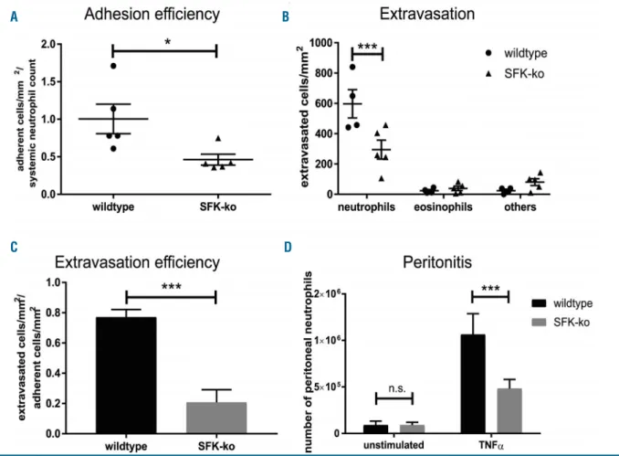
However, after adjusting the number of adherent neu- trophils to the circulating neutrophil count, which was sig- nificantly higher in SFK-ko mice compared to wildtype mice (Online Supplementary Figure S1B and C), the neu- trophil adhesion efficiency calculated as number of adher- ent cells/mm2 divided by the systemic neutrophil count was significantly reduced in SFK-ko mice (0.48±0.04) com- pared to wildtype mice (1.04±0.08) (Figure 1A). This was not due to altered surface expression of rolling and adhe- sion relevant surface proteins including CD18, CD11a, CD11b, CD62L, PSGL1, CXCR2, and CD44 as their sur- face expression was equal between wildtype and SFK-ko neutrophils (Online Supplementary Figure S1D). We there- fore conclude that SFK are critical for neutrophil adhesion in vivo.
Efficient neutrophil extravasation into tissue is required for proper host defense. Recently, Kovács et al. demon- strated in the K/B×N serum-transfer arthritis model that SFK deficiency resulted in lower levels of cytokine produc- tion and release leading to impaired leukocyte extravasa- tion.15 To investigate a cell intrinsic adhesion defect in SFK-ko mice, we injected TNFαinto the mouse scrotum.
This approach circumvents possible effects of a reduced inflammatory environment and enables us to focus on neutrophil recruitment itself. Performing Giemsa staining of TNFα stimulated cremaster muscle whole mounts revealed strongly reduced numbers of extravasated neu- trophils in SFK-ko mice compared to wildtype mice (306.51±43.7 vs. 578.6±72.9, respectively) (Figure 1B and Online Supplementary Figure S1E). Moreover, we calculated the extravasation efficiency as total number of extravasat-
ed leukocytes divided by the total number of adherent leukocytes to differentiate between reduced extravasation due to decreased adhesion versusa specific extravasation defect. Surprisingly, we observed a strongly reduced extravasation efficiency in SFK-ko mice (0.21 in SFK-ko mice vs. 0.77 in wildtype mice) (Figure 1C), indicating that SFK depletion does not only affect neutrophil adhesion, but also interferes with extravasation itself. Finally, we also compared neutrophil cell numbers in the peritoneal cavity 2 hours (h) after TNFα-induced peritonitis (Figure 1D). In SFK-ko animals, peritoneal neutrophil numbers were reduced to 50% compared to wildtype mice (0.42x10⁶ vs. 106, respectively). Taken together, these experiments demonstrate an important role for SFK in neutrophil extravasation.
Adhesion strengthening is severely impaired in Src family kinase-knockout neutrophils in vitro
Our experiments suggest that SFK-deletion in neu- trophils results in an extravasation defect in the presence of the proinflammatory cytokine TNFα. After firm adhe- sion to the inflamed endothelium, neutrophils start to polarize and crawl along the endothelium to find a suit- able spot for extravasation. These changes are highly dependent on β2 integrins LFA1 (αLβ2) and Mac1 (αMβ2)
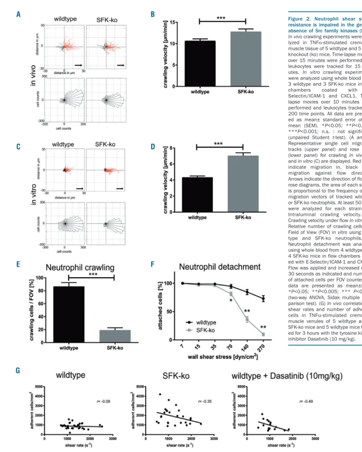
and integrin outside-in signaling.20 We analyzed neu- trophil crawling using time-lapse microscopy in TNFα- stimulated cremaster muscle venules of SFK-ko and wild- type mice. We observed no differences in crawling direc- tion (Figure 2A), but, interestingly, crawling velocity was significantly increased in SFK-ko mice (12.3 µm/min) com- pared to wildtype mice (10.7 μm/min) (Figure 2B), sug- gesting defective adhesion strengthening. Subsequently, we performed ex vivoflow chamber assays using glass cap- illaries coated with E-Selectin/ICAM-1/CXCL1 to mimic the inflamed endothelium. Plots of single cell tracks dis- played more SFK-ko neutrophils crawling in flow direc- tion compared to wildtype neutrophils (Figure 2C).
Crawling velocity was increased in SFK-ko leukocytes (Figure 2D), verifying our in vivoresults. Interestingly, only 20% of SFK-ko neutrophils were able to crawl under flow (Figure 2E and Online Supplementary Mov1), compared to 80% of wildtype cells, suggesting that SFK-ko neutrophils are unable to maintain stable adhesion to the substrate.
Together, these findings demonstrate a decreased capabil- ity of SFK-ko neutrophils to withstand shear forces, indi- cating that SFK are necessary for adhesion strengthening during post-arrest modifications. To further investigate a shear stress dependent adhesion defect in the absence of neutrophil SFK, we conducted detachment assays in the
Figure 1. Src family kinases (SFK) are required for slow rolling, neutrophil adhesion and extravasation. All data are presented as mean± standard error of the mean (SEM). *P<0.05; ***P<0.001; n.s. : not significant (unpaired Student t-test or two-way ANOVA, Sidak multiple comparison test). (A) Adhesion efficiency from wildtype or SFK-knockout (ko) mice: n= 5 wildtype; n=5 SFK-ko mice. (B) In vivoleukocyte extravasation in TNFα-stimulated venules of mouse cremaster muscle.
Differential total cell counts of neutrophils, eosinophils and other cells: n= 4 wildtype; n=5 SFK-ko mice. (C) Extravasation efficiency was calculated by the number of extravasated cells/mm2divided by number of adherent cells/mm2. (D) Total neutrophil numbers after peritoneal lavage of the unstimulated or TNFα–stimulated peritoneal cavity of wildtype and SFK-ko mice: n=7 wildtype and n=6 SFK-ko mice for unstimulated controls and n=6 wild-type and n=6 SFK-ko mice for TNFα.
A B
C D
flow chamber using increasing shear forces. Adherent neutrophils were assessed for each shear stress level and related to the number of adherent neutrophils under low flow conditions (Figure 2F). Loss of SFK resulted in a decrease in shear stress resistant adhesion compared to
wildtype cells. At wall shear stress levels of 140 dyn/cm2, only 36% of SFK-ko leukocytes could adhere to the flow chamber, while 84% of wildtype leukocytes were still attached. To analyze if these findings also apply under in vivo conditions, we correlated in vivo shear rates to
Figure 2. Neutrophil shear stress resistance is impaired in the genetic absence of Src family kinases (SFK).
In vivocrawling experiments were ana- lyzed in TNFα-stimulated cremaster muscle tissue of 5 wildtype and 5 SFK- knockout (ko) mice. Time-lapse movies over 15 minutes were performed and leukocytes were tracked for 15 min- utes. In vitro crawling experiments were analyzed using whole blood from 3 wildtype and 3 SFK-ko mice in flow chambers coated with E- Selectin/ICAM-1 and CXCL1. Time- lapse movies over 10 minutes were performed and leukocytes tracked for 200 time points. All data are present- ed as mean± standard error of the mean (SEM). *P<0.05; **P<0.005;
***P<0.001; n.s. : not significant.
(unpaired Student t-test). (A and C) Representative single cell migration tracks (upper panel) and rose plots (lower panel) for crawling in vivo (A) and in vitro(C) are displayed. Red lines indicate migration in, black lines migration against flow direction.
Arrows indicate the direction of flow. In rose diagrams, the area of each sector is proportional to the frequency of the migration vectors of tracked wildtype or SFK-ko neutrophils. At least 50 cells were analyzed for each strain. (B) Intraluminal crawling velocity. (D) Crawling velocity under flowin vitro.(E) Relative number of crawling cells per Field of View (FOV) in vitro using wild- type and SFK-ko neutrophils. (F) Neutrophil detachment was analyzed using whole blood from 4 wildtype and 4 SFK-ko mice in flow chambers coat- ed with E-Selectin/ICAM-1 and CXCL1.
Flow was applied and increased every 30 seconds as indicated and numbers of attached cells per FOV counted. All data are presented as mean±SEM.
*P<0.05; **P<0.005; *** P<0.001 (two-way ANOVA, Sidak multiple com- parison test). (G) In vivocorrelation of shear rates and number of adherent cells in TNFα-stimulated cremaster muscle venules of 5 wildtype and 5 SFK-ko mice and 5 wildtype mice treat- ed for 3 hours with the tyrosine kinase inhibitor Dasatinib (10 mg/kg).
A B
C D
E
G
F
absolute numbers of adherent cells/mm2in TNFα-stimu- lated post-capillary cremaster muscle venules of SFK-ko and wildtype mice (Figure 2G). We found a similar shear rate dependent decrease in the number of adherent cells/mm2in SFK-ko mice in vivo compared to wildtype mice. Additionally, we used a pharmacological approach and administered the broad-spectrum tyrosine kinase inhibitor Dasatinib before TNFα-stimulation. Dasatinib has been used in various studies as a SFK inhibitor and led to promising results as an immune modulator.21 Similar to the results in SFK-ko mice, we found a decrease in adher- ent cells with increasing shear rates. Importantly, SKF-ko mice receiving Dasatinib did not show any further decrease in adherent cells at high shear stress levels, indi- cating that Dasatinib exerts its observed effects on adhe- sion via inhibition of SFK (Online Supplementary Figure S1F). Taken together, these in vivoand in vitrofindings indi- cate that loss of SFK in neutrophils leads to a shear force dependent inability to firmly adhere to the inflamed endothelium, a prerequisite for subsequent extravasation.
Reduced phosphorylation of cytoskeleton-associated proteins in Src family kinase-deficient neutrophils
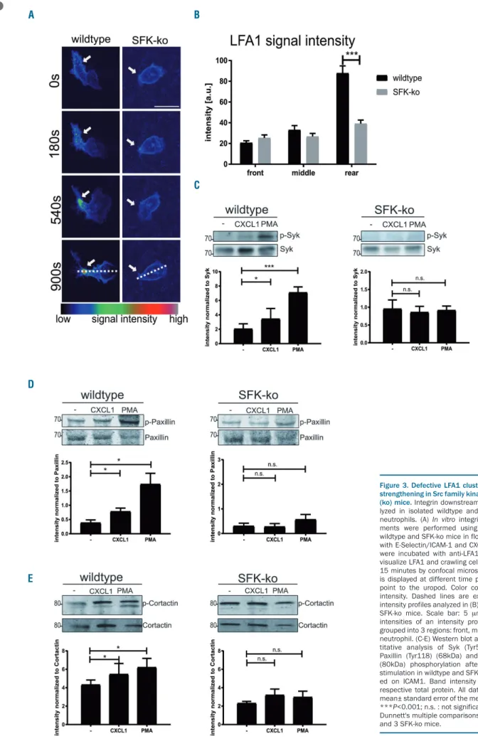
To further elucidate the molecular mechanisms of SFK dependent neutrophil extravasation, we analyzed differ- ent steps of integrin outside-in signaling that occur after integrins bind to their receptor. This includes integrin clus- tering and cytoskeletal rearrangements.22 To analyze inte- grin clustering, we performed time-lapse confocal microscopy and studied LFA1 clustering in SFK-ko and wildtype neutrophils in flow chambers coated with E-selectin/ICAM-1/CXCL1 (Figure 3A and Online Supplementary Mov2). We observed spreading and polariz- ing wild-type neutrophils which showed an accumulating LFA1 signal (clustering) over time at the neutrophil uropod (Figure 3B). In contrast, SFK-deficient neutrophils appeared round and unpolarized with no obvious LFA1 clustering. Several cytoskeleton-associated proteins and signaling molecules are known to be tyrosine phosphory- lated upon integrin ligation.23 We investigated the phos- phorylation and activation of the direct SFK target24,25Syk in SFK-ko and wildtype neutrophils plated on ICAM-1 and then stimulated with CXCL1 or PMA. Western blot analysis of phospho-Syk (Tyr519/520) revealed a strong upregulation of phosphorylation following stimulation of wildtype cells. In contrast, SFK deficiency prevented the upregulation of phosphorylation following stimulation with CXCL1 or PMA (Figure 3C), indicating that Syk acti- vation is defective in the absence of SFK. Moreover, we tested the phosphorylation of the adaptor protein Paxillin, one of the critical proteins for cell adhesion, migration and podosome formation, that is known to be tyrosine phos- phorylated upon β2 integrin activation.26,27 Western blot analysis showed significant upregulation of Paxillin phos- phorylation when stimulated with CXCL1 or PMA in wildtype cells, while no upregulation was detectable in SFK-ko neutrophils (Figure 3D). In addition, we also inves- tigated the tyrosine phosphorylation of Cortactin, which regulates actin branching and therefore cell migration.
Likewise, we observed no significant upregulation of Cortactin Tyr421 phosphorylation upon CXCL1 or PMA stimulation in SFK-ko cell, in contrast to wildtype neu- trophils (Figure 3E). Because during neutrophil adhesion and migration SFK do not selectively signal viaLFA1, but also viaMac1, we additionally analyzed Paxillin phospho-
rylation after plating the cells on Fibrinogen (Online Supplementary Figure S1G). Similar to ICAM1 we observed a stimulus dependent phosphorylation of Paxillin in wild- type neutrophils, which was absent in SFK-ko neu- trophils. Taken together, these experiments show that β2 -integrin clustering and subsequent outside-in signaling is severely impaired in SFK-ko neutrophils.
Src family kinase are indispensable to pass the vascular basement membrane
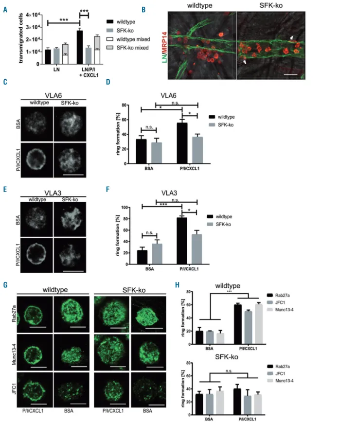
For successful extravasation into inflamed tissue, neu- trophils need to cross the endothelial cell layer and the underlying vascular basement membrane. We aimed to investigate whether SFK are required to perform this last step of extravasation. First, we analyzed transmigration capacity of SFK-ko neutrophils in a transwell assay using CXCL1 as chemoattractant (Online Supplementary Figure S2A). In line with previous results,15we observed no dif- ference in transmigration between SFK-ko neutrophils and wildtype neutrophils. Both groups displayed a proportion- al increase in transmigration with rising CXCL1 concen- trations. As a next step, we coated transwells with laminin-111 (LN1) or a combination of LN1 and PECAM- 1/ICAM-1 and stimulated them with CXCL1 in order to mimic the situation at the vascular BM, as reported previ- ously.6No difference in transmigration through LN alone was detected between wildtype and SFK-ko neutrophils (Figure 4A). Coating with LN1/PECAM-1/ICAM-1 (LN/P/I) together with CXCL1 stimulation induced a strong increase in transmigration of wildtype cells.
However, SFK-ko cells failed to cross the artificial BM.
How exactly neutrophils cross the vascular BM is still a matter of debate, especially, whether the BM is perma- nently modified by extravasating neutrophils. To further address this, we conducted transwell assays as described above, coated with a combination of LN1 and PECAM- 1/ICAM-1 and stimulated with CXCL1. We also used the same total cell number (2x10⁵), but this time mixed SFK- ko x Lyz2GFPneutrophils and wildtype neutrophils in a 1:1 ratio (1x10⁵wildtype and 1x10⁵ SFK-ko x Lyz2GFPneu- trophils). In this manner, transmigrated SFK-ko neu- trophils were identified by their GFP signal. Interestingly, in combination with wildtype neutrophils, SFK-ko neu- trophils were able to overcome the BM to the same extent as wildtype cells, suggesting that wildtype neutrophils facilitate BM penetration by providing exit points for SFK- ko neutrophils (Figure 4A). To confirm these findings in vivo, we performed whole mount staining of TNFα- stimulated cremaster muscles for LN (green) to visualize the BM, and MRP14 (red) to visualize neutrophils, then analyzed the localization of neutrophils by confocal microscopy. In wildtype tissue, we were able to detect local areas of increased extravasation, which is in accor- dance with published literature, describing transmigration hotspots28 (Figure 4B). In contrast, none of these spots could be found in cremaster muscle tissue of SFK-ko mice.
MPR14-positive cells remained located within the vascular compartment and only few extravasated neutrophils could be detected, mainly remaining in close vicinity to the vessel (white arrows). Overall, SFK-ko neutrophils covered less distance away from the vessel wall when compared to wildtype neutrophils (8.0 μm vs. 17.3 μm, respectively) (Online Supplementary Figure S2B). These in vivofindings support our earlier in vitrofindings that SFK are critical for neutrophils to cross the BM.
Figure 3. Defective LFA1 clustering and adhesion strengthening in Src family kinases (SFK)-knockout (ko) mice. Integrin downstream signaling was ana- lyzed in isolated wildtype and SFK-knockout (ko) neutrophils. (A) In vitrointegrin clustering experi- ments were performed using whole blood from wildtype and SFK-ko mice in flow chambers coated with E-Selectin/ICAM-1 and CXCL1. Blood samples were incubated with anti-LFA1-Alexa547 (2D7) to visualize LFA1 and crawling cells were analyzed for 15 minutes by confocal microscopy. LFA1 intensity is displayed at different time points. White arrows point to the uropod. Color code indicates signal intensity. Dashed lines are exemplary for drawn intensity profiles analyzed in (B) n=3 wildtype and 3 SFK-ko mice. Scale bar: 5 μm. (B) Mean signal intensities of an intensity profile of LFA1 signal, grouped into 3 regions: front, middle and rear of the neutrophil. (C-E) Western blot and respective quan- titative analysis of Syk (Tyr519/520) (72kDa), Paxillin (Tyr118) (68kDa) and Cortactin (Tyr421) (80kDa) phosphorylation after CXCL1 and PMA stimulation in wildtype and SFK-ko neutrophils plat- ed on ICAM1. Band intensity was normalized to respective total protein. All data are presented as mean± standard error of the mean (SEM). *P<0.05;
***P<0.001; n.s. : not significant (one-way ANOVA, Dunnett's multiple comparisons test). N=3 wildtype and 3 SFK-ko mice.
A B
C
D
E
Figure 4. Rab27a dependent vesicle transfer is impaired in Src family kinases (SFK)-knockout (ko) neutrophils, leading to an impaired basement membrane pen- etration.(A) Neutrophil transmigration in a transwell assay with or without CXCL1 through filters coated with laminin (LN), or LN, PECAM-1, and ICAM-1 (LN/P/I). All data are presented as mean± standard error of the mean (SEM). *P<0.05; ***P<0.001; n.s. : not significant (two-way ANOVA, Sidak multiple comparison test). N=3 wildtype and 3 SFK-ko x Lyz2GFPmice, transwell assays were performed in duplicates. (B) Maximum projections of confocal microscopic images of venules in TNFα–stimulated cremaster muscle whole mounts from wildtype and SFK-ko mice. Basement membrane was visualized by anti-LN5 (green), neutrophils by anti- MRP14 (red). Surrounding muscle tissue was visualized by bright field microscopy. White arrows point at extravasated, but still attached neutrophils in SFK-ko tissue.
Scale bar: 20 μm. (C) and (E) immunostaining of representative wildtype and SFK-ko neutrophils on BSA- or PECAM-1/ICAM-1/CXCL1–coated (P/I/CXCL1) coverslips for (C) VLA6 and (E) VLA3 analyzed by confocal microscopy. Scale bar: 10 μm. (D) and (F) Quantification of ring formation for VLA6 (D) and VLA3 (F). At least 80 cells from 3 wildtype and 3 SFK-ko mice were analyzed for each condition. (G) Immunostaining of representative wildtype and SFK-ko neutrophils on BSA- or PECAM- 1/ICAM-1/CXCL1–coated coverslips for Rab27a (upper panel), Munc13-4 (middle panel), and JFC1 (lower panel). Scale bar is 5µm. (H) Quantification of ring forma- tion for Rab27a, Munc13-4 and JFC1. At least 80 cells from 3 wildtype and 3 SFK-ko mice were analyzed for each condition. All data are presented as mean±SEM.
*P<0.05; P<0.005; ***P<0.001; n.s. : not significant (two-way ANOVA, Sidak multiple comparison test).
A B
C D
E F
H G
Impaired vesicle transport in the absence of Src family kinase in neutrophils
We and others have previously shown that mobilization of vesicles containing VLA3 and VLA6 to the plasma membrane is an important process during vascular BM penetration.3,4,6 To investigate whether SFK have a role in this process, we analyzed the SFK-dependent transloca- tion of VLA6 and VLA3 to the plasma membrane of SFK- ko and wildtype neutrophils. Bone marrow derived neu- trophils of SFK-ko and wildtype mice were plated on BSA or, again, on a combination of PECAM-1/ICAM-1 and CXCL1 and subsequently stained for VLA6 or VLA3.
Plasma membrane translocation was visualized by confo- cal microscopy as “ring formation” (Figure 4C and E). We observed fewer SFK-ko cells (37%) displaying a ring of VLA6, when plated on PECAM-1/ICAM-1/CXCL1 com- pared to wildtype (55%) (Figure 4D). This observation was also true for VLA3, where only 52% of SFK-deficient neutrophils showed vesicle translocation, compared to 82% of wildtype cells (Figure 4F). To test whether ring for- mation can also be observed in vivo, whole mount cremas- ter muscle stainings were conducted for VLA6, which revealed a similar trend as in vitro. In wildtype whole mounts, extravasated neutrophils displayed a ring-like staining for VLA6, while intravascular neutrophils showed only a diffuse signal. In SFK-ko whole mounts, intravascu- lar and extravasated cells showed no clear peripheral staining (Online Supplementary Figure S2C). Next, we plat- ed wildtype neutrophils on BSA or PECAM-1/ICAM- 1/CXCL1 and stained for SFK. Confocal micrographs revealed diffuse intracellular SFK staining when cells were plated on BSA (Online Supplementary Figure S2D). Upon stimulation with PECAM-1/ICAM-1/CXCL1, we observed a clear SFK signal at the cell border. Again, we quantified SFK distribution and observed a pronounced SFK translocation to the cell periphery when neutrophils were plated on PECAM-1/ICAM-1/CXCL1 (Online Supplementary Figure S2E). These experiments support a critical role for SFK by strongly interfering in vesicle traf- ficking of neutrophils in vitroand in vivo.
Src family kinase regulate Rab27a-dependent vesicle translocation
Vesicle trafficking in neutrophils is mainly regulated by Rab27a GTPases and their corresponding effector pro- teins. In neutrophils, Rab27a works in collaboration with two of its known effectors, JFC1 and Munc13-4, on vesicle trafficking and exocytosis of secretory vesicles and azurophilic granules.8 We analyzed the translocation of Rab27a, JFC1 and Munc13-4 to the cell periphery upon PECAM-1/ICAM-1/CXCL1 stimulation as described above. In wildtype neutrophils, stimulation resulted in translocation of Rab27a and its effectors (Figure 4G).
While 60% of wildtype neutrophils displayed a ring-like structure, only 40.1% showed Rab27a translocation in SFK-ko neutrophils (Figure 4H). Similarly, staining for JFC1 (50.1% vs.29.2%) and Munc13-4 (60.9%vs. 31.3%) revealed an equal trend for wildtype vs. SFK-ko neu- trophils, respectively. Taken together, these experiments show a clear SFK-dependent activation of the secretory machinery in neutrophils.
NE-dependent laminin degradation is defective in Src family kinase-ko neutrophils
Neutrophil extravasation is not only dependent on the
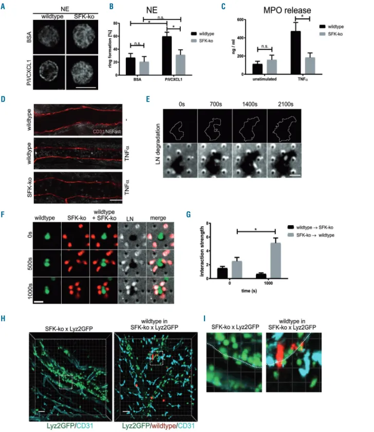
translocation of LN binding β1 integrins, but also on the serine proteinase neutrophil elastase (NE).5,29 We first ana- lyzed the localization of NE in neutrophils plated on either BSA or PECAM-1/ICAM-1/CXCL1 as described above (Figure 5A). Similar to VLA3 and VLA6, we observed an increase in ring formation from 21.9% in BSA stimulated wildtype neutrophils to 59.1% of wildtype neutrophils stimulated on PECAM-1/ICAM-1/CXCL1 (Figure 5B).
This upregulation of vesicle translocation was absent in SFK-ko neutrophils (20.0% for BSA vs. 31.2% for PECAM- 1/ICAM-1/CXCL1 coating). In neutrophils, NE is stored in azurophilic granules together with a variety of other pro- teinases and antimicrobial proteins such as myeloperoxi- dase (MPO). Neutrophils release MPO after stimulation with TNFα.30,31 Thus, we additionally quantified MPO release in the serum of SFK-ko and wildtype mice by ELISA 2 h after TNFα stimulation. While we did not observe upregulation of MPO in the serum of SFK-ko mice 2 h after TNFαstimulation (158.4 ng/mL before vs. 184.0 ng/mL after TNFα stimulation) (Figure 5C), MPO levels were markedly increased in the serum of TNFαstimulated wildtype mice (113.0 ng/mL beforevs. 473.5 ng/mL after TNFα stimulation). This indicates that SFK are required for the release of azurophilic granule content including NE and MPO. Earlier studies revealed that NE is able to pro- teolytically cleave laminins. In addition, NE activates dif- ferent matrix metalloproteinases (MMP) that are involved in matrix degradation.32-34 However, the relevance of these NE functions for neutrophil transmigration are still not completely understood. By applying a NE-fluorescent acti- vatable substrate (NE680FAST) in postcapillary venules with or without TNFα stimulation we were able to inves- tigate NE activity in wildtype and SFK-ko cremaster whole mounts. Compared to unstimulated controls, TNFα stimulation led to a robust NE activity signal in wildtype mice (Figure 5D). As expected, this activity was reduced in SFK-ko venules, indicating that there is reduced NE dependent substrate cleavage in the absence of SFK (Figure 5D).
Next, we investigated whether wildtype neutrophils are able to degrade BM constituents under in vitro conditions.
To this end, we performed transwell experiments in a sys- tem that allows us to image neutrophil crawling and extravasation by spinning disc confocal microscopy.
Neutrophils from wildtype and SFK-ko mice were isolated and stained with different cell trackers (CellTracker™ Red CMTPX Dye and CellTracker™ Green CMFDA Dye,) to image both neutrophil populations in one transmigration chamber. In addition, laminin was visualized with an Alexa-647 coupled antibody. We observed intensive degradation of LN by wildtype neutrophils only, which could be identified by the disappearance of fluorescently labeled LN over time (Figure 5E, Online Supplementary Figure S3A and Online Supplementary Mov3). When we used SFK-ko neutrophils only, no degradation of LN was observed (Online Supplementary Figure S3B). Interestingly we found that SFK-ko neutrophils, which were unable to digest the LN layer, migrated strongly towards wildtype neutrophils during the observation period (Figure 5F and G and Online Supplementary Mov4). We quantified this association of SFK-ko neutrophils around wildtype cells in a randomization approach. Here, the interaction strength is a measure of the degree of dependence of spatial distri- bution between wildtype and SFK-ko neutrophils. We observed an interaction strength of 5.1 after 1,000 seconds
Figure 5. Src family kinases (SFK)-dependent neutrophil elastase (NE) translocation is critical for degradation of the basement membrane.(A) Immunostaining of rep- resentative wildtype and SFK-knockout (ko) neutrophils on BSA- or PECAM-1/ICAM-1/CXCL1–coated (P/I/CXCL1) coverslips for NE analyzed by confocal microscopy.
Scale bar: 10 μm. N=3 wildtype and 3 SFK-ko mice. (B) Quantification of ring formation for NE. At least 80 cells from 3 wildtype and 3 SFK-ko mice were analyzed. All data are presented as mean± standard error of the mean (SEM). *P<0.05; ***P<0.001; n.s. : not significant (two-way ANOVA, Sidak multiple comparison test). (C) Quantitative analysis for myeloidperoxidase (MPO) release in blood plasma samples from wildtype and SFK-ko mice 2 hours after TNFαinjection by quantitative ELISA assay; 3 wildtype and 3 SFK-ko mice were analyzed. All data are presented as mean±SEM. *P<0.05; **P<0.005; ***P<0.001; n.s. : not significant. (D) NE activity (white) within cremaster muscle venules without (upper image) or with TNFα middle and lower image) stimulation from wildtype and SFK-ko mice imaged by confocal microscopy. Venules were visualized using a CD31 antibody (red) (n=3 mice per group). Scale bar: 40 μm (E) Spinning disk confocal micrographs of LN degradation (indi- cated by black areas compared to white LN staining) by wildtype neutrophils at indicated time points (Lower panels). White lines outline the increase in degradation area over time (Upper panels). Scale bar: 10 μm. (F) Spinning disk confocal micrographs of wildtype (green) and SFK-ko (red) neutrophils plated on antibody-labeled LN (white) at indicated time points. Scale bar: 10 μm. (G) Randomization approach to test for wildtype/SFK-ko interaction strength during migration using the nearest-neigh- bor analysis. (H) Multi photon microscopy of TNFα-stimulated SFK-ko x Lyz2GFPor SFK-ko x Lyz2GFPmice with 1x10⁷injected deep red labeled wildtype neutrophils.
Venules were visualized using a CD31 antibody (turquoise). Dotted squares indicate areas of interest (see panel I) (n=4 mice per group). Scale bar: 30 µm. (I) Close-up of vessel wall of SFK-ko x Lyz2GFPor SFK-ko x Lyz2GFPmice with 1x10⁷injected deep red labeled wildtype neutrophils.
A B C
D E
G F
H I
of observation (Online Supplementary Figure S3C and D), indicating that this accumulation is not random, but high- ly dependent. These experiments strongly support the concept of SFK-dependent BM degradation by extravasat- ing wildtype neutrophils. In addition, our results indicate that LN degradation fragments might exert chemotactic activity.
To finally show that these in vitrofindings are also rele- vant under in vivo conditions, we injected labeled (CellTracker™ Deep Red Dye) SFK-ko or wildtype neu- trophils into Lyz2GFP or SFK-ko x Lyz2GFP mice, respectively, and observed neutrophil behavior using multi-photon microscopy of the mouse cremaster muscle vasculature.
Compared to SFK-ko x Lyz2GFP mice, where neutrophils accumulated in the vessel or were stuck to the abluminal side, we observed strongly elevated numbers of extravasated neutrophils in the periphery, when wildtype neutrophils were present (Figure 5H and Online Supplementary Mov5 and 6). Additionally, we were able to visualize SFK-ko x Lyz2GFP neutrophils accumulating around adherent wildtype cells, confirming the observed behavior of SFK-ko neutrophils in the in vitro setting (Figure 5I). Also, in the experiment in which we inverted the procedure, by injecting fluorescently labeled SFK-ko neutrophils into Lyz2GFP mice, we could observe extravasated SFK-ko neutrophils (Online Supplementary Figure S3E and Online Supplementary Mov7 and 8). These experiments strengthen the concept of wildtype neu- trophils creating a path through the BM which enables SFK-ko neutrophils to extravasate into inflamed tissue in vivo.
Discussion
Integrin outside-in signaling is critical for neutrophil firm adhesion to and extravasation through the vessel wall into inflamed tissue. The short intracellular integrin tail has no enzymatic activity and acts exclusively as a binding site for recruited proteins, which, in turn, activate a broad range of pathways, spanning from cytoskeletal rearrange- ments and integrin clustering to vesicle trafficking. It has been widely accepted that tyrosine phosphorylation of ITAM motives by members of the Src family kinases is one way of integrin outside-in signaling.35 Therefore, SFK triple knockout mice (Hck-/-Fgr-/-Lyn-/-, here named SFK-ko) have been used to study the function of these kinases in neutrophils in an in vivo model of acute inflammation.
Interestingly, Hck-/-Fgr-/- neutrophils displayed no adhesion defect in earlier static adhesion experiments on ICAM-
1,13,36 while our findings show a dramatic decrease in neu-
trophil adhesion efficiency in inflamed cremaster muscle venules in vivo implying that SFK-dependent neutrophil adhesion is sensitive to shear stress. We also found that neutrophil extravasation in SFK-deficient mice was dra- matically decreased when compared to wildtype mice, which was caused by an SFK-dependent intrinsic extrava- sation defect. This observation is in line with various reports in different inflammation models with SFK-ko
mice14,15,21where decreased numbers of neutrophils were
observed. Lowell et al. linked low PMN numbers in the liver during endotoxemia to a failure of Hck-/-Fgr-/- neu- trophils to rearrange their actin cytoskeleton, while cytokine production was unchanged. We clearly demon- strate that in the absence of SFK, neutrophils are unable to
strongly adhere to the substrate under flow and detach in vitro and in vivo, due to defective LFA1 clustering and poor adhesion strengthening. The application of the tyro- sine inhibitor Dasatinib led to a comparable shear rate dependent decrease in adherent neutrophils in inflamed venules in vivo. This strengthens previous findings of an improved outcome during sepsis after Dasatinib applica- tion, where leukocyte infiltration into the inflamed peri- toneum was reduced in a dose-dependent manner.21 Interestingly, most in vitro studies were performed under steady state conditions, where integrin clustering and crawling were unaffected in SFK deficient leukocytes,15,37 neglecting the impact of shear stress on neutrophil adhe- sion. This phenomenon was previously observed in WASP- and mAbp1-deficient mice, where integrin cluster- ing and migration defects only arose when neutrophils were analyzed under shear stress.38,39 Our study demon- strates that neutrophil adhesion strengthening under flow conditions is dependent on SFK which mediate clustering of integrins, the rearrangement of the actin cytoskeleton along with polarization of the cell.40 Several cytoskeleton associated proteins required for adhesion are reported phosphorylation targets of SFK (directly or indirectly via Syk) like Paxillin and Cortactin.41-43Here we clearly show reduced phosphorylation of these adhesion-relevant pro- teins.
We also aimed to answer the question as to whether SFK are required for the extravasation process beyond the endothelial layer. It was suggested that leukocytes pre- dominantly cross the vascular BM at LN low expression regions (LER),44 and recent publications, including those from our lab, showed that neutrophils need to translocate vesicles containing VLA3, VLA6 and NE to their surface in a MST1-Rab27a dependent manner in order to overcome the BM.3-6,45,46 Our results identify for the first time SFK to be an additional critical component of this process. We show that SFK-ko neutrophils fail to pass an artificial LN barrier, and even when stimulated with PECAM-1 and ICAM-1, SFK-ko neutrophils are unable to release azurophilic granules and translocate integrin containing secretory vesicles. Exocytosis and vesicle transfer in neu- trophils is strongly regulated by the GTPase Rab27a and its two effectors JFC1 and Munc13-4.8,47 We demonstrate that PECAM-1/ICAM-1 and CXCL1 stimulation facilitates the translocation of all three proteins to the membrane of wildtype neutrophils, suggesting that SFK regulate vesicle transport in a Rab27a-dependent manner. How MST1 and SFK co-operate in this process is still unclear and needs further investigation. Of note, MPO release was not defec- tive in MST1 deficient neutrophils (M Sperandio, 2019, unpublished data/personal communication) compared to SFK- ko neutrophils, where a marked impairment in MPO release could be observed, suggesting that SFK are involved in neutrophil vesicle trafficking independently of MST1.
The role of NE during neutrophil extravasation is still controversial, as is the role of BM degradation by elastases or MMP.5,48-52 However, recent findings suggest that this might, at least in part, be related to the experimental model used. Reichel et al. could clearly demonstrate decreased extravasation into inflamed cremaster muscle tissue after blockage of gelatinases by the specific inhibitor MMP-2/-9 inhibitor III.53 Furthermore, Young et al. were able to show NE-dependent extravasation using the same model.29 It was also suggested that NE/MMP-
dependent LN degradation creates peptides that are chemoattractant for neutrophils.33In line with these find- ings, we were able to show diminished LN degradation by SFK-ko neutrophils in vitro using spinning disk confocal microscopy. Surprisingly, we observed that SFK-ko neu- trophils, which are unable to degrade the LN barrier them- selves, arrange around wildtype neutrophils, strongly sup- porting the hypothesis of the chemoattractant function of LN-fragments. Although our data seem to partly contrast with the study by Kovács et al., in which no intrinsic neu- trophil migration defect was observed in the genetic absence of SFK,15we propose that this is due to the differ- ent models used. It is also likely that the rescued extrava- sation into the synovial joint in their mixed chimeric mice (50% SFK-ko, 50% wildtype neutrophils) is in part due to restored NE or protease secretion and subsequent BM degradation by wildtype neutrophils.
Taken together, we have demonstrated a critical role for SFK in various steps along the neutrophil adhesion cas- cade during acute inflammation. While its effect on adhe- sion strengthening is the direct consequence of its promi- nent role directly downstream of integrin ligation and sig-
naling, the regulation of vesicle trafficking during neu- trophil extravasation is unexpected and provides new insights on the extravasation process through the vascular BM, supporting the concept of neutrophil-mediated vas- cular BM digestion along this route.
Acknowledgments
The authors would like to thank Susanne Bierschenk and Nadine Schmidt for their excellent technical assistance, as well as Andreas Thomae and Steffen Dietzel (Core Facility BioImaging, BioMedical Center, Ludwig-Maximilians-Universität München) for their help with confocal and multi-photon microscopy. The authors thank Thomas Graf for providing the Lyz2GFPmice.
Funding
This work was supported by grants from the European Community’s Seventh Framework Programme (FP7-2007- 2013) under grant agreement HEALTH-F4-2011-282095 (TARKINAID to MS and AM), Deutsche Forschungsgemeinschaft (SFB 914, project B01 [MS], Z03 [MS]) and the Else Kröner-Fresenius-Stiftung (grant 2015_A68 [IR]).
References
1. Ley K, Laudanna C, Cybulsky MI, Nourshargh S. Getting to the site of inflam- mation: the leukocyte adhesion cascade updated. Nat Rev Immunol. 2007;7(9):678- 689.
2. Sperandio M, Gleissner CA, Ley K.
Glycosylation in immune cell trafficking.
Immunol Rev. 2009;230(1):97-113.
3. Hyun Y-M, Sumagin R, Sarangi PP, et al.
Uropod elongation is a common final step in leukocyte extravasation through inflamed vessels. J Exp Med. 2012;209(7):1349-1362.
4. Dangerfield JP, Wang S, Nourshargh S.
Blockade of α6 integrin inhibits IL-1-βbut not TNF-α-induced neutrophil transmigra- tion in vivo. J Leukoc Biol. 2005;77:159-165.
5. Wang S, Dangerfield JP, Young RE, Nourshargh S. PECAM-1, alpha6 integrins and neutrophil elastase cooperate in mediat- ing neutrophil transmigration. J Cell Sci.
2005;118(Pt 9):2067-2076.
6. Kurz ARM, Pruenster M, Rohwedder I, et al.
MST1-dependent vesicle trafficking regu- lates neutrophil transmigration through the vascular basement membrane. J Clin Invest.
2016;126(11):4125-4139.
7. Catz SD. The role of Rab27a in the regula- tion of neutrophil function. Cell Microbiol.
2014;16(9):1301-1310.
8. Brzezinska AA, Johnson JL, Munafo DB, et al. The Rab27a effectors JFC1/Slp1 and munc13-4 regulate exocytosis of neutrophil granules. Traffic. 2008;9(12):2151-2164.
9. Thomas SM, Brugge JS. Cellular Functions Regulated By Src Family Kinases. Annu Rev Cell Dev Biol. 1997;13(1):513-609.
10. Lowell CA. Inhibitory Pathways in Innate Immune Cells : Signaling Cross Talk. Cold Spring Harb Perspect Biol. 2011;1-16.
11. Futosi K, Mócsai A. Tyrosine kinase signal- ing pathways in neutrophils. Immunol Rev.
2016;273(1):121-139.
12. Zarbock A, Abram CL, Hundt M, Altman A, Lowell CA, Ley K. PSGL-1 engagement by E-selectin signals through Src kinase Fgr and
ITAM adapters DAP12 and FcR gamma to induce slow leukocyte rolling. J Exp Med.
2008;205(10):2339-2347.
13. Lowell CA, Fumagalli L, Berton G.
Deficiency of src family kinases p59/61hck and p58c-fgr results in defective adhesion- dependent neutrophil functions. J Cell Biol.
1996;133(4):895-910.
14. Lowell CA, Berton G. Resistance to endotox- ic shock and reduced neutrophil migration in mice deficient for the Src-family kinases Hck and Fgr. Proc Natl Acad Sci U S A. 1998;95 (13):7580-7584.
15. Kovács M, Németh T, Jakus Z, et al. The Src family kinases Hck, Fgr, and Lyn are critical for the generation of the in vivo inflamma- tory environment without a direct role in leukocyte recruitment. J Exp Med. 2014;
211(10):1993-2011.
16. Faust N, Varas F, Kelly LM, Heck S, Graf T.
Insertion of enhanced green fluorescent pro- tein into the lysozyme gene creates mice with green fluorescent granulocytes and macrophages. Blood. 2000;96(2):719-726.
17. Lowell CA, Soriano P, Varmus HE.
Functional overlap in the src gene family:
Inactivation of hck and fgr impairs natural immunity. Genes Dev. 1994;8(4):387-398.
18. Chan VWF, Meng F, Soriano P, DeFranco AL, Lowell CA. Characterization of the B lym- phocyte populations in lyn-deficient mice and the role of lyn in signal initiation and down-regulation. Immunity. 1997;7(1):69- 81.
19. Pruenster M, Kurz ARM, Chung K-J, et al.
Extracellular MRP8/14 is a regulator of β2 integrin-dependent neutrophil slow rolling and adhesion. Nat Commun. 2015;6:6915.
20. Ley K, Zarbock A. Hold on to Your Endothelium: Postarrest Steps of the Leukocyte Adhesion Cascade. Immunity.
2006;25(2):185-187.
21. Gonçalves-de-Albuquerque CF, Rohwedder I, Silva AR, et al. The Yin and Yang of tyro- sine kinase inhibition during experimental polymicrobial sepsis. Front Immunol. 2018;
9:901.
22. Abram CL, Lowell CA. The ins and outs of
leukocyte integrin signaling. Annu Rev Immunol. 2011;27339-27362.
23. Yamada KM GB. Molecular interactions in cell adhesion complexes. Curr Opin Cell Biol. 1997;9(1):76-85.
24. Fernandez R, Suchard SJ. Syk activation is required for spreading and H2O2 release in adherent human neutrophils. J Immunol.
1998;160(10):5154-5162.
25. Gallet C, Rosa JP, Habib A, Lebret M, Lévy- Tolédano S, Maclouf J. Tyrosine phosphory- lation of cortactin associated with Syk accompanies thromboxane analogue- induced platelet shape change. J Biol Chem.
1999;274(33):23610-23616.
26. Deakin NO, Turner CE. Paxillin comes of age. J Cell Sci. 2008;121(15):2435-2444.
27. Fuortes M, Jin WW, Nathan C. β2 Integrin- dependent tyrosine phosphorylation of pax- illin in human neutrophils treated with tumor necrosis factor. J Cell Biol. 1994;
127(5):1477-1483.
28. Wang S, Voisin M-B, Larbi KY, et al. Venular basement membranes contain specific matrix protein low expression regions that act as exit points for emigrating neutrophils.
J Exp Med. 2006;203(6):1519-1532.
29. Young RE, Voisin MB, Wang S, Dangerfield J, Nourshargh S. Role of neutrophil elastase in LTB 4-induced neutrophil transmigration in vivo assessed with a specific inhibitor and neutrophil elastase deficient mice. Br J Pharmacol. 2007;151(5):628-637.
30. Burt HM, Jackson JK. The priming action of tumour necrosis factor-alpha (TNF-a) and granulocyte-macrophage colony-stimulating factor (GM-CSF) on neutrophils activated by inflammatory microcrystals. Clin Exp Immunol. 1997;108(3):432-437.
31. Brandt E, Petersen F, Flad HD. Recombinant tumor necrosis factor-alpha potentiates neu- trophil degranulation in response to host defense cytokines neutrophil-activating pep- tide 2 and IL-8 by modulating intracellular cyclic AMP levels. J Immunol. 1992;
149(4):1356-1364.
32. Okada Y and Nakanishi I. Activation of matrix metallorproteinase 3 (stromelysin)
by human neutrophil elastase and cathepsin G. FEBS Lett. 1989;249(2):353-356.
33. Mydel P, Shipley JM, Adair-Kirk TL, et al.
Neutrophil elastase cleaves laminin-332 (laminin-5) generating peptides that are chemotactic for neutrophils. J Biol Chem.
2008;283(15):9513-9522.
34. Ferry G, Lonchampt M, Pennel L, De Nanteuil G, Canet E, Tucker GC. Activation of MMP-9 by neutrophil elastase in an in vivo model of acute lung injury. FEBS Lett.
1997;402(2-3):111-115.
35. Zarbock A, Lowell CA, Ley K. Syk signaling is necessary for E-selectin-induced LFA-1- ICAM-1 association and rolling but not arrest. Immunity. 2007;26(6):773-783.
36. Mócsai A, Ligeti E, Lowell CA, Berton G.
Adhesion-dependent degranulation of neu- trophils requires the Src family kinases Fgr and Hck. J Immunol. 1999;162(2):1120-1126.
37. Giagulli C, Ottoboni L, Caveggion E, et al.
The Src family Kinases Hck and Fgr are dis- pensable for inside-out, chemoattractant- induced signaling Regulating β2 Integrin affinity and valency in neutrophils, but are required for β2 integrin-mediated outside-in signaling involved in sustained adhesion. J Immunol. 2006;177(1):604-611.
38. Zhang H, Schaff UY, Green CE, et al.
Impaired integrin-dependent Function in Wiskott-Aldrich syndrome protein-deficient murine and human neutrophils. Immunity.
2006;25(2):285-295.
39. Schymeinsky J, Gerstl R, Mannigel I, et al. A fundamental role of mAbp1 in neutrophils : impact on β2 integrin – mediated phagocy- tosis and adhesion in vivo. Blood. 2009;
40. Begandt D, Thome S, Sperandio M, Walzog B. How neutrophils resist shear stress at blood vessel walls: molecular mechanisms, subcellular structures, and cell–cell interac- tions. J Leukoc Biol. 2017;102(3):699-709.
41. Suen PW, Ilic D, Caveggion E, Berton G, Damsky CH, Lowell CA. Impaired integrin- mediated signal transduction, altered cytoskeletal structure and reduced motility in Hck/Fgr deficient macrophages. J Cell Sci.
1999;112:4067-4078.
42. Mócsai A, Zhou M, Meng F, Tybulewicz VL, Lowell CA. Syk is required for integrin sig- naling in neutrophils. Immunity. 2002;
16(4):547-558.
43. Fumagalli L, Zhang H, Baruzzi A, Lowell CA, Berton G. The Src family kinases Hck and Fgr regulate neutrophil responses to N- formyl-methionyl-leucyl-phenylalanine. J Immunol. 2007;178(6):3874-3885.
44. Voisin MB, Pröbstl D, Nourshargh S.
Venular basement membranes ubiquitously express matrix protein low-expression regions: characterization in multiple tissues and remodeling during inflammation. Am J Pathol. 2010;176(1):482-495.
45. Lerman YV, Lim K, Hyun YM, et al. Sepsis lethality via exacerbated tissue infiltration and TLR-induced cytokine production by neutrophils is integrin a3β1-dependent.
Blood. 2014;124(24):3515-3523.
46. Dangerfield J, Larbi KY, Huang M-T, Dewar A, Nourshargh S. PECAM-1 (CD31) Homophilic interaction up-regulates a6β1 on transmigrated neutrophils in vivo and plays a functional role in the ability of a6 Integrins to mediate leukocyte migration
brane. J Exp Med. 2002;196(9):1201-1212.
47. Munafó DB, Johnson JL, Ellis BA, Rutschmann S, Beutler B, Catz SD. Rab27a is a key component of the secretory machin- ery of azurophilic granules in granulocytes.
Biochem J. 2007;402(2):229-239.
48. Young RE, Thompson RD, Larbi KY, et al.
Neutrophil elastase (NE)-deficient mice demonstrate a nonredundant role for NE in neutrophil migration, generation of proin- flammatory mediators, and phagocytosis in response to zymosan particles in vivo. J Immunol. 2004;172(7):4493-4502.
49. Sorokin L. The impact of the extracellular matrix on inflammation. Nat Rev Immunol.
2010;10(10):712-723.
50. Delacourt C, Hérigault S, Delclaux C, et al.
Protection against acute lung injury by intra- venous or intratracheal pretreatment with EPI-HNE-4, a new potent neutrophil elastase inhibitor. Am J Respir Cell Mol Biol. 2002;
26(3):290-297.
51. Hirche TO, Atkinson JJ, Bahr S, Belaaouaj A.
Deficiency in neutrophil elastase does not impair neutrophil recruitment to inflamed sites. Am J Respir Cell Mol Biol. 2004;
30(4):576-584.
52. Huber AR, Weiss SJ. Disruption of the subendothelial basement membrane during neutrophil diapedesis in an in vitro construct of a blood vessel wall. J Clin Invest. 1989;
83(4):1122-1136.
53. Reichel CA, Rehberg M, Bihari P, et al.
Gelatinases mediate neutrophil recruitment in vivo: evidence for stimulus specificity and a critical role in collagen IV remodeling. J Leukoc Biol. 2008;83(4):864-874.