of January 27, 2020.
This information is current as
Breed in Homozygous Form
Severely Neutropenic Mice That Survive and
Attila Gácser and Attila Mócsai
Csonka, Balázs L. Barátki, Dorottya Kövesdi, You-Wen He, Kása, Tamás Németh, Edina Simon, Szabina Fodor, Katalin Janka Zsófia Csepregi, Anita Orosz, Erik Zajta, Orsolya
http://www.jimmunol.org/content/201/12/3793 doi: 10.4049/jimmunol.1701803
November 2018;
2018; 201:3793-3803; Prepublished online 21 J Immunol
Material Supplementary
3.DCSupplemental
http://www.jimmunol.org/content/suppl/2018/11/16/jimmunol.170180
References
http://www.jimmunol.org/content/201/12/3793.full#ref-list-1
, 15 of which you can access for free at:
cites 50 articles This article
average
*
4 weeks from acceptance to publication Fast Publication!
•
Every submission reviewed by practicing scientists No Triage!
•
from submission to initial decision Rapid Reviews! 30 days*
•
Submit online.
? The JI Why
Subscription
http://jimmunol.org/subscription
is online at:
The Journal of Immunology Information about subscribing to
Permissions
http://www.aai.org/About/Publications/JI/copyright.html Submit copyright permission requests at:
Author Choice
Author Choice option
The Journal of Immunology Freely available online through
Email Alerts
http://jimmunol.org/alerts
Receive free email-alerts when new articles cite this article. Sign up at:
Print ISSN: 0022-1767 Online ISSN: 1550-6606.
Copyright © 2018 The Authors All rights reserved.
1451 Rockville Pike, Suite 650, Rockville, MD 20852 The American Association of Immunologists, Inc.,
is published twice each month by The Journal of Immunology
at Szegedi Tudomanyegyetem / University of Szeged on January 27, 2020http://www.jimmunol.org/Downloaded from at Szegedi Tudomanyegyetem / University of Szeged on January 27, 2020http://www.jimmunol.org/Downloaded from
Myeloid-Specific Deletion of Mcl-1 Yields Severely Neutropenic Mice That Survive and Breed in
Homozygous Form
Janka Zso´fia Csepregi,*
,†,1Anita Orosz,*
,†,1Erik Zajta,
‡Orsolya Ka´sa,*
,†Tama´s Ne´meth,*
,†Edina Simon,*
,†Szabina Fodor,
xKatalin Csonka,
‡Bala´zs L. Bara´tki,
{Dorottya Ko¨vesdi,
{,‖You-Wen He,
#Attila Ga´cser,
‡and Attila Mo´csai*
,†Mouse strains with specific deficiency of given hematopoietic lineages provide invaluable tools for understanding blood cell function in health and disease. Whereas neutrophils are dominant leukocytes in humans and mice, there are no widely useful genetic models of neutrophil deficiency in mice. In this study, we show that myeloid-specific deletion of the Mcl-1 antiapoptotic protein inLyz2Cre/CreMcl1flox/flox(Mcl1DMyelo) mice leads to dramatic reduction of circulating and tissue neutrophil counts without affecting circulating lymphocyte, monocyte, or eosinophil numbers. Surprisingly,Mcl1DMyelomice appeared normally, and their survival was mostly normal both under specific pathogen-free and conventional housing conditions.Mcl1DMyelomice were also able to breed in homozygous form, making them highly useful for in vivo experimental studies. The functional relevance of neutropenia was confirmed by the complete protection ofMcl1DMyelomice from arthritis development in the K/B3N serum- transfer model and from skin inflammation in an autoantibody-induced mouse model of epidermolysis bullosa acquisita.
Mcl1DMyelomice were also highly susceptible to systemic Staphylococcus aureusorCandida albicans infection, due to defective clearance of the invading pathogens. Although neutrophil-specific deletion of Mcl-1 inMRP8-CreMcl1flox/flox(Mcl1DPMN) mice also led to severe neutropenia, those mice showed an overt wasting phenotype and strongly reduced survival and breeding, limiting their use as an experimental model of neutrophil deficiency. Taken together, our results with theMcl1DMyelomice indicate that severe neutropenia does not abrogate the viability and fertility of mice, and they provide a useful genetic mouse model for the analysis of the role of neutrophils in health and disease. The Journal of Immunology, 2018, 201: 3793–3803.
G
enetically manipulated mice lacking a certain hemato- poietic lineage (1–11) have strongly contributed to our understanding of immune and inflammatory processes in health and disease. The best example is the deficiency of the re- combination activating genesRag1 or Rag2, which lack B and T lymphocytes and, therefore, are widely used to test the role of the adaptive immune response in in vivo biological processes (1).Additional mutations result in the deficiency of B cells (2), T cell subtypes (3, 4), NK-cells (4), eosinophils (7), basophils (8), mast cells (9, 10), or certain macrophage lineages (11), allowing the analysis of those lineages in the immune and inflammatory
process. The usefulness of such models is determined by the ex- tent and selectivity of the deficiency of the given lineage as well as general characteristics, such as the survival and breeding of the mutant mice.
Neutrophils are the most abundant circulating leukocytes in humans and a predominant leukocyte population in experimental mice. Neutrophils are critically involved in the innate immune response, but they also contribute to tissue damage upon inap- propriate activation of the cells (12–15). There are a number of mouse strains that show reduced numbers of neutrophils due to mutations in the genes encoding the Gfi1 transcription factor
*Department of Physiology, Semmelweis University School of Medicine, 1094 Buda- pest, Hungary;†MTA-SE “Lendu¨let” Inflammation Physiology Research Group of the Hungarian Academy of Sciences and Semmelweis University, 1094 Budapest, Hungary;
‡Department of Microbiology, University of Szeged, 6726 Szeged, Hungary;xDepart- ment of Computer Science, Corvinus University of Budapest, 1093 Budapest, Hungary;
{Department of Immunology, Eo¨tvo¨s Lora´nd University, 1117 Budapest, Hungary;
‖Office of Supported Research Groups of the Hungarian Academy of Sciences, 1051 Budapest, Hungary; and#Department of Immunology, Duke University Medical Center, Durham, NC 27710
1J.Z.C. and A.O. contributed equally to this work.
ORCIDs: 0000-0002-8009-139X (J.Z.C.); 0000-0002-6839-964X (A.O.); 0000- 0002-7493-9569 (O.K.); 0000-0001-6854-4301 (T.N.); 0000-0002-2224- 0836(D.K.).
Received for publication January 2, 2018. Accepted for publication October 9, 2018.
This work was supported by the Lendu¨let program of the Hungarian Academy of Sciences (LP2013-66 to A.M.), the European Commission’s Horizon 2020 Frame- work Program (Grant 777357, RTCure Project), and the Hungarian National Agency for Research, Development and Innovation (K-NVKP_16-1-2016-0152956 and VEKOP-2.3.2-16-2016-00002 to A.M. and GINOP-2.3.2-15-2016-00015 to E.Z., K.C., and A.G.). A.M. was a recipient of a Wellcome Trust International Senior Research Fellowship (Grant 087782). T.N. was a recipient of a Bolyai Research Fellowship from the Hungarian Academy of Sciences. E.Z. was supported by the
National Talent Programme of the Hungarian Ministry of Human Resources (NTP- NFTO¨ -17-B-0382). D.K. was supported by the Hungarian Academy of Sciences Premium Post Doctorate Research Program.
J.Z.C. and A.M. initiated the study and designed the majority of the experiments.
J.Z.C., A.O., O.K., T.N., and E.S. performed the majority of the experiments. E.Z., K.C., and A.G. designed and performed the in vivo infection experiments. B.L.B. and D.K.
designed and performed the analysis of B cell subpopulations. S.F. analyzed the survival and breeding data. J.Z.C., A.O., E.Z., O.K., T.N., S.F., A.G., D.K., and A.M. analyzed and interpreted the data. Y.-W.H. provided experimental tools. J.Z.C., A.O., and A.M.
wrote the manuscript with input from all authors. A.M. supervised the project.
Address correspondence and reprint requests to Prof. Attila Mo´csai, Department of Physiology, Semmelweis University School of Medicine, T}uzolto´ utca 37-47, Post Office Box 259, 1094 Budapest, Hungary. E-mail address: mocsai.attila@med.
semmelweis-univ.hu
The online version of this article contains supplemental material.
Abbreviations used in this article: BHI, brain–heart infusion; CVII, type VII colla- gen; Mcl-1, myeloid cell leukemia 1;Mcl1DMyelo,Lyz2Cre/CreMcl1flox/flox;Mcl1DPMN, MRP8-CreMcl1flox/flox; YPD, yeast extract/peptone/dextrose.
This article is distributed under the terms of theCC BY 4.0 Unported license.
CopyrightÓ2018 The Authors
www.jimmunol.org/cgi/doi/10.4049/jimmunol.1701803
at Szegedi Tudomanyegyetem / University of Szeged on January 27, 2020http://www.jimmunol.org/Downloaded from
(16–18), G-CSF (19), G-CSF receptor (20), or the Foxo3A tran- scription factor (21). Unfortunately, all those models have sub- stantial limitations, such as poor specificity (16, 17), partial neutrophil deficiency, (18–21) or limited survival of the affected animals (16, 17, 19, 20). In addition, although it is widely believed that severe neutropenia is inconsistent with life, this has never been appropriately tested in experimental mice.
Mcl-1 (myeloid cell leukemia 1) is an antiapoptotic member of the Bcl-2 family protein present in various tissues (22, 23). We have previously shown that Mcl-1 is required for the survival of neutrophils (24), likely because these short-lived cells lack other antiapoptotic Bcl-2 family members able to control the intrinsic proapoptotic program of neutrophils (25). In contrast, the survival of other myeloid cells, such as macrophages, does not rely on Mcl-1 expression (24), likely because those cells also express antiapoptotic proteins other than Mcl-1.
Given the critical role of Mcl-1 in neutrophil but not macrophage survival, we hypothesized that myeloid-specific deletion of Mcl-1 would lead to selective loss of neutrophils but not of monocytes/
macrophages or nonmyeloid lineages. Indeed, Cre/lox–mediated myeloid-specific deletion of Mcl-1 led to very severe neutropenia without affecting other hematopoietic lineages. Surprisingly, the survival and fertility of these mice was mostly normal, indicating that mice are able to survive with very low circulating neutrophil numbers. This mouse strain may be suitable for the analysis of the role of neutrophils in various in vivo biological processes in health and disease.
Materials and Methods
Animals
Mice carrying the Mcl1tm1Ywh (Mcl1flox) floxed allele of the Mcl-1–
encoding gene (24) were crossed to mice carrying the Lyz2tm1(cre)Ifo
(Lyz2Cre; also known as LysM-Cre) knock-in strain expressing the Cre recom- binase in the entire myeloid compartment (26) to generateLyz2Cre/CreMcl1flox/flox mutants (referred to asMcl1DMyelo mice). The mutations were mostly maintained by breeding Mcl1DMyelo with Lyz2Cre/CreMcl1flox/+ mice, yieldingMcl1DMyelohomozygous animals andLyz2Cre/CreMcl1flox/+litter- mate controls. Several other breeding strategies (including breeding in the Mcl1DMyelohomozygous form) were also used (seeResults). To generate a more neutrophil-specific Mcl-1 deletion,Mcl1flox/floxmice were crossed to MRP8-Cre transgenic animals (27) to generate MRP8-CreMcl1flox/flox (referred to as Mcl1DPMN) mice. G-CSF receptor–deficient (20) (Csf3rtm1Link/tm1Link
; Csf3r2/2) mice were purchased from The Jackson Laboratory. The genotype of all mice was tested by allele-specific PCR.
All mice were on the C57BL/6 genetic background. Control C57BL/6 animals were obtained from our breeding colony.
Mice were kept in individually sterile ventilated cages (Tecniplast), either in a specific pathogen-free facility or an adjacent conventional facility. The conventional facility has historically been infected with murine hepatitis virus, Theiler murine encephalomyelitis virus, and murine norovirus as well as withHelicobacter, Entamoeba,Hexamastix,Syphacia obvelata, and Mycoptes musculinus species. All experiments were approved by the Animal Experimentation Review Board of Semmelweis University or the University of Szeged. Mice of both genders at 2–6 mo of age were used for the experiments.
Bone marrow chimeras were generated by i.v. injection of unfractionated bone marrow cells into B6.SJL-Ptprcarecipients carrying the CD45.1 allele on the C57BL/6 background lethally irradiated by 11.5 Gy from a [137Cs]
source using a Gamma-Service Medical (Leipzig, Germany) D1 irradiator.
Four weeks after transplantation, peripheral blood samples were stained for Ly6G and CD45.2 and analyzed by flow cytometry. Bone marrow chimeras were used 4–10 wk after the transplantation.
Abs
The following Abs (all from BD Biosciences, except 7/4 from Abcam and IgM from Jackson Immonoresearch) were used for flow cytometry:
CD3 (17A2), CD11b (M1/70), CD45R/B220 (RA3-6B2), CD45.2 (104), Ly6C (AL-21), Ly6G (1A8), Siglec-F (E50-2440), Gr1 (RB6-8C5), 7/4 (ab53453), c-Kit (2B8), B220 (RA3-6B2), IgM (polyclonal, catalog no.
115-606-020), IgD (11-26c.2a), CD21 (7g6), and CD23 (B3B4).
Cell preparation, flow cytometry, and cytospin
Blood samples were obtained from tail vein incisions, washed, stained, and then resuspended in BD Biosciences FACS lysing solution. Bone marrow and spleen cell samples were obtained by flushing the bone marrow or crushing the spleen through a 70-mm cell strainer, followed by RBC lysis with eBioscience RBC Lysis Buffer, staining, and resuspension in PBS containing 5% FBS. Samples were kept at 4˚C during the entire procedure.
Specified volumes were used throughout, allowing a precise determination of absolute cell counts.
Flow cytometry was performed using a BD Biosciences FACSCalibur and analyzed by FCS Express 6 (De Novo Software). The different leu- kocyte populations were identified within their typical forward and side scatter gates as follows: neutrophils as CD11b+Ly6G+Siglec-F2, mono- cytes as CD11b+Ly6G–Siglec-F2, eosinophils as Ly6G2Siglec-F+, T cells as CD3+, and B cells as B220+cells. Blood monocyte subpopulations were differentiated by Ly6C staining.
For cytospin assays, bone marrow cells were obtained by flushing the bone marrow, followed by RBC lysis with eBioscience RBC Lysis Buffer.
Cell counts were adjusted and cytospined onto SuperFrost slides (Thermo Fisher Scientific) for 5 min at room temperature using Shandon Cytospin 3 Cytocentrifuge cytospin equipment. After drying, slides were stained with the May–Gru¨nwald method and analyzed by a Leica DMI6000B inverted microscope.
In vitro culture and PCR analysis of macrophages
Bone marrow cells were obtained by flushing the bone marrow. Cells were washed and resuspended in a-MEM supplemented with 10% FBS, 1%
penicillin/streptomycin, 10 mM HEPES (pH 7.4), 1%L-glutamine, and 10 ng/ml recombinant murine M-CSF. Cells were plated on tissue culture–
treated plates and cultured for 3 d in a humidified CO2incubator. Cells in suspension were then collected, centrifuged, and resuspended in the above- mentioned medium containing 40 ng/ml recombinant murine M-CSF. Four days later, adherent cells were collected and prepared for flow cytometry using the F4/80 marker or isolation of genomic DNA. ForMcl1genomic PCR analysis, the 59-GGT TCC CTG TCT CCT TAC TTA CTG TAF-39 forward primer was used along with the 59-TCG AGA AAA AGA TTT AAC ATC GCC-39reverse primer (Mcl1Dallele;∼600-bp product length) or the 59-CTC CTA ACC ACT GTT CCT GAC ATC C-39reverse primer (Mcl1WTor Mcl1flox allele; ∼260- and 380-bp product length, respec- tively). ForItgb2(CD18) PCR analysis, the 59-GCC CAC ACT CAC TGC TGC TTG-39forward primer was used along with the 59-CCC GGC AAC TGC TGA CTT TGT-39reverse primer (Itgb2WTallele;∼480-bp product length).
Thioglycolate-induced peritonitis
Peritonitis was induced by i.p. injection of 1 ml 3% thioglycolate (Liofilchem) or PBS. After 4 h, mice were euthanized, and the peritoneum was flushed by 5 ml ice-cold PBS containing 5% FBS. The lavage samples were washed, resuspended in PBS containing 5% FBS, and maintained at 4˚C until staining for flow cytometry.
Survival and fertility
An online database (specific pathogen-free facility) and hand-written re- cords (conventional facility) were used for the analysis of the survival, fertility, and breeding behavior of our mice. Data were analyzed using a custom-made software. Body weight of a smaller cohort was measured once weekly from the age of 2 wk.
K/B3N serum transfer arthritis
Serum from KRN transgene-positive (arthritic) K/B3N and transgene- negative (nonarthritic) B3N mice was obtained as described previously (28, 29). Arthritis was induced by i.p. injection of 300ml K/B3N (ar- thritic) or B3N (control) serum, followed by daily scoring of clinical signs of arthritis and measurement of ankle thickness for 2 wk as described previously (29–32).
Autoantibody-induced skin-blistering model
The murine model of human epidermolysis bullosa acquisita was triggered by systemic administration of rabbit polyclonal Abs against type VII collagen (CVII) as described previously (31–34). Twelve milligrams of pathogenic IgG in PBS per mouse or PBS alone was injected s.c. under isoflurane anesthesia every second day between 0 and 8 d (60 mg total IgG per mouse). The disease onset and progression were followed by clinical assessment every second day as described previously (31, 32, 34).
at Szegedi Tudomanyegyetem / University of Szeged on January 27, 2020http://www.jimmunol.org/Downloaded from
In vivo infection models
Staphylococcus aureus strain ATCC25923 andCandida albicans strain SC5314 originated from the Szeged Microbial Collection (World Federa- tion of Culture Collections no. 987).
S. aureuswas maintained on brain–heart infusion (BHI) agar and grown overnight at 37˚C in liquid BHI medium prior to experiments. Mice were infected i.p. with 23107or 13107S. aureusbacteria in 100ml PBS per mouse for survival assays and bacterial burden assessment, respectively.
C. albicanswas maintained on yeast extract/peptone/dextrose (YPD) agar and grown overnight at 30˚C in liquid YPD medium prior to exper- iments. Mice were infected i.v. through the tail vein with 13105yeast cells in 100ml PBS per mouse.
Bacterial and fungal burdens were determined by a conventional CFU counting method 12 h post infection. Kidneys, spleens, livers, and brains were collected, weighed, and homogenized in sterile PBS. Blood was also collected from the retro-orbital venous plexus. Peritoneal lavage was collected by washing the peritoneum with 5 ml sterile PBS. Samples were plated in serial dilutions on BHI or YPD agar plates and incubated for 1 d at 37˚C or for 2 d at 30˚C, respectively, followed by CFU counting.
Presentation of data and statistical analysis
Experiments were performed the indicated number of times. Bar graphs and kinetic curves show mean and SEM of all mice or samples from the in- dicated number of independent experiments. Tissue cell numbers were calculated for the entire spleen, the entire peritoneum, or the bone marrow of both femurs and both humeri combined. Statistical analysis was performed with StatSoft Statistica software. The analysis of blood, bone marrow, and splenic leukocyte populations and bacterial or fungal CFU counts was performed by Studentt test. Peritonitis, arthritis, and dermatitis experi- ments were analyzed by two-way factorial ANOVA. A Mann–WhitneyU test was used to analyze the body-weight curves. Survival studies were analyzed by the Kaplan–Meier method and logrank statistics. Apvalue, 0.05 was considered statistically significant.
Results
Myeloid-specific deletion of Mcl-1 leads to severe neutropenia To test the effect of myeloid-specific deletion of Mcl-1, we have generatedMcl1DMyelomice, which leads to Cre-mediated deletion ofMcl1in the myeloid compartment. Control mice included wild type C57BL/6 animals, Lyz2Cre/Cre or Mcl1flox/flox single-gene mutants, orLyz2Cre/CreMcl1flox/+littermate controls.
Whereas the peripheral blood of wild type animals contained a clear population of neutrophils (Ly6G+ cells with intermediate forward scatter and high side scatter characteristics), this pop- ulation was missing fromMcl1DMyelo mice (Fig. 1A, 1B). This was in line with our previously reported experiments with these animals (24, 35). Quantitative analysis (Fig. 1C) revealed that the circulating neutrophil count in the Mcl1DMyelo mutants was re- duced by 98.1% relative to wild type animals (p= 8.0310223).
No signs of neutropenia were observed in mice carrying mutations only in theLyz2 or Mcl1gene (Supplemental Fig. 1A). Severe neutropenia was also confirmed by staining peripheral blood neutrophils using the 7/4 or RB6-8C5 (Gr1) markers (Supplemental Fig. 1C, 1D).
Specificity of the effect of the Mcl1DMyelomutation
We next tested the effect of the Mcl1DMyelo mutation on other leukocyte lineages. As shown in Fig. 1D and 1E, circulating monocyte (CD11b+Ly6G2Siglec-F2; p = 0.96), eosinophil (Siglec-F+Ly6G2;p= 0.49), and cell (B220+;p= 0.86) numbers were normal, and T cell (CD3+) numbers were even moderately elevated (p = 0.012) in Mcl1DMyelo mice. Analysis of Ly6C+ (“inflammatory”) and Ly6C2 (“patrolling”) monocyte subpop- ulations within the CD11b+Ly6G2Siglec-F2 monocyte gate (Fig. 1F, 1G) indicated normal numbers of Ly6C+ monocytes (p = 0.73) and a moderate although statistically significant re- duction of Ly6C2monocyte counts (p= 0.0039). No substantial differences in those lineages were observed when only theLyz2or
Mcl1genes were mutated (Supplemental Fig. 1A, 1B). No changes in RBC count or blood hemoglobin concentration was observed in Mcl1DMyelomice either (data not shown).
Analysis of tissue leukocytes and in vitro–differentiated macrophages
We next tested the effect of the Mcl1DMyelo mutation on tissue leukocyte numbers. As shown in Fig. 2A, the number of Ly6G+ neutrophils in the bone marrow was strongly reduced in the Mcl1DMyeloanimals (96% reduction;p= 1.131025). This is also reflected in the strong reduction of the number of cells with neutrophil-like donut-shaped nuclear morphology in cytospin preparations of bone marrow cells (Supplemental Fig. 2A). More detailed analysis of Ly6G expression (Supplemental Fig. 2B) in the bone marrow has revealed that although the Ly6Ghigh pop- ulation was practically absent in Mcl1DMyelomice the Ly6Gmed/dim populations were not reduced, suggesting that theMcl1DMyelomuta- tion does not eradicate the myeloid progenitor or early neutrophil lineage cell compartment.
In contrast to neutrophils, no reduction of monocytes or T cells could be observed inMcl1DMyelomice (Fig. 2B;p= 0.20 and 0.48, respectively). However, the number of bone marrow B cells was clearly reduced (p= 4.031024), despite the fact that circulating B cell numbers were not affected (compare Figs. 1E, 2B). Further analysis of the B cell compartment revealed that this reduction affected all tested B cell populations (proB/preB1, immature, and recirculating B cells; Supplemental Fig. 2C). The fact that even the recirculating B cell counts were reduced despite normal cir- culating (Fig. 1D, 1E) and splenic (Supplemental Fig. 2D) B cell numbers suggests that the reduced bone marrow B cell counts are likely due to a disturbed bone marrow B cell niche (rather than an intrinsic B cell defect) and that this bone marrow phenotype is well compensated in the periphery. Finally, the analysis of bone marrow macrophages and dendritic cells did not reveal any difference between wild type and Mcl1DMyelo mice either (Supplemental Fig. 2E).
We have also tested various splenic leukocyte populations. As shown in Fig. 2C, splenic neutrophil numbers were strongly re- duced inMcl1DMyelo animals (93% reduction;p= 1.531026).
However, as shown in Fig. 2D, the number of splenic T or B cells was not affected (p = 0.77 and 0.092, respectively). Further analysis of splenic B cells (Supplemental Fig. 2D) also failed to reveal a defect in any of the splenic B cell populations tested.
Additional studies on splenic macrophages and dendritic cells failed to reveal any reduction in their numbers inMcl1DMyelomice (Supplemental Fig. 2F). However, the number of splenic macro- phages was significantly increased in Mcl1DMyelo animals (Supplemental Fig. 2F), which correlated with the size of the spleen in those mice (i.e., the difference disappeared after nor- malization for the weight of the spleen). Therefore, we believe that the increased macrophage number is related to splenomegaly in those mice (see below), reflecting the fact that macrophages rep- resent one of the predominant cell types in this organ.
The number of tissue neutrophils under inflammatory conditions was assessed in thioglycolate-induced peritonitis. As shown in Fig. 2E, thioglycolate injection triggered a robust neutrophil in- filtration in wild type animals, whereas no such infiltration could be observed inMcl1DMyelomice (97% reduction;p= 1.331024).
Therefore, the severe neutrophil deficiency inMcl1DMyelomice is also evident under inflammatory conditions.
We have also tested the in vitro differentiation of macrophages from Mcl1DMyelo bone marrow cells. We did not observe any difference between the number of bone marrow–derived macro- phages generated from wild type orMcl1DMyelobone marrow cells
at Szegedi Tudomanyegyetem / University of Szeged on January 27, 2020http://www.jimmunol.org/Downloaded from
(Supplemental Fig. 2G), and the morphology and F4/80 expres- sion profile was also similar between those genotypes (data not shown). In contrast, PCR analysis of genomic DNA confirmed effective deletion of theMcl1floxallele in bone marrow–derived macrophage cultures, whereas only a marginal deletion (likely because of the presence of tissue macrophages or osteoclasts) was seen in tail biopsy samples (Supplemental Fig. 2H; see further explanation in the figure legend). Those results indicate thatMcl1 deletion does not affect the proliferation, differentiation, or overall morphology of macrophages.
Survival of Mcl1DMyelomice
Although it is generally believed that severe neutropenia is incon- sistent with life, this has never been tested in mice, in part because of the limitations of currently existing neutropenic mouse models (16–20). Therefore, we tested the survival of theMcl1DMyelomice during a prolonged period of time.
Surprisingly, and in contrast to our previous assumptions, the survival ofMcl1DMyelo mice under specific pathogen-free condi- tions was not dramatically different from that of wild type animals
(Fig. 3A). Although there was a moderate reduction of the survival ofMcl1DMyelomice compared with wild type animals (84% versus 92% at 6 mo and 66% versus 78% at 12 mo of age, respectively) and this was statistically highly significant (p,0.00001) due to the very large number of mice tested (.600 per genotype), this difference was not at all dramatic, especially at the early age range when most animal experiments are performed.
The effect of theMcl1DMyelo mutation under more real-world conditions was tested on a smaller cohort of mice in a conven- tional animal facility (Fig. 3B). Importantly, the survival of Mcl1DMyeloanimals was again only slightly below that of the wild type mice (88 and 93% at 6 mo of age, respectively;p= 0.032), indicating that the survival ofMcl1DMyelomice is not dramatically affected even under conventional conditions.
We did not see any substantial difference between the general appearance or behavior of wild type andMcl1DMyelomice (data not shown). Body weight measurements revealed a slight reduction in Mcl1DMyelomice (Fig. 3C, 3D;p= 0.22 and 2.031026for males and females, respectively). The only consistent difference found during dissection was splenomegaly inMcl1DMyeloanimals, which FIGURE 1. Myeloid-specific deletion of Mcl-1 leads to neutrophil deficiency in peripheral blood. (A) Flow cytometric analysis of peripheral blood leukocytes in wild type (WT) andMcl1DMyelomice. Ly6G+cells are indicated with red color. (B) Histogram of Ly6G staining of WT andMcl1DMyelo peripheral blood leukocytes. (C) Quantitative analysis of the number of mature neutrophils (CD11b+Ly6G+Siglec-F2cells). Flow cytometric profiles (D) and quantitative analysis (E) of other leukocyte populations (red, neutrophils; green, monocytes; blue, eosinophils; magenta, B cells; orange, T cells). Flow cytometric histograms (F) and quantitative analysis (G) of monocyte subpopulations. Dot plots and histograms are representative of and quantitative data show mean and SEM from 21 to 28 (A–E) or 13 to 14 (FandG) mice per group from seven (A–E) or five (FandG) independent experiments.
at Szegedi Tudomanyegyetem / University of Szeged on January 27, 2020http://www.jimmunol.org/Downloaded from
appeared to become more severe in older animals (data not shown).
Mcl1DMyelomice breed in homozygous form
We also tested the breeding behavior ofMcl1DMyeloanimals in our specific pathogen-free facility. As shown in Fig. 3E, new pups were born from all mating strategies (even when both parents were ofMcl1DMyelogenotype) although the overall productivity of the breeding was reduced whenMcl1DMyelofemales were used. Most importantly, breeding Mcl1DMyeloin homozygous form still yielded a comparable number of offspring as wild type breeding pairs, and the moderate reduction was not substantially more se- vere than what is usually observed during breeding of other ge- netically manipulated mice. We were also able to breed a smaller cohort ofMcl1DMyelo mice in homozygous form in our conven- tional facility (data not shown). Analysis of the genotype of the offspring was also very close to the expected Mendelian ratios in all cases (Fig. 3F), indicating normal embryonic and early post- natal survival ofMcl1DMyelomice.
Taken together, our results indicate that theMcl1DMyelomice are viable and fertile even in homozygous mutant form, both under specific pathogen-free and conventional conditions. In addition to the surprising finding of practically normal survival in the almost complete absence of circulating neutrophils (Figs 1, 2), these results also indicate that the Mcl1DMyelomouse strain may be relatively easy to maintain and, therefore, may be a technically very useful model for the in vivo analysis of neutrophil function. This is par- ticularly true given that no individual offspring genotyping is needed upon homozygous breeding and that most mouse experi- ments are performed on younger animals, in which the survival effect of theMcl1DMyelomutation is marginal.
Defective autoantibody-mediated inflammation in Mcl1DMyelomice
The functional relevance of neutropenia inMcl1DMyelomice was tested in two autoantibody-induced, supposedly neutrophil-dependent in vivo inflammation models.
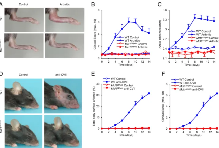
Mice were first subjected to K/B3N serum-transfer arthritis, an autoantibody-induced in vivo arthritis model (36, 37) previously suggested to be mediated by neutrophils (38, 39). As shown in Fig. 4A, K/B3N serum injection triggered robust arthritis in wild type mice, whereasMcl1DMyelomutants appeared to be completely protected. Kinetic analysis of clinical score (Fig. 4B; p= 4.2 3 1025) and ankle thickness (Fig. 4C; p= 0.0059) has confirmed those findings. The protection of Mcl1DMyelo mice was not due to deletion of LysM by theLyz2Cre/Creknock-in mutation because K/B3N serum-transfer arthritis developed normally inLyz2Cre/Cre mice (Supplemental Fig. 3).
Neutrophils have been proposed to be critical for the develop- ment of anti-CVII Ab–induced dermatitis, a mouse model of the rare human-blistering skin disease epidermolysis bullosa acquisita (33, 40, 41). Anti-CVII Abs triggered severe skin inflammation in wild type mice, whereas no signs of the disease could be observed in Mcl1DMyelo animals (Fig. 4D). Kinetic analysis revealed that Mcl1DMyelomice were completely protected from skin inflamma- tion, both in terms of the affected body surface (Fig. 4E;p= 3.83 10211) and of a more elaborate clinical scoring system (Fig. 4F;
p= 3.9310210).
Taken together, our results indicate that Mcl1DMyelo mice are completely protected from two separate, neutrophil-mediated autoantibody-induced inflammation models.
Increased susceptibility to bacterial and fungal infection Although Mcl1DMyelo mice resisted the microbial burden of their commensal flora (Fig. 3), we wanted to test their sus- ceptibility to experimentally induced infections. Therefore, we subjected our mice to systemic S. aureusorC. albicans infection.
Neutrophils are the major players in the host defense against infections byS. aureus, a Gram-positive pathogen able to cause skin and respiratory tract infection, abscess formation, and bacteremia/sepsis (42, 43). As shown in Fig. 5A, whereas wild type animals survived i.p. infection with 23107S. aureus, more FIGURE 2. Tissue leukocytes in Mcl1DMyelomice. Tissue neutrophils and other leukocytes were analyzed in wild type (WT) andMcl1DMyelomice from bone marrow, spleen, and peritoneal lavage samples by flow cytometry. Mature neutrophils were identified as CD11b+Ly6G+cells. Bar graphs show the absolute number of neutrophils (AandC) or other leukocytes (BandD) from the bone marrow (AandB) or the spleen (CandD). (E) Quantitative analysis of peritoneal lavage neutrophils after thioglycolate-induced peritonitis or control treatment. Data show mean and SEM from five to six (A–D) or six to eight (E) mice per group from three independent experiments. at Szegedi Tudomanyegyetem / University of Szeged on January 27, 2020http://www.jimmunol.org/Downloaded from
than 80% ofMcl1DMyelomice succumbed to the same infectious challenge (p= 1.031025). Analysis of the bacterial burden 12 h after the infection with 1 3 107 bacteria revealed a more than 100-fold increase of bacterial colony counts in the spleen (p= 0.0015), kidneys (p= 0.023), and liver (p= 9.031025) and significant increases in the brain (p = 0.028) and in the blood (p= 0.0038) but not in the peritoneum (p= 0.098) ofMcl1DMyelo mice (Fig. 5B, 5C).
Neutrophils are among the critical immune cells protecting the host from infection byC. albicans, a fungal pathogen able to cause superficial or systemic infections and one of the most prevalent causes of hospital-acquired infections (44). As shown in Fig. 5D, i.v. infection with 105C. albicanscaused lethality in 27% of wild type animals, whereas the same infection caused rapid lethality in 95% ofMcl1DMyelo mice (p, 0.00001). Analysis of the fungal burden at 12 h revealed a more than 10-fold increase in fungal counts in the liver (p= 1.0 3 1024) of Mcl1DMyelo mice, with moderate increase also in the spleen (p = 0.0096) and in the kidneys (p= 5.231027) but not in the brain (p= 0.97) of the animals (Fig. 5E).
Taken together,Mcl1DMyelo mice are highly susceptible to in- fectious challenge by bacterial or fungal pathogens, such as S. aureus orC. albicans, likely because of defective neutrophil- mediated elimination of the pathogens.
Analysis of Mcl1DMyelobone marrow chimeras
It is often difficult to obtain larger homogeneous cohorts of mice for in vivo experiments from small breeding colonies. When studying neutrophil function, this problem may be overcome by transplanting bone marrow cells to larger cohorts of recipient mice. To test that possibility, we transplanted wild type orMcl1DMyelobone marrow cells into lethally irradiated wild type recipients carrying the CD45.1 allele.
As shown in Fig. 6D, circulating neutrophils from such chimeras consisted practically exclusively of CD45.2-expressing cells (i.e., donor cells carrying the CD45.2 allele from the C57BL/6 genetic background), indicating successful replacement of the recipients’ he- matopoietic compartment by donor-derived cells. As shown in Fig. 6A, circulating neutrophil numbers ofMcl1DMyelobone marrow chimeras was strongly reduced compared with parallel-generated wild type chimeras (98.3% reduction;p= 1.8310214).Mcl1DMyelobone FIGURE 3. Survival and fertility of Mcl1DMyelomice. (AandB) Survival of wild type (WT) andMcl1DMyelomice under specific pathogen-free (SPF) (A) or conventional (B) conditions. (CandD) The body weight of WT andMcl1DMyelomale (C) and female (D) mice. (E) Breeding behavior of WT and Mcl1DMyelomice. Breeding was considered productive when pups were born from a given mating. (F) Genotype distribution of offspring from different breeding strategies. Survival curves show data of 611–977 (A) or 31–52 (B) mice per group, whereas body weight analysis shows mean and SD from 7 to 28 (C) or 9 to 26 (D) mice per group. Data from 193 breeding pairs and 1520 pups were used for the analysis of breeding behavior and offspring genotype.
at Szegedi Tudomanyegyetem / University of Szeged on January 27, 2020http://www.jimmunol.org/Downloaded from
marrow chimeras were also completely protected from K/B3N serum-transfer arthritis, both in terms of clinical score (p= 2.13 1025; Fig. 6B) and ankle thickness changes (p = 2.2 3 1026; Fig. 6C). Therefore, bone marrow transplantation can be used to generate larger cohorts of mice with neutropenia caused by the Mcl1DMyelomutation.
Neutrophil-specific deletion of Mcl-1 leads to neutropenia with severe survival defects
The above experiments were performed usingMcl1DMyelomice in which Mcl-1 was deleted from the entire myeloid compartment.
To test the effect of Mcl-1 deletion in a more neutrophil-specific manner, we have crossed theMcl1flox/floxmice to mice carrying the MRP8-Cre transgene, which drives Cre expression specifically in the neutrophil compartment (45).
Mcl1DPMNmice showed dramatic (99.1%, p = 9.8 3 10212) reduction of circulating neutrophil counts (Fig. 7A, 7B) that was even more severe than the reduction seen inMcl1DMyeloanimals (98.1%; see Fig. 1). TheMcl1DPMNmutation did not affect cir- culating monocyte (p = 0.60), eosinophil (p = 0.99), B cell (p = 0.21), or T cell (p= 0.58) numbers (Fig. 7C, 7D) or the distribution of monocytes into Ly6C+(inflammatory) or Ly6C2 (patrolling) monocytes (p= 0.24 and 0.26, respectively; Fig. 7E, 7F). Therefore, similar to theMcl1DMyelomutation, theMcl1DPMN mutation also leads to severe and selective neutropenia.
Analysis of the survival of Mcl1DPMNmice (Fig. 7G) revealed a steady and substantial loss ofMcl1DPMNanimals, leading to 58% sur- vival at 6 mo and only 30% survival at 12 mo of age (p,0.000001).
Mcl1DPMNmice were also clearly distinguishable from their wild type littermates and often showed a severe wasting phenotype (data not shown).Mcl1DPMNmice also showed a very poor breeding productivity (Fig. 7H). Taken together, our data suggest that the limited survival and breeding capacity makesMcl1DPMNmice rather difficult to maintain.
This is further complicated by the fact that such poor breeders need to be maintained in heterozygous form; therefore, all offspring need to be individually genotyped, and only a fraction of the pups (25% in the most sensibleMRP8-CreMcl1flox/+3Mcl1flox/floxbreeding strategy) are ex- pected to be of the desiredMcl1DPMNgenotype.
Bone marrow transplantation experiments revealed thatMcl1DPMN bone marrow chimeras also showed a severe wasting phenotype and succumbed to death 3–8 wk after transplantation (data not shown).
Although initial results indicated complete protection ofMcl1DPMN mice from K/B3N serum-transfer arthritis, the limited availability and fragile health status of those mice did not allow us to complete a sufficient number of those experiments (data not shown). The same issue also prevented us from performing more detailed analysis of the tissue leukocyte populations inMcl1DPMNmice. Nevertheless, it is interesting to note that in contrast to Mcl1DMyelomice the few Mcl1DPMNanimals we were able to dissect did not show an overt splenomegaly phenotype (data not shown).
FIGURE 4. Autoantibody-induced arthritis and skin-blistering disease in Mcl1DMyelomice. (A–C) Wild type (WT) orMcl1DMyelomice were injected with control (B3N) or arthritic (K/B3N) serum on day 0. Arthritis development was followed by photographing on day 7 (A), clinical scoring of the hind limbs (B) and ankle thickness measurement (C). (D–F) Skin-blistering disease was triggered in wild type (WT) orMcl1DMyelomice by systemic injection of control IgG or CVII-specific (anti-CVII) Abs. Skin disease was followed by photographing on day 14 (D) and clinical assessment of the total body surface affected (E) and the overall disease severity (F). Images are representative of and quantitative data show mean and SEM from five to nine control and 9 to 15 arthritic serum–treated individual mice per group from three independent experiments (A–C), or from three to four control and three to four anti-CVII–
treated mice per genotype from two independent experiments (D–F).
at Szegedi Tudomanyegyetem / University of Szeged on January 27, 2020http://www.jimmunol.org/Downloaded from
Taken together, our results indicate that although theMcl1DPMN mutation leads to severe and specific neutropenia, the poor and fragile health status, limited survival and fertility, and non- homozygous nature of those animals makes them hardly suitable for larger-scale in vivo experiments.
Partial neutropenia in G-CSF receptor–deficient mice
We have also tested G-CSF receptor–deficient (Csf3r2/2) mice (20) as a reference neutropenic mouse strain. Csf3r2/2 mice showed only partial neutropenia (p= 1.131024; Supplemental Fig. 4A, 4B), which was not nearly as severe and consistent as in Mcl1DMyelo or Mcl1DPMN animals (Figs. 1, 7). Similar to the Mcl1DMyeloand Mcl1DPMNmutations, theCsf3r2/2mutation did not affect other circulating leukocyte populations either (Supplemental Fig. 4C, 4D). Although the survival ofCsf3r2/2 mice was not substantially reduced, we also had difficulties breedingCsf3r2/2 mice in homozygous form (data not shown).
Those results indicate severe limitations of theCsf3r2/2mutation as a neutropenia model.
Discussion
Our results indicate thatMcl1DMyelo mice lacking Mcl-1 in the myeloid lineage are severely neutropenic but survive and breed in homozygous form. Those mice may, therefore, be highly useful in analyzing the role of neutrophils in in vivo processes in health and disease. Our results also indicate that mice are able to survive almost normally when the circulating neutrophil numbers are re- duced to ,2% of their normal values, necessitating the re- evaluation of the role of neutrophils in rodent survival.
Currently available tools for reducing neutrophil numbers have substantial limitations. Although Ab-mediated depletion (e.g., by the RB6-8C5 or NIMP-R14 anti-Gr1 or the 1A8 anti–Ly6G Abs) has clear benefits, such as easy availability and suitability to be
used on transgenic strains without breeding delay, it suffers from limited specificity (especially when using anti-Gr1 Abs), very high reagent costs, and the temporary nature of the depletion. Prior reports of neutropenic mice (16–21) also revealed phenotypes that strongly limit their use as in vivo neutropenia models. Besides severe neutropenia,Gfi1-deficient mice also show various defects in the T and B cell compartment and have a median survival time of ∼8–10 wk (16, 17), in line with the severe neutropenia and lymphocyte defects caused by dominant negativeGFI1muta- tions in human patients (46). The so-called “Genista” mice carrying a chemically induced Gfi1 mutation show incomplete neutrophil deficiency and are only partially protected in a neutrophil-dependent in vivo inflammation model (18). Mice lacking G-CSF (19) or the G-CSF receptor (20) are only moderately neutropenic (see also Supplemental Fig. 4), and the latter strain also shows breeding de- fects (data not shown). Deficiency of the Foxo3A transcription factor causes accelerated neutrophil apoptosis at the site of inflammation but does not affect circulating neutrophil numbers (21). In contrast to those genetic and pharmacological models, the Mcl1DMyelo mice show consistent, severe, and fairly specific neutropenia and survive and breed in homozygous form, making them quite useful as an in vivo neutropenia model.
The specificity of reduced neutrophil numbers in Mcl1DMyelo mice is due to two factors: the deletion of the antiapoptotic Mcl-1 protein in the entire myeloid lineage (including macrophages) and the specific requirement for Mcl-1 for the survival of neutrophils but not of the cells of the monocyte/macrophage lineage (24, 47).
This is also indicated by the normal number and overall ap- pearance of macrophages differentiated from Mcl1DMyelo bone marrow cells, despite effective deletion of the Mcl1flox allele (Supplemental Fig. 2G, 2H). We have also testedMcl1DPMNmice in whichMcl1deletion is achieved by theMRP8-Cre transgene, which is more specific for neutrophils than theLyz2Creknock-in FIGURE 5. Mcl1DMyelomice are highly susceptible to bacterial and fungal infections. (A–C) Survival curves (A) or analysis of the bacterial burden from the indicated tissues (BandC) of wild type (WT) andMcl1DMyelomice following i.p. injection with 23107(A) or 107(BandC)S. aureusbacteria. (DandE) Survival curves (D) or analysis of the fungal burden from the indicated tissues (E) of WT andMcl1DMyelomice following i.v. injection with 105C. albicans.
Survival curves show the data of 16 (A) or 19–22 (D) mice per group from three independent experiments. Bar graphs show mean and SEM from 9 to 10 (BandC) or 10 to 11 (E) mice per group from three (BandC) or four (E) independent experiments.
at Szegedi Tudomanyegyetem / University of Szeged on January 27, 2020http://www.jimmunol.org/Downloaded from
mutant (45). Although theMcl1DPMNmutations also strongly re- duced circulating neutrophil counts and appeared to be specific over several other leukocyte lineages (Fig. 7), the limited sur- vival and poor breeding of those mice make them very difficult to use as an in vivo neutropenia model. Although it is at present unclear why theMcl1DMyeloandMcl1DPMNmice have different survival and breeding characteristics, one of the possible ex- planations is that the remaining ∼2% of neutrophils in Mcl1DMyelo mice is sufficient to control the commensal flora, whereas the ∼1% of remaining neutrophils in the Mcl1DPMN mutants is below the threshold of neutrophil levels required for normal survival. It would theoretically also be possible that the survival of theMcl1DMyelomice is due to some genetic drift in our mouse colony, although our heterozygous breeding strategy argues against that possibility. Understanding the exact reason for the different survival of Mcl1DMyelo and Mcl1DPMN mice would require substantial additional experiments, including detailed apoptosis and in vitro progenitor differentiation/
proliferation assays.
Although the Mcl1DMyelo mutation causes severe neutropenia both in the peripheral blood and in various tissues (Figs. 1, 2), it is at present not entirely clear at which stage the mutation interferes with neutrophil development and/or survival. The fact that the number of Ly6Gmed/dimcells in the bone marrow is not reduced (Supplemental Fig. 2B) suggests that the Mcl1DMyelo mutation affects cells in the latest stage of neutrophil development. This is also supported by the fact that HoxB8-transduced myeloid pro- genitors were unable to engraft the bone marrow of Mcl1DMyelo mice (A.O. and A.M., unpublished observations), suggesting that the myeloid progenitor niche is preoccupied by endogenous cells in those animals.
Although theMcl1DMyelomutation proved to be fairly specific for neutrophils, we have consistently observed reduced B lineage cell
numbers in the bone marrow ofMcl1DMyelomice (Fig. 2B). The relevance of this finding is at present unclear, especially given the normal circulating (Fig. 1E) and splenic (Fig. 2D) B cell counts. More detailed analysis of the B cell compartment (Supplemental Fig. 2C, 2D) has revealed that even recirculating B cell numbers are reduced in the bone marrow of Mcl1DMyelo animals, suggesting disturbance of the bone marrow B cell niche.
Alternatively, this observation may be due to the expression of LysM in the early B cell lineage as indicated by the ImmGen database (www.immgen.org). It should also be noted that more splenic macrophages (Supplemental Fig. 2F) were observed in Mcl1DMyelo mice, which likely reflects splenomegaly in those animals.
To our knowledge, this is the first detailed characterization and validation of the Mcl1DMyelo mice as a suitable experimental neutropenia model. In particular, our study provides the most detailed lineage analysis of those animals, reports large-scale as- sessment of their survival and fertility, and validates the mutant mice on known neutrophil-dependent in vivo inflammation and infection models. To our knowledge, we also provide the first detailed analysis ofMcl1DPMNmice and a side-by-side compari- son of the Mcl1DMyelo, Mcl1DPMN, and Csf3r2/2 mutants. It should, nevertheless, be noted that we have already used the Mcl1DMyelomodel in the recent past to test the role of neutrophils in various disease models, such as graft-versus-host disease (48), contact hypersensitivity (35), gout (49), and experimental lupus (50). All those reports and further ongoing studies have confirmed the usefulness of this model for the in vivo analysis of neutrophil function.
Taken together, our results indicate that the unique combination of severe and fairly specific neutropenia, mostly normal survival, and capability for breeding in homozygous form make the Mcl1DMyelo mutation highly suitable for the analysis of the role FIGURE 6. Neutrophil deficiency and autoantibody-induced arthritis in Mcl1DMyelo bone marrow chimeras. (A) Number of circulating neutrophils (CD11b+Ly6G+Siglec-F–cells) of wild type (WT) orMcl1DMyelobone marrow chimeras by flow cytometry. (BandC) Analysis of the clinical score (B) and ankle thickness (C) of WT andMcl1DMyelobone marrow chimeras injected with control (B3N) or arthritic (K/B3N) serum on day 0. (D) Representative flow cytometric analysis of donor marker (CD45.2) expression in circulating neutrophils (Ly6G+ gate) from intact (nonchimeric) mice of the CD45.1-expressing recipient strain as well as from wild type (WT) orMcl1DMyelobone marrow chimeras. A representative histogram from a large number of experiments is shown. Quantitative data show mean and SEM from 17 chimeras (A) or from eight control and nine arthritic serum–treated chimeras per group from two (A) or three (BandC) independent experiments.
at Szegedi Tudomanyegyetem / University of Szeged on January 27, 2020http://www.jimmunol.org/Downloaded from
of neutrophils in in vivo models of normal and pathological processes in experimental mice. Our results also indicate that rodents are able to survive and breed when their circulating neu- trophil counts are dramatically reduced.
Acknowledgments
We thank Miklo´s Kova´cs, Dona´t Szikszai, Krisztina Futosi, No´ra Kiss, Kata Szilveszter, and Zsolt Sze´kely for help with the experiments;
Cassian Sitaru and Oana Virtic for the preparation of anti-CVII antiserum;
La´szlo´ Papp and Kla´ra Papp for colony management; Diane Mathis and Christophe Benoist for the KRN transgenic animals; Ga´bor Ba´nhegyi and Anna Sebestye´n for access to equipment; and Steve Edwards for inspiring discussions.
Disclosures
The authors have no financial conflicts of interest.
References
1. Spanopoulou, E. 1996. Cellular and molecular analysis of lymphoid develop- ment using Rag-deficient mice.Int. Rev. Immunol.13: 257–288.
2. Kitamura, D., J. Roes, R. Ku¨hn, and K. Rajewsky. 1991. A B cell-deficient mouse by targeted disruption of the membrane exon of the immunoglobulinm chain gene.Nature350: 423–426.
3. Mombaerts, P., A. R. Clarke, M. A. Rudnicki, J. Iacomini, S. Itohara, J. J. Lafaille, L. Wang, Y. Ichikawa, R. Jaenisch, M. L. Hooper, et al. 1992. Mutations in T-cell antigen receptor genesaandbblock thymocyte development at different stages.
Nature360: 225–231.
4. Itohara, S., P. Mombaerts, J. Lafaille, J. Iacomini, A. Nelson, A. R. Clarke, M. L. Hooper, A. Farr, and S. Tonegawa. 1993. T cell receptordgene mutant FIGURE 7. Neutrophil-specific deletion of Mcl-1 leads to neutrophil deficiency with survival and breeding defects. (A) Flow cytometric histo- grams of Ly6G staining of wild type (WT) andMcl1DPMNmouse peripheral blood leukocytes. (B) Quantitative analysis of the number of mature neutrophils (CD11b+Ly6G+Siglec-F cells) in WT andMcl1DPMNmice. Flow cytometric profiles (C) and quantitative analysis (D) of other leukocyte populations (red, neutrophils; green, monocytes; blue, eosinophils; magenta, B cells; orange, T cells). Flow cytometric histograms (E) and quantitative analysis (F) of monocyte subpopulations. (G) Survival of WT and Mcl1DPMN mice under specific pathogen-free conditions. (H) Breeding behavior of WT andMcl1DPMNmice. Flow cytometry dot plots and histograms are representative of and quantitative data show mean and SEM from, 10–22 (A–D) or 8–14 (EandF) mice per group from four (A–D) or three (EandF) independent experiments. Survival curves (G) show the data of 138–469 mice per genotype. Eighty-six breeding pairs were used for the analysis of breeding behavior (H).
at Szegedi Tudomanyegyetem / University of Szeged on January 27, 2020http://www.jimmunol.org/Downloaded from