Research Article
HUWE1 is a critical colonic tumour suppressor gene that prevents MYC signalling, DNA damage
accumulation and tumour initiation
Kevin B Myant
1,2,*, Patrizia Cammareri
1, Michael C Hodder
1, Jimi Wills
2, Alex Von Kriegsheim
2, Balázs Gy} orffy
3,4, Mamun Rashid
5, Simona Polo
6, Elena Maspero
6, Lynsey Vaughan
7, Basanta Gurung
8, Evan Barry
8, Angeliki Malliri
7, Fernando Camargo
8, David J Adams
5, Antonio Iavarone
9,
Anna Lasorella
10& Owen J Sansom
1,**Abstract
Cancer genome sequencing projects have identified hundreds of genetic alterations, often at low frequencies, raising questions as to their functional relevance. One exemplar gene isHUWE1, which has been found to be mutated in numerous studies. However, due to the large size of this gene and a lack of functional analysis of identified mutations, their significance to carcinogenesis is unclear. To deter- mine the importance ofHUWE1, we chose to examine its function in colorectal cancer, where it is mutated in up to15per cent of tumours.
Modelling of identified mutations showed that they inactivate the E3 ubiquitin ligase activity of HUWE1. Genetic deletion ofHuwe1rapidly accelerated tumourigenic in mice carrying loss of the intestinal tumour suppressor geneApc, with a dramatic increase in tumour initi- ation. Mechanistically, this phenotype was driven by increased MYC and rapid DNA damage accumulation leading to loss of the second copy ofApc. The increased levels of DNA damage sensitisedHuwe1- deficient tumours to DNA-damaging agents and to deletion of the anti-apoptotic protein MCL1. Taken together, these data identify HUWE1as abona fidetumour suppressor gene in the intestinal epithe- lium and suggest a potential vulnerability ofHUWE1-mutated tumours to DNA-damaging agents and inhibitors of anti-apoptotic proteins.
Keywordscolorectal cancer; DNA damage; HUWE1; MCL1; MYC Subject Categories Cancer; Digestive System
DOI10.15252/emmm.201606684| Received9June2016| Revised15November 2016| Accepted21November2016| Published online21December2016 EMBO Mol Med (2017)9:181–197
Introduction
The sequencing of human cancer genomes has led to a paradigm shift in our understanding of oncogenesis (Vogelsteinet al, 2013).
These studies have identified hundreds of genetic alterations that broadly segregate into two distinct groups, a small number of
“mountains” (genes commonly mutated) and a large number of
“hills” (gene mutated at low frequency). Whereas the causative role of frequently mutated genes is often clear, the role of those less commonly mutated can be difficult to separate from muta- tional “noise”. Determining the relevance of low-frequency muta- tions is important for providing a comprehensive understanding of the processes driving tumourigenic. To date, most attempts at determining the relevance of such mutations have relied on computational approaches, mining large multi-cancer databases (Alexandrov et al, 2013; Lawrence et al, 2014). Whilst important, these approaches provide only correlative evidence and, as such, direct, functional testing remains the key determinant of the onco- genic potential of somatic mutations (Kadoch & Crabtree, 2013;
Lewis et al, 2013).
HUWE1is an X-linked E3 ubiquitin ligase mutated at moderate frequencies (up to 15%) in a wide range of cancers including colorectal, uterine, gastric, cervical, melanoma and lung (Hodis et al, 2012; TCGA, 2012, 2014). HUWE1 catalyses the attachment of both lysine 48 (K48)- and lysine 63 (K63)-linked polyubiquitin chains, impacting on the function of a number of proteins involved in tumourigenic. The outcome of HUWE1-mediated K48 and K63 ubiquitination is quite different. For example, HUWE1 regulates the
1 Cancer Research UK Beatson Institute, Garscube Estate, Bearsden, Glasgow, UK
2 Cancer Research UK Edinburgh Centre, The Institute of Genetics and Molecular Medicine, Western General Hospital, Edinburgh, UK 3 MTA TTK Lendület Cancer Biomarker Research Group, Budapest, Hungary
4 2nd Department of Pediatrics, Semmelweis University, Budapest, Hungary 5 Wellcome Trust Sanger Institute, Hinxton, Cambridge, UK
6 IFOM, The FIRC Institute for Molecular Oncology, Milano, Italy
7 Cancer Research UK Manchester Institute, The University of Manchester, Withington, Manchester, UK 8 Boston Children’s Hospital, Boston, MA, USA
9 Departments of Neurology and Pathology, Institute for Cancer Genetics, Irving Comprehensive Research Center, New York, NY, USA 10 Departments of Pediatrics and Pathology, Institute for Cancer Genetics, Irving Comprehensive Research Center, New York, NY, USA
*Corresponding author. Tel: +44 131 651 8635; E-mail: kevin.myant@igmm.ed.ac.uk
**Corresponding author. Tel: +44 141 330 3953; E-mail: o.sansom@beatson.gla.ac.uk
stability of MCL1 and TP53 via addition of K48-linked polyubiquitin chains (Chen et al, 2005; Zhong et al, 2005). Additionally, two recent studies have described a role for HUWE1 in DNA damage response and the regulation of genomic stability, again via K48- linked modulation of H2AX and PCNA stability (Atsumiet al, 2015;
Choe et al, 2016). In contrast, HUWE1 regulates the function of DVL, a component of the WNT signalling pathway, via K63-linked ubiquitination (de Groot et al, 2014). This attachment prevents multimerisation of DVL, which is necessary for its role in binding AXIN2 during WNT signalling activation. Thus, HUWE1-mediated ubiquitination of DVL suppresses WNT activation. Perhaps most controversial is the role of HUWE1 in regulating MYC function. It has been reported that HUWE1 mediates K63 ubiquitination promoting the transcriptional activity of MYC, providing a pro- oncogenic function (Adhikary et al, 2005). However, HUWE1 has also been suggested to regulate the stability of both MYC during skin carcinogenesis and MYCN during brain development via K48 ubiquitination (Zhao et al, 2008; Inoue et al, 2013).
Together, these studies produce contrasting predictions, one where HUWE1 may drive cancer via activation of MYC function and one where loss of HUWE1 would promote cancer by increas- ing levels of MYC, DNA damage and genomic instability. Thus far, evidence from cell line and xenograft studies exists to support both models, but genetic evidence from a skin cancer model showed Huwe1 deletion accelerated tumourigenic suggest- ing a tumour suppressor role (Inoueet al, 2013). Mechanistically, the authors reportHuwe1 deletion leading to accumulation of the MYC/MIZ1 complex which, via direct promoter binding, suppresses expression of the anti-proliferative P21and P15genes (Inoue et al, 2013). Thus, in this model Huwe1 suppresses tumourigenic primarily via anti-proliferative effects. However, the relevance of this model to human cancer is unclear as HUWE1 mutations have not yet been observed in the currently limited, cutaneous squamous cell carcinoma sequencing studies (Lee et al, 2014).
Colorectal cancer (CRC), the second most common cause of cancer-related mortality, is a disease characterised by WNT signal- ling activation. Around 80% of tumours carry inactivating muta- tions in theAPCgene, a key inhibitor of the WNT pathway. These mutations lead to a deregulation of WNT signalling that drives transformation of the intestinal epithelium. Particularly pertinent are our previous studies that show thatMyc(a WNT target gene) and its downstream signalling targets are essential for the phenotype of deletion ofApc in vivo(Sansomet al, 2007; Myantet al, 2013;
Faller et al, 2015). Indeed, haploinsufficiency for Myc can reduce the phenotypes of Apc loss and slow tumourigenic (Athineos &
Sansom, 2010). This, in concert with the most frequent mutation of HUWE1 in CRC, makes it an ideal model to characteriseHUWE1 function. Here, we robustly characterise the role of HUWE1 in CRC initiation. We find inactivating HUWE1 mutations in human CRC and that deletion of Huwe1 in CRC mouse models leads to rapid tumourigenic and hugely increased tumour initiation. MYC protein levels are increased and drive increased tumour proliferation but are not the primary cause of the increased tumour initiation. Rather, accumulation of DNA damage, characterised by accumulation of c-H2AX leading to acceleratedApcloss, promotes tumour initiation.
These tumours display increased sensitivity to DNA-damaging agents and are dependent on high levels of MCL1 for their survival
suggesting a potential therapeutic vulnerability ofHUWE1-mutated tumours. Together, these data defineHUWE1as a critical intestinal tumour suppressor gene that restrains cellular proliferation and DNA damage accumulation.
Results
HUWE1is a colonic tumour suppressor
HUWE1 is a pleiotropic E3 ubiquitin ligase that modulates the func- tion of several proteins involved in oncogenesis and DNA damage response including MYC, MYCN, MCL1 and H2AX (Adhikaryet al, 2005; Zhong et al, 2005; Zhao et al, 2008; de Groot et al, 2014).
Previous sequencing studies have identified somatic mutations throughout the HUWE1 gene in up to 15% of colorectal tumours (Wood et al, 2007; Seshagiri et al, 2012; TCGA, 2012; Fig EV1A).
HUWE1 is a large gene (~15,000-bp cDNA), and thus, its frequent mutation could simply be a product of mutational “noise”.Huwe1 was also identified as a high-ranking positive hit in a CRC trans- poson mutagenesis screen (rank 66/752) indicating its mutation may be important to colorectal tumourigenic, but to date direct functional determination of this is lacking (March et al, 2011).
Among many cancer types, CRC harbours the highest frequency of HUWE1mutations (Fig EV1B). However, the consequence of these mutations on the function of HUWE1 remains undetermined. To decipher the functional significance ofHUWE1mutations, we inter- rogated the activity of two CRC-specific mutations targeting the HECT domain ofHUWE1(R4082H and K4204del; Woodet al, 2007;
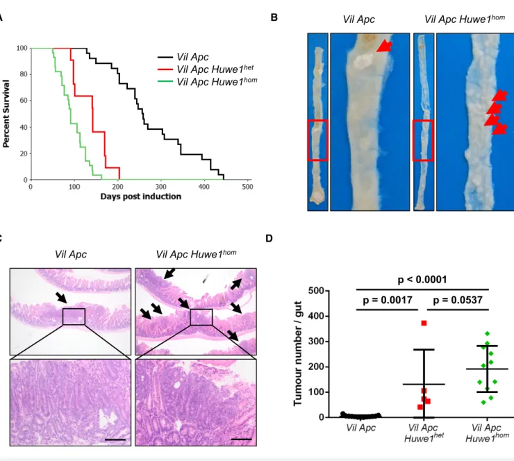
Jones et al, 2008). Following expression and purification of the wild-type and mutant HECT domains of HUWE1, we found that both mutations led to marked inhibition of the ability of the HUWE1-HECT domain to bind to the E2-ubiquitin-conjugating enzyme UbcH7, thus indicating that the two mutants perturb the assembly of the HUWE1 ubiquitin ligase complex (Fig EV1C). As HUWE1is an X-linked gene that is transcriptionally silenced on the inactive X, mutation of a single allele would be sufficient to disrupt its activity (Carrel & Willard, 2005). Thus, we have identified poten- tially functionally inactivating mutations of HUWE1 in primary colorectal tumours. We next addressed the consequence ofHUWE1 loss of function upon intestinal tumour development. We generated a cohort of mice carrying an inducible floxed allele ofApc580S(from here on referred to asApcfl) under the control of thevillin-Cre-ERT2 transgene (vil-Cre-ERT2 Apcfl/+— Vil Apc). Following recombina- tion, these mice lose the wild-type Apc allele spontaneously and succumb to small intestinal and colonic tumourigenic. We crossed these mice to those carrying a conditional deletion allele ofHuwe1 (Huwe1fl) to generate experimental cohorts of both female vil-Cre- ERT2Apcfl/+Huwe1fl/+(Vil Apc Huwe1het) and female or malevil- Cre-ERT2 Apcfl/+ Huwe1fl/fl/Huwe1fl/y mice (Vil Apc Huwe1hom), respectively. Following Cre induction with tamoxifen, we aged these mice until signs of intestinal tumourigenic became apparent (pale feet, hunching and weight loss). Whereas control mice survived to a median of ~250 days, both Huwe1 mutant cohorts succumbed to rapid tumourigenic (Huwe1het ~140 days, Huwe1hom ~90 days;
Fig 1A). Strikingly, macroscopic and microscopic analysis of the guts from sacrificed mice indicated a dramatic increase in tumour number upon Huwe1 deletion (Figs 1B–D and EV1D). Unlike Vil
Apcmice that developed around five tumours, those from both the Vil Apc Huwe1hetandVil Apc Huwe1homcohorts developed around 200 (Fig 1D). This significant increase in tumour number was also observed in the colons of homozygous deleted animals (Fig EV1D). Immunohistochemical analysis of these tumours demonstrated nuclear accumulation of b-catenin in Huwe1hom tumours at a similar level as control tumours indicating that tumour initiation was due to activation of WNT signalling (Fig EV2A–C). These data define Huwe1 as an intestinal and
colonic tumour suppressor in the context of Apc heterozygosity whose loss of function leads to increased tumour initiation.
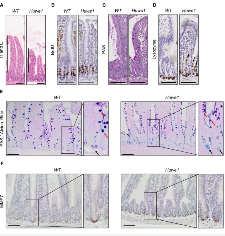
Huwe1deletion leads to perturbed intestinal homoeostasis Given this profound impact on tumour formation, we next analysed whetherHuwe1deletion impacted on intestinal homoeostasis. Histo- logical examination of Huwe1-deficient intestines 14 days post- induction showed modest changes in crypt/villus architecture with D
A
p < 0.0001
p = 0.0017 p = 0.0537 Vil Apc Vil Apc Huwe1
homC
B
Vil Apc
Vil Apc Huwe1
hetVil Apc Huwe1
homVil Apc Vil Apc Huwe1
homFigure1. Huwe1is an intestinal tumour suppressor gene.
A Kaplan–Meier survival plot of cohorts of inducedVil Apc,Vil Apc Huwe1hetandVil Apc Huwe1hommice. Deletion ofHuwe1led to a significant reduction in survival of these animals (Vil ApcversusVil Apc Huwe1het/Vil Apc Huwe1hom, log rank,P<0.001,n≥10).
B Wholemount isolation of small intestines fromVil ApcandVil Apc Huwe1hommice culled at clinical endpoint. Note the huge numbers of tiny macroscopic adenomas visible in theVil Apc Huwe1homintestine (red arrows).
C H&E staining of intestinal cross sections fromVil ApcandVil Apc Huwe1hommice demonstrating the increased adenoma burden followingHuwe1deletion. Black arrows indicate individual adenomas. Scale bars =200lm.
D Quantification of total tumour numbers per gut in sacrificedVil Apc,Vil Apc Huwe1hetandVil Apc Huwe1hommice. Deletion ofHuwe1led to a significant increase in the number of tumours per gut (Mann–Whitney,n≥5). Mean and standard deviation are plotted.
slightly shortened villi, but both cellular proliferation and apoptosis were unchanged (Fig 2A and B, and Appendix Figs S3A and S6A).
We next analysed whetherHuwe1loss altered intestinal cellular dif- ferentiation by examining markers of different intestinal cell popula- tions. Interestingly, whilst we observed no gross changes in the number of goblet cells by periodic acid–Schiff (PAS) staining (Fig 2C), we observed lysozyme expression away from the intestinal crypt base (Fig 2D and Appendix Fig S3B and C). This could indi- cate either mislocalisation of Paneth cells or perturbed secretory intestinal differentiation. To investigate this more closely, we carried out double staining with PAS and alcian blue which marks Paneth cell secretory vesicles and goblet cells. This demonstrated that Paneth cell secretory vesicles are maintained at the crypt base inHuwe1-deficient intestines, suggesting that the lysozyme-positive cells may be cells from a different lineage expressing lysozyme (Fig 2E). Additionally, immunohistochemical analysis indicated that expression of the Paneth cell marker MMP7 was retained at the crypt base following HUWE1 deletion (Fig 2F). Furthermore, under closer scrutiny the lysozyme-positive cells further up the crypt–
villus axis appear to have the morphology of goblet cells (Fig EV3D) suggesting alterations in secretory cell marker expression. Both Paneth cell differentiation (Andreuet al, 2008) and EPHB/EPHRINB gradient-mediated localisation (Batlle et al, 2002; Sansom et al, 2004) are controlled by WNT signalling. A recent study has suggested a role for HUWE1 as a negative regulator of WNT signal- ling (Dominguez-Braueret al, 2016), so we analysed the expression of a number of WNT target genes 14 days after Huwe1 deletion.
Interestingly, we observed a modest increase in expression of a number of WNT target genes, includingEphb3, which is a critical mediator of Paneth cell localisation (Fig EV3E; Batlleet al, 2002).
However, not all of the analysed WNT target genes showed increased expression, and onceApcwas deleted, we no longer saw a HUWE1-dependent modification of WNT target genes, with the robust induction of targets caused byApcloss masking any impact of HUWE1 (Fig EV3F). Thus, it is possible these changes reflect secondary consequences of Huwe1 deletion effects on crypt homoeostasis. We next performed microarray analysis comparing wild-type to HUWE1-deficient intestines to assess whether WNT signalling was globally deregulated followingHuwe1deletion. This was conducted at 4 days post-Huwe1 deletion to reduce potential secondary consequences ofHuwe1loss. We identified 586 and 415 genes whose expression was significantly up- or down-regulated, respectively (Appendix Table S1). The upregulated data set included a number of WNT-responsive intestinal stem cell marker genes includingAscl2, Lect2, Slc14a1andNrn1. Gene set enrichment anal- ysis identified a highly significant overlap of transcriptional markers of intestinal stem cells (chi-squared test with Yates’s correction, P<0.0001; Munozet al, 2012) between the gene lists, demonstrat- ing elevated expression of some members of the intestinal stem cell signature following Huwe1 deletion. However, well-known WNT target genes such asAxin2andMycwere not upregulated and gross changes in nuclear b-catenin localisation were not observed (Fig EV3G), suggesting that HUWE1 loss only modifies the expres- sion of subset of WNT target genes associated with stem cell mark- ers. Together, these data identify a role for HUWE1 loss in moderate amplification of intestinal WNT signalling, expression of some stem cell-related genes and perturbed homoeostasis. However, given the redundancy of both stem cell markers (e.g. Lgr5) and Paneth cells
for intestinal homoeostasis and tumourigenic (de Lauet al, 2011;
Durand et al, 2012), we felt these could not explain the marked increase in tumourigenic we observed.
MYC drives increased tumour proliferation following Huwe1deletion
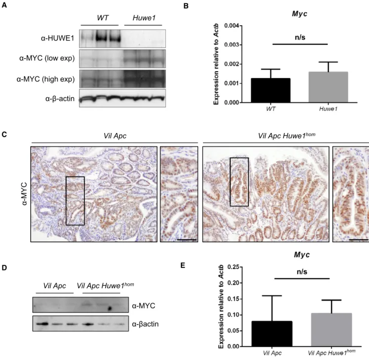
We next wanted to address the mechanism via which HUWE1 suppresses tumourigenic. Of particular relevance is the proposed role of HUWE1 in regulating MYC, a key downstream mediator of HUWE1 function (Inoueet al, 2013). As MYC is a critical modulator of intestinal tumourigenic, we hypothesised that the modulation of MYC stability may be a key tumour-suppressive function of HUWE1 within the gut (Sansomet al, 2007; Athineos & Sansom, 2010). We addressed this by determining MYC expression in epithelial extrac- tions of control andHuwe1-deficient intestines. We found a signifi- cant increase in MYC protein, but not transcript, following HUWE1 loss (Fig 3A and B). This increase in MYC protein was also observed in Huwe1-deficientVil Apc Huwe1 tumours compared to controls and was independent of transcriptional effects (Fig 3C–E). These data indicate that HUWE1 controls MYC protein abundance in both normal intestine and intestinal tumours.
We next sought to determine whether MYC function is important for driving the dramatic phenotypes we observe uponHuwe1dele- tion. This was determined using both loss- and gain-of-function alle- les ofMyc. AsMycdeletion rescues the phenotypes ofApc loss in the small intestine, complete ablation ofMycin the context of addi- tional Huwe1 deletion would likely yield the same phenotype.
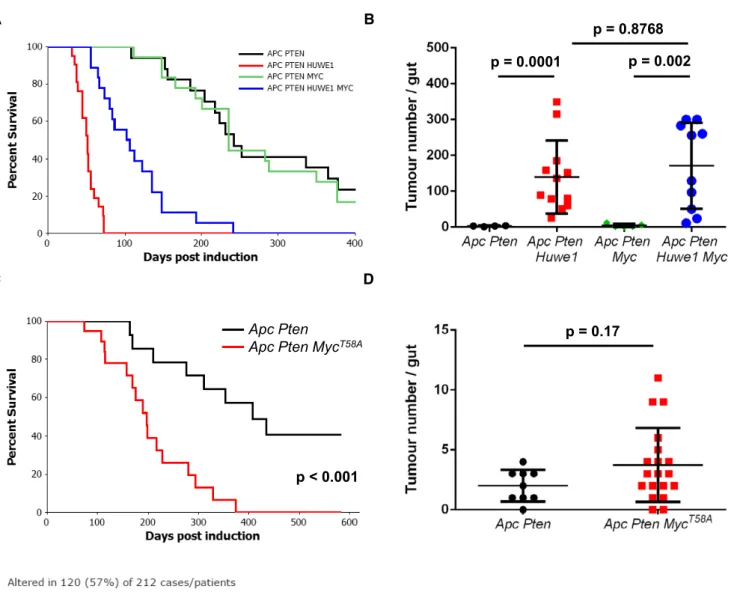
Moreover, as Myc haploinsufficiency slows Apc-dependent tumourigenic, one would expect it to also slow theVil Apc Huwe1 phenotype. Therefore, we wanted to find a system where tumouri- genic is insensitive to a 50% reduction inMycto permit assessment of the role of Huwe1in this context. In CRC, mutation and epige- netic inactivation of the PI3K–PTEN pathway occur in up to 40% of cases, and we have shown that Pten deletion rapidly accelerates tumourigenic and drives tumour progression (Marsh et al, 2008).
Ptenheterozygosity, however, only causes a relatively mild increase in tumourigenic. We therefore thought this model would be an ideal candidate to examine (i) whether Huwe1 loss can accelerate tumourigenic in multiple spontaneous models and (ii) whetherMyc is functionally important for this. We first confirmed that deletion of Huwe1accelerates tumourigenic in a spontaneous model with addi- tionalPtendeletion by ageing cohorts ofvil-Cre-ERT2Apcfl/+Ptenfl/+
(Vil Apc Pten) andvil-Cre-ERT2Apcfl/+Ptenfl/+Huwe1fl/fl(Vil Apc Pten Huwe1) mice. Consistent with our previous model,Huwe1dele- tion led to a dramatic shortening of lifespan and a marked increase in tumour number and increased tumour cell proliferation (Figs 4A and B, and EV4A and B). We next determined whether this model is sensitive to reducedMycexpression by comparing survival of theVil Apc Ptenmice to those with additional deletion of a singleMycallele (vil-Cre-ERT2Apcfl/+Ptenfl/+Mycfl/+—Vil Apc Pten Myc). Interest- ingly, we found neither survival of mice with Pten deletion nor tumour cell proliferation was sensitive to loss of a singleMycallele (Figs 4A and B, and EV4A and B). Therefore, this model is ideal for defining the functional relevance of elevated MYC levels inHuwe1- deficient tumours. We generated vil-Cre-ERT2 Apcfl/+ Ptenfl/+
Huwe1fl/flMycfl/+ (Vil Apc Pten Huwe1 Myc) mice and compared their survival to the Vil Apc Pten Huwe1cohort. Strikingly, in the
context ofHuwe1deletion, reduced expression ofMycled to a signifi- cant increase in survival, thus indicating that the tumour suppressor function of HUWE1 is, at least in part, mediated through the control
of MYC stability (Fig 4A). However, despite the enhanced survival, Vil Apc Pten Huwe1 Myc mice showed no reduction in tumour number (Fig 4B). They did, however, show decreased tumour C
WT Huwe1
H and E
A B
WT Huwe1
BrdU
WT Huwe1
Lysozyme
D WT Huwe1
PAS
1 e w u H T
W
PAS / AlcianBlue
E
1 e w u H T
W
MMP7
F
Figure2. Huwe1deletion leads to perturbed intestinal homoeostasis.
A H&E staining of control andHuwe1-deleted intestinal epithelium. Shortened villi inHuwe1-deficient tissue are indicated.
B BrdU IHC of control andHuwe1-deleted intestinal epithelium.
C PAS staining identifying goblet cells. No gross changes were observed.
D Lysozyme staining (Paneth cell marker) of control andHuwe1-deleted small intestine. Note the occurrence of lysozyme-positive cells away from the crypt base (black arrows).
E Dual periodic acid–Schiff/alcian blue staining to identify Paneth cell secretory vesicles (light blue/pink, marked with black arrows) and goblet cells (dark blue/purple, marked with red arrows). Note Paneth cell secretory vesicles are restricted to crypt base in both control andHuwe1-deficient small intestines (inset, black arrows).
F MMP7staining of control andHuwe1-deleted small intestine. Note MMP7staining is restricted to crypt base inHuwe1-deficient intestines.
Data information: Scale bars=100lm.
proliferation rates compared to tumours fromVil Apc Pten Huwe1 mice (Fig EV4A and B). These data suggest that MYC accumulation in this model promotes tumour growth, but the residual MYC levels in the Mycfl/+ background are sufficient for efficient tumour
initiation. We further characterised the role of increased MYC protein during tumourigenic by overexpressing two copies of a proteolyti- cally stable MYC mutant (Rosa-lsl-MycT58A) in this model (Apc Pten MycT58A). Consistent with our loss-of-function experiments, A
WT Huwe1
α-HUWE1 n/s
B
α-MYC (low exp) α-MYC (high exp) α-β-actin
C
V li A p c V li A p c H u w e 1
homα -MYC α
D E
n/s
Vil Apc Vil Apc Huwe1
homα-MYC α-βactin
Figure3. Increased MYC protein expression followingHuwe1deletion.
A HUWE1and MYC Western blot analysis of epithelial cell extractions from control andHuwe1-deficient intestines. Levels of MYC protein are significantly increased in normal intestines lacking HUWE1(Mann–Whitney,P=0.04,n=3).
B qRT–PCR analysis ofMycexpression in control andHuwe1-deficient intestines. Mann-Whitney,n=4vs6. Data plotted are mean and SD.
C MYC IHC in tumours fromVil ApcandVil Apc Huwe1hommice. Scale bars =50lm.
D MYC Western blot in protein extracts fromVil ApcandVil Apc Huwe1homtumours. Levels of MYC protein are significantly increased in tumours lacking HUWE1 (Mann–Whitney,P=0.04,n=3).
E qRT–PCR analysis ofMycexpression in tumours fromVil ApcandVil Apc Huwe1hommice. Mann-Whitney,n=3vs3. Data plotted are mean and SD.
Source data are available online for this figure.
overexpression of MYCT58Asignificantly decreased survival but had no effect on overall tumour number (Fig 4C and D). In human CRC, we found that HUWE1 mutations were mutually exclusive with amplifications, gains and transcriptional upregulation of MYC (P=0.042; Fig 4E). This suggests thatHUWE1mutation in human
CRC might facilitate increased levels of MYC and bypass the require- ment for genomic amplification. However, it is also clear from our work, and others, that MYC levels are sufficiently high for transfor- mation in the presence of HUWE1 and reduction or overexpression of MYC does not have a profound impact on tumour initiation.
B A
p = 0.002 p = 0.8768 p = 0.0001
D C
p < 0.001
p = 0.17
E
Apc Pten
Apc Pten MycT58A
Figure4. MYC upregulation followingHuwe1deletion drives accelerated tumourigenic.
A Kaplan–Meier survival plot of cohorts of inducedVil Apc Pten,Vil Apc Pten Huwe1, Vil Apc Pten MycandVil Apc Pten Huwe1Mycmice. Heterozygous deletion of Mycled to a specific and significant increase in survival ofHuwe1-deleted animals (Vil Apc Pten Huwe1versusVil Apc Pten Huwe1Myc,log rank,P<0.001,n≥16).
B Quantification of total tumour numbers per gut in sacrificedVil Apc Pten,Vil Apc Pten Huwe1, Vil Apc Pten MycandVil Apc Pten Huwe1Mycmice. Heterozygous deletion ofMycdid not reduce the number of tumours inHuwe1-deleted mice (Vil Apc Pten Huwe1versusVil Apc Pten Huwe1Myc, Mann–Whitney,n≥10). Data plotted are mean and SD.
C Kaplan–Meier survival plot of cohorts of inducedAhCre-ERTApc PtenandAhCre-ERTApc Pten MycT58Amice. Overexpression of proteolytically stabilised MYC led to a significant reduction in survival (log rank,P<0.001,n=14versus21).
D Quantification of total tumour numbers per gut in sacrificedApc PtenandApc Pten MycT58Amice. Overexpression of proteolytically stabilised MYC did not increase the number of tumours inApc Ptenmice (Mann–Whitney,n=9versus19). Data plotted are mean and SD.
E cBioportal OncoPrint showing mutual exclusivity ofHUWE1mutations and increasedMYCcopy number and/or RNA expression in CRC samples (log odds ratio: 1.144 [some tendency towards mutual exclusivity],P=0.042).
Taken together, the above findings indicate that, besides MYC, other substrates of HUWE1 contribute to intestinal tumourigenesis in Huwe1-null mice.
Huwe1deletion leads to increased levels of DNA damage
The massively increased tumour number we observed was reminis- cent of mice displaying a mutator phenotype, such as those deficient forMlh1orMsh2(Reitmairet al, 1996; Edelmannet al, 1999). We reasoned that increased tumour initiation in mice heterozygous for Apcloss would most likely occur via increased DNA damage and/or mutation rate. Two recent reports have described a role for HUWE1 in regulation of DNA damage response and genomic stability via degradation of H2AX and PCNA, respectively (Atsumiet al, 2015;
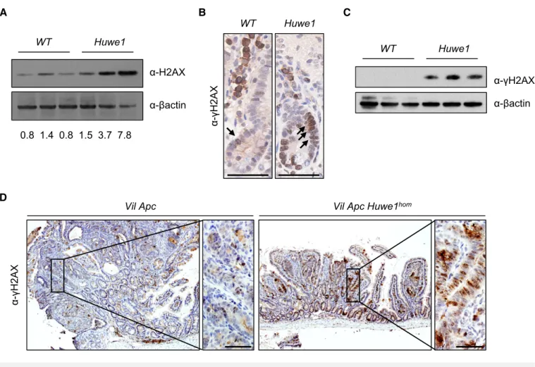
Choe et al, 2016). To attempt to define the mechanism by which Huwe1deletion drives increased tumourigenic, we carried out mass spectrometry analysis to compare the proteome of normal and Huwe1-deficient intestinal tissue. Interestingly, this analysis identi- fied a significant increase in H2AX levels followingHuwe1deletion that we confirmed by Western blot (Appendix Table S2 and Fig 5A).
Thus, loss ofHuwe1function could lead to accumulation of DNA damage via accumulation of H2AX leading to inefficient resolution of c-H2AX foci (Atsumi et al, 2015). To test this hypothesis, we analysed DNA damage levels following Huwe1 deletion. Interest- ingly, we observed increased levels ofc-H2AX in normal intestine and tumour tissue deficient forHuwe1(Fig 5B–D and Appendix Fig S5A). We next used PCR analysis of theApclocus to determine the mechanism by whichHuwe1-deficient cells loseApc.The majority of both control andHuwe1-deficient tumours demonstrated loss of heterozygosity at theApc locus indicating that Apcloss is driving tumour initiation (Fig EV5B). A common property of cells with increased levels of DNA damage is an increased sensitivity to DNA- damaging agents (Farmeret al, 2005; Hayet al, 2005). To investi- gate this further, we treated both control and tumour-bearing mice with the DNA-cross-linking agent cisplatin. Consistent with the observed increase in DNA damage, Huwe1-deficient tumours displayed increased sensitivity to this treatment with significantly higher levels of apoptosis (Fig 6A and B). Together, these data demonstrate that loss of Huwe1 leads to accumulation of DNA damage, loss of Apc and subsequent tumour initiation. We next analysed tumour data from TCGA to determine whether HUWE1 loss of function can confer a similar DNA damage phenotype in human disease. We found that tumours carryingHUWE1mutations displayed a far higher somatic mutational burden than those with wild-typeHUWE1(Fig 6C). Importantly, this increase was indepen- dent of MLH1 silencing, a key determinant of mutational burden indicating this is not merely a consequence of a general increased mutation rate (Fig 6D). Thus, increased DNA damage appears to be a property ofHUWE1 mutation in human CRC and may confer a therapeutic vulnerability on these tumours during tumourigenic.
One prediction of increased sensitivity to DNA-damaging agents uponHUWE1 loss would be that tumours that had low levels of HUWE1 might respond better to therapy. There are currently no data publically available on the treatment response of HUWE1- mutated patients, so we analysed this with respect to HUWE1 expression levels. We found that patients whose tumours express low levels ofHUWE1 have significantly increased overall survival following chemotherapy treatment than those expressing higher
levels (Fig 6E). Interestingly, there were no significant survival dif- ferences between patients who received no adjuvant chemotherapy treatment based onHUWE1expression levels (Fig 6F). These data are consistent with an increased DNA damage burden in human tumours depleted of HUWE1 and may suggest that patients with HUWE1 mutations may respond better to chemotherapeutic treatment.
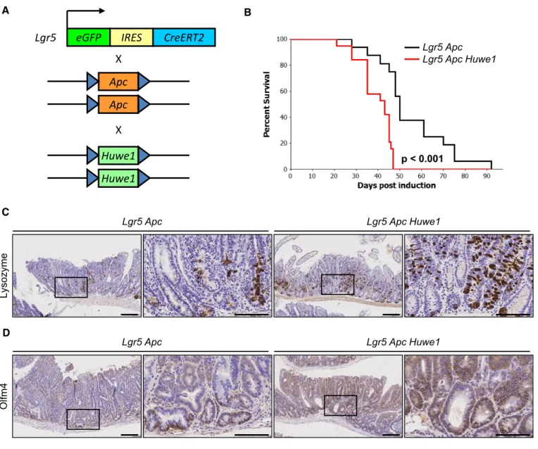
Given this increased DNA damage even in intestines that were wild-type for Apc, this could suggest that Huwe1 loss increases tumour initiation by promoting loss of the wild-typeApcallele. To test whetherHuwe1loss accelerates tumourigenic independently of the requirement for loss of wild-typeApc, we utilised a model initi- ated by homozygous deletion ofApc.Using theLgr5-cre-ERT2knock- in mouse, both copies ofApcalone or in combination withHuwe1 can be deleted specifically in stem cells (Fig 7A). This model permits rapid tumourigenic originating from LGR5-positive intesti- nal stem cells. We generated cohorts ofLgr5-cre-ERT2Apcfl/fl(Lgr5 Apc) and Lgr5-cre-ERT2 Apcfl/fl Huwe1fl/fl (Lgr5 Apc Huwe1) mice and aged them until they showed signs of tumourigenic (Fig 7B). In this model, there was a less dramatic (median survival 50 versus 43 days) though significant decrease in survival ofLgr5 Apc Huwe1 mice compared toLgr5 Apc(P<0.001; Fig 7B). This indicates that HUWE1 suppresses intestinal tumourigenic at multiple levels, in part by stopping tumour initiation but (and consistent with the data on MYC above) also at the level of tumour growth. To determine whether the lysozyme expression and stem cell phenotypes were maintained in tumour tissue, we analysed lysozyme and OLFM4 expression in these tumours. We observed both increased lyso- zyme-positive cell abundance and zone of OLFM4 expression in Huwe1-deficient tumours (Fig 7C and D). Thus, even in the absence ofApcloss ofHuwe1perturbed differentiation.
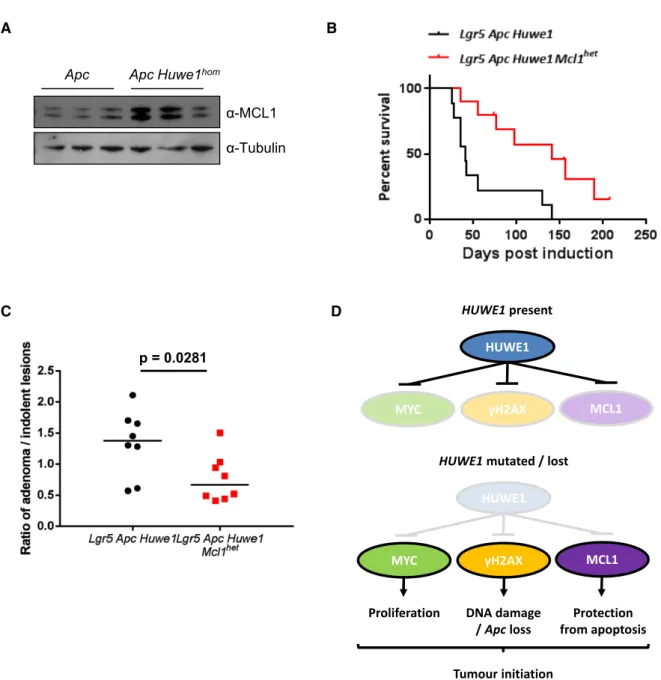
One very surprising observation was that despite the dramati- cally increased DNA damage burden, we did not observe an increase in apoptosis inHuwe1-deficient crypts (Fig EV6A). Intesti- nal epithelial cells are extremely sensitive to DNA damage-induced apoptosis and equivalent levels ofc-H2AX would normally lead to high levels of apoptosis, suggesting thatHuwe1-deficient cells are protected from cell death (Phesseet al, 2014). HUWE1 has previ- ously been implicated in modulating the stability of the anti-apop- totic protein MCL1 and so we hypothesised that increased levels of MCL1 may protectHuwe1-deficient cells from death (Zhonget al, 2005). We observed increased levels of MCL1 in tumour tissue defi- cient for Huwe1 (Fig 8A). To determine whether increased MCL1 levels protect Huwe1-deficient cells from apoptosis, we generated VilCreERT2 Huwe1fl/fl Mcl1fl/+ (Huwe1 Mcl1) mice and induced simultaneous deletion ofHuwe1and one copy ofMcl1. The intesti- nes of these mice displayed significantly higher levels of apoptosis than singly deletedHuwe1mice demonstrating a role for MCL1 in protecting Huwe1-deficient cells from apoptosis in homoeostatic conditions (Fig EV6A). Importantly, deletion of one copy of Mcl1 alone did not induce an apoptotic response suggesting this observa- tion was specific toHuwe1loss-mediated DNA damage (Fig EV6A).
We next addressed whether this impacted on tumour development by generating a cohort of tumour-inducible mice carrying the same Mcl1fl/+ allele. Similar to our observations in normal tissue, we found that heterozygous deletion of Mcl1 reduced Huwe1 loss- driven tumourigenic (Fig 8B). Mice heterozygous forMcl1displayed a greater propensity to form indolent lesions than the mice wild-type
forMcl1.The ratio of canonical adenomas to indolent lesions, iden- tified as a spherical lesion usually contained entirely within a single villus, was significantly different between the genotypes (Fig 8C and Appendix Fig S6B and C). Together, these data indicate thatHuwe1 loss of function leads to increased levels of MCL1 protein and this protects cells from DNA damage-induced apoptosis facilitating onco- genic transformation.
Discussion
The identification of hundreds of somatic mutations in cancer genomes has raised critical questions as to their functional rele- vance. Despite efforts to analyse such mutations computationally, our work demonstrates the importance of direct functional testing, in particular of large genes mutated at moderate levels. Our work shows, by robust genetic characterisation, thatHuwe1is a tumour suppressor in the small intestine and colon. Together with our iden- tification ofHUWE1mutations present in human CRC that perturb
its ubiquitin ligase activity, this strongly suggests it is abona fide colonic tumour suppressor gene.
This is particularly important as previous work on HUWE1’s tumourigenic role had proven controversial. HUWE1 is an E3 ubiq- uitin ligase that controls the stability of MCL1, MYC and MYCN functions which would suggest a tumour-suppressive role. Indeed, work using chemically induced skin cancer mouse models has indi- cated this is the case (Inoue et al, 2013). However, via K63- mediated ubiquitination, HUWE1 is required for the transactivation function of MYC and tumour cell proliferation. It is also overex- pressed in a number of different cancers including colorectal, supporting a pro-oncogenic function (Adhikaryet al, 2005). Using intestinal and colonic specific gene deletion, we show that loss of Huwe1leads to significantly accelerated tumourigenic characterised by a massive increase in tumour incidence. We observe coincident increased MYC protein levels and, using conditional co-deletion of Myc, we demonstrate this increase in MYC is an important mediator of this phenotype. The mutual exclusivity ofHUWE1mutation and MYC genomic amplification we observe in human CRC strongly D
α-γH2AX
1 e w u H c p A li V c
p A li
V hom
C
WT Huwe1
α-γH2AX α-βactin WT Huwe1
B
α-γH2AX
α-H2AX
α-βactin
WT Huwe1
0.8 1.4 0.8 1.5 3.7 7.8 A
Figure5. Huwe1deletion leads to an increase in DNA damage.
A H2AX Western blot in protein extracts from control andHuwe1-deficient intestinal epithelial cells. Band intensity relative tob-actin displayed under each lane (Mann–Whitney,P=0.04,n=3versus3).
B c-H2AX IHC showing increased positivity inHuwe1-deleted intestinal cells (black arrows). Scale bars =50lm.
C c-H2AX Western blot in protein extracts from control andHuwe1-deficient intestinal epithelial cells (Mann–Whitney,P=0.04,n=3versus3).
D c-H2AX IHC ofVil ApcandVil Apc Huwe1homtumours; note increased positivity inHuwe1-deficient tumours. Scale bars =50lm.
Source data are available online for this figure.
A
Cisplatin Control
Lgr5 Apc Lgr5 Apc Huwe1
B
C D
E
No chemotherapy
Chemotherapy
Fp = 0.0002
p = 0.0179
p = 0.0007
Figure6.
implicates them as critical mediators of colorectal tumourigenic.
Thus, our findings are consistent with a tumour-suppressive role of HUWE1 in destabilising MYC post-translationally. The reason for these discrepancies is not immediately clear although it is worth noting the elevated levels of DNA damage we observed following Huwe1deletionin vivo. It is possible that some of the anti-prolifera- tive effects the authors observe followingHUWE1knockdown are in fact due to DNA damage-induced growth arrest that is more appar- ent in cell culture conditions. Interestingly,Mycdeletion does not impact on the increased tumour initiation phenotype; rather, it dampens the elevated tumour cell proliferation observed inHuwe1- deficient tumours. This has the consequence of slowing the acceler- ated tumour growth followingHuwe1loss but has no impact on the ability of tumours to form. Thus, HUWE1 clearly impacts on tumourigenic via multiple pathways.
Our observation that lysozyme expression becomes mislocalised and the stem/progenitor cell zone was expanded is consistent with a recent paper identifying HUWE1 in regulating the intestinal stem cell niche via direct modulation of EPHB3 stability (Dominguez- Braueret al, 2016). However, we believe that the impact of Paneth cell marker mislocalisation due to increased EPHB3 stabilisation is insufficient to explain the tumourigenic phenotype observed in both studies. Firstly, although expression of lysozyme was mislocalised, Paneth cell secretory vesicles and MMP7 expression were not, suggesting this phenotype may be the consequence of perturbed lineage commitment rather than gross changes in Paneth cell locali- sation. In addition, previous studies have shown induction of Paneth cell mislocalisation via perturbation of EPHB/EPHRINB gradient leads to modest changes in tumourigenic with the most striking aspect being CRC progression. Of note, we did not observe any invasive carcinomas in our Apcfl/+ Huwe1-deficient mice.
Rather, we observed an ~40-fold increase in tumour number together suggesting changes in EPHB3 stabilisation are not the primary cause of tumour initiation following Huwe1 deletion.
Notably, we also observed increased tumour formation initiated directly from LGR5-positive stem cells inHuwe1-deficient intestines.
Using a stem cell-specific cre, we were able to show not only accel- erated tumourigenic, but also maintenance of the lysozyme expres- sion phenotype observed in normal tissue. As stem cells are proposed to be the cell of origin of colorectal cancer (Barkeret al, 2009), the increased propensity of those lackingHuwe1to undergo transformation may in part explain its tumour-suppressive role.
However, as yet there is no evidence that increased numbers of intestinal stem cells would lead to transformation. Indeed, one
might predict that increased stem cell number might increase stem cell competition, so the chance that a second mutation in APC is fixed is reduced (Vermeulen et al, 2013). Moreover, we did not observe tumour formation in intestines deficient forHuwe1alone, even a year post-induction suggesting that disruption of homoeosta- sis (e.g. deregulation of stem cell markers and Paneth cells) was not sufficient in driving carcinogenesis. The significance of previously reported roles of HUWE1 and WNT signalling is still to be deter- mined. We only saw activation of a subset of ISC signature WNT targets and not general targets such as AXIN2, and there were no changes in nuclearb-catenin. More importantly, all tumours lost the second copy ofApc, and once this occurred, WNT target expression was similar between wild-type and Huwe1-deficient intestines.
Together, this would suggest that neither the disruption of homoeostasis nor WNT signalling can explain the rapid tumouri- genic observed. Thus, we think it is most likely that tumours arise due to different tumour-suppressive mechanisms exerted by HUWE1.
Particularly pertinent to this was our observation of high levels of DNA damage inHuwe1-deficient tissue and tumours. Our finding that, in addition to increased levels of DNA damage, Huwe1- deficient tumours display additional sensitivity to DNA-damaging agents is in agreement with previous reports (Farmeret al, 2005;
Hay et al, 2005). A number of previous studies have identified HUWE1 as an important mediator of DNA repair via modulation of proteins such as MUTY, BRCA1, POLB and TP53 (Chenet al, 2005;
Parsons et al, 2009; Dornet al, 2014; Wang et al, 2014). Interest- ingly, alongside mediating DNA damage response, a recent paper has identified a mechanism via whichHuwe1loss can driveDNA damage accumulation. Choe and colleagues reported HUWE1 promotes replication of damaged DNA via interaction with PCNA leading to efficient H2AX signalling (Choe et al, 2016). In the absence of Huwe1, DNA damage accumulates and, similar to our own findings, cells show increased sensitivity to DNA-damaging agents. This is in agreement with another study identifying H2AX as a direct target for HUWE1-mediated degradation (Atsumi et al, 2015) and our own findings that c-H2AX accumulates in intestinal epithelial cells following Huwe1 deletion. Together, these data suggest HUWE1 is a critical mediator of DNA damage response and repair and loss of Huwe1 can drive DNA damage accumulation.
They also indicate an additional important facet ofHUWE1-mutated colorectal tumours—that they may be exquisitely sensitive to DNA-damaging agents and may therefore respond better to them.
This is supported by our finding that patients whose tumours
◀
Figure6. Huwe1-deficient tumours are sensitive to cisplatin treatment.A Caspase-3IHC of tumours fromLgr5ApcandLgr5Apc Huwe1mice either untreated or treated with7.5mg/kg cisplatin6h post-treatment. Arrows identify caspase- 3-positive cells. Scale bars =50lm.
B Quantification of cisplatin treatment showing a significant increase in apoptosis inHuwe1-deficient tumour cells (Mann–Whitney,n=3versus5). Data plotted are mean and SD.
C Comparison of somatic mutation rate (mutations/Mb) between human tumours carryingHUWE1mutations or not. Note the significant increase in mutational burden inHUWE1-mutated tumours (Mann–Whitney,P=0.0002,n≥15). Data plotted are mean and SD.
D Comparison of somatic mutation rate (mutations/Mb) between human tumours carryingHUWE1mutations or not grouped according to MLH1status. Note that the increased mutation rate is found primarily in tumours where MLH1is not silenced indicating increased mutation rate (andHUWE1mutation itself) is not due to silencing of MLH1(Mann–Whitney,P=0.0007,n≥11). Data plotted are mean and SD.
E Survival analysis of colorectal cancer patients treated with adjuvant chemotherapy divided byHUWE1expression levels. Note the lowestHUWE1-expressing quartile of patients respond significantly better to chemotherapy.
F Survival analysis of colorectal cancer patients not treated with adjuvant chemotherapy divided byHUWE1expression levels. Note the lowestHUWE1-expressing quartile of patients do not survive significantly longer than those expressing higher levels ofHUWE1if not treated with chemotherapy.
express low levels ofHUWE1expression respond better to adjuvant chemotherapy treatment than those with high levels of HUWE1.
Due to the relatively high prevalence of HUWE1 mutations in a number of cancer types, this may indicate an important therapeutic window for their treatment.
One unexpected finding was that cells in the small intestine toler- ated accumulation of DNA damage followingHuwe1deletion with- out undergoing apoptosis. This appears in conflict with previous reports of sensitivity of intestinal cells to numerous DNA-damaging agents (Phesseet al, 2014). Using genetic deletion of a singleMcl1 allele, we found that protection against cell death is conferred by increased levels of the anti-apoptotic protein MCL1, a previously
described HUWE1 target (Zhong et al, 2005). Thus, tumour cell survival in the context of increased DNA damage followingHuwe1 deletion is partially dependent on increased levels of MCL1.
Currently, there is much excitement of the use of BCL2 family inhi- bitors in hematopoietic malignancies such as CLL. Thus far, there has not been such strong preclinical evidence that inhibition of BCL2 family proteins will be effective for the treatment of late-stage epithelial cancers. Recent studies have shown that intestinal stem cells have high levels of BCL2 and that loss of BCL2 could slow tumourigenic from Lgr5 stem cells (van der Heijden et al, 2016).
Our work here would suggest increased expression of MCL1 (abona fide HUWE1 target) in Huwe1-deficient cells might protect these A
Huwe1 Huwe1
X Apc Apc
eGFP IRES CreERT2 Lgr5
X
p < 0.001 Lgr5 Apc Lgr5 Apc Huwe1 B
1 e w u H c p A 5 r g L c
p A 5 r g L
Lysozyme
C
Olfm4
1 e w u H c p A 5 r g L c
p A 5 r g D L
Figure7. Huwe1loss promotes intestinal stem cell transformation.
A Schematic outliningLgr5Apc Huwe1stem cell transformation/tumour model.
B Kaplan–Meier survival plot of cohorts of inducedLgr5ApcandLgr5Apc Huwe1mice. Deletion ofHuwe1led to a significant reduction in survival of these animals (log rank,P<0.001,n≥15).
C Lysozyme IHC demonstrating increased lysozyme-positive cell numbers in tumours fromLgr5Apc Huwe1mice. Scale bars =200lm (low magnification),100lm (high-magnification inset).
D OLFM4IHC demonstrating expanded stem cell population inLgr5Apc Huwe1tumours. Scale bars =200lm (low magnification),100lm (high-magnification inset).
cells from death as haploinsufficiency for MCL1 markedly slowed tumourigenic. Currently, MCL1 inhibitors are in development and compounds are entering phase 1 trials. Thus, HUWE1 mutations might allow enrichment for responders to MCL1 inhibition either alone or in combination with other BCL2 family inhibitors or chemotherapy.
Overall, we find that loss of Huwe1 function drives a potent tumour initiation phenotype in the context of Apc heterozygosity.
This phenotype is mediated by a number of key pro-oncogenic events including increased MYC stability, elevated DNA damage and increased levels of MCL1 promoting stem cell transformation and protection from cell death (Fig 8D). Together, our work defines A
Apc Apc Huwe1hom
α-MCL1 α-Tubulin
B
HUWE1
MYC γH2AX MCL1
HUWE1 present
HUWE1
MYC γH2AX MCL1
HUWE1 mutated / lost
Proliferation DNA damage / Apc loss
Protection from apoptosis
Tumour initiation D
C
p = 0.0281
Figure8. Huwe1-deficient tumours tolerate high levels of DNA damage due to elevated MCL1.
A MCL1Western blot in protein extracts fromVil ApcandVil Apc Huwe1homtumours. Levels of MCL1protein are significantly increased in tumours lacking HUWE1 (Mann–Whitney,P=0.04,n=3).
B Kaplan–Meier survival plot of cohorts of inducedLgr5Apc Huwe1andLgr5Apc Huwe1Mcl1hetmice. Deletion of one copy ofMcl1led to a significant increase in survival of these animals (log rank,P=0.0052,n=9versus10).
C Ratio of adenoma/indolent lesions observed inLgr5Apc Huwe1andLgr5Apc Huwe1Mcl1hetmice. Note the decreased ratio of adenoma/indolent lesions observed uponMcl1deletion (Mann–Whitney,P=0.0281,n=8versus8).
D Schematic outlining the tumour-suppressive role of HUWE1. When HUWE1is expressed, the amounts of MYC,c-H2AX and MCL1are maintained at low levels.
FollowingHUWE1mutation or loss MYC,c-H2AX and MCL1levels are increased leading to increased levels of proliferation and DNA damage. This leads to loss of Apcand tumour initiation. Increased MCL1is critical to protect these cells from apoptosis induced by the high levels of DNA damage.
Source data are available online for this figure.
a critical tumour suppressor pathway and provides the basis for further dissection of its role in CRC.
Materials and Methods
Mouse experiments
All animal experiments were performed under UK Home Office regulations (licence 70/8646) which underwent local ethical review at Glasgow University prior to being carried out. The backgrounds of mice used were mixed (50% C57BL/6J, 50%
S129). Mice of both sexes were used during these studies at approximately equal ratios except for experiments analysing Huwe1 heterozygous deletion where only females were used.
Animals were bred in positively pressurised IVC caging, and all animal handling took place in change station or CAT11 hoods.
Cages were autoclaved prior to use, with irradiated diet and 0.1- lm filter drinking water supplied by Hydropac. Animal holding rooms are supplied with HEPA-filtered air and the rooms are posi- tively pressurised in relation to the other parts of the facility. For experimentation, animals were caged in conventional cages. These cages were autoclaved prior to use, with irradiated diet and auto- claved water supplied in bottles. Details of animal numbers used for each experiment are included in the relevant figure legend, and those were generally >10 for tumour survival experiments (both control and experimental groups) and >3 for short-term (up to 14 days) experiments. Mice were induced between 6 and 8 weeks of age and when weighing over 20 g. Genetic alleles used throughout this study were as follows: vil-Cre-ERT2 (el Marjou et al, 2004), Apcfl (Shibata et al, 1997), Huwe1fl (Zhao et al, 2008), Mycfl (de Alboran et al, 2001), Ptenfl (Suzuki et al, 2001), Mcl1fl (Opferman et al, 2003), Lgr5-cre-ERT2 (Barker et al, 2007) and Ah-cre-ERT (Kemp et al, 2004). No randomisation was used, and for scoring experiments, blinding of sample genotype to the scorer was carried out. The experimental unit was designated as single animals. Mice were housed (DETAILS). Recombination in vil-Cre-ERT2 tumour models was induced using a single intraperitoneal (IP) injection of 80 mg/kg tamoxifen. Recombina- tion in vil-Cre-ERT2 short-term models (days 4 and 14) was induced using a single IP injection of 80 mg/kg tamoxifen for two consecutive days. Recombination in Lgr5-cre-ERT2 tumour models was induced with a single IP injection of 120 mg/kg tamoxifen.
Recombination inAh-cre-ERT tumour models was induced with a single IP injection of 80 mg/kg tamoxifen and 80 mg/kg b- naphthoflavone for four consecutive days. For cisplatin treatment, tumour-bearing mice were injected with 7.5 mg/kg cisplatin IP and sacrificed 6 h post-injection. For proliferation analysis, mice were injected with 250ll of BrdU (Amersham Biosciences) 2 h before being sacrificed.
Immunohistochemistry
We used standard immunohistochemistry techniques during this study. Details of primary antibodies and concentrations used can be found in Appendix Supplementary Methods. We performed staining on at least three mice of each genotype. Following blind scoring, representative images were selected for each scoring.
Tissue sample scoring
Tissue samples were scored following various immunohistochemi- cal staining. For normal tissue, the number of positive cells per crypt or half-crypt was scored where appropriate. At least three mice were used for each genotype. For tumour tissue, at least 20 images were captured at 40×magnification. The percentage of posi- tive cells was scored for each image and the values averaged. At least three mice were used for each genotype.
UbcH7pulldown
In vitro binding assays were performed with the HUWE1-HECT domain (amino acids 4,015–4,374 of human Huwe1) produced as glutathione S-transferase (GST) fusion proteins. GST–HECT fusion proteins were purified for wild-type Huwe1, the catalyti- cally inactive Huwe1 mutant C4341A (C/A) and the two human colorectal carcinoma-specific mutants (R4082H and K4204del).
The E2 subunit UbcH7 was produced as a His6 fusion protein and eluted from Ni-NTA Agarose beads (Qiagen). The binding reactions were performed as described (Zhao et al, 2008), and the fraction of bound His-UbcH7 was measured by anti-histidine Western blot.
Microarray analysis
One microgram of total intestinal RNA was reverse-transcribed to cDNA and hybridised to Affymetrix Mouse Genome 430 2.0 microar- rays. CEL files of six samples were normalised and analysed in Partek Genomics Suite software. RMA normalisation and log2 trans- formation of the data were followed by differential expression analy- sis using ANOVA andpost hoclinear contrasts between all pairs of experimental conditions. Multiple test corrections were carried out forP-values calculated. The fold change values for ranking genes of interest were then considered (Appendix Table S1). Gene set enrich- ment analysis was performed with chi-squared test with Yates’s correction.
Mass spectrometry analysis
Tissue samples were lysed in 2% SDS by sonication. Lysates were washed, reduced, alkylated, digested with trypsin and analysed on a Q-Exactive mass spectrometer as previously reported (Farrellet al, 2014). Proteins were identified and quantified by label-free quan- tification (LFQ) in the MaxQuant software suite (Coxet al, 2014) by searching against the mouse Uniprot database, with carbamylation of cysteines as a fixed modification and N-terminal acetylation and methionine oxidation as variable modifications. The LFQ values were transformed (log2) and grouped, 0 values imputed (normal distribution shifted 2p) and statistically distinct protein groups iden- tified (permutation-based FDR, 0.05) by using the Perseus software suite (Tyanovaet al, 2016).
HUWE1expression analysis in human patients
We assembled an integrated database of colon cancer patient samples measured by Affymetrix HGU133A, HGU133Aplus2 and HGU133Av2 gene chips by employing the keywords “colon”,
“cancer”, “GPL96”, “GPL571” and “GPL570” (respective platform accession numbers for each above gene chip) in NCBI GEO (http://
www.ncbi.nlm.nih.gov/geo/). Only studies presenting raw data, clinical data including survival length, and at least 30 patients were included. Redundant samples (n=777) were identified using the ranked expression of all genes and removed from the final combined database. The raw files were MAS5-normalised in the R environment using the Affy Bioconductor library (http://www.bioconductor.org).
In the final analysis, samples with and without chemotherapy (n=244 andn=318, respectively) were analysed separately.
Further experimental details are provided in Appendix Supple- mentary Methods.
Expanded Viewfor this article is available online.
Acknowledgements
All authors are supported by Cancer Research UK. K.B.M. is funded by an AICR grant, a University of Edinburgh Chancellor’s Fellowship and a Cancer Research UK Career Development Fellowship. The research leading to these results has received funding from the European Union Seventh Framework Programme FP7/2007-2013under grant agreement number278568. Thank you to histology, microscopy and the BSU for enabling this work to be performed.
Author contributions
OJS, KBM, PC, MCH, AI, AL, DJA, FC, AVK and AM conceived and designed the project. KBM, PC, MR, JW, BGy, SP, EM, BG, EB and LV performed the experi- ments and analysed the data. KBM, OJS, PC, AI, AL, DJA and MR interpreted the data. KBM, OJS, PC, MCH, AI, AL and DJA wrote the manuscript.
Conflict of interest
The authors declare that they have no conflict of interest.
References
Adhikary S, Marinoni F, Hock A, Hulleman E, Popov N, Beier R, Bernard S, Quarto M, Capra M, Goettig Set al(2005) The ubiquitin ligase HectH9 regulates transcriptional activation by Myc and is essential for tumor cell proliferation.Cell123:409–421
de Alboran IM, O’Hagan RC, Gartner F, Malynn B, Davidson L, Rickert R, Rajewsky K, DePinho RA, Alt FW (2001) Analysis of C-MYC function in normal cells via conditional gene-targeted mutation.Immunity14:45–55 Alexandrov LB, Nik-Zainal S, Wedge DC, Aparicio SA, Behjati S, Biankin AV,
Bignell GR, Bolli N, Borg A, Borresen-Dale ALet al(2013) Signatures of mutational processes in human cancer.Nature500:415–421
Andreu P, Peignon G, Slomianny C, Taketo MM, Colnot S, Robine S, Lamarque D, Laurent-Puig P, Perret C, Romagnolo B (2008) A genetic study of the role of the Wnt/beta-catenin signalling in Paneth cell differentiation.Dev Biol324:288–296
Athineos D, Sansom OJ (2010) Myc heterozygosity attenuates the phenotypes of APC deficiency in the small intestine.Oncogene29:2585–2590 Atsumi Y, Minakawa Y, Ono M, Dobashi S, Shinohe K, Shinohara A, Takeda S,
Takagi M, Takamatsu N, Nakagama Het al(2015) ATM and SIRT6/SNF2H mediate transient H2AX stabilization when DSBs form by blocking HUWE1 to allow efficient gammaH2AX foci formation.Cell Rep13:2728–2740 Barker N, van Es JH, Kuipers J, Kujala P, van den Born M, Cozijnsen M,
Haegebarth A, Korving J, Begthel H, Peters PJet al(2007) Identification of stem cells in small intestine and colon by marker gene Lgr5.Nature449: 1003–1007
Barker N, Ridgway RA, van Es JH, van de Wetering M, Begthel H, van den Born M, Danenberg E, Clarke AR, Sansom OJ, Clevers H (2009) Crypt stem cells as the cells-of-origin of intestinal cancer.Nature457:608–611 Batlle E, Henderson JT, Beghtel H, van den Born MM, Sancho E, Huls G,
Meeldijk J, Robertson J, van de Wetering M, Pawson Tet al(2002) Beta- catenin and TCF mediate cell positioning in the intestinal epithelium by controlling the expression of EphB/ephrinB.Cell111:251–263
Carrel L, Willard HF (2005) X-inactivation profile reveals extensive variability in X-linked gene expression in females.Nature434:400–404
Chen D, Kon N, Li M, Zhang W, Qin J, Gu W (2005) ARF-BP1/Mule is a critical mediator of the ARF tumor suppressor.Cell121:1071–1083
Choe KN, Nicolae CM, Constantin D, Imamura Kawasawa Y, Delgado-Diaz MR, De S, Freire R, Smits VA, Moldovan GL (2016) HUWE1interacts with PCNA to alleviate replication stress.EMBO Rep17:874–886
Cox J, Hein MY, Luber CA, Paron I, Nagaraj N, Mann M (2014) Accurate proteome-wide label-free quantification by delayed normalization and maximal peptide ratio extraction, termed MaxLFQ.Mol Cell Proteomics13: 2513–2526
Dominguez-Brauer C, Hao Z, Elia AJ, Fortin JM, Nechanitzky R, Brauer PM, Sheng Y, Mana MD, Chio II, Haight Jet al(2016) Mule regulates the intestinal stem cell niche via the Wnt pathway and targets EphB3for proteasomal and lysosomal degradation.Cell Stem Cell19:205–216 Dorn J, Ferrari E, Imhof R, Ziegler N, Hubscher U (2014) Regulation of human
MutYH DNA glycosylase by the E3ubiquitin ligase mule.J Biol Chem289: 7049–7058
Durand A, Donahue B, Peignon G, Letourneur F, Cagnard N, Slomianny C, Perret C, Shroyer NF, Romagnolo B (2012) Functional intestinal stem cells after Paneth cell ablation induced by the loss of transcription factor Math1(Atoh1).Proc Natl Acad Sci USA109:8965–8970
Edelmann W, Yang K, Kuraguchi M, Heyer J, Lia M, Kneitz B, Fan K, Brown AM, Lipkin M, Kucherlapati R (1999) Tumorigenesis in Mlh1and Mlh1/ Apc1638N mutant mice.Cancer Res59:1301–1307
The paper explained Problem
Cancer sequencing efforts have identified a large number of genes mutated at low frequency whose function during tumour initiation and growth is unknown. It is important to know whether these mutated genes play a role in how tumours develop and respond to treatments as this could guide how we choose to treat patients with these mutations.
Results
We identify a key role during colorectal tumour initiation for a gene called HUWE1 that is mutated in7–15% of colorectal cancer cases.
Using animal cancer models, we demonstrate that Huwe1 is a key tumour suppressor gene whose loss drives increased DNA damage.
Importantly, this increased DNA damage phenotype sensitisesHuwe1- deficient tumours to treatment with DNA-damaging agents and depletion of the anti-apoptotic protein MCL1.
Impact
This work demonstrates that infrequently mutated genes do play a functional role in colorectal cancer development. Importantly, by dissecting the mechanism via which Huwe1 suppresses colonic tumourigenic, we identify a potential vulnerability of these tumours to DNA-damaging agents and anti-apoptotic inhibitors.