Maintained cGMP levels improve endothelial and vascular function after oxidative stress
Ph.D. Doctoral Dissertation
Dr. Péter Hegedűs
Doctoral School of Basic Medicine Semmelweis University
Supervisor: Prof. Dr. Szabó Gábor, Ph.D.
Official reviewers:
Dr. Szekeres Mária, Ph.D.
Dr. Szokodi István, D.Sc.
Head of the Final Examination Committee:
Prof. Dr. Sándor Péter, D.Sc.
Members of the Final Examination Committee:
Dr. Ivanics Tamás, Ph.D.
Dr. Jancsó Gábor, Ph.D.
Budapest
2015
1 Contents
1 Contents ... 2
2 List of abbreviations ... 5
3 Introduction ... 7
3.1 Epidemiology of cardiovascular diseases ... 7
3.2 Ischemia reperfusion injury ... 8
3.3 Endothelium and oxidative stress ... 9
3.3.1 Endothelial physiology ... 9
3.3.2 Changes under ischemic conditions ... 10
3.3.3 Endothelial pathophysiology in oxidative stress ... 11
3.3.3.1 Reactive oxygen species... 12
3.3.3.2 eNOS uncoupling ... 16
3.3.3.3 Other endogenous sources of ROS... 16
3.3.3.4 Lipid peroxidation ... 17
3.3.4 Ischemia reperfusion injury in organ preservation ... 18
3.4 NO - cGMP - PKG pathway in the vascular wall... 19
3.4.1 The therapeutic perspective of the NO - sGC - cGMP pathway ... 22
3.4.1.1 Cinaciguat... 22
3.4.1.2 Vardenafil ... 23
4 Objectives ... 25
5 Methods ... 26
5.1 Experimental models ... 26
5.1.1 In vitro model of vascular dysfunction induced by peroxynitrite exposure ... 26
5.1.2 In vitro model of vascular dysfunction induced by long term cold preservation, reoxygenation and hypochlorite exposure ... 26
5.2 Animals ... 26
5.3 Experimental groups and treatment protocols ... 27
5.3.1 Experimental groups and treatment for cinaciguat experiments ... 27
5.3.2 Experimental groups and treatment for vardenafil experiments ... 27
5.4 Preparation of isolated aortic rings ... 28
5.5 In vitro organ bath experiments ... 28
5.6 Histopathological processing ... 29
5.6.1 Immunohistochemical staining ... 30
5.6.1.1 Nitrotyrosin immunohistochemical staining ... 30
5.6.1.2 Cyclic GMP immunohistochemical staining ... 30
5.6.2 Terminal deoxynucleotidyl transferase-mediated dUTP nick-end labeling (TUNEL) assay ... 31
5.6.3 Quantification of immunostainings and TUNEL assay ... 31
5.7 Quantitative Real-Time Polymerase Chain Reaction (PCR) ... 32
5.8 Western-blot analysis ... 32
5.9 Statistical Analysis ... 33
5.10 Preparation and application of chemical reagents ... 33
6 Results ... 35
6.1 Vascular dysfunction induced by peroxynitrite in vitro - effects of cinaciguat ... 35
6.1.1 Contractile responses of aortic rings ... 35
6.1.2 Endothelium-dependent vasorelaxation of aortic rings ... 36
6.1.3 Endothelium-independent vasorelaxation of aortic rings ... 36
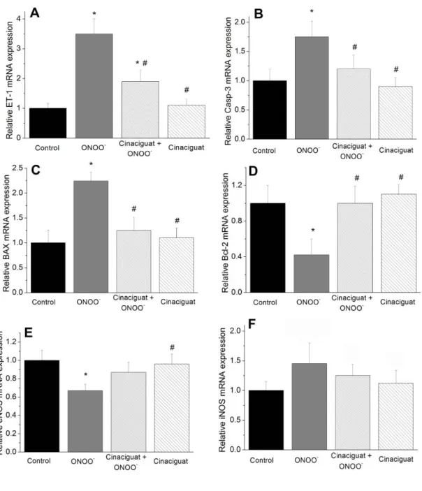
6.1.4 Cinaciguat decreases nitro-oxidative stress and DNA strand breaks in aortic rings exposed to peroxynitrite ... 38
6.1.5 Cinaciguat increases cGMP levels in aortic rings exposed to peroxynitrite ... 38
6.1.6 Cinaciguat regulates gene expression ... 40
6.1.7 Effect of cinaciguat on cleaved caspase-3 level, Bax and Bcl-2 protein expression ... 41
6.2 Vascular dysfunction induced by cold preservation, in vitro reoxygenation and hypochlorite - effects of vardenafil ... 44
6.2.1 Contractile responses of aortic rings ... 44
6.2.2 Endothelium-dependent vasorelaxation of aortic rings ... 44
6.2.3 Endothelium-independent vasorelaxation of aortic rings ... 45
6.2.4 Vardenafil decreases DNA strand breaks in aortic rings ... 50
6.2.5 Vardenafil increases cGMP levels in aortic rings ... 50
6.2.6 Vardenafil regulates aortic gene expression ... 50
6.2.7 Effect of vardenafil on cleaved caspase-3 level, Bax and Bcl-2 protein
expression ... 51
7 Discussion ... 56
7.1 In vitro model of vascular dysfunction induced by peroxynitrite ... 56
7.2 Effect of cinaciguat on peroxynitrite induced vascular dysfunction ... 57
7.3 In vitro model of I/R injury caused vascular dysfunction ... 60
7.4 Effect of vardenafil on I/R induced vascular dysfunction ... 61
7.5 The possible protective down-stream mechanisms behind cGMP accumulation ... 62
7.6 Therapeutic perspectives of maintained cGMP levels against oxidative stress ... 64
8 Conclusion ... 66
9 Summary ... 67
10 Összefoglalás ... 68
11 Bibliography ... 69
12 Bibliography of the candidate's publications ... 88
List of publications related to the dissertation ... 88
List of publications not related to the dissertation ... 88
2 List of abbreviations
ACh acetylcholine
ADMA asymmetric dimethylarginine ADP adenosine diphosphate AMP adenosine monophosphate ATP adenosine triphosphate
BH2/BH4 dihydrobiopterin / tetrahydrobiopterin
BAX Bcl-2-like protein 4; pro-apoptotic regulator factor Bcl-2 B-cell lymphoma 2; anti-apoptotic regulator factor cAMP cyclic adenosine monophosphate
CAT catalase
cGMP cyclic guanosine monophosphate CO3-
carbonate radical CVD cardiovascular disease DNA deoxyribonucleic acid
eNOS endothelial nitric oxide synthase ERK extracellular signal-regulated kinase ET-1 endothelin-1
GSH-Px glutathione peroxidase GTP guanosine triphosphate H2O2 hydrogen peroxyde HIF hypoxia-inducible factor
HTK histidine-triptophane-ketoglutarate I/R ischemia reperfusion
ICAM intercellular cell adhesion molecules IHD ischemic heart disease
Il Interleukin
iNOS inducible nitric oxide synthase K+ATP ATP sensitive K+ channel LTB leukotrien B
MDA malondialdehyde MMA monomethylarginine MMP matrix metalloproteinase
MnSOD manganese superoxide dismutase
mPTP mitochondrial permeability transition pore NADH nicotinamide adenine dinucleotide (reduced)
NADPH nicotinamide adenine dinucleotide phosphate (reduced) NaOH sodium hydroxide
NF-κB nuclear factor kappa b NO. nitric oxide
NO2 nitrogen dioxide NOS nitric oxide synthases O2-. superoxide anion OCl- hypochlorite anion
.OH hydroxide anion ONOO- peroxynitrite anion PAF platelet activating factor PARP poly(ADP-ribose) polymerase PCR polymerase chain reaction PDE phosphodiestherase PE phenylephrine
PKG cGMP dependent protein kinase Rmax maximal relaxation
ROS reactive oxygen species S.E.M. standard error of the mean sGC soluble guanylate cyclase SNP sodium nitroprusside TNF tumor necrosis factor
TUNEL terminal deoxynucleotidyl transferase-mediated dUTP nick-end labeling VCAM vascular cell adhesion molecule
VEGF vascular endothelial growth factor VSMC Vascular smooth muscle cell
3 Introduction
3.1 Epidemiology of cardiovascular diseases
Cardiovascular disease (CVD) is the cause of more than 4 million deaths in Europe and over 1.9 million deaths in the European Union each year. Since decades, it is the leading cause of morbidity and mortality statistics in the developed part of the world. According to epidemiological studies, risk factors such as smoking, diabetes, hyperlipidemia, nutrition factors and age dispose changes in the cardiovascular system that in time may lead to CVD. The common in these changes are the atherosclerotic alterations in the vasculature. In most cases CVD manifests also in form of coronary artery disease, which is characterized by reduced blood supply to the heart. In Hungary, more than 10% of the population suffers in circulatory disorders of which half suffers in ischemic heart disease (IHD). Currently IHD is responsible for every fourth death and is prognosed to become the leading cause of death worldwide in ten years [1,2]. The mortality caused by the acute manifestations of IHD has been decreased by the high niveau invasive treatment of percutan coronary intervention, of which in Hungary more than 15 thousand performed annually. However, with the decrease of acute IHD mortality the prevalence of the chronic forms (heart failure) is increasing. It is estimated, that 26 million people have heart failure worldwide up to 6 million Europeans among them, with an incidence of 1 million people in the European Union and the USA alone. According to the approximations shared by the Hungarian Society of Cardiology, 3% of the adult population suffers in heart failure, 6-10% of elderly people above the age of 65. The overall CVD is estimated to cost the European Union nearly 196 billion Euro per year. The total cost of CVD in the EU consists of numerous sub-components: approximately just 54% is due to direct health care costs, 24% of it is due to productivity losses and 22% of it is known to be caused by the informal care of people with CVD [3].
The pathophysiology of cardiovascular diseases associated to hypercholesterolemia, atherosclerosis, hypertension, diabetes, cardiac insufficiency and the phenomenon of restenosis, ischemia-reperfusion injury and sepsis share many common attributes [4-8].
Growing evidence indicates, that in vivo formation of free radicals in the vascular wall
initiation and progression of these diseases [9, 10]. The vascular system, especially the endothelium itself is very sensitive to oxidative stress. Resulting from an exceeding production of reactive oxygen species, the increased oxidative stress has a critical role in determining vascular injury.
3.2 Ischemia reperfusion injury
Historically, the first suspicion of a severe injury caused by reperfusion dates back to the second world war, when Bywaters and Beal [11] noted fatal metabolic dysfunction followed by death after the release of crushed limbs in air-raid casualties. In full extent, tissue injury induced by reperfusion was described by Parks and Granger in 1986 [12]. Reimer and his colleagues [13] introduced the term lethal reperfusion injury in 1989 as they interpreted cell necrosis resulting from reperfusion and the role of free radicals and neutrophils in reperfusion injury.
The reperfusion of an ischemic organ is fundamental for its viability and functional recovery. However, the arrival of blood oxygen leads to a series of lesions. Reperfusion injury is a complex and paradoxical phenomenon, where cellular dysfunction and cellular death exacerbates after the restoration of blood supply to the preliminary ischemic tissues. When blood flow is reestablished to an organ or tissue exposed to prolonged ischemia, renewed blood flow aggravates tissue damage by causing additional injury. The arterial clamping/unclamping in vascular surgery, cardiac surgery with or without extracorporeal circulation, transplant surgery, the use of tourniquet in orthopedic surgery, haemorrhagic shocks, septicaemia and low blood flow states are only some examples from the operating theatre which illustrate daily ischemia- reperfusion injury in clinical practice [14].
The reperfusion stage can be broken up into two phases. First, initial return of blood to ischemic tissue results in the return of aerobic respiration. Due to molecular changes and the drop in the antioxidant capacities in the cells during hypoxic stage, enzyme systems in their modified state start the rapid generation of reactive oxygen species damaging cellular functions. The second phase of reperfusion injury elicits through these biochemical and cellular changes an acute inflammatory response that is
characterized by inflammation mediated by interactions among neutrophils, platelets and endothelium, resulting in further oxidant production and endothelial barrier failure [15, 16]. While a massive ischemic injury through the deprivation of oxygen more likely manifests in cellular necrosis, reperfusion injury is rather dominated by cellular apoptosis.
3.3 Endothelium and oxidative stress
Endothelial cells have multiple vital duties: they control vascular tonicity and local blood flow, modulate coagulation and inflammation, intervene in the immune system, control the transfer of micro- and macromolecules towards the interstitial region, convert prohormones into active hormones (angiotensin II) and intervene in the formation of new blood vessels. For the understanding of how oxidative stress leads to the damage of the endothelium and how endothelial dysfunction leads to impaired vascular function, the prior discussion of endothelial physiology, the source of reactive oxygen species, and their effect on cellular function is essential.
3.3.1 Endothelial physiology
Endothelial cells show vasodilator, anti-coagulant and anti-adhesive properties thus contributing to the upkeep of the balance between the pro- and anti-inflammatory mediators, haemostatic balance, cell-proliferation and vascular permeability [17]. The vasomotor tone is regulated by the release of vasodilator agents (such as nitric oxide, prostacyclin, bradykinin, endothelium derived hyperpolarizing factor), and vasoconstrictors (endothelin-1, angiotensin II, thromboxane). The predominant nitric oxide is a free radical with short, only a few seconds long biological half-life. Its synthesis is catalyzed by a family of nitric oxide synthases (NOSs) from the aminoacid L-arginine in a reaction requiring tetrahydrobiopterin (BH4) as a cofactor, and leads to the relaxation of smooth muscle cells by increasing intracellular cyclic guanosine- monophosphate levels. The constitutive members of the NOS family (endothelial nitric oxide synthase and neuronal nitric oxide synthase) produce constantly a small amount
aggregation and adhesion of leukocytes. Namely, endothelin-1 could be the counterpart of NO- by causing vasoconstriction, vascular smooth muscle cell proliferation through ET-A receptor, and release of inflammatory mediators (Il-1, 6, 8). The most important physiologic trigger of the constitutive NO. synthase of endothelial cells (eNOS) is shear stress (tangential distortion of endothelial cells) generated by blood flow [18]. A constant shear stress maintains the homeostasis of vascular endothelium, while the acute loss of shear stress (e.g. in ischemia) results in the depolarization of the cell and within short to the inactivation of eNOS [17].
Under normal oxygen tension, oxidative phosphorylation is the major source of energy production by mitochondria in the cell. This results in formation of ATP, which serves as energy currency of the cell. ATP is converted to ADP, and the energy released is used to maintain intracellular ion homeostasis and to cover the energy need of metabolic reaction. The complex process of ATP synthesis is carried out by the respiratory chain (electron transport chain) in the mitochondria. The respiratory chain is a set of enzymes embedded in the mitochondrial inner membrane. Each enzyme is responsible for a specific step, which causes release of electrons that are sequentially transferred down the chain; ATP is the end product. NADH dehydrogenase and cytochrome oxidase are two of the most important enzyme complexes involved in the electron transfer chain.
The superoxides generated by them are only produced in small amounts, and are quickly eliminated by the several scavenger mechanisms including enzymes such as enzyme manganese superoxide dismutase (MnSOD), catalase and glutathione peroxidase, and non enzymatic antioxidants such as vitamin E, vitamin C, beta-carotine and heme binding proteins [15, 19].
3.3.2 Changes under ischemic conditions
Hypoxia induces changes in various enzymes participating in the energy metabolism of the cells, and directly to the mitochondrial mechanisms themselves, causing uncoordinated production of ROS on resumption of oxygen supply during reperfusion.
Enzyme systems affected by I/R injury include cytochrome oxidase, xanthine oxidase, reduced nicotinamide adenine dinucleotide (phosphate) oxidase and the mitochondrial electron transport chain. I/R injury causes the jumble in the function of NADH
dehydrogenase and cytochrome oxydase enzymes, leading to excess superoxides that rapidly depleting stores of MnSOD fails to neutralize.
More than hypoxia, that refers to the absolute or relative reduction of oxygen supply, the term ischemia (Greek isch: restriction, and haema: blood) translates as the absolute or relative reduction, or cessation of blood flow to and from a tissue and generally coincides with the reduced tissue oxygen delivery/demand ratio of 2:1 or below. The severity of its impact on the parenchyma depend on the intensity and duration of the ischemia, on the type and metabolic need of the affected cell. Oxygen is essential for the homeostasis of human cells. The ultimate consumer of oxygen is the oxidative phosphorylation reaction in the mitochondria, that supplies the cell with energy in the form of ATP. The large number of metabolic reactions requiring these high-energy phosphates make cells dependent of their oxygen supply. Within a few seconds after the cessation of blood flow, the oxygen content of oxyhaemoglobin, myoglobin or neuroglobin is consumed [20, 21]. Low level of ATP production is maintained by the less effective anaerobic glycolysis mainly supplied by glycogenolysis, but the demand quickly exceeds the anaerobic production. As the cytosol turns to acidic by the accumulation of lactate and protons and by the reduction in the oxidation of nicotinamide adenine nucleotide phosphate (NADPH+) by mitochondria acidifies the cell, anaerobic glycolysis slows down [22]. The lack of ATP promotes the split of high energy phosphate from ADP and finally from AMP. In apoptotic cells pannexin hemichannels contribute to the release of ADP and AMP nucleotides out of the dieing cell, of which the 5’ecto-nucleotidase enzyme generates free adenosine in the extracellular space [23]. The generated adenosine can freely diffuse in and out of the cell, thus increasing the pool of xanthine oxidase substrates.
3.3.3 Endothelial pathophysiology in oxidative stress
Endothelial dysfunction is a corner-stone of cardiovascular diseases associated to hypercholesterolemia, hypertension, diabetes mellitus, ischemia reperfusion injury and sepsis [9, 10]. Under normal circumstances in healthy cells, a balance exists between the
During oxidative stress, the disruption of this balance favours ROS production thus impairing cellular functions on multiple levels via a wide range of processes: impaired vasodilator responses, oxidizing proteins and lipids in higher concentrations, causing the oxidative damage of the DNA and leading to apoptosis, autophagy or necrosis of the cell.
Oxidative stress through the inactivation of the constitutive nitric oxide synthase results in the decreased production of NO. and increased release of vasoconstrictors, expression of adhesion molecules (P-selectin), inflammatory mediators (PAF), leukotriene B4 (LTB4) and cytokines (Il-8) thus promoting a pro-inflammatory, pro- coagulant and proliferative state [24]. This condition is called endothelial activation.
The superoxide anion scavenges nitric oxide and forms peroxynitrite, triggering proinflammatory signals and inhibiting endothelial repair [25]. Superoxide has been demonstrated to play a key role in apoptosis through a reaction with nitric oxide to form peroxynitrite, which in turn induces tyrosine nitration and deleterious protein changes [26].
Methylarginine substratespőü, like asymmetric dimethylarginine (ADMA) and monomethylarginine (MMA) are the endogenous inhibitors of NOS, derived from the proteolysis of methylated arginine residues in various (nuclear) proteins by a group of enzymes (protein-arginine methyl transferases). These methylarginines are subsquently degraded to L-citrulline and dimethylamine by the enzyme dimethylarginine dimethylamine hydrolase (DDAH). The enzymatic activity of dimethylamine hydrolase has been shown to be decreased by oxidative stress causing the accumulation of NOS inhibiting ADMA and thus to decreased NO. production [27].
3.3.3.1 Reactive oxygen species
Reactive oxygen species include superoxide (O2-.), hydrogen peroxide (H2O2), hypochlorite (OCl-), hydroxyl ions (.OH) and peroxynitrite (ONOO-) which is one of the most harmful oxidant species from the reaction of superoxide and nitric oxide. They are produced by various exogenous sources such as neutrophyl granulocytes, macrophages or circulating xanthine oxidase, and by endogenous systems as well. Although NAD(P)H oxidase can be found in endothelial cells too, in neutrophil granulocytes and
platelets it is one of the most important exogenous source of ROS, especially involved in ischemia reperfusion injury [28]. The NAD(P)H oxidase-induced ROS production is more aggressive in neutrophils (oxidative burst) as compared with slower release in endothelial cells [29]. This enzyme produces superoxide that may interact with NO. [30]
thus producing reactive nitrogen species. By the oxidation of specific enzymes and co- factors, reactive oxygen species are capable to create self-perpetuating mechanisms to intensify their own production.
Figure 1. Generation of peroxynitrite and hypochlorite and subsequent free roots H2O2: hydrogen-peroxide; MPO: myeloperoxidase; NADP(H+): Nicotinamide adenine dinucleotide phosphate (reduced); NO.: nitric oxide; NOS: nitric oxide synthase; O2-.
: superoxide anion; .OH: hydroxide anion; R-NH2: organic amino group; R-NHCl:
organic N-chloramines; (HO)SCN-: (hypo)thiocyanous acid;
In some cases ROS might also act as a second messenger and have significant effect on signaling pathways involving mitogen-activated protein kinase (MAPK), extracellular signal-regulated kinase (ERK) Jun N-terminal kinase (JNK) as well as on regulatory proteins like NF-κB, hypoxia-inducible factor-1 (HIF-1), or activator protein- 1 (AP-1) [31]. In endothelial cells NF-κB is responsible for the up-regulation of cell adhesion molecules (VCAM-1, ICAM-1), ET-1, MMPs and VEGF thus enhancing leukocyte and platelet adhesion to the endothelial surface and leukocyte excavation into the vascular wall. Recruited leukocytes serve as exogenous sources of ROS thus further accelerating the oxidative stress [32]. Endothelial cells in response to TNF-α, Il-1β, platelet derived growth factor can generate endogenous ROS by cyclo-oxygenase and NADPH oxidase.
3.3.3.1.1 Peroxynitrite
The peroxynitrite (ONOO-) anion is a short-lived reactive oxidant species that is produced by the reaction of nitric oxide (NO.) and superoxide (O2-.
) radicals at diffusion controlled rates (Figure 1). The sites of peroxynitrite formation is associated with the sources of superoxide (such as plasma membrane NAD(P)H oxidases or mitochondrial respiratory complexes) in space and time, because though NO. is a relatively stable and highly diffusible free radical, superoxide has a much shorter lifetime and restricted diffusion across biomembranes [33]. On the other hand, peroxynitrite is able to cross cell membranes thus despite its short half-life at physiological pH (~10 ms) peroxynitrite generated from a cellular source can influence surrounding target cells within one or two cell diameters (~5-20 µm). In biological systems one fundamental reaction of ONOO- is to react with carbon dioxide (in equilibrum with physiological levels of bicarbonate anion) thus forming carbonate (CO3-) and nitrogen dioxide (NO2-) radicals [34]. These one-electron oxidants can oxidize amino acids thus creating radicals such as cysteinil from cysteine or tyrosyl from tyrosine. NO2-
can also undergo radical- radical termination reactions with biomolecules in a diffusion-controlled manner, resulting in nitrated compounds. Another fundamental but significantly slower reaction of peroxynitrite is the homolytic fission of its protonated form (ONOOH) to generate one-electron oxidant hydroxyl (HO-) and NO-2 radicals. Although this proton-catalysed
decomposition of ONOO- is a modest component of the in vivo reactivity of peroxynitrite, .OH and NO-2 radicals gain relevance in hydrophobic phases, resulting in the initiation of lipid oxidation, nitration and protein tyrosine nitration processes.
Moreover, ONOOH in the membranes may undergo direct reactions with transition metal centres such as hemin, membrane-associated thiols and lipids [25, 35].
Deoxyribose and purine nucleotides of the DNA are also vulnerable targets of peroxynitrite producing 8-oxo and 8-nitroguanine as major products, but it can also cause deoxyribose oxidation and single strand breaks [36]. Single strand DNA breakage is the obligatory inducer of the poly(ADP-ribose) polymerase (PARP) pathway.
Peroxynitrite formation results in cardiovascular dysregulation through various mechanisms; reduction of NO. biovailability, the inhibition of prostacycline synthase, MnSOD, mitochondrial NAD dehydrogenase (complex I), sarcoplasmic reticular calcium-ATP-ase are only a few examples [37].
Due to the very short half-life the steady-state concentration of peroxynitrite is low and cannot be directly measured in vivo. Indirectly, protein-3-nitrotyrosine (NT) as footprints of peroxynitrite give reliable information about the oxidative load of peroxynitrite [38].
3.3.3.1.2 Hypochlorite
Hypochlorous acid is a highly reactive cytotoxic agent generated by activated polymorphonuclear leukocytes. It has major role in immune defence, however hypochlorous acid is a major contributor of endothelial oxidative damage during reperfusion injury. Activated phagocytes, neutrophils release the heme enzyme myeloperoxidase (MPO), while their membrane-bound NADPH oxidase generates superoxide radicals (O2-.) and hence H2O2, via an oxidative burst (Figure 1). The reaction of MPO with H2O2 in the presence of chloride ions generates HOCl (the physiological mixture of hypochlorous acid and its anion present at pH 7.4). HOCl is reactive toward a variety of biological substances such as ascorbate, amines, thiols, sulfides and disulfides, nucleotides, DNA, proteins and unsaturated fatty acids.
Exposure of amino groups to hypochlorite leads to generation of long-lived and reactive
chloramide decomposition and glycosaminoglycan fragmentation. HOCl interacts with DNA in a rather slow, but very efficient manner, assumed by the concomitant denaturation. Unlike by .OH, denaturation is not primarily due to DNA fragmentation but by chlorination of amino- and heterocyclic NH-groups of the bases, with the consequences that the double strand dissociates into single strands due to the loss of hydrogen bonds. However, even a partial chlorination of DNA bases may interfere with vital biological functions and activate PARP pathway. The presence of low-valent transition metal ions (Cu2+ or Fe2+) facilitates the fragmentation of both the DNA and glycosaminoglycans by one-electron reduction of the chloramides [39-42].
3.3.3.2 eNOS uncoupling
Endothelial NOS is a dimeric enzyme with a reductase domain for NADPH and an oxidase domain for the co-factor oxygen and L-arginine. The co-factor BH4 is essential to catalyse the reaction of generating NO. and L-citrulline. When BH4 levels are inadequate, "eNOS uncoupling" may occur: uncoupling of NADPH oxidation and NO. synthesis, with oxygen instead of L-arginine as terminal electron acceptor, resulting in the formation of superoxide [43]. Moreover, the interaction between NO. and superoxide leads to the formation of peroxynitrite that further oxidises BH4 to BH2 which competitively replace eNOS-bound BH4 thus promoting eNOS uncoupling. Hence eNOS uncoupling is greatly dependent on intracellular BH4:BH2 ratio and by the decrease of BH4 relative availability eNOS becomes the most important endogenous ROS generator [44]. The salvage re-synthesis pathway of BH4 by the dihydrofolate- reductase requires NADPH. Peroxynitrite directly oxidizes this co-factor while creating H2O2, thus cytoplasmic NADPH level is rapidly decreasing in oxidative stress. Not only superoxide and peroxynitrite lead to eNOS uncoupling. Through the chlorination of L- arginine OCl- may damage NO. synthesis [45].
3.3.3.3 Other endogenous sources of ROS
Parallel to eNOS, increased oxidative stress causes the jumble in the function of cyclooxygenase, mitochondrial NADH dehydrogenase and cytochrome oxydase
enzymes, leading to excess superoxides that scavenger mechanisms are unable to neutralize.
The inducible NOS (iNOS) stimulated by different noxas affecting the endothelium (inflammation, oxidative stress) produces larger and more persistent amount of nitric oxide, thus leading to its detrimental actions: hypotension and transformation to peroxynitrite [17].
Xanthine dehydrogenase is an enzyme responsible for the safe transformation of hypoxanthine to uric acid. When oxidized by ROS, xanthine dehydrogenase is converted to xanthine oxidase and uses oxygen as a substrate for converting hypoxanthine to urate, meanwhile superoxides are produced as a by-product of the reaction [46]. This pathway gains special significance in ischemia reperfusion injury.
Reactive ferryl species have also been recognized as contributors to oxidative stress [47, 48] in trauma settings when myoglobin and haemoglobin are released into the plasma.
In vitro experiments have shown that under conditions as they prevail in IR injury, haemoglobin is oxidized to an intermediate reactive ferryl form and causes lipid peroxidation in endothelial cells [49]. The iron-dependent formation of highly reactive species is triggered by a cold-induced increase in the cellular chelatable iron pool [50, 51]. This is particularly important in cardiac surgery when hearts are perfused and stopped with cold preservation solution. As vascular grafts are also stored in hypothermic saline or preservation solutions, they often exhibit significant loss of structurally intact endothelium following reperfusion.
3.3.3.4 Lipid peroxidation
Lipid peroxidation is the direct damage of cellular and organelle membranes resulting in structural damage and release of various autolytic enzymes. Regardless of the nature of the free radicals, they attack the phospholipids of the cell and organelle membranes culminating in irreversible structural and functional damage.
Polyunsaturated fatty acids are more vulnerable to this insult than monounsaturated fatty acids [52]. Experimental evidence proves the prevalence of this mechanism in almost every organ system exposed to oxidative stress: kidney, retina, blood vessels,
process, biologically active by-products are formed, and as mediators, contribute to the self-perpetuating nature of this injury. One such secondary by-product produced is malonaldehyde (MDA). MDA level can be used as a marker of suffered oxidative stress in various disease conditions, such as eclampsia and renal ischemia reperfusion injury [59].
3.3.4 Ischemia reperfusion injury in organ preservation
In transplantation and cardiac surgery I/R injury is one of the determining factors of acute and chronic graft failure. Organs are preferably stored in cold (4ºC) preservation solutions of which a wide scale is known in the clinical practice, such as histidine-triptophane-ketoglutarate (HTK or Custodiol), or University of Wisconsin solutions. Vascular grafts are also often stored in heparinised blood or saline. The basic concept behind the usual cold storage is to slow down the ATP consuming metabolic activities of the cells during the ischemic period. However, there are some deleterious consequences of cold [60].
Cellular swelling: the impaired activity of Na+/K+ ATP dependent pump leads to changes in cellular substructures, to swelling and to formation of protruding pockets [61]. Na+ enters the cell creating a hyperosmolar enviroment and water influx.
Acidosis: Even at low temperature of preservation intracellular ATP content is depleting. In order to maintain ATP production for cellular processes under ischemic conditions the switch to the less effective anaerobic metabolism is promoted thus generating lactic acid and acidosis. Severe acidosis activates proteases and phospholipases causing lysosomal damage and cell death.
Calcium: Through the drop of cellular ATP content and the disturbances in Na+ balance, the active transport mechanisms cannot maintain Ca2+ homeostasis. In the cold phase accumulation leads to activation of calcium-dependent calpain activation and protein kinase C signaling, and to loss of cell structure by breakdown of cytoskeletal
spectrin [62]. Calpain activity increases in cold-stored cells, and further increases during rewarming [63].
Enzymes: proteases and MMPs may be activated due to cold preservation, leading to detachment of endothelial cells from the underlaying matrix [64, 65]. The other relevant family of enzymes activated during cold preservation is the apoptosis-related caspases.
During cold ischemia free iron is released form cytochrome P-450 [66]. In combination with hydrogen-peroxide free iron leads to severe ROS production.
3.4 NO - cGMP - PKG pathway in the vascular wall
It has been widely discussed previously, that NO. plays a crucial role in regulation of vascular relaxation, thrombocyte activation and clotting, but it is also responsible for the intracellular homeostasis of endothelial cells. At physiologically relevant concentrations NO. was shown to inhibit mitochondrial cytochrome oxidase [67], to inhibit activation of NF-kB [68], while enzymes such as caspase and cysteine protease, that are involved in apoptosis, were inhibited by S-nitrosylation in the presence of NO.[69].
The impairment of nitric oxide production by oxidative stress through NO. scavenging, eNOS uncoupling, or its overproduction by iNOS leads to endothelial and vascular dysfunction. The regulation of eNOS activity is complex due to a variety of factors, including shear stress, bradykinin, histamine, VEGF, thrombin, estrogen.
Acetylcholine increases eNOS activity in endothelial cells by binding to its muscarinic- 3 receptor thus causing Ca2+ signal. eNOS activity is regulated on transcriptional level and by post-transcriptional interactions and modifications, such as binding of calcium- dependent calmodulin, phosphorylation, acylation, sub-cellular compartmentalisation [70]. It has been shown, that NOS activation can inhibit NADPH oxidase via interfering with its assembly [71, 72] and changing its expression [73-75].
Soluble guanylate cyclase (sGC) is the downstream molecule in the NO-cGMP signaling pathway, and is responsible for the conversion of GTP to the messenger
heme-containing B-subunit. In normal conditions, NO. generated by the endothelial constitutive NOS acts as a paracrine mediator and diffuses into the neighbouring cells, such as vascular smooth muscle cells, platelets, even heart or brain cells. In these target cells NO. activates sGC by binding to its ferrous heme iron (Fe2+). By the binding of NO. the sGC-catalyzed conversion of GTP to cGMP is activated approximately 200- fold [76]. The generated cGMP activates cGMP-dependent phosphodiesterases, protein kinases, ion channels, thus exerting its effects such as the reduction of cytosolic calcium concentration and/or calcium desensitization of the contractile apparatus, which result in smooth muscle relaxation and vascular relaxation. The activation of the cGMP dependent protein kinase G (PKG) leads to the phosphorylation of proteins at the so- called maxi-potassium channels (large conductance calcium-activated potassium channel). This results in an outflow of potassium ions into the extracellular space with subsequent hyperpolarization, with inhibition or blockade of voltage-dependent calcium channels and therefore a decrease in intracellular Ca2+ ion concentrations [77]. The availability of the messenger cGMP is regulated by not only its synthesis, but through its degradation by phosphodiestherases, which are thereby also cornerstone regulators of the pathway. From eleven currently known members of the phosphodiestherase family more than seven may interact with cGMP, but in the cardiovascular system (including vessel wall) the cGMP selective phosphodiestherase-5 (PDE-5) is responsible dominantly for its metabolism. cGMP facilitates its own degradation by negative feed- back through the up-regulation and marked activation of PDE-5 [78].
Though the role of NO. in vasodilation is clearly established, the mechanism of action of cGMP and PKG still seems less defined. An early and consistent finding was that NO. donor drugs, cGMP analogues, and PKG all lower intracellular Ca2+ levels, especially when elevated with a Ca2+-mobilizing agonist [79]. Ca2+ is the major activation signal of the myosin light chain (MLC) kinase and cross-bridge cycling in SMCs.
Smooth muscle contraction and relaxation are tightly coupled to the phosphorylation and dephosphorylation, respectively, of the regulatory myosin light chain. Myosin chain phosphorylation state is determined by the relative activities of myosin light chain kinase (MLCK) and myosin light chain phosphatase (MLCP). MLCK phosphorylates
MLC leading to contraction, and MLCP dephosphorylates MLC, leading to relaxation.
Both MLCK and MLCP are highly regulated, MLCK is activated by the binding of calcium/calmodulin and thus is the primary mechanism linking intracellular calcium concentration to smooth muscle contractility. MLCP is regulated by both vasodilator and vasoconstrictor stimuli, and is therefore responsible for much of the calcium- independent regulation of contractility [80]. PKG interacts directly with the regulatory myosin binding subunit of MLCP, which is critical for the PKG dependent activation of MLCP.
There are at least three major known mechanisms by which PKG appears to lower intracellular Ca2+ levels in VSMCs. First, PKG activates Ca2+ sensitive potassium (K+Ca) channels, leading to hyperpolarization and to an inhibition of voltage-dependent Ca2+ channels. The second well described mechanism is that PKG increases the uptake of Ca2+ into intracellular stores such as sarcoplasmatic and endoplasmatic reticulum through the phosphorylation of phospholamban and activation of Ca2+ ATP-ases [81, 82]. The importance of phospholamban phosphorylation in vivo VSMC relaxation has been questioned using studies on phospholamban-deficient mice, where VSMCs demonstrated predictable enhanced sensitivity to agonist-evoked contraction. On the other hand, the animals do not display impaired vascular relaxation in response to NO. donor drugs [83]. However, the critical role of phospholamban phosphorylation in the K+Ca regulation has also been evidenced [84]. Thirdly, through the inhibition of PIP- IP3-Ca2+ signaling pathway. It has been shown, that PKG catalyzes phosphorylation of the type I inositol 1,4,5- trisphosphate receptor in the SR in vitro in rat aorta [85-87]
which decreases Ca2+ release from the intracellular compartment. Many of the PKG substrate proteins are yet uncharacterized. Some of these are relatively small molecular weight entities. One such protein is the heat-shock protein (HSP) -20 [88]. HSP-20 has been described to play a crucial role in the ischemic preconditioning in rat and dog hearts, and in the NO-cGMP dependent relaxation as a regulator of myosine phosphorylation and thin filament function [89].
3.4.1 The therapeutic perspective of the NO - sGC - cGMP pathway
Impaired endothelial function is correlated with cardiovascular diseases, therefore therapeutic strategies aimed at limiting vascular oxidative stress and improving endothelial function may have clinical benefits. Organic nitrates act as a source of NO., but drug tolerance develops when used as sustained therapy [90]. Their efficacy is limited by the absence of clinically relevant anti-platelet activity [91] and the inability to activate NO-insensitive sGC. Oxidative stress affects the heme-containing NO. receptor of sGC by both decreasing its expression and potentially impairing NO- induced activation [92]. Oxidative stress leads to the oxidation of sGC and to the dissociation of the heme thereby inactivating the enzyme and destines to degradation [93]. Moreover, in cardiovascular diseases due to oxidative stress significantly increased PDE-5 expression was detected to accelerate cGMP degradation [78]. As a result, NO. donors and other pharmacological agents that protect vascular function through NO- dependent activation of sGC may not be as beneficial in the setting of oxidative stress or I/R injury. As an alternative therapeutic approach, a novel class of drugs is aimed to modulate the NO-sGC-cGMP signal transduction pathway (Figure 2A).
3.4.1.1 Cinaciguat
Cinaciguat (BAY 58-2667, molecular mass: 565.697 g/mol) is a compound that activates NO- and heme-independent sGC but devoid tolerance and potential cytotoxic actions of NO. (Figure 2B). It triggers selectively a state of sGC that is indistinguishable from the NO-insensitive oxidized/heme-free state of the enzyme [94].
In preclinical studies, the sGC activator cinaciguat has been shown to bypass the impaired NO-sGC-cGMP pathway by activation of the oxidized (Fe3+)/heme-free forms of sGC and to preferentially dilate the diseased versus non-diseased vasculature [76, 95- 97]. In a phase I clinical trial in healthy human participants, intravenously administered cinaciguat had a favourable safety profile and was well tolerated [98]. Moreover the phase IIa clinical study proved its effectivity in patients with acute decompensated heart failure [99].
3.4.1.2 Vardenafil
Vardenafil (molecular mass: 488.604 g/mol) is a selective PDE-5 inhibitor, an exciting small molecule, that hinders the degradation of cGMP by inhibiting its predominant regulator enzyme (Figure 2C).
Vardenafil is a drug approved by the US Food and Drug Administration (FDA) with the primary indication of the treatment of erectile dysfunction in men, and its effectivity is supported by clinical studies [100]. However, its cardiovascular effects has already been widely discussed. As previously mentioned, oxidative stress leads to increased PDE-5 activity. Intracellular cGMP accumulation is proven to reduce tissue injury in conditions associated with increased free radical release and oxidative stress [101, 102]. Vardenafil was shown to have beneficial effects against myocardial I/R injury after preconditioning-like treatment in rabbits [103] and to have advantageous protective effect on vascular endothelium [104]. The protective effect of PDE-5 inhibition on endothelial dysfunction following I/R injury was also demonstrated in a human study, nevertheless sildenafil was used [105].
A)
B) C)
Figure 2. (A) The schematic figure of the eNOS-sGC-PKG pathway with the pharmacologic targets of cinaciguat and vardenafil; molecular structure of (B) Cinaciguat (C36H39NO5) and (C) Vardenafil (C23H32N6O4S);
NO.: nitric oxide; NOS: nitric oxide synthase; O2-.
: superoxide anion; ONOO-: peroxynitrite; PKG: cGMP dependent protein kinase; PDE-5: phosphodiestherase-5;
sGC: soluble guanylate cyclase;
4 Objectives
Based upon the described mechanisms how oxidative stress leads to endothelial and vascular dysfunction, the present studies investigate whether increased cGMP levels contribute to the protection of vascular function and structure against acute oxidative stress.
1. The aims of the first in vitro model of vascular oxidative stress induced by peroxynitrite incubation was:
the investigation of vascular dysfunction and the contribution of decreased cGMP level to it after an acute oxidative stress;
testing the effect of the soluble guanylate cyclase activator cinaciguat on vascular dysfunction induced by peroxynitrite and underlying cellular and molecular changes in the vessel wall;
2. The aim of the second model of vascular oxidative stress induced by the model of in vitro ischemia and reperfusion was:
the investigation of I/R injury on vascular function, structure and cGMP levels;
testing the effect of the selective phosphodiesterase -5 inhibitor vardenafil-maintained cGMP levels on vascular dysfunction induced by in vitro I/R injury;
As a summary, the main goal of the studies was to establish novel potent therapeutic strategies facilitating the NO-sGC-cGMP pathway for ameliorating the endothelial and vascular dysfunction associated with acute oxidative stress.
5 Methods
5.1 Experimental models
5.1.1 In vitro model of vascular dysfunction induced by peroxynitrite
exposure
Thoracic aortic rings were isolated from rats. In organ bath experiments for isometric tension the effect of in vitro peroxynitrite exposure on vasoconstriction, endothelium-dependent and independent vasorelaxation was measured as described detailed below. Endothelial injury was induced by incubating the isolated aortic rings in peroxynitrite (200 µmol/L) for 30 minutes.
5.1.2 In vitro model of vascular dysfunction induced by long term cold
preservation, reoxygenation and hypochlorite exposure
Thoracic aortic rings were isolated from rats and incubated in cold hypoxic solution for 24 hours. In organ bath experiments for isometric tension the effect of in vitro hypochlorite exposure on vasoconstriction, endothelium-dependent and independent vasorelaxation was measured as described below. Endothelial injury was induced by reoxygenation and incubating the isolated aortic rings in hypochlorite (200 µmol/L) for 30 minutes.
5.2 Animals
Male Sprague-Dawley rats (250-330 g, Charles River, Sulzfeld, Germany) were housed in a room at constant temperature of 22 ± 2 °C with 12-hour light/dark cycles and were fed a standard laboratory rat diet and water ad libitum. All procedures concerning animals were conformed to the Guide for the Care and Use of Laboratory Animals prepared by the Institute of Laboratory Animal Resources and published by the National Institutes of Health (NIH Publication No. 86–23, revised 1996). The
investigations were reviewed and approved by the local Ethical Committee for Animal Experimentation.
5.3 Experimental groups and treatment protocols
5.3.1 Experimental groups and treatment for cinaciguat experiments
Rats were treated orally 2 times at an interval of 17 hours with vehicle (1%
methylcellulose solution) or with the sGC activator cinaciguat (10 mg/kg). One hour after the last treatment, animals were exsanguinized. After excision and preparation of the descending thoracic aorta (as described below), aortic rings were placed in Krebs- Henseleit solution (118 mmol/L NaCl, 4.7 mmol/L KCl, 1.2 mmol/L KH2PO4, 1.2 mmol/L MgSO4, 1.77 mmol/L CaCl2, 25 mmol/L NaHCO3, and 11.4 mmol/L glucose;
pH=7.4) at 37 °C, aerated with 95% O2 and 5% CO2 and divided into 4 groups (n= 12- 15 rings in each group from N= 4-5 animals) as follows: Control group (rats pretreated orally with methylcellulose vehicle, then aortic rings were incubated for 30 minutes in 4,7% NaOH vehicle), Peroxynitrite group (pretreatment of rats with methylcellulose, aortic rings were exposed to 200 µmol/L peroxynitrite), Cinaciguat + peroxynitrite group (pretreatment of rats with cinaciguat, then aortic rings exposed to peroxynitrite), and Cinaciguat group (pretreatment of rats with cinaciguat, exposure of aortic rings to NaOH). The concentration of peroxynitrite was chosen on the basis of previous studies [106].
5.3.2 Experimental groups and treatment for vardenafil experiments
Rats were euthanized by an overdose of sodium pentobarbital before exsanguination. After the excision and preparation of the descending thoracic aorta (as described below), aortic rings were divided into 5 groups (n= 15-20 in each group form N= 4-5 animals): Saline group (aortic rings were incubated in physiological saline for 24 hours at 4 ºC), Vardenafil groups (aortic rings were divided and incubated in 4 different concentration (10-12 mol/L, 10-11 mol/L, 10-10 mol/L, 10-9 mol/L) of PDE-5
ischemic storage, and received no hypochlorite exposition). The solutions were previously equilibrated for 15 minutes with nitrous oxide to extrude oxygen from the solution.
5.4 Preparation of isolated aortic rings
Rats were euthanized with sodium pentobarbital (60 mg/kg intraperitoneally) before exsanguination. The left chamber was pierced with a 4G needle and 20 ml Krebs–Henseleit solution was infused at a speed of 1ml/sec transcardially to wash out any blood from the aorta. Then, thoracic aorta was carefully excised, cleaned from connective tissue and cut transversely into 4 mm wide rings (n=4 from each animal with a mean diameter of 1.7-1.8 mm) under an operation microscope. Special attention was paid to avoid damaging the endothelium.
5.5 In vitro organ bath experiments
Isolated aortic rings were mounted on stainless steel hooks in individual organ baths (Radnoti Glass Technology, Monrovia, CA, USA), containing 25 ml of Krebs–
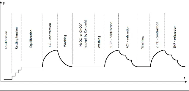
Henseleit solution at 37 °C and aerated with 95% O2 and 5% CO2. Isometric contractions were recorded using isometric force transducers of a myograph (159901A, Radnoti Glass Technology, Monrovia, CA, USA), digitalized, stored and displayed with the IOX Software System (EMKA Technologies, Paris, France). The aortic rings were placed under a resting tension of 2g (found optimal in preliminary experiments [106, 107]) and equilibrated for 60 minutes. Tension was periodically adjusted to the desired level during this period and the Krebs–Henseleit solution was changed in every 30 minutes. At the beginning of each experiment, the maximal contraction forces in response to potassium chloride (KCl, 80 mmol/L) were determined and then aortic rings were washed until the resting tension was obtained again. Afterwards, to simulate free radical burst which occurs usually in vivo during reperfusion [108], determined by the experimental setup, 200 μmol/L hypochlorite or 200 μmol/L peroxynitrite was added to the baths for 30 minutes, then washed out. Aortic preparations were preconstricted with α-adrenergic receptor agonist phenylephrine (10−6 mol/L) until stable plateau was
reached, and relaxation responses were examined by adding cumulative concentrations of endothelium-dependent dilator acetylcholine (10−9–10−4 mol/L). For testing relaxing response of smooth muscle cells, a direct nitric oxide donor, sodium nitroprusside (10−10–10−5 mol/L) was used. Half-maximal response (EC50) values were obtained from individual concentration–response curves by fitting experimental data to a sigmoidal equation using Origin 7.0 (Microcal Software, Northampton, USA). Contractile responses to phenylephrine are expressed as percent of the maximal contraction induced by KCl. The sensitivity to vasorelaxants was assessed by pD2=−log EC50 (mol/L), vasorelaxation (and its maximum [Rmax]) is expressed as percent of the contraction induced by phenylephrine (10−6 mol/L).
Figure 3: Organ bath protocol
The phases of the organ bath experiments on time (t) - force (F) scale.
ACh: acetylcholine; KCl: potassium chloride; NaOCl: sodium hypochloride; PE:
phenylephrine; SNP: sodium nitroprusside;
5.6 Histopathological processing
Aortic segments from each experimental group were fixed in paraformaldehyde solution (4%) and embedded in paraffin. 3-μm-thick sections cut by microtome were placed on adhesive slides.
5.6.1
Immunohistochemical staining
5.6.1.1 Nitrotyrosin immunohistochemical staining
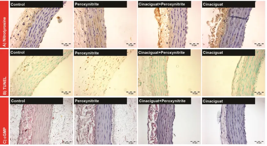
According to previously described methods [109], we performed immonohistochemical staining on aortic rings for nitrotyrosine, a marker of peroxynitrite-mediated damage. Peroxynitrite has very short lifetime, therefore it's generation is usually characterized by the “footprint” of peroxynitrite generation nitrotyrosine, a product of the reaction of peroxynitrite and tyrosine.
Immunohistochemical staining for nitrotyrosine was performed by using polyclonal sheep anti-nitrotyrosine antibody (OXIS, Portland, OR, USA) incubation was performed (1:80) for 2 hours at room temperature. Negative controls were performed by omitting the primary antibody. Sections were counterstained with Gill’s hematoxylin, mounted with Permount, and coverslips were placed on the section.
5.6.1.2 Cyclic GMP immunohistochemical staining
Cyclic GMP immunohistochemical staining was performed for the identification of intracellular cGMP content. Rehydrated sections were blocked (3% goat serum), rabbit polyclonal anti- cGMP primary antibody (Abcam plc, Cambridge, UK) incubation was performed (1:1000) for 2 hours at room temperature. Tissue sections were overnight incubated at 4ºC [110], then secondary biotinylated anti-rabbit immunglobuline E (BioGenex, CA, USA) incubation followed which allowed reacting with alcaline phosphatase-conjugated streptavidin (BioGenex, CA, USA). A red reaction product at the site of the target antigen was formed by use of Fast Red substrate (DakoCytomation, Hamburg, Germany). Negative controls were performed by omitting the primary antibody. Sections were counterstained with Gill’s hematoxylin, mounted with Permount, and coverslips were placed on the section.
5.6.2 Terminal deoxynucleotidyl transferase-mediated dUTP nick-end labeling (TUNEL) assay
TUNEL assay was performed for detection of DNA strand breaks (free 3'-OH DNA ends). The detection was performed using a commercial kit according to the protocol provided by the manufacturer (Chemicon International, Temecula, CA, USA).
Rehydrated sections were treated with 20μg/ml DNAse-free Proteinase K (Sigma- Aldrich, Taufkirchen, Germany) to retrieve antigenic epitopes, followed by 3%
hydrogen-peroxide to quench endogenous peroxidase activity. Free 3′-OH termini were labeled with a reaction mixture of terminal deoxynucleotidyl transferase and digoxigenin-deoxynucleotidyl (dUTP) for 1 hour at 37°C. Incorporated digoxigenin- conjugated nucleotides were detected using a horseradish peroxidase–conjugated anti- digoxigenin antibody and 3,3′-diaminobenzidine. Sections were counterstained with methylgreen. Dehydrated sections were cleared in xylene and mounted with Permount (Fischer Scientific, Germany), and coverslips were applied.
5.6.3 Quantification of immunostainings and TUNEL assay
Semiquantitative histomorphological assessment was performed on all of the stained specimens of cinaciguat and vardenafil projects in a blinded fashion using conventional microscopy. The results were expressed with a scoring system. Section with the most intense labeling signals was used as reference for maximum labeling intensity. All other tissue sections were comparatively evaluated. Colours were measured using densitometry and sorted in four colour classes: one class for background staining and three classes for positively stained areas. On the basis of the measured intensity, the colour classes were coupled with score values 0-4 where 0 meant no positive staining and 4 meant extensive staining. Using the Cell-A software (Olympus Soft Imaging Solutions GmbH, Germany) we measured the area of the objects in each class in each field, and assigned an area score (0 = 0%, 1 = up to 30%
positive cells, 2 = 31–60% positive cells, 3 = 61–90% positive cells, 4 = >91% positive cells), and calculated an average score for the whole picture (intensity score multiplied by area score, 0–16). Finally, each specimen was characterized by 4 adjacent fields.
For the assessment of TUNEL-labeled cells four different fields were pictured with digital camera from each section at a magnification of x200. TUNEL positive and negative cell nuclei were counted and the TUNEL positive cell nuclei were calculated as percentage of total cell number.
5.7 Quantitative Real-Time Polymerase Chain Reaction (PCR)
After homogenization of the chosen aortic rings total RNA was isolated from the RNeasy Fibrous Tissue Mini Kit (Qiagen, Hilden, Germany). RNA concentration and purity were determined at 260, 280, and 230 nm wave-lengths with spectrophotometer.
Reverse transcription was performed with the QuantiTect Reverse Transcription Kit (Qiagen, Hilden, Germany) using 400 μg RNA in a volume of 20 μL. Quantitative real- time PCR was performed on a LightCycler 480 system with the Universal ProbeLibrary probes (Roche, Mannheim, Germany). Primers were obtained from TIB Molbiol (Berlin, Germany), their sequences and UPL probes used are represented on Table I.
Evaluation was performed with LightCycler 480 SW1.5 software (Roche, Mannheim, Germany).
Efficiency of the PCR reaction was confirmed with standard curve analysis. Every sample was quantified in duplicate, normalized to glyceraldehyde-3-phosphate dehydrogenase (GAPDH) expression.
5.8 Western-blot analysis
Proteins were extracted from the tissue homogenisate, and concentration was determined. Homogenates (15 µg) were denatured, separated on sodium dodecyl sulfate polyacrylamide gel, transferred to polyvinylidene fluoride membrane. Membranes were blocked, incubated overnight with primary antibodies specific to p17 caspase-3 fragment (1:1000, Abcam, Cambridge, UK), Bax (1:200, Abcam, Cambridge, UK), Bcl- 2 (1:100, Abcam, Cambridge, UK). Blots were incubated for 1 hour with horseradish peroxydase conjugated secondary antibody (1:5000, Santa Cruz Biothechnology, Heidelberg, Germany). Immunoreactive protein bands were developed using Enhanced Chemiluminescence system (PerkinElmer, Rodgau-Juegesheim, Germany). Band
intensities were detected with Hyperfilm ECL System. For GAPDH housekeeping (1:500, Santa Cruz Biothechnology, Heidelberg, Germany) incubation and detection was repeated after stripping the blots form primary antibodies (Restore Plus Western Blot Stripping Buffer, Thermo Scientific, Rockford, USA). Target protein densities were normalized to housekeeping densities of the same samples, respectively.
5.9 Statistical Analysis
Data were tested for normal distribution (Shapiro-Wilk) and where met the requirements for parametric analysis, means were tested by one-way ANOVA followed by Student's unpaired t-test with Bonferoni’s correction test. For the analysis of PCR results Kruskal-Wallis one-way analysis of variance with Dunn's post hoc test was used.
A p value <0.05 was considered statistically significant.
5.10 Preparation and application of chemical reagents
Cinaciguat (BAY 58-2667), an amino dicarboxylic acid, was kindly provided by Bayer HealthCare (Wuppertal, Germany). It was dissolved in 1% methylcellulose solution vehicle and administered orally at a dose of 10 mg/kg at a volume of 10 ml/kg.
The application and dosage of cinaciguat have been determined according to the pharmacokinetic and dynamic properties [98] as well as to the results of previous rodent experiments [111]. Vardenafil was provided by Bayer HealthCare (Wuppertal, Germany) and was diluted in physiologic saline to 10-12 mol/L, 10-11 mol/L, 10-10 mol/L and 10-9 mol/L. Peroxynitrite (Calbiochem, San Diego, CA, USA) was diluted with 4.7% NaOH. Sodium-hypochlorite solution (AppliChem, Darmstadt, Germany) was diluted with distilled water. Phenylephrine, acetylcholine and sodium nitroprusside (Sigma-Aldrich, Germany) were dissolved in normal saline.
Table I
The sequences for the forward (F) and reverse (R) primers (from 5’ to 3’) and Universal Probe Library (UPL) probes
Assay Sequence UPL probes
BAX F: 5’-TAGCAAACTGGTGCTCAAGG
R: 5’-GCCACCCTGGTCTTGGAT 69
Bcl-2 F: 5’-GTACCTGAACCGGCATCTG
R: 5’-GGGGCCATATAGTTCCACAA 75
Caspase-3 F: 5’-AAACCTCCGTGGATTCAAAA
R: 5’-AGCCCATTTCAGGGTAATCC 56
Endothelin-1 F: 5’-TGTCTACTTCTGCCACCTGGA
R: 5’-CCTAGTCCATACGGGACGAC 115
eNOS F: 5’-TGACCCTCACCGATACAACA
R: 5’-CGGGTGTCTAGATCCATGC 5
GAPDH F: 5’-CTACCCACGGCAAGTTCAAT
R: 5’-ATTTGATGTTAGCGGGATCG 111
iNOS F: 5’-CAGCGGCTCCATGACTCT-3’
R: 5’-ATCTCCTGCATTTCTTCCTGAT-3’ 82
BAX: Bcl-2-like protein 4, Bcl-2: B-cell lymphoma, eNOS: endothelial nitric oxide synthase, GAPDH: glyceraldehyde-3-phosphate dehydrogenase, iNOS: inducible nitric oxide synthase
6 Results
6.1 Vascular dysfunction induced by peroxynitrite in vitro - effects of cinaciguat
6.1.1 Contractile responses of aortic rings
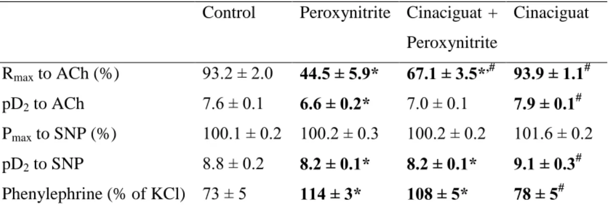
The contractile responses of aortic segments to phenylephrine (10-6 mol/L), an α1- adrenergic agonist are shown in Table II. Incubation of aortic rings with peroxynitrite significantly increased the phenylephrine-induced maximum contraction compared to the control rings. However, treatment of rats with cinaciguat did not significantly reduce increased contractile responses to phenylephrine. In the absence of peroxynitrite, cinaciguat treatment did not have any effect (Table II).
Table II.
Values of maximal relaxation (Rmax, %) and pD2 (affinity) to the vasorelaxant actions of acetylcholine (ACh) and sodium nitroprusside (SNP), and contraction values induced by phenylephrine (PE % of KCl) in percentage of the contraction induced by 0.1 mol/L potassium-chloride caused depolarization in rat thoracic aortic rings.
Control Peroxynitrite Cinaciguat + Peroxynitrite
Cinaciguat
Rmax to ACh (%) 93.2 ± 2.0 44.5 ± 5.9* 67.1 ± 3.5*,# 93.9 ± 1.1# pD2 to ACh 7.6 ± 0.1 6.6 ± 0.2* 7.0 ± 0.1 7.9 ± 0.1# Pmax to SNP (%) 100.1 ± 0.2 100.2 ± 0.3 100.2 ± 0.2 101.6 ± 0.2 pD2 to SNP 8.8 ± 0.2 8.2 ± 0.1* 8.2 ± 0.1* 9.1 ± 0.3# Phenylephrine (% of KCl) 73 ± 5 114 ± 3* 108 ± 5* 78 ± 5# Values represent mean ± S.E.M. of 12-15 experiments.
Statistical relevance is highlighted with bold.
*p < 0.05 versus control;
# p < 0.05 versus peroxynitrite group;
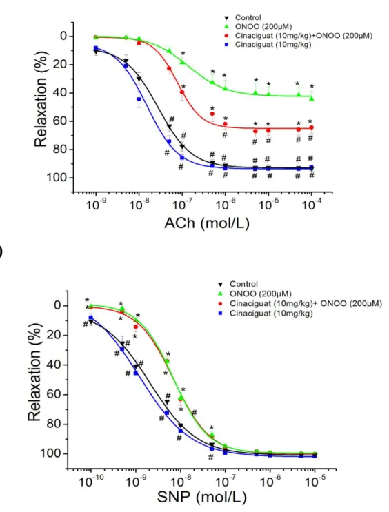
6.1.2 Endothelium-dependent vasorelaxation of aortic rings
In aortic rings precontracted with 10-6 mol/L phenylephrine, 10-9 to 10-4 mol/L acetylcholine induced a concentration-dependent relaxation. In contrast, exposure of aortic rings to the reactive oxidant peroxynitrite (200 µmol/L) for 30 minutes significantly attenuated the maximal relaxation to acetylcholine and decreased pD2 values for the concentration-response curves as compared to control (NaOH only) segments (Table II, Figure 4A). Treatment of rats with cinaciguat significantly improved the acetylcholine-induced, endothelium-dependent, NO-mediated vasorelaxation after exposure of aortic rings to peroxynitrite. In the absence of peroxynitrite, cinaciguat treatment did not alter maximal relaxation and the sensitivity to acetylcholine compared with the control group (Table II, Figure 4A).
6.1.3 Endothelium-independent vasorelaxation of aortic rings
Figure 4B shows concentration-dependent relaxations induced by 10-10 to 10-5 mol/L sodium nitroprusside, an endothelium-independent vasodilator. In contrast to acetylcholine, maximal relaxation did not differ significantly among the different experimental groups (Table II, Figure 4B). However, incubation of aortic rings with peroxynitrite caused a significant shift of the sodium nitroprusside concentration- response curves to the right. Cinaciguat has no effect on this level of damage (Table II, Figure 4B). In the absence of peroxynitrite, treatment of rats with cinaciguat did not alter maximal relaxation and the sensitivity to acetylcholine compared with the control group (Table II, Figure 4B).
A)
B)
Figure 4. Effect of cinaciguat on vascular relaxation in rat thoracic aortic ring after peroxynitrite exposure
(A) Acetylcholine (ACh)-induced endothelium-dependent vasorelaxation; (B) sodium nitroprusside (SNP)-induced endothelium-independent vasorelaxation; Each point of the curves represents mean ± S.E.M. of 12-15 experiments in thoracic aortic rings of the different groups. p < 0.05: * vs. Control; # vs Peroxynitrite group.