https://doi.org/10.1007/s00408-019-00236-1 RESPIRATORY PHYSIOLOGY
Lung Function Changes are More Common in Marfan Patients Who Need Major Thoracic Surgery
Abigel M. Kolonics‑Farkas1 · Bence Agg2,3,4 · Kalman Benke2,3 · Balazs Odler1,5 · Aniko Bohacs1 · Zsuzsanna Kovats1 · Zoltan Szabolcs2,3 · Veronika Müller1
Received: 24 January 2019 / Accepted: 2 May 2019 / Published online: 14 May 2019
© The Author(s) 2019
Abstract
Introduction Marfan syndrome is a genetic disorder affecting the connective tissue. Changes in lung tissue might influence respiratory function; however, a detailed respiratory functional assessment according to the need for major thoracic surgery is missing.
Methods Comprehensive pulmonary examinations were performed in 55 Marfan patients including respiratory symptoms, lung function (LF) testing using European Coal and Steel Community (ECSC) reference values, TLCO and quality of life measurements. Groups included patients who did not need surgery (Mf, n = 32) and those who underwent major thoracic surgery (Mfop, n = 23).
Results Respiratory symptoms affected 20% of patients. Scoliosis was significantly more frequent in the Mfop group. LF demonstrated in all Marfan patients a tendency towards airway obstruction (FEV1/FVC = 0.77 ± 0.10), more prominent in Mfop patients (0.74 ± 0.08 vs. Mf: 0.80 ± 0.11; p = 0.03). Correction of LF values using a standing height modification by arm span (Hcorrected) revealed additional changes in FVC and FEV1. TLCO and quality of life did not differ between groups.
Conclusions Marfan syndrome is associated with airway obstruction, especially in patients who have undergone major tho- racic surgery, indicative of more severe connective tissue malfunction. The use of arm span for height correction is suitable to evaluate LF changes in this special patient group including patients with significant scoliosis.
Keywords Marfan syndrome · Musculoskeletal disorder · Lung function testing · Airway obstruction · Thoracic surgery
Introduction
Marfan syndrome is a systemic, autosomal dominantly inherited connective tissue disorder, first described in 1896 by Antoine Marfan [1, 2]. In 1991, Francesco Ramirez iden- tified the underlying changes in the glycoprotein fibrillin 1, encoded by the FBN1 gene, located on chromosome 15 at position 15q21.1 [3]. In approximately 25% of cases, a de novo mutation can be observed [4]. Fibrillin 1, a principal component of microfibrils, plays a key role in the forma- tion and protection of the extracellular matrix [5]. Microfi- brils support elastin deposition, and are therefore essential components of elastic fibres [6]. The prevalence of Mar- fan disease is about 0.2 ‰ [7]. Since this condition is the consequence of connective tissue weakness, it has diverse symptoms. To ease the diagnostic process, the main symp- toms have been collected to a unified nosology (Ghent cri- teria, 1996) [7]. In 2010, a revision of the criteria abolished major and minor criteria and emphasised the value of genetic
Abigel M. Kolonics-Farkas and Bence Agg contributed equally to this work.
* Abigel M. Kolonics-Farkas
kolonics-farkas.abigel@med.semmelweis-univ.hu
1 Department of Pulmonology, Semmelweis University, Budapest, Hungary
2 Heart and Vascular Centre, Semmelweis University, Budapest, Hungary
3 Hungarian Marfan Foundation, Budapest, Hungary
4 Department of Pharmacology and Pharmacotherapy, Semmelweis University, Budapest, Hungary
5 Clinical Division of Nephrology, Department of Internal Medicine, Medical University of Graz, Graz, Austria
testing [8]. Regarding lung manifestations, little information is available on the effects of connective tissue changes in the respiratory system; only a few pleuropulmonary abnor- malities are known. Chest deformities or dissection of the ascending aorta can affect the mechanics of the ventilatory pump. Structural changes to the lungs can lead to apical blebs and bullae or result in spontaneous pneumothorax [9, 10]. Sleep apnoea is also observed as a consequence of the involvement of the upper airways [11].
Lung function (LF) values measured by spirometry and plethysmography are influenced by thoracic structures such as the airways, lung parenchyma, pleura and muscles; thus, functional changes in LF parameters used in routine clini- cal practice might be influenced by Marfan syndrome [12].
However, the reference values used in patients with the spe- cial body measurements characteristic of Marfan syndrome can be misleading, and comparative measures are lacking [13, 14] . In the present study, our aim was to assess changes to the respiratory system in this rare inherited connective tissue disorder using different reference equations.
Materials and Methods
Study SubjectsThe study had a cross-sectional design. Following a written inquiry, 55 Caucasian patients from the National Marfan Registry (established and supervised by the Hungarian Mar- fan Foundation) agreed to participate in the study. Patients were diagnosed with Marfan syndrome using the revised Ghent nosology [8] and/or genetic confirmation (Table 3).
Study Design
Pulmonary examinations were voluntary. After signing the informed consent, a detailed respiratory assessment was carried out in the Department of Pulmonology, Sem- melweis University, Budapest, Hungary between the 31 March 2015 and 4 September 2017. Exclusion criteria were age < 16 years old and major thoracic surgery within 6 months before the assessment. Major thoracic surgery was usually prophylactic aortic root surgery [15, 16] or chest wall surgery and spine correction.
Data on respiratory symptoms (dyspnoea, cough, sputum, chest pain), smoking history, sex, age, height, bodyweight, body mass index (BMI) and arm span (cm) were collected. All patients underwent arterialised ear- lobe blood gas analysis (Cobas b 221, Roche, Budapest, Hungary), chest X-ray and fluoroscopy, laboratory testing and electrocardiography. The 6-minute walk test (6MWT) was performed to measure exercise capacity according to American Thoracic Society (ATS) guidelines [17]. The
extent of scoliosis was measured according to the Cobb method [18]. To assess general quality of life (QoL), the QoL Visual Analogue Scale (VAS) was used. To iden- tify patient health-related conditions, the COPD Assess- ment Test (CAT®, Hungarian version) [19] and modified Medical Research Council Dyspnoea Scale (mMRC) were applied [20].
The study protocol was approved by the Semmelweis University Regional and Institutional Committee of Sci- ence and Research Ethics (TUKEB 165/2016) in accord- ance with the Declaration of Helsinki.
Lung Function Measurements
LF measurements included forced vital capacity (FVC), forced expiratory volume in the first second (FEV1), FEV1/FVC, forced expiratory flow between 25 and 75%
of FVC (FEF25–75), total lung capacity (TLC), residual volume (RV) and functional residual capacity (FRC) by means of electronic spirometer and body plethysmogra- phy (PDD-301/s, Piston, Budapest, Hungary) according to the European Respiratory Society and ATS guidelines [12]. Three technically acceptable manoeuvres were per- formed and the highest value of them was used. Transfer factor (TLCO) and coefficient (KLCO) of the lung for car- bon monoxide were measured with single breath method (PDD-301/s, Piston, Budapest, Hungary). LF variables are expressed as percentage of predicted values.
As baseline reference values, we used the database of the European Coal and Steel Community (ECSC) set by the spirometry manufacturer [21]. ECSC is used in all Hungarian lung function laboratories. ECSC spirometry reference calculations are the following: FVC men: 5.76H - 0.026A - 4.34; FVC women: 4.43H - 0.026A - 2.89 and FEV1 men: 4.30H - 0.029A - 2.49; FEV1 women: 3.95H - 0.025A - 2.69; (H—height, A—age).
Reference equations using measured height (Hmeasured), age and gender may be inappropriate in Marfan syndrome patients due to their special skeletal features, especially following thoracic surgery. To overcome these thoracic abnormalities, we used arm span to correct for height (Hcorrected) [22]. For homogeneous Caucasian populations, the following equations are recommended by Parker et al.
[23]:
Males: Hcorrected (m) = 68.74 + 0.63008·Arm span (m) − 0.1019A.
Females: Hcorrected (m) = 33.14 + 0.79499·Arm span (m).
We recalculated the spirometric values based on Hcorrected by applying the original ECSC equations. The range of accu- racy in the recommendations for forced expiratory manoeu- vres FVC and FEV1 is ± 3% of reading or ± 0.050 L, which- ever is greater [12].
Statistical Analysis
Statistical analysis was performed with GraphPad soft- ware (Graph Pad Prism 5.0 by Graph Pad Software Inc., San Diego, USA). Data are presented as mean and stand- ard deviation for continuous data and as median and range for categorical data, respectively. Differences between groups for parametric data were compared using Student’s t test, while Fisher’s exact test was applied for analysing non-parametric data. Pearson correlation was performed to test connection between the degree of scoliosis and lung function values. In all cases, p < 0.05 was considered statistically significant.
Results
Patient characteristics are summarised in Table 1. The aver- age age was 38.1 ± 13.1 years. Most patient were never smokers. In the Mfop group, patients had undergone major thoracic surgery mainly due to cardiac causes. Height cor- rection resulted in significantly lower values in Mf patients;
however, this difference was only observed in men.
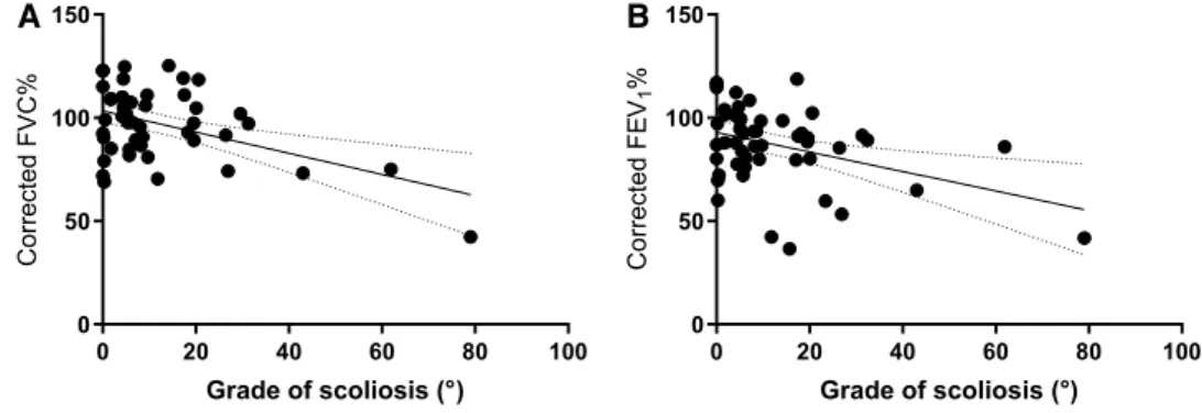
Chest deformities and respiratory symptoms are sum- marised in Table 2. Significantly more patients suffered from scoliosis in the Mfop group. Significant negative correlation between the extent of scoliosis and FVC%
(r = − 0.414, [95% CI − 0.617 to − 0.159], p = 0.0023) and FEV1% (r = − 0.401, [95% CI − 0.607 to − 0.144], p = 0.003) were noted. Similarly, Hcorrected FVC% (r = − 0.463, [95%
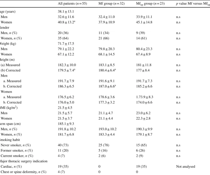
Table 1 Patient characteristics
a Significant difference compared to the value above
All patients (n = 55) Mf group (n = 32) Mfop group (n = 23) p value Mf versus Mfop
Age (years) 38.1 ± 13.1
Men 32.6 ± 11.6 32.4 ± 11.0 33.9 ± 11.1 n.s
Women 40.8 ± 13.2a 37.9 ± 10.9 45.1 ± 14.8 n.s
Gender
Men, n (%) 20 (36) 11 (34) 9 (39) n.s
Women, n (%) 35 (64) 21 (66) 14 (61) n.s
Weight (kg) 71.7 ± 17.5
Men 79.1 ± 22.2 79.8 ± 20.3 80.4 ± 23.3 n.s
Women 67.1 ± 12.2 68.1 ± 14.5 67.4 ± 8.9 n.s
Height (m)
(a) Measured 182.3 ± 10.0 183.1 ± 8.5 181 ± 11.8 n.s
(b) Corrected 179.5 ± 7.4a 180.4 ± 6.4a 177 ± 8.4 n.s
Men
a. Measured 191.7 ± 7.9 191.6 ± 9.1 191.7 ± 7.3 n.s
b. Corrected 186.3 ± 6.5 187.0 ± 6.6a 185.2 ± 6.6 n.s
Women
a. Measured 176.5 ± 6.2 178.6 ± 3.6 1.73.9 ± 8.3 n.s
b. Corrected 176.0 ± 5.0 177.3 ± 3.2 174.0 ± 6.6 n.s
BMI (kg/m2) 21.5 ± 4.5
Men 21.5 ± 5.7 21.1 ± 4.7 23.0 ± 6.2 n.s
Women 21.5 ± 3.7 21.1 ± 4.4 22.3 ± 2.8 n.s
Arm span (cm) 185.1 ± 9.3
Men, n (%) 191.8 ± 10.2 193.0 ± 10.2 190.3 ± 9.9 n.s
Women, n (%) 181.7 ± 6.8 183.3 ± 4.4 179.1 ± 8.7 n.s
Smoking habit
Never smoker, n (%) 40 (73) 25 (78) 15 (65) n.s
Former smoker, n (%) 11 (20) 5 (16) 6 (26) n.s
Current smoker, n (%) 4 (7) 2 (6) 2 (9) n.s
Major thoracic surgery indication
Cardiac, n (%) 19 (35) 0 19 (35) Not analysed
Chest or spine deformity, n (%) 4 (7) 0 0
Table 2 Chest deformities and respiratory symptoms in patients with Marfan syndrome
FBN1 Fibrillin 1
All patients
(n = 55) Mf group (n = 32) Mfop group (n = 23) p value Mf versus Mfop Chest deformities
Pectus carinatum, n (%) 24 (48) 12 (38) 12 (52) n.s
Pectus excavatum, n (%) 14 (28) 6 (19) 6 (26) n.s
Scoliosis, n (%) 36 (72) 15 (47) 21 (91) < 0.01
Asymmetric chest, n (%) 19 (38) 11 (34) 8 (35) n.s
Structural abnormalities of the lung
Spontaneous pneumothorax, n (%) 5 (10) 3 (9) 2 (9) n.s
Apical blebs and bullae, n (%) 4 (8) 3 (9) 1 (4) n.s
Pleuropulmonary symptoms
Cough, n (%) 11 (20) 5 (16) 6 (26) < 0.01
Sputum, n (%) 5 (9) 1 (3) 4 (17) n.s
Dyspnoea, n (%) 10 (18) 3 (9) 7 (30) < 0.01
Chest pain, n (%) 9 (16) 2 (6) 7 (30) 0.03
Ghent nosology, n (%)
Dilatation of the ascending aorta 38 (69) 20 (61) 18 (78) n.s
Dissection of the ascending aorta 7 (13) 2 (7) 5 (22) n.s
Mitral valve prolapse 48 (87) 28 (87) 20 (86) n.s
Dilatation or dissection of descending aorta 1 (2) 0 (0) 1 (6) n.s
Reduced upper-to-lower segment ratio 8 (14) 5 (16) 3 (12) n.s
Increased arm span-to-height ratio 8 (15) 4 (14) 4 (17) n.s
Wrist sign 47 (85) 29 (90) 18 (79) n.s
Thumb sign 51 (92) 28 (86) 23 (100) n.s
Reduced extension at the elbows 5 (9) 1 (3) 4 (18) n.s
Medial displacement of the medial malleolus caus-
ing pes planus 30 (55) 16 (52) 14 (61) n.s
Heel deformity 8 (15) 5 (17) 3 (12) n.s
Protrusio acetabulae of any degree 1 (2) 1 (3) 0 (0) n.s
Joint hypermobility 29 (52) 17 (52) 12 (53) n.s
Highly arched palate with crowding of teeth 35 (63) 20 (62) 15 (65) n.s
Facial appearance 35 (63) 19 (59) 16 (68) n.s
Dolichocephaly 11 (20) 5 (17) 6 (24) n.s
Enophthalmos 12 (22) 7 (21) 6 (24) n.s
Downslanting palpebral fissures 9 (17) 7 (21) 3 (12) n.s
Malar hypoplasia 8 (15) 4 (14) 4 (18) n.s
Retrognathia 17 (30) 8 (24) 9 (41) n.s
Ectopia lentis 15 (28) 7 (23) 8 (35) n.s
Myopia over 3 diopters 29 (52) 16 (50) 13 (56) n.s
Increased axial length of the globe 3 (6) 2 (7) 1 (6) n.s
Hypoplastic iris or hypoplastic ciliary muscle caus-
ing decreased miosis 1 (2) 0 (3) 0 (0) n.s
Lumbosacral dural ectasia 4 (7) 4 (10) 0 (0) n.s
Striae atrophicae (stretch marks) 36 (66) 22 (69) 14 (61) n.s
Positive family history 32 (58) 18 (56) 14 (60) n.s
Sex (male) 20 (36) 11 (34) 9 (39) n.s
Systemic score 8 ± 2 8 ± 2 8 ± 2 n.s
FBN1 mutation identified 40 (73) 21 (62) 19 (84) n.s
CI − 0.661 to − 0.206], p < 0.001) and FEV1% (r = − 0.386, [95% CI − 0.599 to − 0.125], p = 0.005) confirmed the asso- ciation (Fig. 1.).
Respiratory symptoms were present in fewer than 20% of patients. Dyspnoea, cough and chest pain were significantly more frequent Mfop patients. Structural changes assessed by chest CT scans of the lungs were scarce.
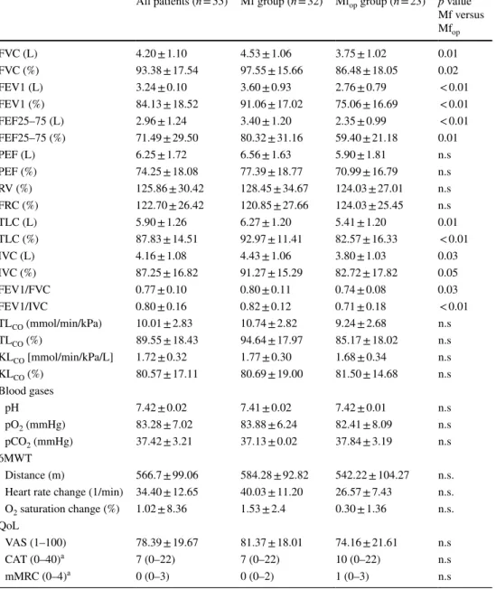
The LF data using the ECSC reference and Hmeasured are summarised in Table 3. Mfop patients had significantly lower FVC, IVC (inspiratory vital capacity) and TLC as com- pared to Mf patients. FEV1/FVC was 0.74 ± 0.08 in Mfop and 0.80 ± 0.11 in Mf patients, suggesting an obstructive ventilatory pattern in operated patients. Obstruction severity in Mfop, expressed as % predicted FEV1, corresponded to moderate changes. Airway obstruction in Mfop patients was confirmed by significantly decreased FEF25–75 values as compared to Mf patients. Increased RV and FRC, both signs of hyperinflation, were observed in both groups.
Diffusion (TLCO and KLCO), blood gases, 6MWT data or QoL were not different between groups (Table 3). CAT® and mMRC showed higher values in the Mfop group with more respiratory symptoms.
Using arm span corrected height, FVC and FEV1%
predicted values increased in all patient groups (Table 4.).
FEV1 remained in the pathological range in Mfop patients ( < 80% predicted) and stayed significantly lower as com- pared to Mf group.
Discussion
Our study is the largest cohort of Marfan patients who were serially assessed for pulmonary involvement. Twenty per- cent of the patients complained about pulmonary symptoms.
Cough, dyspnoea and chest pain were common, affecting a higher proportion of Mfop patients. QoL measures correlated with symptoms.
LF values are usually based on age, sex and standing height, which may be misleading in Marfan syndrome, where the length of the lower limbs contributes dispropor- tionally to height [24]. As height can be corrected by arm
span, we used equations to overcome this height measure- ment bias. This resulted in a significant decrease in the height values of Mf group patients, especially in men, lead- ing to the conclusion that, in many Marfan patients, standard LF reference values are disproportionally high.
In 1960, the ECSC was the first organisation to issue rec- ommendations for the calculation of reference values [25].
The reference values described by the ECSC were based on males working in coal mines and steel works. This was not a representative reference population, and in practice the predicted values were considered to be too high. Although no women had been tested, the ECSC calculated reference values for females at 80% of the values for men [14].
Our data confirmed airway obstruction, mainly affect- ing the small airways, in all Marfan patients. Similar results were previously observed in a study by Streeten and al [26].
The novelty of our study is the subgrouping according to major thoracic surgery. It is of high clinical importance to ensure appropriate lung function during or following exten- sive thoracic interventions. As a majority of Mfop patients had scoliosis, it is not surprising that the measured and corrected heights did not differ in these patients. However, height correction revealed abnormal FVC and FEV1 values.
Airway obstruction was moderate in all patients. This change might result from connective tissue malfunction in this young patient population due to their disease. The changes might also reflect incipient emphysema and/or an increased tendency for the airways to collapse [27]. Due to the pathological structure of fibrillin 1, the development of emphysema can be often observed in these patients. Rob- besom et al. showed that aberrant fibrillin 1 in the lung is significantly associated with the three most important mor- phometric parameters of emphysema: alveolar destruction, airspace enlargement and emphysema-related morphologi- cal abnormalities; [28] experimental data in mice have con- firmed widening of the distal airspaces in Marfan syndrome [29]. As described by Hogg et al., small airways are the main site of obstruction in lungs affected by emphysema [30]. It is suspected that areas with trapped air develop emphysema over time [31]. Combined with the increased tendency of the small airways to collapse in Marfan syndrome [27], it
Fig. 1 Correlation between extent of scoliosis and height corrected FVC% (a) and FEV1% (b)
A B
0 20 40 60 80 100
0 50 100 150
Grade of scoliosis (°) Corrected FEV1%
0 20 40 60 80 100
0 50 100 150
Grade of scoliosis (°)
Corrected FVC%
Table 3 Lung function testing in Mf and Mfop using Hmeasured for the ECSC equations
FVC forced vital capacity, FEV1 forced expiratory volume in the first second, FEF25–75 forced expiratory flow between 25 and 75%, PEF peak expiratory flow, RV residual volume, FRC functional residual capac- ity, TLC total lung capacity
a Data expressed as median (range)
All patients (n = 55) Mf group (n = 32) Mfop group (n = 23) p value Mf versus Mfop
FVC (L) 4.20 ± 1.10 4.53 ± 1.06 3.75 ± 1.02 0.01
FVC (%) 93.38 ± 17.54 97.55 ± 15.66 86.48 ± 18.05 0.02
FEV1 (L) 3.24 ± 0.10 3.60 ± 0.93 2.76 ± 0.79 < 0.01
FEV1 (%) 84.13 ± 18.52 91.06 ± 17.02 75.06 ± 16.69 < 0.01
FEF25–75 (L) 2.96 ± 1.24 3.40 ± 1.20 2.35 ± 0.99 < 0.01
FEF25–75 (%) 71.49 ± 29.50 80.32 ± 31.16 59.40 ± 21.18 0.01
PEF (L) 6.25 ± 1.72 6.56 ± 1.63 5.90 ± 1.81 n.s
PEF (%) 74.25 ± 18.08 77.39 ± 18.77 70.99 ± 16.79 n.s
RV (%) 125.86 ± 30.42 128.45 ± 34.67 124.03 ± 27.01 n.s
FRC (%) 122.70 ± 26.42 120.85 ± 27.66 124.03 ± 25.45 n.s
TLC (L) 5.90 ± 1.26 6.27 ± 1.20 5.41 ± 1.20 0.01
TLC (%) 87.83 ± 14.51 92.97 ± 11.41 82.57 ± 16.33 < 0.01
IVC (L) 4.16 ± 1.08 4.43 ± 1.06 3.80 ± 1.03 0.03
IVC (%) 87.25 ± 16.82 91.27 ± 15.29 82.72 ± 17.82 0.05
FEV1/FVC 0.77 ± 0.10 0.80 ± 0.11 0.74 ± 0.08 0.03
FEV1/IVC 0.80 ± 0.16 0.82 ± 0.12 0.71 ± 0.18 < 0.01
TLCO (mmol/min/kPa) 10.01 ± 2.83 10.74 ± 2.82 9.24 ± 2.68 n.s
TLCO (%) 89.55 ± 18.43 94.64 ± 17.97 85.17 ± 18.02 n.s
KLCO [mmol/min/kPa/L] 1.72 ± 0.32 1.77 ± 0.30 1.68 ± 0.34 n.s
KLCO (%) 80.57 ± 17.11 80.69 ± 19.00 81.50 ± 14.68 n.s
Blood gases
pH 7.42 ± 0.02 7.41 ± 0.02 7.42 ± 0.01 n.s
pO2 (mmHg) 83.28 ± 7.02 83.88 ± 6.24 82.41 ± 8.09 n.s
pCO2 (mmHg) 37.42 ± 3.21 37.13 ± 0.02 37.84 ± 3.19 n.s
6MWT
Distance (m) 566.7 ± 99.06 584.28 ± 92.82 542.22 ± 104.27 n.s.
Heart rate change (1/min) 34.40 ± 12.65 40.03 ± 11.20 26.57 ± 7.43 n.s.
O2 saturation change (%) 1.02 ± 8.36 1.53 ± 2.4 0.30 ± 1.36 n.s.
QoL
VAS (1–100) 78.39 ± 19.67 81.37 ± 18.01 74.16 ± 21.61 n.s
CAT (0–40)a 7 (0–22) 7 (0–22) 10 (0–22) n.s
mMRC (0–4)a 0 (0–3) 0 (0–2) 1 (0–3) n.s
Table 4 Lung function parameters using ECSC with Hmeasured and Hcorrected in Marfan patients
All patients (n = 55) Mf group (n = 32) Mfop group (n = 23) p value Mf versus Mfop FVC%
ECSC Hmeasured (%) 93.38 ± 17.54 97.55 ± 15.66 86.48 ± 18.05 0.02 ECSC Hcorrected (%) 96.68 ± 18.09 101.99 ± 15.18 88.02 ± 19.15 0.01 FEV1%
ECSC Hmeasured (%) 84.13 ± 18.52 91.06 ± 17.02 75.06 ± 16.69 < 0.01 ECSC Hcorrected (%) 86.41 ± 23.49 93.27 ± 16.68 77.25 ± 18.92 < 0.01
can be assumed that, due to connective tissue malfunction, air trapping starts in the small airways, which later might convert into emphysema.
Six of our patients (10,9%) were diagnosed with asthma, 5 of them well controlled (Mf n = 3, Mfop n = 2) without obstructive ventilatory changes at the time of assessment.
One patient awaiting cardiac surgery presented with mixed ventilatory pattern. No further patients had clinical signs of asthma. The extent of scoliosis showed significant nega- tive correlation with FVC% and FEV1%, pointing towards restrictive changes due to thorax abnormalities.
Our data suggest that LF evaluation in patients with atypi- cal anthropometrical features can be difficult. The equations applied in LF testing might give different results and it might be beneficial to reassess results in those who have unusual physical features.
Conclusion
This study performed a complex respiratory functional assessment of a large cohort of Marfan patients, confirming previous data showing mild obstructive ventilatory disorder.
The need for major thoracic surgery was associated with more respiratory symptoms, more severe functional changes and worse QoL. Height correction revealed decreased FVC and FEV1 values in Mfop patients more in line with their clinical symptoms. Small airway obstruction in our patients indicates that particular attention is needed in the follow-up of respiratory status. One weakness of our study is that the reversibility of airway obstruction in Mfop patients was not investigated in the absence of clinical symptoms of asthma.
The extent of scoliosis showed significant negative cor- relation with FVC% and FEV1% suggestive of restrictive changes due to thoracic deformities. Longitudinal data will be needed to evaluate changes of airway function in Marfan syndrome. In daily clinical practice, more attention should be placed on pulmonary involvement and LF assessments when planning or after major thoracic surgery in Marfan patients.
Acknowledgements We thank all the members of the Hungarian Mar- fan Foundation. Open access funding was provided by Semmelweis University (SE).
Funding This work was supported by the National Research, Develop- ment and Innovation Office of Hungary (NKFIH; NVKP-16–1-2016–
0017) and by the “New National Excellence Program of the Ministry of Human Capacities of Hungary” (ÚNKP-17–3-I-SE-31 and ÚNKP- 18-3-I-SE-69; BÁ).
Compliance with Ethical Standards
Conflict of interest The authors declare that they have no conflict of interest.
Informed Consent Informed consent was obtained from all individual participants included in the study.
Open Access This article is distributed under the terms of the Crea- tive Commons Attribution 4.0 International License (http://creat iveco mmons .org/licen ses/by/4.0/), which permits unrestricted use, distribu- tion, and reproduction in any medium, provided you give appropriate credit to the original author(s) and the source, provide a link to the Creative Commons license, and indicate if changes were made.
References
1. Marfan A (1896) Un cas de déformation congénitale des qua- tre membres, plus prononcée aux extrémités, caractérisée par l’allongement des os avec un certain degré d’amincissement Impr Maretheux
2. Judge DP, Dietz HC (2005) Marfan’s syndrome. Lancet 366:1965–1976
3. Colovati ME, da Silva LR, Takeno SS, Mancini TI, Dutra AR, Guilherme RS, de Mello CB, Melaragno MI, Perez AB (2012) Marfan syndrome with a complex chromosomal rearrangement including deletion of the FBN1 gene. Mol Cytogenet 5:5 4. Dyhdalo K, Farver C (2011) Pulmonary histologic changes in
Marfan syndrome: a case series and literature review. Am J Clin Pathol 136:857–863
5. Seyama Y, Hayashi M, Usami E, Yamashita S (1992) Change in elastin structure in human aortic connective tissue diseases.
Amino Acids 3:287–292
6. Kielty CM (2017) Fell-Muir Lecture: Fibrillin microfibrils: struc- tural tensometers of elastic tissues? Int J Exp Pathol 98:172–190 7. De Paepe A, Devereux RB, Dietz HC, Hennekam RCM, Pyeritz
RE (1996) Revised diagnostic criteria for the Marfan syndrome.
Am J Med Genet 62:417–426
8. Loeys BL, Dietz HC, Braverman AC, Callewaert BL, De Backer J, Devereux RB, Hilhorst-Hofstee Y, Jondeau G, Faivre L, Mile- wicz DM et al (2010) The revised Ghent nosology for the Marfan syndrome. J Med Genet https ://doi.org/10.1136/jmg.2009.07278 9. Hao W, Fang Y, Lai H, Shen Y, Wang H, Lin M, Tan L (2017) 5
Marfan syndrome with pneumothorax: case report and review of literatures. J Thorac Dis 9:E1100–E1103
10. Corsico AG, Grosso A, Tripon B, Albicini F, Gini E, Mazzetta A, Di Vincenzo EM, Agnesi ME, Tsana Tegomo E, Ronzoni V et al (2014) Pulmonary involvement in patients with Marfan Syn- drome. Panminerva Med 56:177–182
11. Neuville M, Jondeau G, Crestani B, Taillé C (2015) Respiratory manifestations of Marfan’s syndrome. Rev Mal Respir 32:173–181 12. Miller MR (2005) Standardisation of spirometry. Eur Respir J
26:319–338
13. Quanjer PH, Stanojevic S, Cole TJ, Baur X, Hall GL, Culver BH, Enright PL, Hankinson JL, Ip MSM, Zheng J et al (2012) Multi-ethnic reference values for spirometry for the 3–95-yr age range: the global lung function 2012 equations. Eur Respir J 40:1324–1343
14. Quanjer PH, Stanojevic S, Stocks J, Cole TJ GLI-2012 reference values for spirometry GLI-2012 All-Age Multi-Ethnic Reference
Values for Spirometry Advantages Consequences GLI-2012 refer- ence values for spirometry Interpretation of spirometric data 15. Pearson GD, Devereux R, Loeys B, Maslen C, Milewicz D, Pyer-
itz R, Ramirez F, Rifkin D, Sakai L, Svensson L et al (2008) Report of the national heart, lung, and blood institute and national marfan foundation working group on research in marfan syndrome and related disorders. Circulation 118:785–791
16. Benke K, Ágg B, Szabó L, Szilveszter B, Odler B, Pólos M, Cao C, Maurovich-Horvat P, Radovits T, Merkely B et al (2016) Ben- tall procedure: quarter century of clinical experiences of a single surgeon. J Cardiothorac Surg 11:19
17. ATS Statement (2012) https ://doi.org/10.1164/AJRCC M.166.1.AT110 2
18. Goldberg CJ, Kaliszer M, Moore DP, Fogarty EE, Dowling FE (2001) Surface topography, Cobb angles, and cosmetic change in scoliosis. Spine (Phila Pa 1976) 26:E55–63
19. Jones PW, Harding G, Berry P, Wiklund I, Chen W-H, Leidy NK (2009) Development and first validation of the COPD Assessment Test. Eur Respir J 34:648–654
20. Bestall JC, Paul EA, Garrod R, Garnham R, Jones PW, Wedzicha JA (1999) Usefulness of the Medical Research Council (MRC) dyspnoea scale as a measure of disability in patients with chronic obstructive pulmonary disease. Thorax 54:581–586
21. Piston User Manual. [date unknown]
22. Measuring arm span. [date unknown]
23. Parker JM, Dillard TA, Phillips YY (1996) Arm span-height rela- tionships in patients referred for spirometry. Am J Respir Crit Care Med 154:533–536
24. Pyeritz RE, McKusick VA (1979) The Marfan syndrome: diagno- sis and management. N Engl J Med 300:772–777
25. Jouasset D (1960) Standardization of respiratory function tests in countries of the European coal and steel region. Poumon Coeur 16:1145–1159
26. Streeten E (1987) Pulmonary function in the Marfan syndrome.
Chest. https ://doi.org/10.1378/chest .91.3.408
27. Giske L, Stanghelle JK, Rand-Hendrikssen S, Strøm V, Wilhelm- sen J-E, Røe C (2003) Pulmonary function, working capacity and strength in young adults with Marfan syndrome. J Rehabil Med 35:221–228
28. Robbesom AA, Koenders MM, Smits NC, Hafmans T, Versteeg EM, Bulten J, Veerkamp JH, Dekhuijzen R, Van Kuppevelt TH (2008) Aberrant fibrillin-1 expression in early emphysematous human lung: a proposed predisposition for emphysema. Mod Pathol 21:297–307
29. Uriarte JJ, Meirelles T, Del Blanco DG, Nonaka PN, Campillo N, Sarri E, Navajas D, Egea G, Farré R (2016) Early impairment of lung mechanics in a murine model of Marfan syndrome. PLoS ONE. https ://doi.org/10.1371/journ al.pone.01521 24
30. Hogg JC, Macklem PT, Thurlbeck WM (1967) The resistance of small airways in normal and diseased human lungs. Aspen Emphysema Conf 10:433–441
31. Hogg JC, Paré PD, Hackett T-L (2017) The contribution of small airway obstruction to the pathogenesis of chronic obstructive pul- monary disease. Physiol Rev 97:529–552
Publisher’s Note Springer Nature remains neutral with regard to jurisdictional claims in published maps and institutional affiliations.