IDENTIFICATION OF PROGNOSTIC AND DIAGNOSTIC MARKERS IN THYMIC
EPITHELIAL TUMORS
PhD thesis
Bernhard Moser
Clinical Medicine Doctoral School Semmelweis University
Consultant: Ferenc Rényi-Vámos, MD, PhD Official reviewers: Heiler Zoltán MD, PhD
Zoltán Takácsi-Nagy MD PhD
Head of the Complex Examination Committee: György Losonczy, MD, D.Sc Members of the Complex Examination Committee: Nóra Bittner, MD, PhD
Marcell A Szász, MD, PhD
Budapest
2019
2
Table of Contents
1. Abbreviations ... 6
2. Introduction to Thymic epithelial tumors ... 9
2.1 Epidemiology ... 9
2.2 Etiology and heredity ... 9
2.3 Histology ... 10
2.3.1 Thymoma and TC ... 10
2.3.2 TNETs ... 13
2.3.3 Thymic hyperplasia ... 14
2.4 Paraneoplastic syndromes: ... 14
2.4.1 Myasthenia gravis ... 14
2.5 Molecular biology ... 16
2.6 Screening, symptoms, diagnosis and prognosis ... 17
2.6.1 Screening ... 17
2.6.2 Symptoms ... 17
2.6.3 Diagnosis ... 17
2.6.4 Imaging ... 17
2.7 Tumor staging systems ... 20
2.8 Definition of TET recurrences ... 23
2.9 Treatment of TETs ... 24
2.9.1 National and international Consensus statements ... 24
2.9.2 Management algorithm: resectable disease ... 24
2.9.3 Institutional treatment specifics ... 25
2.9.4 Surgery ... 26
2.9.4.1 Surgical approach: open vs. minimally invasive 26 2.9.4.2 Rationale for extended thymectomy 28 2.9.4.3 Treatment of TETs with pleural involvement 29 2.9.5 Systemic therapy ... 31
2.9.5.1 Chemotherapy (Cht) 31
3
2.9.5.2 Novel systemic therapies: targeted therapy and immunotherapy 32
2.9.6 Radiation therapy (RT) ... 33
2.10 Prognostic factors for patients with TETs ... 34
2.10.1 Outcome measures for TETs ... 34
2.10.2 Pathological predictors of treatment outcome ... 36
2.10.3 Experimental biomarkers for TETs ... 36
2.11 Introduction to the biomarkers of the thesis ... 37
2.11.1 C-reactive protein (CRP) ... 37
2.11.2 Fibrinogen ... 39
2.11.3 NLR and PLR: the role of neutrophils, lymphocytes and platelets in cancer ... 39
3. Objectives ... 41
3.1 Prognostic factors and multi-modal management of TETs treated in a single European thoracic surgery center ... 41
3.2 Prognostic factors for TETs with pleural involvement: an ESTS Thymic Working Group Project ... 41
3.3 CRP as a prognostic marker for TETs ... 42
3.4 Fibrinogen, NLR, and PLR as prognostic markers for TETs ... 42
4. Methods ... 43
4.1 Ethical considerations ... 43
4.2 Research facility ... 43
4.3 Prognostic factors and multi-modal management of TETs: a single center experience ... 43
4.3.1 Study design ... 43
4.3.2 Patients, diagnostic workup and treatment decisions, tumor staging and histology ... 44
4.3.3 Statistical analyses ... 45
4.4 Surgical therapy of thymic tumours with pleural involvement: an ESTS Thymic Working Group Project ... 45
4.4.1 Study design ... 45
4.4.2 Statistical analyses ... 48
4
4.5 Evaluation of CRP as a prognostic marker for TETs ... 49
4.5.1 Study cohort ... 49
4.5.2 Tumor samples, Immunohistochemical analysis ... 50
4.5.3 Statistical analyses ... 50
4.6 Evaluation of fibrinogen, NLR, and PLR as prognostic markers for TETs ... 51
4.6.1 Study cohort ... 51
4.6.2 Blood work and immunohistochemistry on tumor tissue ... 52
4.6.3 Statistical analyses ... 52
5. Results ... 54
5.1 Prognostic factors and multi-modal management of TETs: a single center experience ... 54
5.1.1 Survival is dependent upon tumor stage and resection status but not histology ... 54
5.1.2 Worse survival in advanced cases: recurrences, progressions, and multimodal therapy ... 56
5.1.3 The role of biopsies and surgery ... 60
5.1.4 Myasthenia Gravis was not a prognostic factor ... 61
5.2 Surgical therapy of TETs with pleural involvement: an ESTS Thymic Working Group Project ... 62
5.2.1 TC and incomplete resection predict worse survival ... 62
5.2.2 The prognosis of patients with pleural disease is multifactorial: resection status, histology, primary or recurrent disease, type of pleural surgery and necessity of multimodal treatment ... 65
5.2.3 Clinical data and treatment specifics for TETs with pleural involvement ... 67
5.3 CRP serum concentrations predict poor outcome and tumor recurrence in patients with TETs ... 70
5.3.1 Survival analysis: Increased pretreatment CRP is a predictor of worse FFR ... 70
5.3.2 Pretreatment CRP is a prognostic factor for FFR ... 71
5.3.3 CRP serum concentrations are increased in patients with TETs .... 72
5
5.4 Prognostic and diagnostic impact of fibrinogen, NLR, and PLR on TET
outcome... 76
5.4.1 Survival and freedom from recurrence are associated with fibrinogen plasma concentrations, NLR and PLR ... 76
5.4.2 Fibrinogen and NLR as predictors of survival and recurrence ... 77
5.4.3 NLR and PLR as predictors of tumor recurrence during oncological follow-up ... 80
6. Discussion ... 85
6.1 Pathological prognostic biomarkers for TETs ... 85
6.2 Peripheral blood derived prognostic biomarkers for TETs ... 89
7. Conclusions ... 97
8. Summary ... 99
9. Összefoglalás ... 101
10. Bibliography ... 103
11. Bibliography of the candidate’s publications ... 120
11.1 Publications related to the thesis ... 120
11.2 Publications not related to the thesis ... 121
12. List of figures ... 131
13. List of tables ... 135
14. Acknowledgements ... 137
6
1. Abbreviations
AJCC American Joint Committee on Cancer ARDS acute respiratory distress syndrome AUC area under the curve
CCS Cause-specific survival
ChART Chinese Alliance for Research in Thymomas
Cht Chemotherapy
CI Confidence interval
CIR Cumulative incidence of recurrence
CR complete response
CRP C-reactive protein
CT Computed tomography
DFS Disease-free survival
dNLR derived Neutrophil-to-Lymphocyte ratio
ECG electrocardiography
ELISA Enzyme linked immunosorbent assay EPD Extended Pleurectomy/Decortication
EPP Extrapleural pneumonectomy
ESMO European Society for Medical Oncology
esRAGE endogenous secretory Receptor for Advanced Glycation Endprod- ucts
ESTS European Society of Thoracic Surgeons f:m ratio female:male ratio
FDG-PET 18Fluorine-fluorodeoxyglucose–positron emission tomography
FFR Freedom from recurrence
HMGB1 high mobility group box 1
HR hazard ratio
HSP Heat shock protein
IARC International agency for research on cancer
IASLC International Association for the Study of Lung Cancer ICD-O International Classification of Diseases for Oncology
7
ITMIG International Thymic Malignancies Interest Group JART Japanese Association for Research on the Thymus
LP Local pleurectomy
MEN 1 multiple endocrine neoplasia syndrome type 1
MG Myasthenia gravis
MGFA Myasthenia Gravis Foundation of America MODS multi organ dysfunction syndrome
MRI Magnetic resonance imaging MRI Magnetic resonance imaging NLR Neutrophil-to-Lymphocyte ratio NPV Negative Predictive Value
OS Overall survival
P/D Pleurectomy/Decortication PLR Platelet-to-Lymphocyte ratio PORT postoperative radiation therapy PPV Positive Predictive Value
PR partial response
R0 no residual tumor
R1 microscopic residual tumor
R2 macroscopic residual tumor
RAGE Receptor for Advanced Glycation Endproducts RATS robotic-assisted thoracic Surgery
RECIST criteria Response Evaluation Criteria in Solid Tumor RFS Recurrence-free survival
ROC Receiver operating characteristic curves
RT Radiotherapy
SD standard deviation
SEER Surveillance, Epidemiology, and End Results SEM standard error of the mean
sRAGE Soluble Receptor for Advanced Glycation Endproducts
SST somatostatin
SUVmax maximum standard uptake value
8
TAMG Thymoma-associated MG
TETs Thymic epithelial tumors TNET Thymic neuroendocrine tumor
TNM Tumor Node Metastasis
TP Total pleurectomy
UICC Union for International Cancer control
VATET Video-assisted thorascopic extended thymectomy VATS Video-assisted thoracic surgery
WHO World Health Organization
9
2. Introduction to Thymic epithelial tumors
2.1 Epidemiology
In the European Union non-communicable diseases that affect less than five in 10,000 people are considered rare (European Commission). In a population-based study using two nationwide databases in the Netherlands the incidence of thymic epithelial tumors (TETs) was reported to be 3.2/1,000,000 (de Jong, Blaauwgeers et al. 2008). The overall annual incidence in the United States was reported as 0.15 per 100,000 inhabitants (Engels and Pfeiffer 2003). TETs are thus an orphan disease.
Two distinct forms of TETs can be distinguished: thymomas and thymic carcinomas (TC). In the Netherlands (1994-2003) thymoma incidence was 2.2/1,000,000 and TC in- cidence 0.3/1,000,000 with equal incidence rates for men and women (de Jong, Blaauwgeers et al. 2008). In the Netherlands the median age at diagnosis of TETs was 59 years (de Jong, Blaauwgeers et al. 2008).
Among the rarest forms of TETs are thymic neuroendocrine tumors (TNETs). They ac- counted for 0.4% of carcinoid tumors in the Surveillance, Epidemiology, and End Results (SEER) database of the United States National Cancer Institute (Yao, Hassan et al. 2008, Gaur, Leary et al. 2010) and constitute about 4-7% of all anterior mediastinal tumors (Gaur, Leary et al. 2010).
2.2 Etiology and heredity
To date no risk factors for the development of TETs have been discovered. Regarding the existence of genetic variants a higher risk of thymomas in diverse Asian and Pacific Is- landers was reported. So far no evidence for the contribution of alcohol, tobacco, occu- pational or environmental hazards, dietary factors or ionizing radiation could be detected.
The incidence of TETs seems not to be higher in immunosuppressed HIV patients or organ transplant recipients (Engels and Pfeiffer 2003). Epstein Barr virus (EBV) was de- tected in TCs of lymphoepithelial histology (Mann, Wu et al. 1992, Engels and Pfeiffer 2003).
The familial occurrence of thymomas is a very rare event. In one family thymomas oc- curred in three family members (next to autoimmune diseases in 4 other family members,
10
such as Grave`s disease, pernicious anemia, Sjögren`s diease and autoimmune pancyto- penia). Of the 27 tested family members 11 had a constitutional translocation t(14;20)(q24;p12) which was present in all the thymoma patients of this family. The DNA strand break 14q24 was in a tumor suppressor gene (RAD5I family) known to be involved in tumors such as uterine leiomyoma or pulmonary chondroid hamartoma). The DNA strand break 20p12 was in close proximity (100kb) to BMP2, a TGFβ-family member involved in the differentiation of thymocytes (Nicodeme, Geffroy et al. 2005).
2.3 Histology
2.3.1 Thymoma and TC
In 1999 the WHO defined histopathological criteria for thymomas, namely types A, AB, B1, B2, B3 and for the different TC subtypes collectively as type C (J.). In the year 2002 the new WHO Histologic Classification of TETs was published (Chen, Marx et al. 2002).
In the fourth edition of the WHO Classification of Thymic Tumors (TETs, germ cell tu- mors, lymphomas, dendritic cell and myeloid neoplasms, and soft tissue tumors of the thymus and mediastinum) a comprehensive overview of newly defined tumor entities and their variants as well as refined criteria for the diagnosis of thymomas and thymic squa- mous cell carcinoma (see Table 2; and reference (Marx, Chan et al. 2015)).
Table 1: WHO classification of TETs. Adapted from Table 1: Epithelial Tumors: The 2015 World Health Organization Classification of Tumors of the Thymus: Continuity and Changes (Marx, Chan et al. 2015).
Behavior is coded /0 for benign tumors; /1 for unspecified, borderline, or uncertain behavior; /2 for carci- noma in situ and grade III intraepithelial neoplasia; and /3 for malignant tumors. NUT, nuclear protein in testis. aThese new codes were approved by the IARC/WHO Committee for ICD-0.
Thymoma ICD-O
Type A thymoma, including atypical variant 8581/3a
Type AB thymoma 8582/3a
Type B1 thymoma 8583/3a
Type B2 thymoma 8584/3a
Type B3 thymoma 8585/3a
Micronodular thymoma with lymphoid stroma 8580/1a
Metaplastic thymoma Other rare thymomas 8580/3
Microscopic thymoma 8580/0
Sclerosing thymoma 8580/3
Lipofibroadenoma 9010/0a
Thymic carcinoma
Squamous cell carcinoma 8070/3
Basaloid carcinoma 8123/3
11
Mucoepidermoid carcinoma 8430/3
Lymphoepithelioma-like carcinoma 8082/3
Clear cell carcinoma 8310/3
Sarcomatoid carcinoma 8033/3
Adenocarcinomas
Papillary adenocarcinoma 8260/3
Thymic carcinoma with adenoid cystic carcinoma-like features 8200/3a
Mucinous adenocarcinoma 8480/3
Adenocarcinoma, NOS 8140/3
NUT carcinoma 8023/3a
Undifferentiated carcinoma 8020/3
Other rare thymic carcinomas
Adenosquamous carcinoma 8560/3
Hepatoid carcinoma 8576/3
Thymic carcinoma, NOS 8586/3
Thymic neuroendocrine tumors Carcinoid tumors
Typical carcinoid 8240/3
Atypical carcinoid 8249/3
Large-cell neuroendocrine carcinoma 8013/3
Combined large-cell neuroendocrine carcinoma 8013/3
Small-cell carcinoma (SCC) 8041/3
Combined SCC 8045/3
Combined thymic carcinomas
Table 2: WHO classification of thymic tumors: refined diagnostic criteria. From: The 2015 WHO Classifi- cation of Tumors of the Thymus: Continuity and Changes (Marx, Chan et al. 2015). aPaucity versus abun- dance: any area of crowded immature T cells or moderate numbers of immature T cells in >10% of the investigated tumor are indicative of “abundance”;
Thymoma subtype Obligatory criteria Optional criteria
Type A Occurrence of bland, spindle shaped epithelial cells (at least focally); paucitya or absence of im- mature (TdT+) T cells throughout the tumor
Polygonal epithelial cells CD20+
epithelial cells Atypical type A variant Criteria of type A thymoma; in addition: comedo-
type tumor necrosis; increased mitotic count (>4/2mm2); nuclear crowding
Polygonal epithelial cells CD20+
epithelial cells Type AB Occurrence of bland, spindle shaped epithelial
cells (at least focally); abundancea of immature (TdT+) T cells focally or throughout tumor
Polygonal epithelial cells CD20+epithelial cells
Type B1 Thymus-like architecture and cytology: abun- dance of immature T cells, areas of medullary dif- ferentiation (medullary islands); paucity of polyg- onal or dendritic epithelia cells without clustering (i.e.<3 contiguous epithelial cells)
Hassall’s corpuscles; perivas- cular spaces
Type B2 Increased numbers of single or clustered polygo- nal or dendritic epithelial cells intermingled with abundant immature T cells
Medullary islands; Hassall’s cor- puscles; perivascular spaces Type B3 Sheets of polygonal slightly to moderately atypical
epithelial cells; absent or rare intercellular bridges;
paucity or absence of intermingled TdT+ T cells
Hassall’s corpuscles; perivas- cular spaces
Micronodular thy- momas (MNT) with lymphoid stroma;
Nodules of bland spindle or oval epithelial cells surrounded by an epithelial cell-free lymphoid stroma
Lymphoid follicles; monoclonal B cells and/or plasma cells (rare)
12
Metaplastic thymoma Biphasic tumor composed of solid areas of epithe- lial cells in a background of bland-looking spindle cells; absence of immature T cells
Pleomorphism of epithelial cells;
actin, keratin, or EMA-positive spindle cells
Rare other:Microscopic thymoma; sclerosing thymoma, lipofibroadenoma
Outcome related to WHO histology
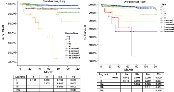
In a study on 200 Chinese patients undergoing surgery for TETs (only 55 patients re- ceived adjuvnat radiotherapy and 8 patients adjuvant chemotherapy) OS was reported as follows: none of the Type A and AB thymomas patients died of tumor; there was one patient with type B1 thymoma who died at 22 months; type B2, B3, and TC (formerly type C thymomas) patients had a significantly worse prognosis: 5-year OS 75.0%, 70.0%, and 48.0%, respectively (see Figure 1). Masaoka-Koga stage was a statistically significant predictor of survival. There was a statistically significant association between WHO his- tologic subtype and stage. WHO histology was an independent predictive factor of OS in stage I and II TETs: type B2, B3, and TC had a worse prognosis than type A, AB, and B1 thymomas (p<0.003) (Chen, Marx et al. 2002).
Figure 1: OS by Stage and WHO histology. Patients with higher stage had a significantly (p<0.001) in- creased risk of death from tumor. Type B2-3 and C thymomas (TCs) had a significantly (p<0.01) increased risk of death from tumor. From (Chen, Marx et al. 2002).
Our group has recently proposed a new subtype of TC: primary thymic adenocarcinoma of enteric type (Moser, Schiefer et al. 2015). The inclusion of this new TC subtype may help prevent misdiagnosis of metastatic disease from an extrathymic primary cancer, par- ticularly metastatic disease from the gastrointestinal tract Figure 2.
13
Figure 2: Primary thymic adenocarcinoma of enteric type. Hematoxylin and eosin staining (A). Immuno- histochemistry for CK20 (B), CDX2 (C), and CEA (D). From (Moser, Schiefer et al. 2015).
2.3.2 TNETs
The current nomenclature of TNETs distinguishes: thymic typical (<2 mitoses/2mm2; no necrosis) and atypical (<2 mitoses/2mm2; with necrosis; or 2–10 mitoses/2mm2; + or − necrosis) carcinoids, large cell neuroendocrine carcinoma (LCNEC; >10 mitoses/2mm2; no small cell features) and small cell carcinoma; and the combination of LCNEC or small cell carcinoma with thymoma of TC (Marx, Chan et al. 2015). Pathognomonic for TNETs is their high biologic aggressiveness and poor prognosis due to high recurrences rates and tumor related deaths (Strobel, Zettl et al. 2014, Filosso, Yao et al. 2015). OS of patients with TNETs in the SEER database at 1-, 3- and 5 years was 89%, 66% and 53% with poorer survival of patients in advanced stages (Gaur, Leary et al. 2010). 5 year OS for localized disease (tumor confined to organ) was 80%, regional disease (local invasion or metastasis to regional lymph nodes) 48% and distant disease 31% (Gaur, Leary et al.
2010). OS was reported to be significantly better in patients undergoing macroscopic complete resection (Ose, Maeda et al. 2018). TNETs can be associated with parane- oplastic syndromes, e.g. Cushing`s syndrome or multiple endocrine neoplasia syndrome type 1 (MEN 1).
A B
A
C D
14
2.3.3 Thymic hyperplasia
Thymic hyperplasia (TH) is caused by non-malignant thymic changes with an increase in constituent cells (Castleman 1955). Pathology distinguishes two inherently different types of TH. True thymic hyperplasia (TTH) is diagnosed if the thymus is of regular micro- scopic histologic architecture but is marked by increased weight and size. Follicular (or lymphoid) TH (FTH) is characterized by the presence of lymphoid follicles with germinal centers in the thymic medulla (Rosai and Levine 1976). The diagnosis benign enlarge- ment of the thymus :TTH and FTH can solely be made from the pathological specimen.
To date there are no established risk factors (Engels 2010) nor biomarkers that can dis- tinguish TH from malignant TETs.
2.4 Paraneoplastic syndromes:
There is a strong association between thymomas and autoimmune diseases: systemic lu- pus erythematosus, autoimmune cytopenias (pure red cell aplasia (PRCA), aplastic ane- mia (AA), autoimmune hemolytic anemia (AIHA), immune thrombocytopenia, autoim- mune neutropenia, thrombotic thrombocytopenic purpura, agranulocytosis, polymyositis, Good`s syndrome, hypogammaglobulinemia, autoimmune thyroid diseases, autoimmune hepatitis, cutaneous autoimmune diseases, paraneoplastic pemphigus or Lichen planus (Bernard, Frih et al. 2016). Many of the observed paraneoplastic disease are neurological:
Myasthenia gravis, Lambert Eaton syndrome, Myositis, Isaac`s syndrome, encephalitis, Morvan`s syndrome, Autoimmune Autonomic Neuropathy, Paraneoplastic cerebellar de- generation, Stiff person syndrome (Evoli and Lancaster 2014). The most frequent para- neoplastic autoimmune neurological disorder is MG. TETs are diagnosed in up to 15%
of patients with MG. Conversely, about 30% of patients with TETs experience symptoms of MG at the time of diagnosis (Marx, Willcox et al. 2010).
2.4.1 Myasthenia gravis
Myasthenia gravis (MG) is a neurological disorder with a prevalence of 10/100,000 peo- ple (Phillips 2003). MG is a B-cell mediated autoimmune disorder (Gilhus and Verschuuren 2015) characterized by autoantibodies against the nicotinic acetylcholine receptor and less common against other proteins of the neuromuscular junction. The au- toimmune destruction of acetylcholine receptors results in impaired transmission at the
15
neuromuscular junction and leads to the patients’ pathognomonic fluctuating muscle weakness (Vincent 2002). MG is more prevalent in patients with B-type thymomas than types A or AB and is not observed in patients with TCs (Chen, Marx et al. 2002, Radovich, Pickering et al. 2018) Table 3.
Table 3: Frequency of the different histological TET subtypes in relation to MG and stage Adopted from Thymic epithelial tumours: ESMO Clinical Practice Guidelines for diagnosis, treatment and follow-up (Girard, Ruffini et al. 2015). The statistics were derived from four publications (Kojima, Ito et al. 2006, Ruffini, Detterbeck et al. 2014, Omasa, Date et al. 2015, Weis, Yao et al. 2015).
Relative frequency Myasthenia gravis Masaoka stage
I II III IVA IVB Type A 12 (3–26) 15 (0–35) 60 31 8 <1 <1
Type AB 28 (15–43) 20 (5–42) 67 26 6 1 1
Type B1 18 (6–53) 40 (5–69) 50 37 9 3 1
Type B2 26 (8–41) 50 (23–73) 32 29 28 8 3
Type B3 16 (3–35) 50 (25–65) 19 36 27 1 3
Carcinoma 18 (1–28) <5 10 10 45 15 20
Intrathymic pathogenesis of MG
MG is a well described autoimmune disease with well identified autoimmune targets and autoimmune effector mechanisms (Levinson 2013). Evidence for the role of thymus in pathogenesis of MG was collected: pathological evidence (germinal center hyperplasia and thymomas), clinical evidence (treatment benefits of surgical thymectomy), immuno- logical evidence (thymic B cells and CD4+ T cells reactive to nicotinic acetylcholine receptors (AChRα), decreased function and number of thymic regulatory T cells, in- creased chemokine expression (CXCL13 and CCL21) of hyperplastic thymi of MG pa- tients) and structural evidence (nicotinic AChR subunits expressed on myoid cells and thymic epithelial cells). Despite the increase in knowledge on MG the events leading to a loss of self tolerance to AChRs remain elusive (Levinson 2013).
Recently, the first randomized controlled trial of thymectomy in non-thymomatous MG confirmed the clinical improvement of patients undergoing thymectomy (Wolfe, Kaminski et al. 2016).
Thymoma-associated MG (TAMG)
Multi-omic analysis of TAMG as part of the “The Cancer Genome Atlas” project ob- served a higher prevalence of aneuploidy in thymomas of MG patients (Radovich,
16
Pickering et al. 2018). Interestingly, the presence of MG was not associated with muta- tions in single genes or methylation patterns in TETs compared to MG negative TETs (e.g. GTF2I). Tumors of TAMG patients revealed an overexpression of genes with lim- ited (NEFM) or extensive (RYR3) sequence similarity of autoimmune targets. Thus thy- momas and MG were linked by the expression of known autoimmune targets (muscle autoantigens) and increased aneuploidy (Radovich, Pickering et al. 2018).
2.5 Molecular biology
Molecular profiling utilizing next generation gene panel sequencing combined with FISH and immunohistochemistry can be utilized in the search for novel therapeutic targets (Enkner, Pichlhofer et al. 2017). Nonsynonymous mutations were identified in TCs by cancer gene sequencing of ALK, ATM, CDKN2A, ERBB4, FGFR3, KIT, NRAS and TP53. Fluorescence in situ hybridization (FISH) detected gene deletions of CDKN2A, TP53 and ATM in TCs, but not in type A thymomas. Differences between TCs and type A thyomomas in total microRNA were detected by sequencing (e.g.C19MC microRNA cluster highly expressed in type, but silenced in TC). Immunohistochemistry showed in- creased PDGFRA in TCs and PD-L1 in type B3 and TCs. The identified differences in cancer gene mutations and differential microRNA expression between type B3 and A thymomas and TCs may spark new developments in drug development (Enkner, Pichlhofer et al. 2017).
The genome of TETs was explored in with a muli-omic platform participating in the “The Cancer Genome Atlas”. The gene GTF2I showed a high mutational frequency (39%) in WHO type A ad AB thymomas. Other recurrent somatic mutations at a lower frequency were described for the genes of HRAS, TP53 and NRAS. The clonality of all four signifi- cantly mutated genes suggested their involvement in early thymic cancer development (Radovich, Pickering et al. 2018). The multi-omics approach could define four robust molecular types of TETs associated with differing survival outcome and revealed that thymomas have the lowest mutational burden of all investigated cancers in adults. The authors suggested that future drug development based on the advances in genomic knowledge of the project will have an impact on TET management (Radovich, Pickering et al. 2018).
17
2.6 Screening, symptoms, diagnosis and progno- sis
2.6.1 Screening
In patients with paraneoplastic syndromes, particularly MG, screening for thymomas is recommended by chest CT (followed by FDG-PET) or integrated FDG-PET/CT (Titulaer, Soffietti et al. 2011).
2.6.2 Symptoms
TETS can present as an incidental finding in asymptomatic people undergoing screening investigations or during radiological workup (Chest X-ray (CXR) or computed tomogra- phy (CT)) for unrelated symptoms or disorders. TETs can present symptoms due to local compression of (thoracic) organs or systemic symptoms from TET associated parane- oplastic diseases.
Local symptoms arising from TETs that are typically located in the bed of the thymus from compression and/or invasion of adjacent thoracic organs (e.g. cough, dyspnea, chest pain). Rare symptoms are superior vena cava syndrome from direct mediastinal mass compression of the superior caval vein or diaphragmatic paralysis due to phrenic nerve invasion/compression.
Thymomas may manifest with a characteristic growth pattern along the serous mem- branes in the chest cavity, the pleura and the pericardium. Pleural and pericardial effu- sions cause local symptoms and are a sign of more advanced disease.
2.6.3 Diagnosis
A preoperative biopsy is obtained in cases of suspicion of lymphoma, germ cell tumors, mediastinal metastasis and patients with suspicion of advanced TETs with infiltration on potentially resectable structures for planning of neoadjuvant therapy. In MG patients with a high suspicion for thymomas surgery is planned without the need for a biopsy.
2.6.4 Imaging
Chest X-rays (CXR)
18
Forty-five to 80% of thymomas were reported to be visible on CXR (Marom 2010) ( See Figure 3). Any anterior mediastinal mass identified on CXR has to be further character- ized by CT. Patients with clinical suspicion for thymoma and normal CXR will have a CT scan because of higher sensitivity (Marom 2010).
Figure 3: Chest X-ray of ADC of the thymus. Adopted from (Moser, Schiefer et al. 2015).
Computed tomography (CT)
CT is the preferred imaging modality for diagnosis and assessment of resectability for patients with TETs (See Figure 4). Intravenous contrast agents are essential for preoper- ative staging and assessment of resectability of invasive tumors (e.g. vessel infiltration) (Marom 2010). Standard report terms for chest CT findings of anterior mediastinal masses suspicious for thymomas were defined by ITMIG (Marom, Rosado-de- Christenson et al. 2011).
Figure 4: Computed tomography of ADC of the thymus. Adapted from (Moser, Schiefer et al. 2015).
In a retrospective study of 133 patients with thymoma who underwent surgical resection (1997-2010) 23 patients (17%) had an incomplete surgical resection. Several preoperative CT imaging characteristics predicting respectability were identified: lobulated tumor con- tour, ≥50% abutment of vessel circumference, thoracic lymphadenopathy, lung changes, pleural nodularity, larger tumor size (mean 9.7 cm). On multivariable analysis only de- gree of vessel abutment and pleural nodularity remained independent prognosticators of
A B
A B
19
incomplete resection. The authors concluded that CT can predict the probability of com- plete surgical resection and might help identify patients benefiting from neoadjuvant treatments (Hayes, Huang et al. 2014).
In a retrospective study on 84 patients with thymoma (1986-2007) associations between computed tomography features of thymomas and their pathological classification (Masaoka-Koga) were reported (Ozawa, Hara et al. 2016). Stage III-IV thymomas were of larger size, displayed a more irregular shape or contour, and necrosis and calcification were more prevalent than in stages I-II. WHO B2 an B3 type thymomas showed irregular contour and shape, invasion of: mediastinal fat, great vessel, pericardium or lung more often than WHO type A, AB or B1 thymomas.
Magnetic resonance imaging (MRI)
MRI has no radiation toxicity but is not recommended for mediastinal masses with un- known etiology because it provides poor resolution of pulmonary parenchyma. It may be employed to assess vessel invasion with or without intravenous contrast agents (Marom 2010).
18Fluorine-fluorodeoxyglucose–positron emission tomography (FDG-PET)
In a retrospective study on 94 patients with anterior mediastinal nodules or masses PET/CT maximum standard uptake value (SUVmax) was found to discriminate thymomas from TC, diffuse large cell B cell lymphoma and Hodgkin lymphoma. The authors sug- gested that a tumor tissue biopsy should be obtained in cases of SUVmax ≥ 7.5 to possibly select patients with TC for neoadjuvant therapy and to avoid futile resections in patients with lymphomas (see Figure 5; (Watanabe, Shimomura et al. 2019)).
20
Figure 5 SUVmax of anterior mediastinal tumors. Distribution of SUVmax (FDG-PET CT) of patients with anterior mediastinal tumors. From (Watanabe, Shimomura et al. 2019). *p < 0.05, ***p < 0.005,
****p < 0.001
Conflicting data regarding the use of SUVmax to distinguish thymoma WHO types were reported (Shibata, Nomori et al. 2009, Otsuka 2012, Watanabe, Shimomura et al. 2019).
FDG-PET CT SUVmax may aide in detecting local invasiveness of thymomas but SUVmax
cut-offs for Masaoka-Koga or TNM stage could not be defined (Luzzi, Campione et al.
2009, Otsuka 2012, Watanabe, Shimomura et al. 2019). In an ITMIG prospective data- base with FDG-PET data of 154 patients SUVmax was reported to predict histologic type and pathologic Masaoka-Koga stage; ROC analysis: area under curve: 0.79; p < 0.001 and 0.81; p < 0.001, respectively (Korst, Fernando et al. 2017).
In a study of 27 patients with advanced or recurrent TETs response to chemotherapy was assessed by 18F-FDG PET-CT according to RECIST criteria (Response Evaluation Cri- teria in Solid Tumor). Percent change of SUVmax in before and after treatment 18F-FDG PET-CT correlated with morphovolumetric response (r = 0.64, p = 0.001). Percent change of SUVmax of -25% was reported to discriminate responders from non-responders (sen- sitivity of 88% and a specificity of 80%) (Segreto, Fonti et al. 2017).
Of additional value are 68Gallium labeled somatostatin (SST) analogues and so- matostatin receptor scintigraphies to evaluate further therapeutic modalities in patients not responding to therapy (Imbimbo, Ottaviano et al. 2018). 68Ga-SST-analogues PET/CT and 18F-FDG-PET/CT showed concordance in 43% of 39 patients with metastasized TETs. In only 5% of patients there was additional information when 68Ga-SST-analogues PET/CT was positive and 18F-FDG-PET/CT was negative (Sollini, Erba et al. 2014).
2.7 Tumor staging systems
Over time diffenent staging systems for patients with TETs have evolved (International Association for the Study of Lung Cancer 2016). A widely used staging system that was used for all studies in this thesis is the Koga modification (Koga, Matsuno et al. 1994) of the Masaoka (Masaoka, Monden et al. 1981) stage classification. Stage definitions for the Koga modification of the Masaoka system (from now on termed Masaoka-Koga) are de- picted in Table 4 (Detterbeck, Nicholson et al. 2011).
21
Table 4: Masaoka-Koga staging sytem. Adapted from Koga et al. and Detterbeck et al (Koga, Matsuno et al. 1994, Detterbeck, Nicholson et al. 2011).
I Grossly and microscopically completely encapsulated tumor IIa Microscopic transcapsular invasion
IIb Macroscopic invasion into thymic or surrounding fatty tissue, or grossly adherent to but not breaking through mediastinal pleura or pericardium
III Macroscopic invasion into neighboring organ (i.e., pericardium, great vessel, or lung) IVa Pleural or pericardial metastases
IVb Lymphogenous or hematogenous metastasis
Although the Masaoka-Koga staging system was the most widely used staging system for TETs, more than 15 different classification systems were in use to stage patients with TETs (Filosso, Ruffini et al. 2014). In an effort to create one official stage classification for TETs, the Union for International Cancer control (UICC) and the American Joint Committee on Cancer (AJCC) have reviewed a retrospective database in- cluding data of 10,808 patients collected by the International Thymic Malignancies Interest Group (ITMIG) and International Association for the Study of Lung Cancer (IASLC) in order to develop a new TNM Clin- ical Classification for TETs (see
Table 5) (Detterbeck, Stratton et al. 2014, Filosso, Ruffini et al. 2014, Kondo, Van Schil et al. 2014, Nicholson, Detterbeck et al. 2014, Carter, Benveniste et al. 2017).
Table 5: IASLC/ITMIG TNM (8th edition) categories and stage. Adapted from the Staging Manual in Tho- racic Oncology, Second Edition, An International Association for the Study of Lung Cancer Publication (International Association for the Study of Lung Cancer 2016).
TX Primary tumor cannot be assessed.
T0 No evidence of primary tumor
T1 Tumor encapsulated or extending into the mediastinal fat, may involve the mediastinal pleura.
T1a No mediastinal pleural involvement T1b Direct invasion of the mediastinal pleura
T2 Tumor with direct involvement of the pericardium (partial or full thickness).
T3 Tumor with direct invasion into any of the following; lung, brachiocephalic vein, superior vena cava, phrenic nerve, chest wall, or extrapericardial pulmonary artery or vein.
T4 Tumor with direct invasion into any of the following; aorta (ascending, arch or descending), arch vessels, intrapericardial pulmonary artery, myocardium, trachea, or oesophagus
NX Regional lymph nodes cannot be assessed N0 No regional lymph node metastasis
N1 Metastasis in anterior (perithymic) lymph nodes N2 Metastasis in deep intrathoracic or cervical lymph nodes M0 No pleural, pericardial or distant metastasis
M1 Distant metastasis
M1a Separate pleural or pericardial nodule(s)
M1b Distant metastasis beyond the pleura or pericardium
Stage I T1 N0 M0
Stage II T2 N0 M0 Stage IIIA T3 N0 M0 Stage IIIB T4 N0 M0 Stage IVA Any T N1 M0
22
Any T N0, N1 M1a Stage IVB Any T
Any T N2 Any N
M0, M1a M1b
A lymph node map was proposed by ITMIG (Detterbeck, Stratton et al. 2014, Carter, Benveniste et al. 2017). The new N descriptor distinguishes the anterior region (N1), in- cluding prevascular, para-aortic, ascending aorta, superior and inferior phrenic, supradi- aphragmatic, and low anterior cervical lymphnodes from the deep region (N2), including internal mammary, upper and lower paratracheal, subaortic, subcarinal, hilar, lower jug- ular and supraclavicular lymph nodes.
The ESMO Clinical Practice Guidelines 2015 recommend the routine removal of N1 nodes (anterior mediastinal and anterior cervical). A systematic lymphadenectomy of N1 and N2 nodes is only recommended for TC histology which presents with higher rates of lymphatic metastases than thymomas (20% vs. 3%) (Girard, Ruffini et al. 2015).
The comparison of OS staged according to the Masaoka-Koga and TNM system is de- picted in Figure 6.
Figure 6: OS of patients with any R resection in different stage by the Masaoka-Koga or the 8th edition TNM staging (Kaplan-Meier survival curves: log-rank test). Adopted from (Liang, Gu et al. 2016).
The Masaoka-Koga and IASLC/ITMIG TNM (8th edition) are displayed side by side in order to hightlight differences (see Table 6).
23
Table 6: Relationship between IASLC/ITMIG TNM (8th edition) categories and Masaoka-Koga staging system. Adopted from (Liang, Gu et al. 2016).
TNM Stage
TNM Definition (involvement of) Masaoka-Koga
Stage I T1aN0M0 Encapsulated or unencapsulated, with or without extension into mediastinal fat
Stage I and II
T1bN0M0 Extension into mediastinal pleura Stage III (partial-pleura)
Stage II T2N0M0 Pericardium Stage III (partial-pericar-
dium) Stage IIIa T3N0M0 Lung, brachiocephalic vein, superior vena cava,
chest wall, phrenic nerve, hilar (extrapericardial) pulmonary vessels
Stage III (partial-complete- ness
of resection) Stage IIIb T4N0M0 Aorta, arch vessels, main pulmonary artery,
myocardium, trachea, or esophagus
Stage III (partial-incom- pleteness
of resection)
Stage IVa TxN1M0 Anterior (perithymic) nodes Stage IVb
TxN0M1a Separate pleural or pericardial nodule(s) Stage IVa TxN1M1a Anterior (perithymic) nodes, Separate pleural or peri-
cardial nodule(s)
Stage IVb Stage IVb TxN2M0 Deep intrathoracic or cervical nodes Stage IVb
TxN2M1a Deep intrathoracic or cervical nodes, Separate pleural or pericardial nodule(s)
Stage IVb TxNxM1b Pulmonary intraparenchymal nodule or distant
organ metastasis
Stage IVb
2.8 Definition of TET recurrences
Recurrences of TETs can occur after all disease was eradicated (after R0 Resection or Radiographic Com- plete Response). Classification of recurrences in local, regional and distant was followed as recommended by ITMIG (see
Table 7) (Huang, Detterbeck et al. 2011).
Table 7: ITMIG Definitions of TET recurrence. Adopted from (Huang, Detterbeck et al. 2011).
Local recurrence—anterior mediastinum
TET occurring in bed of thymus or previously resected TET
Includes pericardial, pleural, or pulmonary tumor that is immediately adjacent to the thymus or previously resected TET
Lymph nodes immediately adjacent to the thymus or previously resected TET (including nodes in the neck immediately adjacent to the upper poles of the thymus)
Recurrence at the site of a previous noncontiguous metastasis (stage IVa)
Regional recurrence—intrathoracic recurrence not contiguous with thymus or previous thymoma
Parietal or visceral pleural nodules
Pericardial nodules
Mediastinal lymph nodes not adjacent to the normal thymus or the previous TET Distant recurrence
Extrathoracic recurrence
Intraparenchymal pulmonary nodules (with rim of normal lung between the nodule and the visceral pleura)
24
2.9 Treatment of TETs
The following descriptions on TET treatment are focused but not limited to the treatment of patients undergoing surgery. All studies in this thesis were performed on TET patients who underwent surgical resection with or without multimodal therapies.
2.9.1 National and international Consensus statements
Different countries have formulated national expert consensus guidelines for the treat- ment of patients with TETs. Exemplary, the French and Italian efforts are briefly ex- plained. In 2012 in France a nationwide network named RYTHMIC (Réseau tumeurs THYMiques et Cancer) for the management and research on patients with TETs was initiated by the French National Cancer Institute. A central pathologic review of all TET specimens as well as a tumor board discussion of all patients are central components of the French network (Hadoux, Girard et al. 2012). In the recent past many national efforts directed at improving patient care for those diagnosed with TETs emerged. In 2014 in Italy a network called TYME (ThYmic MalignanciEs was founded (Imbimbo, Ottaviano et al. 2018). An expert consensus of 66 multi-disciplinary specialists from 27 Italian cen- tres for the management (diagnosis and treatment) of TETs in Italy was published (Imbimbo, Ottaviano et al. 2018).
Other national efforts include the Chinese Alliance for Research in Thymomas (ChART) registry (Wang, Pang et al. 2016) or the Japanese Association for Research on the Thymus (JART) (Okuda, Yano et al. 2014).
International efforts of the ESTS Thymic working group (Ruffini, Falcoz et al. 2018) and ITMIG (Detterbeck 2013) have established large thymic databases necessary to establish guidelines based on higher quality evidence.
2.9.2 Management algorithm: resectable disease
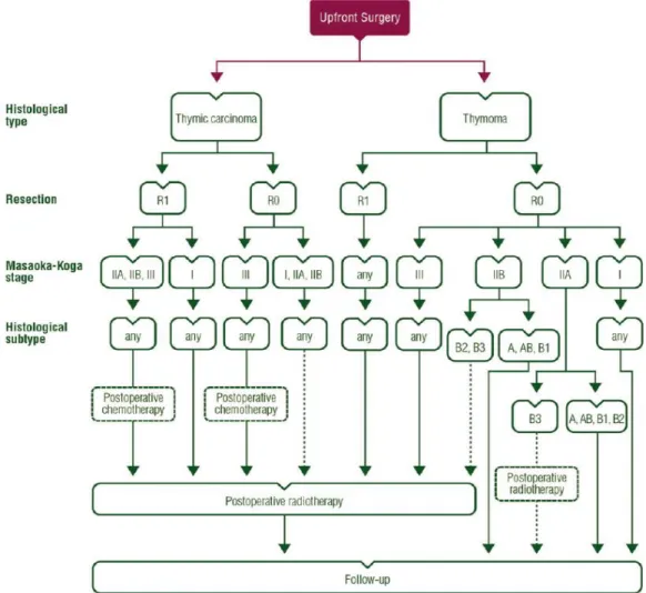
The 2015 ESMO (European Society for Medical Oncology) clinical practice guidelines for patients with resectable TETs (European Society for Medical Oncology) is depicted in Figure 7.
25
Figure 7: Treatment algorithm for resectable thymic tumour (Masaoka-Koga stage I–III, TNM stage I–
IIIA). From: Thymic epithelial tumours: ESMO Clinical Practice Guidelines for diagnosis, treatment and follow-up (14).
2.9.3 Institutional treatment specifics
There are no randomized controlled clinical multicenter trials on the treatment of TETs. The current medical evidence is mostly based on retrospective single center experiences and population studies. Thus the peer- reviewed literature cannot give definitive recommendations for patient care. We recommend that all cases undergo multidisciplinary tumor board review. The treatment scheme applied for TET patients is based on the experience gained from our thoracic surgery center, a multidisciplinary consensus of thoracic surgeons, oncologists, radiotherapists, and pathologists (see
Table 8).
26
Table 8: Therapeutic algorithm at the Division of Thoracic Surgery, Medical University Vienna (Moser, Scharitzer et al. 2014).
WHO type
stagea A, AB, B1 B2, B3 Thymic carcinoma
I - - -
II - - adjuvant therapy?
III nCRT nCRT nCRT
IV nCRT nCRT nCRT
nCRT neoadjuvant Chemo- and/or radiotherapy
aMasaoka-Koga stage
The institutional treatment algorithm stipulates a multimodal therapy regimen for patients in Masaoka–Koga stages III and IV. For TCs neoadjuvant or adjuvant therapy is discussed also in stage II patients. Standard chemotherapy for either neoadjuvant or adjuvant chem- otherapy consists of three cycles of cisplatin 50 mg/m2, doxorubicin 50 mg/m2, and cy- clophosphamide 500 mg/m2 (PAC chemotherapy). Individualized radiation therapy in an adjuvant or neoadjuvant setting is applied with doses up to 50 to 60 Gray. For definitive staging and histology the histopathological examination of the operative TET specimen is reqired. All TET specimens are reviewed by the reference pathology at the Faculty of Mannheim, University of Heidelberg, Germany (second opinion). Decisions on postop- erative RT (PORT) or adjuvant ChT are based on pathological staging.
2.9.4 Surgery
If a complete TET resection (R0) is deemed feasible upfront surgery is indicated. This is usually the case in Masaoka-Koga stages I and II, as well as stage III with invasion of structures that are readily resectable (e.g. pericardium, adjacent mediastinal pleura or lung). In this Chapter different surgical approaches to TETs, the rationale for extended thymectomy and surgery for TETs with pleural involvement are detailed.
2.9.4.1 Surgical approach: open vs. minimally invasive
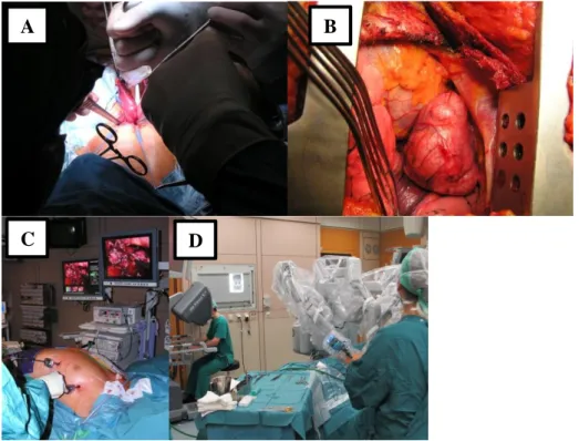
A variety of different open and minimally-invasive surgical approaches for the resection of TETs are in use. The open approaches are: thoracotomy, sternotomy, hemiclamshell and clamshell and cervical incisions. Minimally-invasive techniques include various modifications of VATS and robotic surgery (RATS) (Figure 8). Minimally-invasive tech- niques are established as the treatment standard for early-stage TETs (Masaoka-Koga stages I and II) by specialized centers experienced in thymic surgery (Liu, Lin et al. 2014,
27
Manoly, Whistance et al. 2014, Friedant, Handorf et al. 2016). All the oncological prin- ciples that are followed in open standard thymic surgery have to be met. There are no signs that minimally-invasive procedures are inferior to open surgery considering com- plications, recurrence rates or survival. The reported advantages of minimally-invasive surgery are shorter length of hospital stay, less intraoperative blood loss and improved cosmetic results (Ruffini, Filosso et al. 2018). The state of the art of thymic minimally invasive surgery across Europe (Matilla, Klepetko et al. 2017) as well as our initial expe- rience with a combined sequential left-sided and subxiphoid video-assisted thoracic sur- gery approach for resection of large anterior mediastinal tumors (Matilla JR 2018) was recently reviewed by our group.
Figure 8: Surgical approach. Open surgery: through (A) cervical incision for basic thymectomy. Cervical thymectomy: The two thymic horns are developed through a cervical incision. The part of the surgical thymus shown in the photograph is called basic thymectomy. Left-sided thoracotomy for resection of thy- moma ((B) arrow heads), VATS thymectomy (C), Robotic thymectomy (D). Photographs A-D from the division of thoracic surgery, Medical University Vienna;
A B
C D
28
Rationale for extended thymectomy
Basic thymectomy is the surgical removal of the thymic horns (see Figure 8A and Figure 9).
Figure 9: Surgical anatomy of the thymus. (black,thymus; gray, fat, which may contain islands of thymus and microscopic thymus). From (Sonett and Jaretzki 2008).
Thymomectomy is the removal of the thymoma/TC without removal of the thymus. Ex- tended thymectomy is the removal of all mediastinal fatty tissue between the phrenic nerves along with basic thymectomy. While extended thymectomy is the treatment of choice for patients with myasthenia gravis its role in thoracic surgery for patients with thymomas without MG is still a matter of debate. The rationale to perform extended thy- mectomy in patients with MG with and without TETs is to completely remove all thymic tissue that is dispersed within the mediastinal fatty tissue (see Figure 9 and Figure 10) (Masaoka, Nagaoka et al. 1975, Sonett and Jaretzki 2008). Extended thymectomy for MG is supported by improvements in MG disease activity in patients with residual thymus undergoing extended thymectomy after failure to improve after basic or non-radical thy- mectomy (Masaoka and Monden 1981, Jaretzki, Penn et al. 1988). In cases of patients with TETs without MG current recommendations advocate an extended thymectomy in conjunction with the resection of the TET (Ruffini, Filosso et al. 2018). In support of this more radical approach are the risk of multiple TETs, risk of local recurrences, risk of postoperative newly developed MG, the difficulty to pre- and intraoperatively decide
29
whether the TET is encapsulated and the difficulty to achieve tumor-free resection mar- gins in stage II tumors (Ruffini, Filosso et al. 2018).
Figure 10: Extended thymectomy. Representative CT sections with the corresponding operative specimens of extended thymectomies (picture from the division of thoracic surgery, Medical University Vienna) of (A and C) a patient with MG (Osserman II-b): cervikal & left VATS approach; thymoma - WHO type B2 Masaoka II-1, R0 (B) (B and D) Incidental finding during preoperative radiological workup strumectomy);
thoracotomy; thymoma WHO type AB, Masaoka II-1, R0, no MG; follicular thymitis;
2.9.4.2 Treatment of TETs with pleural involvement
For lower stages of TETs complete surgical resection has become the accepted treatment standard. Patients with advanced-stage TETs presenting with pleural or pericardial dis- semination (Masaoka-Koga Stage IVA (Koga, Matsuno et al. 1994)) are encountered in only 6.8% of all patients with TETs (Koga, Matsuno et al. 1994, Kondo and Monden 2003, Murakawa, Karasaki et al. 2015). Because of the scarcity of existing data in cases with TETs with pleural disease the value of surgical resection remains in question. There are several reasons for low case numbers: TETs with pleural involvement are usually treated in single institutions. Also, there is a wide array of different forms of clinical presentation. While some patients are diagnosed with one or few well defined and local- ized pleural lesions , others present with a diffuse pattern of pleural involvement. A subset
A B
C
D
30
of patients presents the combination of pleural and pulmonary tumor spread. Depending on the disease distribution several surgical techniques were developed for resection: ex- trapleural pneumonectomy (EPP), total pleurectomy (TP) or local pleurectomy (LP). All of the surgical approaches are frequently combined with ChT and/or RT (Ishikawa, Matsuguma et al. 2009, Fabre, Fadel et al. 2011). EPP will be performed for numerous visceral pleural, parietal pleural and pericardial implants (and pulmonary nodules) that cannot be locally resected (Wright 2011). TP removes all parietal (Figure 11), mediastinal and diaphragmatic pleural surfaces and pericardium with or without resection of the dia- phragm. Therefore TP is performed when visceral pleura and lung are not affected by malignant disease. LP is the local resection of pleural implants without removal of all pleural surfaces (metastasectomy) and is performed for mono- or oligometastatic disease.
Pleurectomy/decortications (P/D) is a lung sparing procedure with the intent of removing all macroscopic disease in order to prolong patient survival. It is a TP of the parietal and visceral pleural surfaces (Imanishi, Nabe et al. 2018). Extended P/D (EPD) includes re- section of diaphragm and pericardium in addition to P/D (Bilancia, Nardini et al. 2018, Imanishi, Nabe et al. 2018). In patients with Masaoka-Koga stage IVa (pleural or peri- cardial metastases) current recommendations include major pleural surgery with curative intent such as P/D or EPP usually performed as part of multimodal therapy (Ruffini, Filosso et al. 2018).
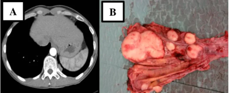
Figure 11: Pleural metastases of WHO type B2 thymoma. Representative CT scan section of the regional recurrence (A) with part of the operative specimen of total pleurectomy showing several pleural implants (B).
A B
31 Debulking surgery
There is an ongoing debate on wheter debulking (reduction in tumor volume) for not completely resectable tumors has a role in TET surgery. Debulking surgery may be indi- cated in patients where resection can alleviate cardiopulmonary compromise (see Figure 12) in order to facilitate systemic treatments or RT. Debulking may reduce the number of local recurrences (Mornex, Resbeut et al. 1995) and reduce radiation field sizes of adju- vant RT (Attaran, Acharya et al. 2012). In cases of reoperation for recurrent thymomas debulking surgery should be limited to selected patients with no other available treatment options (Dai, Song et al. 2015).
Figure 12: Magnetic resonance image (A) of a not completely resectable TC. Because of upper inflow occlusion and severe compromise of cardiorespiratory function the patient was not amenable to ChT. After incomplete resection of the TC regular cardiopulmonary conditions allowed adjuvant ChT (postoperative chest X-ray (B)). From (Moser and Klepetko 2013).
2.9.5 Systemic therapy
In patients with unresectable disease (distant metastases or technical unresectability, e.g.
extensive infiltration of cardiac ventricles) or those unfit for surgery or anasthesia because of comorbidities may undergo individualized therapy. The patients`performance status, tumor stage or symptoms will influence decisions regarding the use RT, chemo- or chemoradiotherapy, or other systemic treatments. As all studies in this thesis were done on patients undergoing surgical resection the following sections will focus on systemic or radiation therapy with regard to patients undergoing surgery for TETs.
2.9.5.1 Chemotherapy (Cht)
ChT has a role in patients with unresectable disease, as neoadjuvant or adjuvant therapy and in metastatic and recurrent disease.
A B
32
In cases of TET invasion of potentially resectable structures (e.g. superior vena cava, aortic arch) when upfront surgery is in doubt to achieve R0 resection borders neo- adjuvant therapy should be considered in order to increase the probability for a complete resection. In a metaanalysis evaluating the effect of induction therapy (cisplatin based ChT) and surgery on OS of patients with advanced TETs reported a pooled response rate of induction therapy of 59%, a pooled rate of complete resection of 73% and pooled 5- and 10-year OS following induction therapy confirming favorable outcomes of this ap- proach (Hamaji, Ali et al. 2015). The highest response rates (70-81%) with neoadjuvant ChT were reported with platinum-based ChT combined with anthracycline (Loehrer, Chen et al. 1997, Berruti, Borasio et al. 1999, Kim, Putnam et al. 2004, Chau, Kim et al.
2010).
PAC ChT for 29 patients with metastatic or recurrent thymoma (intergroup trial) showed three complete responses (CRs) and 12 partial responses (PRs): rate of combined CR+PR:
50% (Loehrer, Kim et al. 1994). Combinations of platinum based ChT without anthracy- cline were reported with inferior response rates: 32-56% (Giaccone, Ardizzoni et al. 1996, Loehrer, Jiroutek et al. 2001, Chau, Kim et al. 2010, Lemma, Lee et al. 2011) but are an option for patients who cannot undergo the most aggressive regimens. ChT with single agents was inferior to the combinations of several chemotherapeutic agents (Chau, Kim et al. 2010). A vast array of second-line chemotherapeutic approaches were undertaken for progression of disease with first line ChT (e.g. pemetrexed (Gbolahan, Porter et al.
2018), ocreotide (Loehrer, Wang et al. 2004)). The role of chemotherapy in advanced TETs was recently reviewed (Schmitt and Loehrer 2010).
2.9.5.2 Novel systemic therapies: targeted therapy and immuno- therapy
The recent experience with immune checkpoint inhibitor therapy (Brown, Dorfman et al.
2003, Merveilleux du Vignaux, Maury et al. 2017, Badiyan, Roach et al. 2018, Saleh, Khalifeh-Saleh et al. 2018, Yokoyama and Miyoshi 2018) and the use of targeted thera- pies for patients with TETs was comprehensively reviewed (Berardi, De Lisa et al. 2014, Ried, Marx et al. 2016).
KIT is a potential target in TCs
KIT mutations are present in up to 12% of TCs (Schirosi, Nannini et al. 2012) . In patients with TCs with reactivity of CD117 (product of proto-oncogene c-kit) immunostaining
33
testing for c-kit is recommended. Several therapeutic possibilities were described as for example imatinib for mutated exons 9 and 11, sunitinib for mutated exons 13, 14 or so- rafenib for mutated exon 17 (Schirosi, Nannini et al. 2012).
Immune checkpoint inhibitor therapy for TCs
Forty patients with recurrent TC progressing after ChT were treated with pembrolizumab, monoclonal antibody with specificity for PD-1 (single-arm phase 2 study), showed 22.5%
objective reponses, including one complete reponse. Despite the observation that TC pa- tients are not typically affected by paraneoplastic autoimmune disorders, a high rate of immune-related events (e.g. myocarditis) was reported (Giaccone, Kim et al. 2018).
2.9.6 Radiation therapy (RT)
Treatment recommendations regarding RT for TETs are mostly based on data of small patient cohorts collected over long periods of time and population-based studies (Fuller, Ramahi et al. 2010). The indications for RT as part of a multimodal approach as well as the optimal protocols for different treatment situations remain a matter of debate. All cases should be discussed in a multidisciplinary tumor board.
In the following treatment situations of patients with TETs there is a role for RT:
unresectable disease (also progressive disease during neoadjuvant ChT)
following incomplete resections of TETs (R1 or R2 resections).
The role of RT with regards to tumor stage was recently reviewed (Fuller, Ramahi et al.
2010). Postoperative radiation therapy (PORT) should be considered for patients after complete surgical resection of Masaoka-Koga stage II-IV TETs (ESMO guidelines) (Girard, Ruffini et al. 2015). RT is currently not recommended in Masaoka-Koga stage I after complete resection. One reason is the low frequency of recurrences. In stage I thy- momas undergoing complete resections and followed-up for 32 years recurrence rates were reported with 2-3% (Awad, Symmans et al. 1998). Another reason is the lack of benefit of PORT in stage I studies. In a Chinese prospective study on 29 patients that were randomized to postoperative radiotherapy for stage I thymomas no survival benefit for either group was detected: 10-year OS: resection plus RT 88% vs. resection alone 92%
(Zhang, Lu et al. 1999). The role of PORT after complete resection in stage II thymoma is still a matter of debate (See also Figure 7: Treatment algorithm for resectable thymic
34
tumour (Masaoka-Koga stage I–III, TNM stage I–IIIA). Several retrospective center ex- periences do not recommend PORT in this situation (Mangi, Wright et al. 2002, Rena, Papalia et al. 2007, Chen, Feng et al. 2010, Berman, Litzky et al. 2011). A meta-analysis including seven retrospective studies (1724 patients with primary thymoma) found a po- tential OS benefit of PORT for patients with locally advanced thymomas (Masaoka-Koga stages III and IV) after macroscopically complete resection compared to surgery alone.
In this analysis PORT did not convey a survival benefit in stage II patients (Lim, Kim et al. 2016). TCs present in higher stages than thymomas and are accompanyied by lym- phatic or hematogeneous metastases in 10-30% (Kondo and Monden 2003, Ruffini, Detterbeck et al. 2014, Hamaji, Shah et al. 2017). A Meta-Analysis of PORT for TC (7 retrospective observational studies, 786 patients) supports PORT for TC (recommenda- tions regarding stage and resection status could not be provided) (Hamaji, Shah et al.
2017).
2.10 Prognostic factors for patients with TETs
2.10.1 Outcome measures for TETs
Overall survival (OS) is an adequate standard outcome measure for many cancers, espe- cially when survival after recurrence is short and the cause of death is the particular can- cer. In the case of thymomas many patients die of causes other than thymomas (see Figure 13) and many patients experiencing a thymoma recurrence have a long life expectancy (Huang, Detterbeck et al. 2011). Because of the indolent behavior of especially early stage thymomas, freedom from recurrence (FFR) reflects best the biology of these tumors after curative treatment. In addition to OS, CSS should be reported because it reflects death from thymoma/TC. The analysis of Time-to-progression (TTP) is recommended for pa- tients with residual disease, although the outcome of this patient population is well re- flected in the analysis of OS (Huang, Detterbeck et al. 2011). Another issue concerning survival analysis is the increased risk of thymoma patients to develop and die from ex- trathymic malignancies (Filosso, Galassi et al. 2013).
35
Figure 13: Overall cause of death after resection of thymomas. From (Huang, Detterbeck et al. 2011).
This is a brief description of the outcome measures recommended by ITMIG (Huang, Detterbeck et al. 2011) and ESTS Thymic Working group (Ruffini, Detterbeck et al. 2014, Ruffini, Detterbeck et al. 2014) that were used in this thesis. OS was calculated as the primary outcome from the date of surgery to the date of death of any cause (censored observations: patients at the last time point known to be alive). The end-point of interest for Cause-specific survival (CSS) was defined as death from TET (censored observations:
unrelated deaths and unknown cause of death) (Huang, Detterbeck et al. 2011). FFR was calculated only in patients after complete surgical resection (R0) from the date of first pleural surgery to the date of recurrence in patients with full information on recurrence status (Ruffini, Detterbeck et al. 2014). Disease-free survival (DFS) was analyzed from the date of surgery to the date of recurrence or death from any cause (Ruffini, Detterbeck et al. 2014). For the determination of time to progression only patients after incomplete resection (R1 or R2) or patients with partial remission or stable disease after chemother- apy and/or radiotherapy were included (Huang, Detterbeck et al. 2011). Figure 14 depicts estimated outcomes for stage III thymomas when different outcome parameter definitions are applied (Huang, Detterbeck et al. 2011).