JAK – STAT inhibition impairs K-RAS-driven lung adenocarcinoma progression
Julian Mohrherr1,2, Marcel Haber2, Kristina Breitenecker1,2, Petra Aigner1,2, Stefan Moritsch3, Viktor Voronin2,
Robert Eferl 3, Richard Moriggl2,4,5, Dagmar Stoiber1,2, Balázs Győrffy6, Luka Brcic7, Viktória László8, Balázs Döme8,9,10,11, Judit Moldvay10,12, Katalin Dezső13, Martin Bilban14,15, Helmut Popper 7, Herwig P. Moll 1*and Emilio Casanova 1,2*
1Department of Physiology, Center of Physiology and Pharmacology & Comprehensive Cancer Center (CCC), Medical University of Vienna, Vienna, Austria
2Ludwig Boltzmann Institute for Cancer Research (LBI-CR), Vienna, Austria
3Institute of Cancer Research, Medical University of Vienna & Comprehensive Cancer Center (CCC), Vienna, Austria
4Institute of Animal Breeding and Genetics, University of Veterinary Medicine, Vienna, Austria
5Medical University of Vienna, Vienna, Austria
6MTA TK Lendület Cancer Biomarker Research Group, Institute of Enzymology, and Second Department of Pediatrics, Semmelweis University, Budapest, Hungary
7Diagnostic & Research Institute of Pathology, Medical University of Graz, Graz, Austria
8Division of Thoracic Surgery, Department of Surgery & Comprehensive Cancer Center (CCC), Medical University of Vienna, Vienna, Austria
9Department of Biomedical Imaging and Image-guided Therapy, Division of Molecular and Gender Imaging, Medical University of Vienna, Vienna, Austria
10Department of Tumor Biology, National Korányi Institute of Pulmonology, Semmelweis University, Budapest, Hungary
11Department of Thoracic Surgery, National Institute of Oncology and Semmelweis University, Budapest, Hungary
12SE-NAP Brain Metastasis Research Group,2nd Department of Pathology, Semmelweis University, Budapest, Hungary
13First Department of Pathology and Experimental Cancer Research, Semmelweis University, Budapest, Hungary
14Department of Laboratory Medicine, Medical University of Vienna, Vienna, Austria
15Core Facilities, Medical University of Vienna, Vienna, Austria
Oncogenic K-RAS has been difficult to target and currently there is no K-RAS-based targeted therapy available for patients suffering from K-RAS-driven lung adenocarcinoma (AC). Alternatively, targeting K-RAS-downstream effectors, K-RAS-cooperating signaling pathways or cancer hallmarks, such as tumor-promoting inflammation, has been shown to be a promising therapeutic strategy. Since the JAK–STAT pathway is considered to be a central player in inflammation-mediated tumorigenesis, we investigated here the implication of JAK–STAT signaling and the therapeutic potential of JAK1/2inhibition in K-RAS-driven lung
*H.P.M. and E.C. are equal senior authors
Additional Supporting Information may be found in the online version of this article.
Key words:non-small cell lung cancer, lung adenocarcinoma (AC), Kirsten rat sarcoma viral proto-oncogene (K-RAS), Janus kinase (JAK), ruxolitinib, cell-line derived xenografts, genetically engineered mouse models, tumor microenvironment (TME), tumor promoting inflammation
Abbreviations:AC: adenocarcinoma; ACTB: actin beta; ANOVA: one-way analysis of variance; CXCL1: chemokine (C-X-C motif) ligand 1;
EGFR: epidermal growth factor receptor; GO: gene ontology; GSEA: gene set enrichment analysis; IL: interleukin; JAK: Janus kinase; K-RAS:
Kirsten rat sarcoma viral proto-oncogene; MDSCs: myeloid derived suppressor cells; mRNA: messenger RNA; NKs: natural killer cells; PD-L1:
programmed cell death 1 ligand 1; RNA-seq: RNA sequencing; STAT: signal transducer and activator of transcription; TBP: TATA-box binding protein; TME: tumor microenvironment; TNF: tumor necrosis factor; VEGF: vascular endothelial growth factor; WT: wild-type
Conflict of interest:The authors declare no potential conflict of interest.
Grant sponsor:Austrian Science Fund;Grant numbers:FWF-P 25599-B19, P29222-B28;Grant sponsor:National Research, Development and Innovation Office, Hungary;Grant numbers:KH-129581, NVKP_16-1-2016-0037
This is an open access article under the terms of the Creative Commons Attribution License, which permits use, distribution and reproduction in any medium, provided the original work is properly cited.
DOI:10.1002/ijc.32624
History:Received 19 Feb 2019; Accepted 22 Jul 2019; Online 12 Aug 2019
Correspondence to:Herwig P. Moll, Department of Physiology, Center of Physiology and Pharmacology, Comprehensive Cancer Center, Medical University of Vienna, AT-1090 Vienna, Austria, Tel.: +43140160-71230, E-mail: herwig.moll@meduniwien.ac.at
Cancer Therapy and Prevention
Int. J. Cancer:145,3376–3388 (2019)©2019 The Authors.International Journal of Cancerpublished by John Wiley & Sons Ltd on behalf of UICC
AC. Our data showed that JAK1and JAK2are activated in human lung AC and that increased activation of JAK–STAT signaling correlated with disease progression and K-RAS activity in human lung AC. Accordingly, administration of the JAK1/2selective tyrosine kinase inhibitor ruxolitinib reduced proliferation of tumor cells and effectively reduced tumor progression in immunodeficient and immunocompetent mouse models of K-RAS-driven lung AC. Notably, JAK1/2inhibition led to the establishment of an antitumorigenic tumor microenvironment, characterized by decreased levels of tumor-promoting chemokines and cytokines and reduced numbers of infiltrating myeloid derived suppressor cells, thereby impairing tumor growth. Taken together, we identified JAK1/2inhibition as promising therapy for K-RAS-driven lung AC.
What’s new?
A drug that inhibits the JAK–STAT pathway may score a hit against K-RAS driven lung cancer. Here, the authors Investigated the JAK STAT pathway as a possible target in lung adenocarcinoma because of its role in inflammation-mediated tumorigenesis.
First, they showed that JAK1and JAK2are both activated in lung adenocarcinoma patients with oncogenic mutations in K-RAS.
Next, they treated the tumors with ruxolitinib, which inhibits JAK1/2. The drug successfully slowed tumor proliferation and progression in immunocompetent mouse models. Furthermore, treatment with ruxolitinib reduced the tumor-promoting factors present in the microenvironment.
Introduction
Lung cancer is still the leading cause of cancer-related deaths worldwide in both men and women.1Histologically, lung cancer can be subdivided into small cell lung cancer and non-small cell lung cancer, with lung adenocarcinoma (AC) being the most abundant among the non-small cell lung cancer subtype.2 Roughly 50% of lung ACs harbor oncogenic mutations in the V- Ki-ras2 Kirsten rat sarcoma viral oncogene homolog (K-RAS) or epidermal growth factor receptor (EGFR).3,4While EGFR tyro- sine kinase inhibitors, such as erlotinib, gefitinib or afatinib, are part of the clinical routine to treat EGFR-mutated lung AC patients,5 the development of K-RAS inhibitors has been less successful so far, with no K-RAS-based targeted therapy being clinically approved.6Hence, targeting alternative tumor drivers fueling K-RAS-mediated lung tumorigenesis such as tumor- promoting inflammation and K-RAS downstream effector path- ways are considered to be a promising treatment strategy.6,7
Given the pivotal role of Janus kinase (JAK) activation in tumor-promoting inflammation and its oncogenic functions, JAKs represent attractive therapeutic targets in tumorigenesis.8 The JAK family is composed of four members (JAK1, JAK2, JAK3 and tyrosine kinase 2) and couples extracellular stimuli with gene transcription. Upon cytokine binding to their cognate receptors, JAKs become activated and phosphorylate downstream signal transducer and activator of transcription (STAT) molecules, ulti- mately leading to nuclear translocation and transcription of vari- ous target genes involved in cell cycle regulation, angiogenesis, apoptosis, tumor invasion and metastasis.9JAK signaling either promotes or suppresses tumorigenesis in a tumor cell intrinsic manner. Indeed, K-RAS-mutated lung AC cells secrete pro- inflammatory cytokines including interleukin (IL)-6 that activate JAK1 and JAK2viaglycoprotein 130 in an autocrine loop, thereby promoting tumor cell survival.7,10,11On the other hand, genetic
deletion of STAT3, a key signaling mediator of JAK1/2, enhances K-RAS-driven lung tumorigenesis, and activation of the inter- feron/JAK/STAT axis induces cell apoptosis and suppresses tumorigenesis in various experimental tumor models.12–16
In addition to these tumor-cell intrinsic effects, JAK signaling is also substantially involved in shaping the tumor microenviron- ment (TME) by modulating T, natural killer (NK) and dendritic cell functions.17As in tumor cells, JAK activation in the TME and its role in tumorigenesis is controversially discussed. While JAK1/2 inhibition impairs NK-cell function and NK mediated antitumor immunity and hence indirectly facilitates tumor meta- static spread,18 targeting the JAK–STAT axis has also been proposed as a potential effective strategy to reduce levels of tumor- promoting immunosuppressive myeloid derived suppressor cells (MDSCs), thereby counteracting malignant progression.19 Fur- thermore, IL6/JAK/STAT3, as well as interferon-γ/JAK/STAT1 signaling pathways have also been described to induce pro- grammed cell death 1 and or programmed cell death 1 ligand 1 (PD-L1) expression and, therefore, to promote tumor immune escape.20,21Taken together, these reports indicate that JAKs may have opposite functions in tumorigenesis depending on the cellular context.
Clinical trials investigating JAK inhibition in particular for EGFR-mutated lung cancers are ongoing. These studies combine the JAK inhibitors ruxolitinib, itacitinib and AZD4205 with the EGFR tyrosine kinase inhibitors erlotinib (NCT02155465), afatinib (NCT02145637) and osimertinib (NCT02917993, NCT03450330).
By contrast, JAK targeting inK-RAS-mutated lung ACs has not been thoroughly investigated so far. Notably, a recent clinical trial showed positive results when combining ruxolitinib and che- motherapy in non-small cell lung cancer patients, but the K-RAS mutation status is not available (NCT02119650). Thus, to comple- ment these clinical studies, we addressed the role of JAK1/2 in
Cancer Therapy and Prevention
K-RAS-driven lung AC pathogenesis. By using human tumor samples and experimental mouse models, we show that JAK1/2 are activated in K-RAS-driven lung AC and promote tumor growth and progression by establishing a protumorigenic TME. Impor- tantly, pharmacological inhibition of JAK1/2 with the tyrosine kinase inhibitor ruxolitinib reverts the protumorigenic TME and impairs tumorigenesis.
Materials and Methods Human data
For gene expression analysis, we took advantage of the publicly available Illumina microarray data set, which contains biopsies of K-RAS-mutated lung AC tissue and adjacent healthy tissue (GSE75037).22To determineJAK1andJAK2mRNA expression levels, we used probes as depicted in Table S1. As a second cohort, we used gene expression data from K-RAS-mutated lung AC patients included in the Cancer Genome Atlas (n= 154).23
Gene set enrichment analysis (GSEA) was performed accor- ding to the provider’s protocol and permutations were set to 1,000.24Gene sets were downloaded from the Molecular Signa- ture Database (http://software.broadinstitute.org/gsea/msigdb/
index.jsp). To determine the prognostic value of chemo/cytokine related genes upon ruxolitinib treatment for human lung ACs pathogenesis, we used the KM-Plotter online tool.25In addition, gene expression data ofK-RAS-mutated lung AC patients with available survival data included in the Cancer Genome Atlas were analyzed (n= 150).23We have run the analysis by evaluating all possible cutoff values and the best performing value was used as the cutoff in thefinal analysis. False discovery rate was computed to correct for multiple testing, and only results having a false dis- covery rate below 1% was accepted as statistically significant.
Analysis was performed using the mean expression of the genes included in the multigene signature, and all utilized Probe-IDs are shown in Table S2.
For immunohistochemical analysis, we generated tissue microarrays from two independent lung AC patient cohorts.
Cohort A includes biopsies from lung AC patients where information about the mutationalK-RASstatus was not avail- able (pJAK1 n= 303, pJAK2 n= 318). Cohort B consists of biopsies of lung AC patients positive for K-RAS mutation (pJAK1n= 26, pJAK2n= 24). Analysis was done with prior approval by the institutional ethical committee. For detection of JAK1 or JAK2 tyrosine phosphorylation the tissue micro- arrays were stained with a pJAK1 (1:200, Y1022, #PA537617, Thermo Fisher Scientific, Waltham, MA) and a pJAK2 (1:200, Y1007 and Y1008, #32101, Abcam, Cambridge, UK) antibody.
Histoscores were determined according to staining intensities and percentage of positive tumor cells by a board-certified pathologist (H.P.).26
Cell lines
The humanK-RAS-mutated lung AC cell lines A549 and A427 were grown in RPMI medium (Gibco, Grand Island, NY) sup- plemented with 10% FBS, 2 mM glutamine (Gibco), penicillin
(50 U/ml, Gibco) and streptomycin (50μg/ml, Gibco). A549 and A427 cell lines were obtained from American Type Culture Col- lection, and cells were profiled for authentication (Eurofins Geno- mics, Luxembourg). Cells were tested to be free of mycoplasma contaminations twice a year. For experiments, cells were harvested at ~70% confluency, and the number of viable cells was deter- mined using a CASY counter (Roche, Basel, Switzerland).
Animal husbandry and experiments
For subcutaneous xenograft experiments using the K-RAS- mutated human lung AC cell lines A549 and A427, 1×10⁶ cells were mixed 1:2 with Matrigel (Corning, Corning, NY) and injected into the right and left flank of immunodeficient NOD scid gamma mice older than 6 weeks of age. Tumor vol- ume was measured using a caliper and tumor volume was cal- culated using the formula [(length×width2)×0.52] as previously established in our laboratory.27
K-rasLSL-G12D (K) mice as well as K-rasLSL-G12D:p53fl/fl (KP) mice, both bred on a C57Bl/6 background, served as geneti- cally engineered mouse models for autochthonous lung AC.28 Genotyping of the transgenes was performed using primers depicted in Table S3. Induction of lung tumors in these geneti- cally engineered mouse models was achieved through intranasal inhalation of a Cre-recombinase expressing adenovirus (Ad.Cre) in 8–12 week old mice using 2.5×10⁷ plaque-forming units.
Inhalation of K and KP mice with Ad.Cre triggersK-rasG12Dacti- vation in lung epithelial cells resulting in formation of p53 expressing (K) or deficient (KP) lung ACs. Ad.Cre was purchased from the Viral Vector Core (University of Iowa). For treatment of mice with the JAK1/2 inhibitor ruxolitinib (LC Labs, Woburn, MA), the inhibitor was suspended in 0.5% (w/v) methylcellulose solution. Mice were treated via oral gavage with vehicle (ctrl, 0.5% Methylcellulose) or ruxolitinib (Ruxo) at 90 mg/kg twice a day (BID), 7 days per week for the indicated experimental time period. Treatment of mice was paused if any signs of distress occurred.18All experimental protocols as well as animal mainte- nance and breeding described above, followed ethical guidelines and were approved by the Austrian Federal Ministry of Science, Research and Economy.
Histology
At the experimental endpoint, subcutaneously engrafted tumor tissue or lung tissue was retrieved and fixed in 2% buffered formaldehyde at 4C for 24 hr and embedded in paraffin after dehydration. Five micron sections were used for analysis. To determine tumor area, lung sections were stained with hematoxy- lin and eosin and subsequently scanned using TissueFaxs (TissueGnostics, Vienna, Austria) software. Tumor area and number were quantified using HistoQuest (TissueGnostics) soft- ware. Tumor grades were determined by a board-certified pathol- ogist Katalin Dezso (K.D.) according to DuPage et al.28 For immunohistochemistry, standard protocols were applied, using antibodies against pSTAT3 (1:200,Y705, #9145, Cell Signaling, Danvers, MA), KI67 (1:400, Cell Signaling #12202 or 1:200,
Cancer Therapy and Prevention
#14-5698-82, eBioscience, San Diego, CA), cleaved caspase-3 (1:200, Cell Signaling #9661), cluster of differentiation 31 (1:400, Cell Signaling #77699), PD-L1 (1:200, Cell signaling #64988). For analysis of stained immunohistochemistry slides HistoQuest (TissueGnostics) software was used.
RNA-sequencing (RNA-seq) analysis of total lungs and Gene Expression Omnibus data set analysis
For RNA-seq analysis, K mice were treated 8 weeks post inhala- tion with Ad.Cre for four consecutive days with vehicle (0.5%
Methylcellulose) or ruxolitinib (90 mg/kg BID). Additionally tumor harboring lungs from KP mice and lungs from wild-type (WT) mice were included in the analysis. At the experimental endpoint, lungs were harvested and RNA was isolated using the RNeasy Kit (Qiagen, Venlo, Netherlands) following on column DNA digestion using RNase-Free DNase (Qiagen). Library prep- aration and sequencing was carried out by the Core facility Geno- mics, Medical University of Vienna, Vienna, Austria. Briefly, sequencing libraries were prepared using the NEBNext®Ultra™
II RNA Library Prep Kit (NEB, Ipswich, MA) according to manu- facturer’s instructions and sequenced on a Illumina NextSeq500 platform in a 75 bp single-read mode. RNA-Seq data were mapped to the Mus musculus/mmc10 assembly of the murine genome using the STAR Aligner.29Differential gene expression was analyzed using DESeq2.30
For GSEA, gene sets were downloaded from the Molecular Signature Database (http://software.broadinstitute.org/gsea/
msigdb/index.jsp). For gene ontology (GO)-molecular func- tion analysis, the Enrichr web tool was used (http://amp.
pharm.mssm.edu/Enrichr/enrich). The top 350 downregulated genes (based on log2 fold changes) in the ruxolitinib treated group were subjected to pathway analysis with Enrichr.31 To investigate the implications of ruxolitinib downregulated chemo/cytokine related genes for K-RAS-driven lung tumori- genesis, we retrieved all genes within the chemo/cytokine related GO gene sets GO0042379, GO0008009, GO0045236, GO0048020 and GO0005125. Data were analyzed using the web-based online tool InteractiVenn,32 and for hierarchical clustering and heatmap illustration, we used the heatmapper.
ca web tool.33 In order to check for the activation status of the JAK–STAT pathway and the expression of chemo/cytokine related genes in primary K-rasG12D-mutated lung alveolar type II (K) cells, we analyzed the publically available GSE113146 data set.27
RNA extraction and real-time quantitative polymerase chain reaction
RNA isolation from the lungs was performed using TRIzol™
Reagent (Invitrogen, Carlsbad, CA) according to the manufac- turer’s protocol. Complementary DNA was transcribed using the RevertAid H Minus Reverse Transcriptase (Fermentas, Waltham, MA) kit. For detection of the transcript, the iTaq Universal SYBR Green reagent (Biorad, Hercules, CA) and a Biorad CFX Connect Real Time polymerase chain reaction system were utilized, using
specific primers depicted in Table S4. For determination of rela- tive gene expression ratios, the Pfafflmethod was applied. Calcu- lation of bestkeeper values was performed using actin beta (ACTB),28Sand TATA-box binding protein (TBP) as house- keeping genes.34
Flow cytometric analysis of total lungs
At the experimental endpoint,K-RAS-mutated tumor harboring lungs were harvested, and the lung tissue was mechanically and enzymatically disrupted for cell isolation. Briefly, lungs were put into isolation medium (RPMI with 5% FBS) and minced with scissors. Minced pieces were then transferred into a falcon tube containing isolation medium supplemented with DNase I (50 U/ml, Sigma, St.Louis, MO) and Collagenase I (Gibco, 150 U/ml), and incubated for 1 hr at 37C. Subsequently, the cell suspension wasfiltered through 70 and 40μm cell strainers. For subsequentflow cytometric analysis, cells were stained with the antibodies indicated in Table S5 using a standard protocol. For identification of Th17 and regulatory T cells, the mouse Th17/
Treg phenotyping kit (#560767, BD Pharmingen, Franklin Lakes, NJ) was used. All cells are CD45+. Stained samples were measured using the BD FACSCanto™ II system. For data analysis, the FlowJo software (TreeStar, Inc., Ashland, OR) was used.
Statistical analysis
All values are presented as meanstandard error of mean. For single outlier detection the Grubbs-test was used, multiple out- liers were defined when values were lower than Q1–1.5×inter- quartile range or greater than Q3 + 1.5×interquartile range (difference between the third quartile [Q3] and first quartile [Q1]). Subsequent statistical analyses were either performed uti- lizing Student’s t-test or Mann–Whitney U-test calculated by GraphPad Prism 5.0 software. For multiple group comparison, one-way analysis of variance (ANOVA) in combination with Tukey’s multiple comparison test was used. For GO-molecular function analysis, the results were ranked according to the adjustedp-value. For Kaplan–Meier analysis, the Log-rank test was used and false discovery rate was computed to combat for multiple hypothesis testing. For all graphs:*p< 0.05;**p< 0.01;
***p< 0.001.
Data availability
All materials will be made available to the scientific commu- nity. RNAseq data are available within the sequence read archive (SRA) with the accession number PRJNA518030.
Results
JAK signaling is activated in humanK-RAS-mutated lung AC during tumor progression
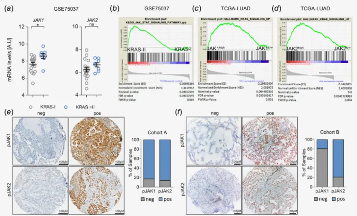
To investigate the implication ofJAK1andJAK2for the patho- genesis of humanK-RAS-mutated lung AC, we took advantage of the publicly available mRNA expression data set GSE75037.22 Comparison ofJAK1andJAK2mRNA expression levels of stage I classifiedK-RAS-mutated lung AC patientsversus those with
Cancer Therapy and Prevention
more advanced disease revealed increased JAK1/2 expression levels in the latter group, which reached significance forJAK1 (Fig. 1a). In line with these data, we found activation of the JAK–
STAT pathway in patients with more advanced lung AC (i.e., in stage II disease), as determined by GSEA (Fig. 1b).24 Next, we confirmed these data inK-RASmutated lung AC patients from a different cohort published by the Cancer Genome Atlas.23 Indeed, we found a significant correlation of JAK1 and JAK2 mRNA expression levels with enrichment of K-RAS signaling in lung AC patients (Figs. 1cand 1d). Moreover, we noticed a signif- icant increase ofJAK1andJAK2mRNA expression in tumors of late stage patients with oncogenic K-RAS mutations compared to patients harboring K-RAS WT tumors, which was not observed in early stage patients (Figs. S1aand S1b).
However, using mRNA expression data of bulk tumor tissue does not allow distinguishing between tumor and stromal cells.
Hence, we performed immunohistochemistry for activated JAK1 and JAK2 in biopsies of lung AC patients from two independent cohorts (cohorts A&B, consisting of unselected and K-RAS
mutated samples, respectively). Indeed, JAK1 and JAK2 were activated/phosphorylated in tumor cells of lung AC patients as determined by a pathologist (H.P.). In the stroma, a positive reac- tion for JAK1 and JAK2 was mainly found in lymphocytes (Figs. 1eand 1f). Taken together, these data indicated that JAK signaling is active in human K-RAS-driven lung AC.
JAK inhibition impairs humanK-RAS-mutated lung AC cell growthin vivo
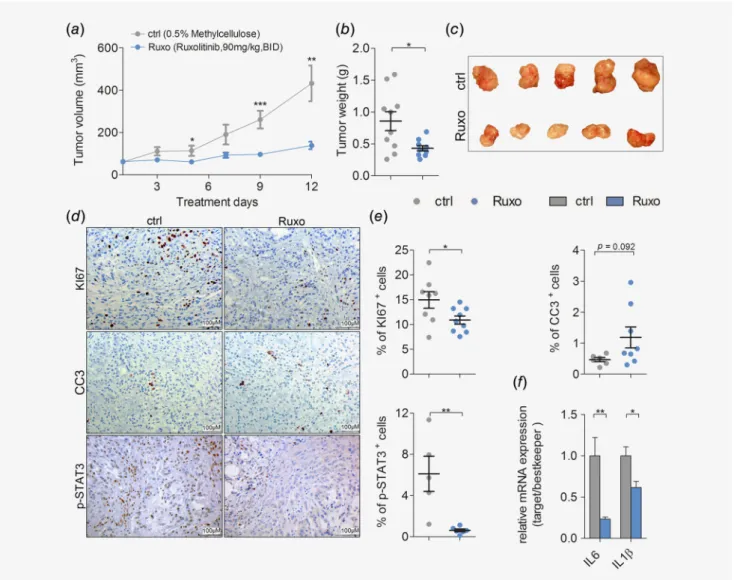
Next, we assessed the potential benefits of pharmacological JAK1/2 inhibition in K-RAS-driven lung AC using the JAK1/2 selective tyrosine kinase inhibitor ruxolitinib. We performed subcutaneous engraftments of human lung AC cell lines harboring oncogenic K- RAS mutations into immunodeficient NOD scid gamma mice. Intriguingly, ruxolitinib treatment significantly attenuated growth of the humanK-RASG12S-mutated lung AC cell line A549 (Figs. 2a–2c), and impaired growth of A427 (K-RASG12D) transplanted cells (Figs. S2a and S2b). Immunohistochemistry analysis of proliferation and apoptosis markers within tumors
Figure1.JAK-mediated signaling is activated in progressing K-RAS-mutated human lung AC. (a) Graph showing relativeJAK1andJAK2mRNA expression levels in humanK-RAS-mutated lung AC tissue of stage I (n=19)versusstage II or higher (≥II,n=8). Data represent meanSEM, A.U (arbitrary units), Student’st-test,*p<0.05. (b) GSEA for Kyoto encyclopedia of genes and genomes-JAK–STAT pathway signature geneset comparing humanK-RAS-mutated tumors of stage Iversusstage II, using tumorversushealthy parenchyma mRNA expression ratios. Data in (a) and (b) were retrieved from the Gene Expression Omnibus (GSE75037). (c) GSEA using gene expression data of the Cancer Genome Atlas-LUAD cohort and HALLMARK_KRAS_SIGNALING_UP geneset, stratifying patients according toJAK1and (d)JAK2expression levels (n=154). (e) Representative pictures of lung AC biopsies of patients included in cohort A (unknownK-RASmutation status; pJAK1n=303, pJAK2n=318) and (f) cohort B (K-RASmutated; pJAK1n=26, pJAK2n=24) with negative and positive staining reactions for pJAK1and pJAK2in tumor cells.
Graphs represent percentage of positive and negative cases within the respective cohort. Staining intensities and percentage of positive tumor cells were determined by a board-certified pathologist (H.P.). [Colorfigure can be viewed at wileyonlinelibrary.com]
Cancer Therapy and Prevention
revealed that JAK inhibition significantly reduced proliferation and slightly increased apoptosis of transplanted lung AC cells (Figs. 2d and 2e). JAK1/2 inhibition in tumor cells of ruxolitinib treated mice was efficient, since activation of the downstream effector STAT3 was almost absent after tyrosine kinase inhibitor treatment (Figs. 2dand 2e). Accordingly, ruxolitinib treatment impaired a well-described autocrine oncogenic activation loop in this model.10 Indeed, tumor cell-derived expression of pro-inflammatory cyto- kinesIL-6andIL-1βwas significantly decreased in the ruxolitinib treated group, as verified by quantitative real-time PCR using primers specific for the human transcripts of these cytokines (Fig. 2f). Notably, the ruxolitinib dose used in our experiments was generally well tolerated by the experimental mice, and we did not
observe any significant body weight loss in the treated groups (Fig. S2c). Since abrogation of JAK signaling may also impair angiogenesis,35we checked expression of the angiogenic markers VegfαandHif1αand evaluated numbers of cluster of differentia- tion 31+vessels, but we did not observe changes upon ruxolitinib treatment (Figs. S2dand S2e).
Ruxolitinib attenuates tumorigenesis of autochthonousK- RAS-driven lung AC
Although cell-line-derived xenograft models have the advantage to study tumors of human origin, a major drawback of these models is the absence of a functional immune system.36However, this is a critical factor in our study, since ruxolitinib was reported
Figure2.JAK inhibition impairs growth of human K-RAS-mutated lung AC cellsin vivo. (a) Mean volumes of xenografted A549derived tumors in mice treated with vehicle control (ctrl) or ruxolitinib (Ruxo) at90mg/kg body weight, seven times per week, BID, and (b) the endpoint tumor weight. (c) Representative pictures of A549tumor-derived xenografts after ctrl or Ruxo treatment. (d) Representative pictures of immunohistochemical stainings for KI67, cleaved caspase3(CC3) and p-STAT3staining of A549cell line derived xenograft tumors upon ctrl and Ruxo treatment (scale bar:100μm), and (e) quantitation of positive cells for respective stainings. (f) Relative mRNA expression of human transcript variants of indicated genes normalized to human housekeeping genes (28S,TBP,ACTB) in A549derived xenografted tumors. (d–f) n=≥5tumors per group. For all graphs Student’st-test:*p<0.05,**p<0.01,***p<0.001. For all graphs data presented as meansSEM.
[Colorfigure can be viewed at wileyonlinelibrary.com]
Cancer Therapy and Prevention
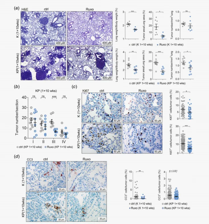
Figure3.Ruxolitinib attenuates tumorigenesis of autochthonous K-RAS-driven lung AC. (a) Left panel: representative pictures of hematoxylin and eosin stained lung sections of vehicle control (ctrl) or ruxolitinib (Ruxo) treatedK-rasG12D(K) mice (n=7per group) andK-rasG12D:p53fl/fl (KP) mice (n=8per group). Treatment was started1week post tumor initiation, and continued for10weeks, with ruxolitinib being
administered at90mg/kg body weight, seven times per week, BID (scale bars:400μm). Right panel: quantitation of hematoxylin and eosin stained lung sections from K mice (upper) and KP-mice (lower) treated with ctrl or Ruxo started1week post tumor initiation and continued for10weeks. (b) Graph depicts tumor numbers per section stratified by tumor grades. Per mouse, one section was analyzed (n=8mice per group). (c) Left panel: representative pictures of immunohistochemical stainings for KI67positive cells of lungs stemming from ctrl and Ruxo treated K and KP mice. Right panel: quantitation of indicated stainings. (d) Left panel: cleaved caspase-3positive cells of lungs of ctrl and Ruxo treated K and KP mice. Right panel: quantitation of indicated stainings (scale bars:50μm). (c,d) Mann–WhitneyU-test,*p<0.05,
**p<0.01,***p<0.001. Others: Student’st-test.*p<0.05,**p<0.01,***p<0.001. For all graphs data presented as meansSEM. [Color figure can be viewed at wileyonlinelibrary.com]
Cancer Therapy and Prevention
to promote immunosuppression and could thereby eventually enhance tumor progression.18Thus, we addressed the effects of JAK1/2 inhibition in fully immunocompetent mouse models of autochthonous K-RAS-driven lung AC. Therefore, we took advantage of the K-rasLSL-G12D (K) and K-rasLSL-G12D:p53fl/fl (KP) mice. Inhalation of K and KP mice with a Cre-recombinase expressing adenovirus (Ad.Cre) triggersK-rasG12Dactivation in lung epithelial cells, resulting in formation of p53 expressing
(K) or deficient (KP) lung ACs. Of note, KP mice develop lung ACs showing features of advanced grade tumors.28,37First, we validated the mouse models by isolating primary lung alveolar type II cells derived from K mice and inducing the expression of oncogenic KRASin vitro. Intriguingly, global gene expression analysis revealed thatK-rasG12Dexpression resulted in activation of the JAK–STAT pathway (Figs. S3aand S3b).27Encouraged by these data, we treated K and KP mice 1 week after tumor
Figure4.Legend on next page.
Cancer Therapy and Prevention
induction with ruxolitinib or vehicle control for a period of 10 weeks, euthanized them and analyzed their lungs. We noticed a significant decrease in tumor burden in ruxolitinib treated mice compared to vehicle-treated control mice, as indicated by reduced lung weight to body weight and tumor to lung area ratios, con- comitant with reduced tumor numbers (Fig. 3a). Of note, sex dif- ferences in treatment response were not observed in our mouse models (Fig. S4a). Furthermore, grading of tumors by a patholo- gist (K.D.) revealed that ruxolitinib blocked the progression of K- RAS-driven lung tumors. Indeed, ruxolitinib treatment resulted in a significant reduction of high grade tumors compared to vehi- cle control, which reached significance for grade III tumors (Fig. 3b and Fig. S4b). No differences in body weights were observed between control and treatment groups (Figs. S4cand S4d). Similar to the engrafted human lung AC cells, ruxolitinib significantly attenuated tumor cell proliferation in these mice (Fig. 3c). Intriguingly, we observed a higher rate of tumor cell apoptosis in the vehicle treated compared to ruxolitinib group in both K and KP mouse models (Fig. 3d). Importantly, there were no differences in the expression levels of the angiogenic factors Hif1α and Vegfα between control and ruxolitinib treatment groups (Figs. S4eand S4f), suggesting that impairment of tumor cell proliferation is responsible for the reduced tumor burden upon ruxolitinib treatment in these models.
JAK inhibition impairs progression of establishedK-RAS- mutated lung AC and modulates the TME
Having demonstrated that ruxolitinib effectively reduces tumori- genesis when administered at early stages of tumor formation, we questioned whether ruxolitinib prevents progression of already established K-RAS-driven lung AC in a therapeutic setting. There- fore, we treated K mice with ruxolitinib starting 8 weeks post Ad.
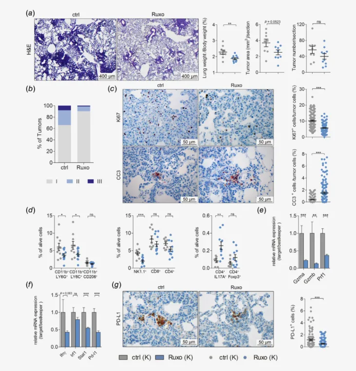
Cre inhalation for five consecutive weeks. In this model, we observed a reduction in lung to body weight ratios and total tumor area per lung section after ruxolitinib treatment as compared to vehicle control treatment, while tumor numbers remained unaltered (Fig. 4a). Notably, analysis of tumor to lung area ratios did not show differences between the treatment groups, most likely because total lung areas were smaller upon ruxolitinib treat- ment (Figs. S5a–S5c). Strikingly, grading of tumors revealed a
reduction of high-grade tumors, namely tumors of grades II and III (Fig. 4b). In addition, ruxolitinib treatment of mice with established tumors also triggered significant reduction in tumor cell proliferation and increased apoptosis (Fig. 4c). Similar to the previous models, differences in mRNA expression levels of angio- genic markers Hif1α and Vegfα were not observed and body weights showed no significant differences between control and ruxolitinib treatment groups (Figs. S5dand S5e). These data sug- gests that ruxolitinib impairs the progression of established K- RAS-driven lung AC. Since JAK signaling plays a crucial role in regulating the immune system, we analyzed if JAK1/2 inhibition affected the composition of the TME by performing flow cytometric analysis of lung tumors.17Indeed, the abundance of tumor promoting granulocytic CD45+CD11b+LY6G+and mono- cytic CD45+CD11b+LY6C+MDSC38was significantly reduced in the lungs of tumor-bearing, ruxolitinib-treated mice. Further- more, the number of tumor promoting CD45+CD11b+CD206+ M2 macrophages was lowered upon JAK1/2 inhibition (Fig. 4d).
With respect to the lymphoid lineage, our analysis revealed signifi- cantly diminished NK cell numbers upon ruxolitinib administra- tion in tumor bearing lungs of these mice (Fig. 4d). These data are in line with a previous report using a preclinical breast cancer model.18In addition, we observed a slight reduction in infiltrating CD8+ and CD4+ T cells upon ruxolitinib treatment, but an increase in CD4+IL17A+Th17 cells as well as CD4+Foxp3+regula- tory T cells (Fig. 4d). Moreover, JAK inhibition lead to a signifi- cant impairment of NK and CD8+T cell activation, but on the other hand also abrogated interferon-γ-mediated STAT1 signal- ing and hence reduced PD-L1 expression (Figs. 4e–4g). In sum- mary, these data indicated that ruxolitinib altered the myeloid compartment of the TME toward an antitumorigenic state, which out-competed the suppressing effects of ruxolitinib on infiltrating cytotoxic NK and CD8+T cells.
JAK inhibition abrogates expression of oncogenic chemokines and cytokines
Next, we thought to unravel the transcriptional changes, leading to the establishment of an antitumorigenic TME upon ruxolitinib treatment. Therefore, we performed RNA-seq analysis of short- term (4 days) treated tumor-bearing lungs from K mice with
Figure4.JAK inhibition impairs progression of established K-RAS-mutated lung AC and alters TME. (a) Left panel: representative pictures of hematoxylin and eosin stained lung sections of vehicle control (ctrl) or ruxolitinib (Ruxo) treatedK-rasG12D(K) mice. Right panel: quantitation of hematoxylin and eosin stained lung sections of ctrl or Ruxo treatedK-rasG12D(K) mice. Treatment was started8week post tumor initiation, and continued for5weeks (8+5wks), with ruxolitinib being administered at90mg/kg body weight, seven times per week, BID (scale bars:
400μm) (n≥9per group). (b) Graph represents the percentage of tumors with indicated grades in vehicle control and ruxolitinib treated K mice. (c) Left panel: representative pictures of immunohistochemical stainings for KI67and cleaved caspase-3positive cells, comparing lungs of ctrl or Ruxo treated K mice (8+5weeks). Right panel: quantitation of indicated stainings (scale bars:50μm). (d) Results offlow cytometric analysis depicting the percentage of myeloid cells (left), lymphoid cells (middle) and CD4+subsets (right) in tumor-harboring lung lysates of ctrl and Ruxo treated K mice (8+5weeks). (e) Graph displaying relative mRNA expression of the indicated genes normalized to housekeeping genes (28s,Tbp,Actb) in tumor harboring lungs of ctrlversusRuxo treated K mice (8+5weeks). (f) Graph indicating relative mRNA expression of the indicated genes normalized to housekeeping genes (28s,Tbp,Actb) in tumor harboring lungs of ctrlversusRuxo treated K mice (8+5weeks). (g) Left panel: representative pictures of immunohistochemical staining for PD-L1positive cells of lungs stemming from ctrl and Ruxo treated K mice (8+5weeks). Right panel: quantitation of indicated staining. (c,g) Mann–WhitneyU-test.
Others: Student’st-test. For all graphs data presented as meansSEM.*p<0.05,**p<0.01,***p<0.001. [Colorfigure can be viewed at wileyonlinelibrary.com]
Cancer Therapy and Prevention
already established lung ACs. As expected, short-term administra- tion of ruxolitinib significantly diminished activation of JAK–
STAT signaling in the lung lysates of these mice (Fig. 5a and
Fig. S6a). Intriguingly, GSEA analysis also revealed that ruxolitinib administration significantly reduced K-RAS activity in these lungs (Fig. 5b). Notably, within the top 350 genes negatively regulated
Figure5.Legend on next page.
Cancer Therapy and Prevention
by ruxolitinib (Table S6), we noticed an over-representation of factors found within GO clusters for chemokine and cytokine activity (Fig. 5c and Fig. S6b). Accordingly, most of the genes annotated in these gene sets were downregulated upon ruxolitinib treatment compared to vehicle control (Fig. 5d and Table S6).
Importantly, 84 of these 109 genes downregulated by ruxolitinib were either upregulated inK-rasG12D-mutated lung alveolar type II (K) cells compared to WT cells (67 genes) or in tumor harbor- ing lungs of KP mice compared to healthy WT lungs (52 genes).
Notably, 35 hits (Tables S2 and S6) were common in all three groups (Fig. 5eand Figs. S6cand S6d). Among these 35 genes, key pro-inflammatory and pro-tumorigenic factors includingll6, Tnfαand chemokine (C-X-C motif) ligand 1 (Cxcl1) were found.
The anti-inflammatory properties of ruxolitinib were also observed in the long-term treatment models, as evidenced by decreased expression of these genes in treated K and KP mice (Fig. 5f).
Next, we evaluated the implication of these chemo/cytokines for human lung AC pathogenesis. Therefore, we took advantage of a meta-analysis based tool for the assessment of potential bio- markers in lung AC.25Multigene classifier analysis revealed that lower mRNA expression of the signature comprising of 35 genes was associated with a better survival prognosis in these patients at a false discover rate < 1% (Fig. S6eand Table S2). Moreover, lower mRNA levels oflL6,TNFαandIL8, the human orthologue to mouseCxcl1(Fig. 5g), could be associated with a better prog- nosis in lung AC patients harboringK-RASmutations.39Taken together, we provide here evidence that ruxolitinib treatment succesfully impairs K-RAS activity in lung tumors and leads to the establishment of an anti-inflammatory and antitumorgenic tumor microenviroment.
Discussion
Being a well-defined culprit in myeloproliferative disorders, the JAK/STAT pathway is a well-known target for pharmaceutical industry. Ruxolitinib was already Food and Drug Administration approved in 2011 based on the positive results of the COMFORT-I clinical trial.40 Somatic mutations rendering JAK2 constitutively active like the V617F mutation are often found in hematologic
malignancies. However, solid cancers including lung cancer lack such activating somatic mutations.23,41Nevertheless, clinical trials for lung AC patients were launched using JAK inhibitors ruxolitinib, in particular for patients with activatingEGFR-mutations upstream of JAK1 and JAK2 (NCT02155465, NCT02145637, NCT02917993, NCT03450330). A recent clinical trial specifically excluding patients with EGFR-mutations showed a longer overall survival (5.9 vs.
7.5 months for placebo treated vs. ruxolitinib treated, hazard ratio = 0.877) when combining ruxolitinib and chemotherapy in non-small cell lung cancer patients (NCT02119650). However, the K-RASmutation status of the involved patients was not disclosed.
Therefore the potential benefit of a JAK1/2 inhibition for K-RAS- driven lung AC pathogenesis cannot be extrapolated. Hence, as in other clinical trials when using for example mitogen-activated pro- tein kinase kinase 1 or rapidly acceleratedfibrosarcoma inhibitors, it would be important for JAK1/2 targeting clinical trials to stratify for theK-RASmutational status.
Here, we found increased expression of JAKs and activation of downstream STAT signaling in patients harboring advanced K- RAS lung AC. Increased JAK levels also correlated with enhanced K-RAS activity in human lung AC patient samples, suggesting that JAK/STAT signaling fuels the proliferation ofK-RAS-mutated lung AC. Accordingly, ruxolitinib treatment markedly reduced tumor cell proliferation of humanK-RAS-mutated A549 cells engrafted in immunodeficient NOD scid gamma mice and reduced a K-RAS activation gene signature. The engrafted tumors showed decreased expression of tumor cell-derived, pro-oncogenic cytokines IL-1β andIL-6, suggesting that ruxolitinib interferes with feed-forward cytokine signaling which promotes K-RAS tumorigenesis.10Fur- thermore, ruxolitinib treatment resulted in tumor growth reduction in immunocompetent experimental mouse models. Ruxolitinib impaired K-RAS-driven lung AC not only at early tumor stages but also in established tumors, independently of the p53 status. Early treatment of tumors with ruxolitinib resulted in reduced tumor cell proliferation and reduced apoptosis levels. By contrast, we observed reduced cell proliferation and increased apoptosis rates in established tumors upon ruxolitinib treatment. This discrepancy can be interpreted as the result of a higher cell turnover in early tumorigenesis or by a tumor-compensatory mechanism upon
Figure5.JAK inhibition abrogates expression of oncogenic chemokines and cytokines. (a) GSEA of RNA-seq data of lungs ofK-rasG12D (K) mice8weeks post tumor induction, treated with vehicle control (ctrl) or ruxolitinib (Ruxo) for four consecutive days before harvesting (n=4per group). The used genesets are HALLMARK_IL6_STAT3_SIGNALING and (b) HALLMARK_KRAS_SIGNALING_UP, comparing ctrl and Ruxo treatment. (c) Graph depicting the adjusted p-values of enrichment scores of indicated GO-molecular function pathways in ctrlversus Ruxo treated K mice. The top350genes downregulated in lungs of Ruxo treated mice compared to ctrl treated mice were included in the analysis. (d) Graph shows DESeq calculated log2fold changes in mRNA expression of indicated up- or downregulated chemokine/cytokine related genes in tumor bearing lungs of Ruxo treated compared to ctrl treated K mice. (e) Venn diagram depicting amount of overlapping chemokine and cytokine related genes in all three groups. The defined35common genes were found by intersecting the after gene data sets: genes downregulated in tumor bearing lungs of Ruxo treated mice compared to ctrl treated mice (RUXO-DOWN), genes upregulated inK- rasG12Dmutated alveolar type-II cells compared to WT alveolar type-II cells (KRAS-UP, GSE113146) and genes upregulated inK-ras-
p53-mutated tumor-harboring lungs compared to WT lungs from KP mice (KRAS-P53-UP). (f) Relative mRNA expression of the indicated genes normalized to housekeeping genes (28s,Tbp,Actb) in tumor harboring lungs of vehicle controlversusruxolitinib treated KP mice (upper) and K mice (lower). (g) Graph depicts prognostic value of indicated genes in humanK-RAS-mutated lung AC samples. Patients were stratified according to auto best cut-off selection. Data were retrieved from the Cancer Genome Atlas (n=150). Log-rank test was used for statistical analysis. For (f) Student’st-test.*p<0.05,**p<0.01,***p<0.001. Data presented as meansSEM. [Colorfigure can be viewed at wileyonlinelibrary.com]
Cancer Therapy and Prevention
JAK1/2 inhibition present at early tumor stages. In agreement with previous reports, ruxolitinib treatment diminished tumor infiltrat- ing CD8+and CD4+T cells and significantly lowered NK cell num- bers but did not result in tumor metastasis, even in the absence of p53.17,18,42 In agreement with others, JAK inhibition abrogated interferon-γ-mediated STAT1 signaling and reduced PD-L1 expression levels.43Nevertheless, NK and CD8+T cell activation markers were reduced in lungs of ruxolitinib treated mice.
Furthermore, ruxolitinib elevated numbers of tumor infiltrating CD4+IL17A+Th17 and CD4+Foxp3+regulatory T cells. Of note, T cells potentially play a minor role in our experimental mouse models due to a lack of tumor immunogenicity. In this sense, assess- ment of JAK inhibitors in T-cell-dependent lung AC experimental models44may be a better option and can be more informative to design and interpret future and ongoing clinical trials, especially those combining JAK inhibitors and immune checkpoint blockers (e.g., NCT03425006).
On the other hand, systemic JAK inhibition triggered a slight reduction of tumor promoting M2 macrophages and significantly reduced the abundance of infiltrating granulocytic and monocytic MDSCs, which is in agreement with an induction of an anti- tumorigenic TME and the antitumor effects of ruxolitinib.
Importantly, MDSCs and M2 tumor-associated macrophages not only impede tumor immune surveillance but also support tumor cell proliferationviacytokine production, and could thereby fos- ter growth of K-RAS-driven lung AC.38,45In line with these data, ruxolitinib treatment of mouse autochthonous lung tumors mainly affected the expression of pro-tumorigenic chemokines and cytokines. This chemokine/cytokine milieu is well-known to be a central modulator of the cancer microenvironment and it has been established as one of the hallmark drivers of cancer.46 Among these prominently downregulated chemokines and cyto- kines upon ruxolitinib treatment, we foundll6,TnfαandCxcl1.
All of them are well-described pro-inflammatory and pro- tumorigenic chemo- and cytokines. Reduced IL6 levels were shown to trigger a switch in the TME toward an antitumoral phe- notype.47 TNFα directly endorses tumor progression and
metastasis.48Furthermore, CXCL1 has been reported to facilitate lung cancer cell growth due to the recruitment of MDSCs and tumor associated neutrophils.13,49Importantly, all of these factors were also found to be upregulated inK-rasG12D-mutated lung alveolar type II (K) cells compared to WT cells or in tumor har- boring lungs from KP mice compared to non-tumorous lungs from KP mice. In agreement with this data, low mRNA expres- sion levels oflL6,TnfαandIL8(the human orthologue to murine Cxcl1) were also associated with a better prognosis in lung AC patients harboringK-RASmutations.39Thus, these data indicate thatlL6,TnfαandCxcl1play a central role for K-RAS driven lung AC both in human and mice and provides a solid rationality for using agents blocking inflammation-driven tumorigenesis, such as ruxolitinib, to treat lung AC.7
In summary, we demonstrated here that JAK-mediated signal- ing is involved in progression of K-RAS-driven lung tumorigene- sis and that pharmacological inhibition of JAK/STAT signaling blocks the cytokine-mediated feed-forward loop responsible for tumor cell proliferation. Additionally, JAK inhibition reprograms the TME toward an anti-inflammatory/antitumorigenic state.
Therefore, our data suggest that ruxolitinib treatment represents an attractive therapeutic opportunity for treatment of K-RAS- mutated lung AC.
Acknowledgements
This project was supported by the Austrian Science Fund (FWF):
P 25599-B19 to EC. H.P.M. was supported by the Initiative Krebsforschung research grant and the Fellinger Krebsforschungsverein. B.G. was supported by the NVKP_16-1-2016-0037 and KH-129581 grants of the National Research, Development and Innovation Office, Hungary. R.E. was supported by the Austrian Science Fund Doktoratskolleg grant“Inflamma- tion and Immunity”and P29222-B28, and K.D. was supported by the Jànos Bolyai Research Scholarship, Hungarian Academy of Sciences. Funders had no role in study design, data collection and analysis, decision to publish or in the preparation of the manuscript. The authors thank Safia Zahma for help with the preparation of tissue sections and Markus Jeitler and Sophia Derdak (Core Facilities, Medical University of Vienna, Vienna, Austria) for help with RNA-Seq analysis.
References
1. Siegel RL, Miller KD, Jemal A. Cancer statistics, 2016.CA Cancer J Clin2016;66:7–30.
2. Zappa C, Mousa SA. Non-small cell lung cancer:
current treatment and future advances.Transl Lung Cancer Res2016;5:288–300.
3. Cancer Genome Atlas Research Network.
Comprehensive molecular profiling of lung adenocarcinoma.Nature2014;511:543–50.
4. Swanton C, Govindan R. Clinical implications of genomic discoveries in lung cancer.N Engl J Med 2016;374:1864–73.
5. Yang Z, Hackshaw A, Feng Q, et al. Comparison of gefitinib, erlotinib and afatinib in non-small cell lung cancer: a meta-analysis.Int J Cancer 2017;140:2805–19.
6. Roman M, Baraibar I, Lopez I, et al. KRAS onco- gene in non-small cell lung cancer: clinical
perspectives on the treatment of an old target.
Mol Cancer2018;17:33.
7. Golay HG, Barbie DA. Targeting cytokine net- works in KRAS-driven tumorigenesis.Expert Rev Anticancer Ther2014;14:869–71.
8. Buchert M, Burns CJ, Ernst M. Targeting JAK kinase in solid tumors: emerging opportunities and challenges.Oncogene2016;35:939–51.
9. O’Shea JJ, Schwartz DM, Villarino AV, et al. The JAK-STAT pathway: impact on human disease and therapeutic intervention.Annu Rev Med 2015;66:311–28.
10. Zhu Z, Aref AR, Cohoon TJ, et al. Inhibition of KRAS- driven tumorigenicity by interruption of an autocrine cytokine circuit.Cancer Discov2014;4:452–65.
11. Kitajima S, Thummalapalli R, Barbie DA. Inflam- mation as a driver and vulnerability of KRAS
mediated oncogenesis.Semin Cell Dev Biol2016;
58:127–35.
12. Looyenga BD, Hutchings D, Cherni I, et al.
STAT3 is activated by JAK2 independent of key oncogenic driver mutations in non-small cell lung carcinoma.PLoS One2012;7:e30820.
13. Grabner B, Schramek D, Mueller KM, et al. Dis- ruption of STAT3 signalling promotes KRAS- induced lung tumorigenesis.Nat Commun2015;6:
6285.
14. Caetano MS, Hassane M, Van HT, et al. Sex spe- cific function of epithelial STAT3 signaling in pathogenesis of K-ras mutant lung cancer.Nat Commun2018;9:4589.
15. Song L, Rawal B, Nemeth JA, et al. JAK1 activates STAT3 activity in non-small-cell lung cancer cells and IL-6 neutralizing antibodies can suppress
Cancer Therapy and Prevention
JAK1-STAT3 signaling.Mol Cancer Ther2011;10:
481–94.
16. Meissl K, Macho-Maschler S, Muller M, et al. The good and the bad faces of STAT1 in solid tumours.Cytokine2017;89:12–20.
17. McLornan DP, Khan AA, Harrison CN. Immuno- logical consequences of JAK inhibition: friend or foe?Curr Hematol Malig Rep2015;10:370–9.
18. Bottos A, Gotthardt D, Gill JW, et al. Decreased NK-cell tumour immunosurveillance consequent to JAK inhibition enhances metastasis in breast cancer models.Nat Commun2016;7:12258.
19. de Haas N, de Koning C, Spilgies L, et al. Improving cancer immunotherapy by targeting the STATe of MDSCs.Oncoimmunology2016;5:e1196312.
20. Johnson DE, O’Keefe RA, Grandis JR. Targeting the IL-6/JAK/STAT3 signalling axis in cancer.Nat Rev Clin Oncol2018;15:234–48.
21. Ribas A, Wolchok JD. Cancer immunotherapy using checkpoint blockade.Science2018;359:
1350–5.
22. Girard L, Rodriguez-Canales J, Behrens C, et al.
An expression signature as an aid to the histologic classification of non-small cell lung cancer.Clin Cancer Res2016;22:4880–9.
23. Campbell JD, Alexandrov A, Kim J, et al. Distinct patterns of somatic genome alterations in lung adenocarcinomas and squamous cell carcinomas.
Nat Genet2016;48:607–16.
24. Subramanian A, Tamayo P, Mootha VK, et al.
Gene set enrichment analysis: a knowledge-based approach for interpreting genome-wide expression profiles.Proc Natl Acad Sci U S A2005;102:
15545–50.
25. Gyorffy B, Surowiak P, Budczies J, et al. Online survival analysis software to assess the prognostic value of biomarkers using transcriptomic data in non-small-cell lung cancer.PLoS One2013;8:
e82241.
26. Ali A, Brown V, Denley S, et al. Expression of KOC, S100P, mesothelin and MUC1 in pancreatico- biliary adenocarcinomas: development and utility of a potential diagnostic immunohistochemistry panel.BMC Clin Pathol2014;14:35.
27. Moll HP, Pranz K, Musteanu M, et al. Afatinib restrains K-RAS-driven lung tumorigenesis.Sci Transl Med2018;10:eaao2301.
28. DuPage M, Dooley AL, Jacks T. Conditional mouse lung cancer models using adenoviral or lentiviral delivery of Cre recombinase.Nat Protoc 2009;4:1064–72.
29. Dobin A, Davis CA, Schlesinger F, et al. STAR:
ultrafast universal RNA-seq aligner.Bioinformat- ics2013;29:15–21.
30. Love MI, Huber W, Anders S. Moderated estima- tion of fold change and dispersion for RNA-seq data with DESeq2.Genome Biol2014;15:550.
31. Kuleshov MV, Jones MR, Rouillard AD, et al.
Enrichr: a comprehensive gene set enrichment analysis web server 2016 update.Nucleic Acids Res 2016;44:W90–7.
32. Heberle H, Meirelles GV, da Silva FR, et al. Inter- actiVenn: a web-based tool for the analysis of sets through Venn diagrams.BMC Bioinformatics2015;
16:169.
33. Babicki S, Arndt D, Marcu A, et al. Heatmapper:
web-enabled heat mapping for all.Nucleic Acids Res2016;44:W147–53.
34. PfafflMW, Tichopad A, Prgomet C, et al. Deter- mination of stable housekeeping genes, differen- tially regulated target genes and sample integrity:
BestKeeper--Excel-based tool using pair-wise cor- relations.Biotechnol Lett2004;26:509–15.
35. Xue C, Xie J, Zhao D, et al. The JAK/STAT3 sig- nalling pathway regulated angiogenesis in an endothelial cell/adipose-derived stromal cell co-culture, 3D gel model.Cell Prolif2017;50:
e12307.
36. Hidalgo M, Amant F, Biankin AV, et al. Patient- derived xenograft models: an emerging platform for translational cancer research.Cancer Discov 2014;4:998–1013.
37. Jackson EL, Olive KP, Tuveson DA, et al. The dif- ferential effects of mutant p53 alleles on advanced murine lung cancer.Cancer Res2005;65:10280–8.
38. Ostrand-Rosenberg S, Sinha P. Myeloid-derived suppressor cells: linking inflammation and cancer.
J Immunol2009;182:4499–506.
39. Zlotnik A, Yoshie O, Nomiyama H. The chemokine and chemokine receptor superfamilies and their molecular evolution.Genome Biol2006;7:243.
40. Verstovsek S, Mesa RA, Gotlib J, et al. A double- blind, placebo-controlled trial of ruxolitinib for myelofibrosis.N Engl J Med2012;366:799–807.
41. Levine RL, Wadleigh M, Cools J, et al. Activating mutation in the tyrosine kinase JAK2 in polycy- themia vera, essential thrombocythemia, and mye- loid metaplasia with myelofibrosis.Cancer Cell 2005;7:387–97.
42. Heine A, Held SAE, Daecke SN, et al. The JAK- inhibitor ruxolitinib impairs dendritic cell func- tion in vitro and in vivo.Blood2013;122:
1192–202.
43. Luo N, Formisano L, Gonzalez-Ericsson PI, et al.
Melanoma response to anti-PD-L1 immunotherapy requires JAK1 signaling, but not JAK2. Oncoimmunology2018;7:
e1438106.
44. DuPage M, Cheung AF, Mazumdar C, et al.
Endogenous T cell responses to antigens expressed in lung adenocarcinomas delay malig- nant tumor progression.Cancer Cell2011;19:
72–85.
45. Caux C, Ramos RN, Prendergast GC, et al.
A milestone review on how macrophages affect tumor growth.Cancer Res2016;76:6439–42.
46. Hanahan D, Weinberg RA. Hallmarks of cancer:
the next generation.Cell2011;144:646–74.
47. Unver N, Delgado O, Zeleke K, et al. Reduced IL- 6 levels and tumor-associated phospho-STAT3 are associated with reduced tumor development in a mouse model of lung cancer chemopreven- tion with myo-inositol.Int J Cancer2018;142:
1405–17.
48. Shang GS, Liu L, Qin YW. IL-6 and TNF-alpha promote metastasis of lung cancer by inducing epithelial-mesenchymal transition.Oncol Lett 2017;13:4657–60.
49. Yuan M, Zhu H, Xu J, et al. Tumor-derived CXCL1 promotes lung cancer growth via recruit- ment of tumor-associated neutrophils.J Immunol Res2016;2016:6530410.