SEMMELWEIS EGYETEM DOKTORI ISKOLA
Ph.D. értekezések 2037.
LAKATOS KINGA
Szív- és érrendszeri betegségek élettana és klinikuma című program
Programvezető: Dr. Merkely Béla, egyetemi tanár Témavezető: Dr. Merkely Béla, egyetemi tanár
The effect of hypoxia and hypoxic preconditioning on human bone marrow mesenchymal stem cells
Kinga Lakatos MD.
Basic Medicine Doctoral School Semmelweis University
Supervisor: Prof. Dr. Béla Merkely MD. DSc.
Opponents: Dr. Anita Zádori PhD.
Dr. László Cervenak PhD.
Head of the Final Examination Committee: Prof. Dr. Gábor Varga DSc.
Members of the Final Examination Committee: Dr. Katalin Német PhD.
Dr. Levente Kiss PhD.
Budapest
2017
1
Table of Contents
List of Abbreviations ... 4
1. Introduction... 10
1.1 Stem cells ... 10
1.2 Mesenchymal stem cells ... 12
1.2.1 Developmental origins of MSC ... 13
1.2.2 In vivo position of MSC ... 13
1.2.3 The putative roles of MSC ... 15
1.3 The modulation of tissue response to injury by MSC... 17
1.4 Adult stem cells for cardiovascular repair – translational aspects ... 20
1.4.1 Bone marrow cells ... 21
1.4.2 Mesenchymal stem cells ... 21
1.4.3 Cardiac progenitor and cardiosphere derived cells ... 23
1.5 Improving the efficacy of stem cell therapy: focus on hypoxia and hypoxic preconditioning ... 24
2 Aims of the study ... 27
3 Methods ... 28
3.1.1 Isolation and culture of mesenchymal stem cells ... 28
3.1.2 Immunophenotypic characterization of mesenchymal stem cells ... 28
3.1.3 Studies with hypoxia... 28
3.1.4 Cell proliferation analysis ... 29
3.1.5 Cell cycle analysis ... 29
3.1.6 Osteogenic and adipogenic differentiation assays ... 30
3.1.7 In vitro cell survival and apoptosis detection ... 31
2
3.1.8 In vivo retention study ... 32
3.1.9 Glucose and lactate measurements ... 32
3.1.10 Ultrahigh pressure liquid chromatography- quadrupole time-of-flight tandem mass spectrometry (UHPLC- QTOF-MS/MS) ... 33
3.1.11 Measurement of DG levels by enzyme-linked immunosorbent assay (ELISA)... 34
3.1.12 Detection of angiogenic factors ... 35
3.1.13 Wound/scratch assay ... 35
3.1.14 Statistical analysis and presentation of data ... 36
4 Results... 37
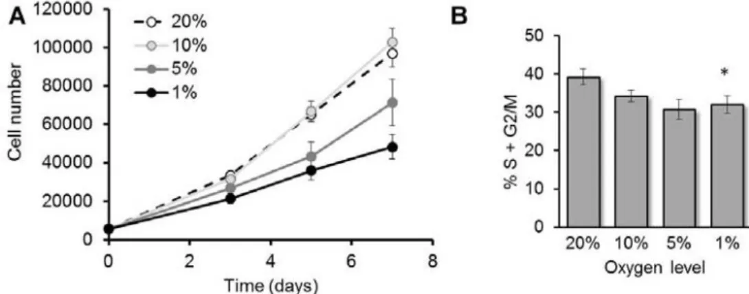
4.1 Proliferation of MSC is inhibited by hypoxia in a dose-dependent manner ... 37
4.2 Differentiation of MSC is inhibited by hypoxia in a dose-dependent manner ... 37
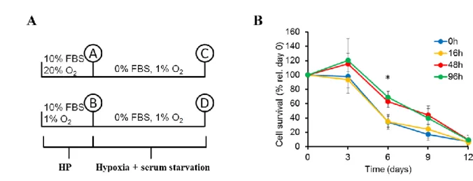
4.3 Hypoxic pre‐conditioning increases survival of MSC in vitro ... 38
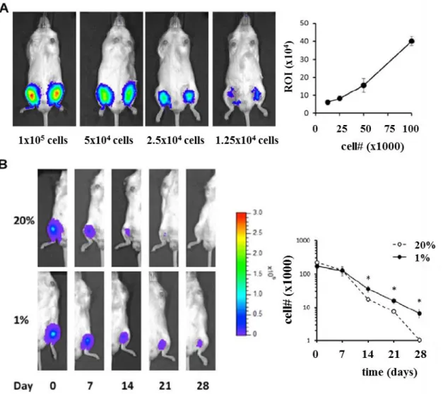
4.4 Hypoxic preconditioning enhances retention of MSC in vivo ... 40
4.5 Hypoxic pre-conditioned MSC have more glucose available during serum deprivation in vitro ... 42
4.6 Lipid composition of MSC ... 44
4.7 Hypoxia induces changes in the lipid composition of MSC and significantly increases diacylglycerols levels ... 45
4.8 Reduction of DG in MSC limits their potential to induce migration of endothelial cells ………49
5 Discussion ... 51
5.1 The effect of hypoxia and hypoxic preconditioning on MSC... 51
5.2 Study of the lipid composition of MSC in normoxia and hypoxia ... 55
6 Conclusions... 62
7 Summary ... 66
3
8 Összefoglalás ... 67
9 References... 68
10 List of Publications ... 90
10.1 Publications related to the dissertation: ... 90
10.2 Publications not related to the dissertation: ... 90
11 Acknowledgements... 91
4
List of Abbreviations
ALP alkaline phosphatase
AMI acute myocardial infarction
Ang-1 angiopoietin-1
Ang-2 angiopoietin-2
ANOVA analysis of variance
ARS Alizarin Red S
bFGF basic fibroblast growth factor
BM bone marrow
BMC bone marrow cells
BMNC bone marrow mononuclear cells
BM-MSC bone marrow mesenchymal stem cells
bsp bone sialoprotein
Ca calcium
CABG coronary artery bypass grafting
CAR CXCL-12-abundant reticular cells
CD cluster of differentiation
CFU-F colony forming unit fibroblast
CM conditioned media
CoCl2 cobalt-chloride
CSC cardiac stem cells
5
CXCL 12 C-X-C-motif chemokine 12
CXCR-4 C-X-C chemokine receptor type 4
DAPI 4',6-diamidino-2-phenylindole
DG diacylglycerols
DGAT diacylglycerol acyltransferase
DNA deoxyribonucleic acid
ECM extracellular matrix
eGFP enhanced green fluorescent protein
ELISA enzyme- linked immunosorbent assay
EPC endothelial progenitor cells
ESC embryonic stem cells
EthD- III ethidium-homodimer III
FBS fetal bovine serum
FFA free fatty acid
F-FDG 18F- fluoro-deoxy-D-glucose
FGF-1 fibroblast growth factor 1
FGF-2 fibroblast growth factor 2
FGF-4 fibroblast growth factor 4
GAPDH glyceraldehyde 3-phosphate dehydrogenase
Glut-1 glucose transporter 1
GPCR G protein coupled receptor
HE heamatopoietic microenvironment
HIF-1 hypoxia inducible factor 1
HGF hepatocyte growth factor
6
HLA human leukocyte antigen
MHC-I major histocompatibility antigen I
HP hypoxic preconditioning
HSL hormone sensitive lipase
HSPC haematopoietic stem- and progenitor cells
HUVEC human umbilical vein endothelial cell
IL-3 interleukin 3
IL-6 interleukin 6
IL-8 interleukin 8
IP3 inositol triphosphate
iPSC induced pluripotent stem cells
ISCT International Society of Stem Cell Therapy
IVIS in vivo imaging system
LDH-A lactate dehydrogenase A
LepR Leptin Receptor
LPC lysophosphatidylcholine
LPE lysophosphatidylethanolamine
LVEF left ventricular ejection fraction
LVESV left ventricular end systolic volume
MACE major adverse cardiac event
MCAM melanoma cell adhesion molecule
7
MCP-1 monocyte chemoattractant protein 1
MEM α minimal essential medium alpha
MGAT monoacylglycerol acyltransferase
MI myocardial infarction
miRNA micro ribonucleic acids
mRNA messenger ribonucleic acid
MSC Mesenchymal Stem Cells
mTIC total ion chromatogram/current
NaCl sodium-chloride
NCT national clinical trial
NF-kB nuclear factor κB
NG-2 neural/glycan antigen 2
NO nitric oxide
NOD/SCID-IL2Rγ-/- (NSG) non-obese diabetic/ severe combined immune deficiency – interleukin 2 receptor gamma chain null
PBS phosphate buffer saline
PC phosphatidyl choline
PCNA proliferating cell nuclear antigen
PC-PLC phosphatidylcholine-specific phospholipase C
PDGF platelet derived growth factor
8
PDGF-Rβ platelet derived growth factor receptor beta
PE phosphatidyl ethanolamine
PECAM-1 platelet/endothelial cell adhesion molecule 1
PI phosphatidyl-inositol
PIP2 phosphatidylinositol-4,5-bisphosphate
PI-PLC phosphatidylinositol-specific phospholipase C
PKC protein kinase C
PLA2 phospholipase A2
PlGF placental growth factor
PPH phosphatide phosphatase
PS phosphatidyl-serine
PVDF polyvinylidene fluoride
QC quality control
RIPA buffer Radioimmunoprecipitation assay buffer
RNA ribonucleic acid
SCF-1 stem cell factor 1
SDF-1 stromal cell derived factor 1
SEM standard error of the mean
siRNA small interfering RNA
SMS sphingomyelin synthase
STEMI ST segment elevation myocardial infarction
STRO-1 stromal cell precursor antigen 1
9
TG triglycerides
TGL triacylglycerol lipase
TGF-β transforming growth factor β
TMCAO transient medial cerebral artery occlusion
UHPLC – QTOF MS/MS ultrahigh pressure liquid chromatography – quadrupole time of flight tandem mass spectrometry
VE - cadherin vascular endothelial cadherin
VEGF vascular endothelial growth factor
vWF von Willebrand factor
10
1. Introduction 1.1 Stem cells
Stem cells are defined by the ability to give rise to various mature cell types, meanwhile maintaining the capacity to divide and create another stem cell. Stem cells have long been regarded as undifferentiated and capable of indefinite proliferation, self-renewal, and provision of a large number of differentiated cells (Blau et al., 2001, Ramalho-Santos and Willenbring, 2007). Both the development of the embryo and the regeneration of adult tissues throughout the lifetime are possible because of stem cells (Fuchs et al., 2000).
It is difficult to strictly define categories of stem cells, but the most widely used groups are embryonic stem cells (ESC), induced pluripotent stem cells (iPSC) and adult/somatic stem cells (Genetic Science Learning Center). Embryonic stem cells only exist at the beginning of development, and are isolated from the inner cell mass of the blastocyst of the pre-implantation mammalian embryo, i.e. the cells contributing to the embryo proper. A. J. Thomson and colleagues were the first to successfully isolate and maintain human embryonic stem cells in culture (Thomson et al., 1998). Embryonic stem cells have the capacity to proliferate extensively in culture, while maintaining pluripotency.
Unique properties compared to other cell types include the lack of contact inhibition in culture, specific cell cycle regulation and the ability to form teratomas (tumors that contain cells from all three germ layers) after injection in vivo. Embryonic stem cells can contribute to virtually all cell types in the chimeric offspring, including germ line cells, when injected into a host embryo at the blastocyst stage (Rao, 2013). In culture, ESC can be differentiated into various cell types, most commonly through the embryoid body formation method.
Generally, 10-20% of cells in the culture can be navigated toward the intended cell type, though differentiated cells are often not the same as adult cells, as they are not fully mature in phenotype and function, but rather fetal or embryonic (Rao, 2013, Avior et al., 2016).
Induced pluripotent stem cells were generated from adult mouse, then human skin fibroblasts (Takahashi and Yamanaka, 2006, Takahashi et al., 2007, Yu et al., 2007). The factors that could induce pluripotency in fully differentiated somatic cells were Oct-3/4,
11
Sox-2, Klf-4 and c-Myc (Takahashi and Yamanaka, 2006), or Lin28 and Nanog instead of Klf-4 and c-Myc (Yu et al., 2007). Besides the original pluripotency factors, several other transcription factors, small molecules, micro ribonucleic acids (miRNA) and proteins have been used to effectively reprogram somatic cells (Brouwer et al., 2016). Genome- integrative (retrovirus, lentivirus, transposon, bacteriophages and Zinc-finger nucleases) and non-integrative (adenovirus, Sendai virus, minicircle deoxyribonucleic acid (DNA), messenger RNA (mRNA), episomal vectors and proteins) methods are now used to induce pluripotency. Integrative techniques carry the risk of insertional mutagenesis, ineffective silencing of somatic genes and transgene reactivation later on, also, retroviruses require cell division for integration and are highly cell-type specific. Non-integrating techniques are generally less effective or can require multiple transfections to achieve successful reprogramming (Brouwer et al., 2016). The iPSC can be remarkably useful in disease modeling, and in the understanding of disease pathogenesis: patient- derived iPSC can be differentiated into mature cell types demonstrating disease- specific phenotype. Fibroblasts (Huangfu et al., 2008), keratinocytes (Aasen et al., 2008), neural stem cells (Kim et al., 2009), peripheral blood cells (Loh et al., 2009) and cord blood cells (Giorgetti et al., 2010) have been reported as sources of human iPSC. The iPSC can also be a valuable tool for drug screening, replacing animal models (Avior et al., 2016).
Given the almost indefinite capacity to proliferate and self-renew, and the ability to form almost any mature cell type of mammalian organs, embryonic and induced pluripotent stem cells have become incredibly promising candidates for regenerative cell therapy (Fuchs et al., 2000). However, there are major hurdles that have prevented the wide-spread clinical application of both ESC and iPSC for regenerative purposes. The most common issue is the risk of teratoma formation after in vivo transplantation of pluripotent cells (Thomson et al., 1998). Other issues include variability in differentiation potentials (Huangfu et al., 2008), the epigenetic memory of the original somatic cell (Kim et al., 2010), iPSC-specific genetic abnormalities (Hussein et al., 2011, Gore et al 2011., Lister et al., 2011), and that the efficiency of reprogramming somatic cells into iPSC is typically less than 1% (Yamanaka, 2009). In a non-human primate model of myocardial infarction (MI),
12
the transplantation of ESC- or iPSC-derived cardiomyocytes significantly increased the incidence of sustained ventricular arrhythmias in cell treated animals compared to controls (Chong et al., Nature 2014, Shiba et al., Nature 2016).
Adult stem cells originate in the developing embryo and are characterized by the loss of pluripotency during development and tissue- specific differentiation capacity (Kfoury and Scadden, 2015). Adult or tissue stem cells are far from pluripotent embryonic stem cells, but can be found in the adult organism in an undifferentiated, quiescent state (Rao, 2013, Morrison and Spradling, 2008). Certain cells in the tissues retain their ability to divide and self-renew, and have been categorized based on their differentiation capacity:
hematopoietic, bone, epithelial muscle-, neural-, and hepatic stem cells, and so on. Adult stem cells are able to give rise to the cells of their tissue of residence. Progenitor cells are committed to a particular lineage, are the immediate precursors of a mature, specialized cell, with their differentiation capacities being limited to just one cell type and having a defined amount of lifetime before programmed senescence and cell death (Young and Black 2013).
1.2 Mesenchymal stem cells
Mesenchymal stem cells (MSC) were first found in the rodent bone marrow (BM), as non-hematopoietic, multipotent progenitors. MSC are adherent to plastic in culture, capable of colony formation and differentiation into osteoblasts, adipocytes and chondrocytes in vitro (Friedenstein et al., 1970). A single colony forming unit fibroblast (CFU-F), when transplanted ectopically (subcutaneously), can differentiate into all the cell types of a miniature bone-organ (ossicle), is able to recreate the hematopoietic microenvironment (HME) and capable of self-renewal, during serial transplantation (Sacchetti et al., 2007). The International Society for Cell Therapy (ISCT) definition states that, MSC are adherent to plastic, capable of trilineage differentiation in vitro, express CD105, CD73 and CD90 on the cell surface and lack expression of CD45, CD34, CD14 or CD11b, CD79a or CD19 and human leukocyte antigen (HLA) class II (Dominici et al., 2006).
13 1.2.1 Developmental origins of MSC
MSC in the developing organism can be found around the changing sites of hematopoiesis (Kfoury and Scadden, 2015). Accordingly, the earliest site of MSC isolation was the aorta-gonad-mesonephros near the dorsal aorta of the mouse embryo (Mendes et al., 2005). Sox-1+ neuroepithelial cells have also been identified as the source of the earliest batch of MSC (Takashima et al., 2007). In a mouse tooth model system, peripheral nerve- associated glial cells contributed to a significant population of MSC during the development, self-renewal and repair of a tooth. Schwann cells gave rise to MSC in the adult mouse incisor, and these MSC produced odontoblasts and dental pulp cells (Kaukua et al., 2014). However, in the postnatal development, MSC from neural origin are replaced by MSC from other sources: based on the analysis of typical mesenchymal markers and the unique Hox genes (that define positional identity during embryonic development and point out anatomical origin of cells in postnatal life) in murine MSC from various tissues, it was concluded that MSC in different organs originate from the post-segmentation mesoderm and develop in a parallel manner in different body segments (Ackema and Charité, 2008, Sági et al., 2012). In conclusion, it is now widely accepted that mesenchymal stem cells present in the adult organism derive from the mesoderm (Bianco et al., 2008), probably colonize tissues along invading blood vessels, and occupy their mural position in response to signals from endothelial cells as it was demonstrated in human MSC transplants (Hellström et al., 1999, Bianco et al 2013, Armulik et al., 2005).
1.2.2 In vivo position of MSC
Stromal cells with colony forming and multipotent differentiation capacity were isolated from a perivascular position in many postnatal organs. Crisan and colleagues prospectively isolated perivascular cells from various tissues of human fetal and adult tissue, which uniformly expressed neural/glycan antigen-2 (NG- 2), CD 146/ MCAM (melanoma cell adhesion molecule) and platelet derived growth factor receptor beta (PDGF-Rβ), and did not express the endothelial markers CD 144 (vascular endothelial (VE) - cadherin), von Willebrand factor (vWF), CD 34 (hematopoietic progenitor cell antigen) and CD 31 (PECAM-1, platelet/endothelial cell adhesion molecule 1). The same
14
phenotype was found in the perivascular region in skeletal muscle, pancreas, placenta, white adipose tissue, skin, lung, brain, bone marrow sections and was exclusive for perivascular cells. CD 146high/ CD 34 - / CD 45 - / CD 56 - cells were then isolated from muscle and non- muscle tissues, were found to form myotubules in vitro, and generated human spectrin- and dystrophin- expressing myofibers in vivo. Perivascular cells after long term culture (up to 11-14 passages) retained their original phenotypes. They also showed strong chemotaxis towards digested extracellular matrix (ECM) components. Most importantly, long-term cultured perivascular cells prospectively isolated based on typical MSC marker expression in their native arrangement, were positive for CD 73, CD 90, CD 105, and negative for CD 34 and HLA- DR among others, and differentiated into adipocytes, osteoblasts and chondrocytes in the respective differentiation media. This study provided strong evidence for the existence of a uniform perivascular cell type in a wide range of human fetal and adult tissues which show MSC features (Crisan et al 2008).
Two main concepts regarding the identity of MSC have emerged, termed
‘fundamental’ and ‘revisionist’ by Bianco in 2013 (Bianco et al., 2013). The ‘fundamental’
concept relies on the original work of Friedenstein et al., where the existence of a committed multipotent skeletal progenitor in the bone marrow has been proven solidly, transplantable locally, but not necessarily systematically. According to the ‘revisionist’
concept, a common progenitor exists not just for skeletal, but for all non-epithelial, non- hematopoietic tissue derived from mesoderm, not only residing in the bone marrow, but also in many other postnatal organs (da Silva Meirelles et al., 2006). The ‘revisionist’
concept was established mainly by Caplan (Caplan, 2008), who first used the term
‘mesenchymal stem cell’. The terms mesenchymal stem cell or mesenchymal stromal cell are both used in the literature (since MSC are not truly multipotent). For the sake of consistency, I use mesenchymal stem cell in this work.
15 1.2.3 The putative roles of MSC
1.2.3.1 Hematopoietic niche cells
The main role of MSC is to maintain stem cell niches (Kfoury and Scadden, 2015).
The most well described is the hematopoietic stem cell niche. Stromal cells that were selected from bone marrow and enriched for CFU-Fs, supported the generation of mature hematopoietic cells from CD34+ cells in vitro (Simmons and Torok-Storb, 1991). The in vivo support of hematopoiesis by a bone marrow stromal cell- line was first demonstrated in mice following total body irradiation (Anklesaria et al., 1989). There are functionally distinct subpopulations of MSC in the bone marrow, with different cell surface marker expression (nestin, leptin receptor (LepR), and CXCL-12), different spatial distribution, and the capacity to differentiate into cell types of the skeletal system (Kudlik et al., 2015).
LepR, C-X-C-motif chemokine 12/ stromal cell derived factor 1 (CXCL-12/ SDF-1), stem cell factor 1 (SCF-1) and Angiopoietin 1 (Ang-1) all play an indispensable role in hematopoietic stem and progenitor cell (HSPC) maintenance (Mendelson and Frenette, 2014). Nestin-expressing cells were identified in mouse bone marrow stroma, were exclusively perivascular in localization and expressed high levels of CXCL-12 and Ang-1 (Méndez-Ferrer et al., 2010). In this regard, they are similar to the previously described CXCL-12-abundant reticular (CAR) cells, also playing a crucial role in quiescent HSPC maintenance (Sugiyama et al., 2006, Omatsu et al., 2010). Nestin+ cells were proven to be bona fide MSC, could form ‘mesenspheres’ (indicating neuroectodermal origin), which had multilineage differentiation potential in vitro and were able to self-renew in serial transplantation experiments of heterotopic ossicle formation. Nestin+ MSC also contributed to skeletal tissue turnover in vivo by differentiation into osteoblasts, osteocytes and chondrocytes. The nestin+ MSC were physically associated with HSC, expressed high levels of HSC maintenance genes, and the depletion of nestin+ cells significantly reduced HSC numbers in bone marrow, while HSC rapidly homed near nestin+ MSC in lethally irradiated mice (Méndez-Ferrer et al., 2010). Based on the level of nestin expression, the distribution of nestin-bright and nestin-dim cells was further analyzed. Nestin-bright cells were rare in the BM, localized exclusively around arterioles, were responsible for most of
16
the CFU-F activity, while nestin-dim cells were more abundant, localized around sinusoids and overlapped with LepR+ cells (Kunisaki et al., 2013). LepR labels a population of cells that produce a large proportion of SCF-1 and CXCL-12 in the bone marrow, have trilineage differentiation ability and expresses low levels of nestin. The crucial role of MSC in HSPC maintenance was also demonstrated by Raaijmakers et al. (2010). Cytokine-treated MSC homing to the BM of irradiated NOD/SCID (non-obese diabetic/ severe combined immune deficiency) mice and hematologic recovery was shown, as well as the crucial role of the SDF-1/ C-X-C chemokine receptor type 4 (CXCR-4) axis in the stem cell niche reconstitution (Shi et al., 2007). MSC are also able to transfer and recreate the hematopoietic environment, through the formation of heterotopic ossicles (Sacchetti et al., 2007). Further studies will hopefully shed light on the the cell surface expression, spatial distribution and functional role of the different MSC subpopulations in the bone marrow.
1.2.3.2 Other stem cell niches
MSC provide stem cell niches other than the hematopoietic niche, where tissue resident stem cells exist and proliferate throughout the lifespan of the adult organism (Kfoury and Scadden, 2015). Mesenchymal stem cells have been isolated from tissues with slow turnover, such as skeletal muscle, where they contributed little to normal tissue homeostasis, but proliferated after toxin-induced muscle injury and upregulated myogenic differentiation-inducing gene expression (Joe et al., 2010). MSC have been also isolated from hair follicle (Wang et al., 2015), and it has been shown in mice that a specific subset of adipose-forming mesenchymal cells is necessary and sufficient for skin stem cell activation, and mice that lack adipocyte precursors showed defects in follicular stem cell activation (Festa et al., 2011).
1.2.3.3 Angiogenesis support
MSC have also been proposed to have a key role in the support of blood vessel formation and regeneration, given their perivascular position and the still growing body of evidence (Watt et al., 2013). MSC have been shown to secrete a wide range of pro- angiogenic factors: Ang-1, Angiopoietin-2 (Ang-2), Endothelin-1, fibroblast growth factor 1 (FGF-1), 2 (FGF-2) and 4 (FGF-4), HGF, interleukin 8 (IL-8), members of the platelet
17
derived growth factor (PDGF) family, placental growth factor (PlGF), and vascular endothelial growth factor (VEGF), to name a few, as assessed by angiogenic antibody arrays (Watt et al., 2013), and demonstrated by numerous other investigators (Williams et al., 2011). MSC have been shown to contribute to vasculogenesis, the de novo assembly of new and stable vasculature from endothelial colony forming cells in in vitro and in vivo models (Kachgal and Putnam, 2011, Au et al., 2008). MSC also promote endothelial and vascular smooth muscle cell proliferation in vitro (Kinnaird et al., 2004). When transplanted along with human endothelial progenitor cells (EPC) into immune deficient mice, they rapidly formed extensive networks of erythrocyte-containing human blood vessels (Melero-Martin et al., 2008), and in a similar study, when co-implanted with human umbilical vein endothelial cells (HUVECs) into mice, MSC formed blood vessels and retained a perivascular position (Au et al., 2008). Of note, when MSC were implanted alone (without HUVECs), functional vasculature was not formed. In addition, murine MSC from four different tissue sources (bone marrow, white adipose tissue, myocardium and skeletal muscle) promoted endothelial cell proliferation, migration and tube formation in vitro, and when co-transplanted with endothelial colony- forming cells, formed vascular networks and occupied perivascular position in vivo, regardless of the tissue source (Lin et al., 2012). MSC also support arteriogenesis, during which collateral vessels grow in diameter and lead to the revascularization of ischemic tissues (Potente et al., 2011). In this regard, MSC have been injected into mouse hind limb ischemia models and were shown to significantly increase blood flow, collateral number and total collateral cross- sectional area of blood vessels (Kinnaird et al., 2004), and also improved blood flow and enhanced muscle regeneration (Lian et al., 2010).
1.3 The modulation of tissue response to injury by MSC
Besides the support of angiogenesis, cultured MSC were shown to modulate the response to injury by numerous studies (Williams and Hare, 2011). Besides the hind limb ischemia model, regenerative capacity of MSC has been assessed in animal models of MI, cardiomyopathies and stroke (Ranganath et al., 2012). For example, membrane- and nuclear- labeled allogeneic bone marrow MSC were injected after surgically induced MI in
18
swine, and MSC were still detectable after 8 weeks, expressed muscle specific proteins and were present in vascular structures, expressing vWF and VEGF, while extracardiac engraftment and ectopic tissue formation was not observed (Amado et al., 2005). In a canine model of chronic myocardial ischemia, allogenic MSC injection increased left ventricular ejection fraction (LVEF) and vascular density, and there was a trend towards reduced fibrosis in treatment group compared to controls. Also, MSC labeling co-localized with endothelial cells and smooth muscle cells but not with myocytes (Silva et al., 2005). In a swine model, high dose autologous MSC treatment 3 months after MI caused significant scar size reduction, increased infarct region wall thickness, myocardial blood flow and LVEF (Schuleri et al., 2009). In another swine model of chronic myocardial ischemia, male swine MSC administration into female animals resulted in cell engraftment and co- localization of Y-chromosome with cardiac transcription factors and structural myocyte proteins, while cell engraftment correlated most with improved regional contractility. Scar size reduction, infarct region wall thickening and improved myocardial blood flow was also observed (Quevedo et al., 2009).
Given the low rate of MSC engraftment after cell therapy (reported to be 0.44% of intramyocardially injected cells by Toma et al. in 2002), several other mechanisms have been proposed and demonstrated to be involved in tissue repair by MSC: enhancement of vasculogenesis, cardioprotection (pro-survival effect on cardiomyocytes as well as enhancement of endogenous repair) and attenuation of fibrosis. It seems most likely that through differentiation into vascular cell types as well as the secretion of potent angiogenic factors, MSC promote angiogenesis/vasculogenesis in vitro and in vivo in various ischemic and non-ischemic settings (Williams and Hare, 2011). Indeed, conditioned medium from MSC overexpressing the pro-survival gene Akt, protected cardiomyocytes from apoptosis during hypoxia in vitro and reduced infarct size and improved cardiac function compared to controls after MI in rat hearts (Mangi et al., 2003). MSC have been shown to secrete VEGF, basic fibroblast growth factor (bFGF), PlGF, and monocyte chemoattractant protein-1 (MCP-1) in vitro and improved limb perfusion, increased the number and total cross sectional area of collaterals in vivo, while higher VEGF and bFGF levels were shown
19
in the vicinity of injected MSC compared to controls in a hind limb ischemia model in mice (Kinnaird et al., 2004).
Cardioprotection by MSC was shown when Akt-modified MSC enhanced survival of cardiomyocytes after acute myocardial infarction (AMI) through Wnt-signaling (Gnecchi et al., 2006). MSC also promote the proliferation and differentiation of endogenous c-kit+ cardiac stem cells (CSC) in the heart: in a swine model of AMI, there was a 20-fold increase in the number of c-kit+ CSC, 6-fold in GATA-4+ CSC and 4-fold in the number of mitotic myocytes after MSC injection compared to untreated animals. The MSC feeder layer enhanced CSC proliferation in an organotypic culture from the heart, and resulted in Nkx2.5 and troponin I positive cardioblast appearance in vitro (Hatzistergos et al., 2010). Human bone marrow MSC and CSC have also been implanted together following MI in immunosuppressed swine by Williams et al. (2013), which resulted in a 2- fold greater reduction in infarct size than with either cell type alone, while all cell therapy groups decreased infarct size compared to placebo group. In cell-treated pigs, LVEF was restored to baseline, while placebo group had decreased left ventricular function. Co- transplantation of the two types of stem cells also led to a 7-fold enhanced engraftment of stem cells compared to either cell type alone. After a MI, the process of remodeling starts in the injured heart, where necrotic myocardium is replaced by scar tissue, while reduction of fibrosis enhances endogenous myogenesis (Serrano et al., 2011). MSC can influence the existing amount of extracellular matrix by secreting proteinases and tilting the balance towards matrix degradation (Molina et al., 2009), and also attenuating fibrosis by suppression of fibroblast proliferation after MI (Mias et al., 2009).
MSC have been shown to contribute to cardiovascular repair via mitochondrial transfer, exosomes, and connexin-43 on the cellular level. Exosomes are secreted by cells, can contain mRNA, miRNA and other non-coding RNAs, and have important roles in programmed cell death, angiogenesis (Deregibus et al., 2007), inflammation and coagulation (Théry et al., 2002, Mittelbrunn et al., 2011). Purified exosomes derived from MSC reduced infarct size in a mouse model of myocardial ischemia/reperfusion injury (Lai et al., 2010). Gentamicin-induced kidney injury in rats was treated by injections of bone
20
marrow MSC (BM-MSC), conditioned media (CM), CM treated with trypsin or ribonuclease (RNase), or exosome-like microvesicles extracted from the CM. BM-MSC transplantation, its CM or exosome treatment prevented the increase of serum creatinine and urea and also had beneficial effects on necrosis, apoptosis and cell proliferation. If the CM or the microvesicles were treated with RNase, the protective effects were diminished (Reis et al., 2012). MSC released miR-133b-containing microvesicles and increased the level of miR-133b in astrocytes and neurons isolated from mouse brains after transient medial cerebral artery occlusion (TMCAO), and exosome-enriched fractions from MSC exposed to 72 hours to post-TMCAO brain extracts significantly increase the neurite branch number and total neurite length of cultured neurons (Xin et al., 2012). Mitochondrial transfer have been demonstrated by Spees et al., when A549ρ° cells (devoid of mitochondrial DNA), were co-cultured with MSC or skin fibroblasts and some of the A549ρ° cells acquired functional mitochondria. Rescued A549ρ° cells were able to proliferate rapidly, mitochondrial network was restored and 97% of the clones contained mitochondrial DNA from the donor cells, while genomic DNA was from the original cells (Spees et al., 2006). Recently, the comprehensive proteomic analysis of MSC exosomes revealed the presence of various angiogenic proteins within, and nuclear factor κB (NF-kB) signaling was identified as a key mediator of MSC exosome induced angiogenesis in endothelial cells in an in vitro model (Anderson et al., 2016). MSC also contribute to the reconstitution of the endogenous stem cell niche in the heart (Mazhari and Hare, 2007) 1.4 Adult stem cells for cardiovascular repair – translational aspects
Although MSC have made one of the fastest transitions from the bench to the clinic, MSC therapy has yielded modest results in terms of efficacy in the cardiovascular field (Karantalis and Hare, 2015). Various approaches have been used: exogenous cell transplants (skeletal myoblasts, bone marrow cells (BMC), mesenchymal stem cells and cardiac progenitor cells), the activation of endogenous cardiac cells, direct reprograming and enhancement of proliferation (Chen et al., 2015). The rationale behind skeletal myoblast therapy was the well-known capacity of skeletal muscle to regenerate through the activation of satellite cells (Brack and Rando, 2012). Two clinical trials, MAGIC
21
(Menasché et al., 2008) and MARVEL (Povsic et al., 2011) have been conducted, where autologous myoblast transplantation in chronic ischemic heart disease failed to show efficacy, but ventricular arrhythmias were induced, probably due to the lack of electromechanical coupling between cardiomyocytes and transplanted myoblasts, forestalling further studies with this cell type.
1.4.1 Bone marrow cells
Despite evidence that differentiation into cardiomyocyte lineage does not happen in an unselected BMC population (Murry et al 2004), both unselected and selected bone marrow cells have been injected in acute or chronic myocardial ischemia. CD 34 has been the most widely used marker for selection, for example, in the TOP-CARE AMI study, where autologous CD 34+ cells were injected into the infarct-related artery at 1.5- 5 days post-AMI, and showed limited improvement in LVEF and reduction in left ventricular end systolic volume (LVESV), although no control group was created (Schächinger et al., 2004). Due to the considerable heterogeneity in cell type and study design in BMC transplantation clinical trials, meaningful benefit could not be shown in myocardial ischemia (Simari et al., 2014).
1.4.2 Mesenchymal stem cells
Mesenchymal stem cells were selected from bone marrow cells and were quickly translated into clinical trials. The main advantages of MSC over other types of stem cells are their ready isolation from patients, the easy expansion in culture with maintenance of differentiation potential, their genetic stability (Bernardo et al., 2007), the low risk of immune rejection by host organism, and the lack of ethical concerns plaguing the field of embryonic stem cell research. Of note, MSC isolated from different tissue sources and injected in various scenarios are most likely a heterogeneous population of progenitor cells, where the differentiation potentials may differ (Nombela-Arrieta et al., 2011). In a phase I randomized study, allogeneic MSC were injected 7-10 days after AMI, and were found to be safe: adverse events were similar in the cell treatment and placebo group, no ectopic tissue growth was found with whole body computed tomography (CT), and in addition, a 4- fold decrease in arrhythmias in the MSC group was observed compared to placebo (Hare et
22
al., 2009). In the APOLLO trial, freshly isolated, adipose-derived MSC were used in 14 patients in the setting of AMI. Intracoronary injection of MSC did not cause microvascular obstruction or alteration in coronary flow, and resulted in perfusion defect improvement and scar size reduction by 50% (Houtgraaf et al., 2012). MSC are most convincing as promoters of functional recovery in chronic ischemic cardiac disease, where the mechanisms of action defined in preclinical models seem to be at work: antifibrosis and neoangiogenesis, with the most prominent result being a 30-50% reduction in scar size (Karantalis et al., 2014, Heldman et al., 2014). PRECISE was the first clinical trial to use adipose-derived MSC for ischemic heart failure, where left ventricular infarcted muscle mass remained the same during follow-up in the cell therapy group, but increased in the placebo group, and perfusion and contractility increased in the cell group (Perin et al., 2014). The strongest effect was observed at the site of MSC injection in the POSEIDON study, where autologous vs allogeneic MSC from BM were compared in ischemic cardiomyopathy patients (Suncion et al., 2014). There was no immunologic reaction following allogeneic stem cell injection and no ventricular arrhythmias or new ischemia occurred during the first 30 days after cell injection, also, ectopic tissue formation did not occur in any of the study patients. Scar size reduction and reverse remodeling was apparent as assessed by CT scan. In the C-CURE trial, autologous MSC were harvested from chronic ischemic cardiomyopathy patients, culture expanded and treated with a cardiogenic cocktail. Following intramyocardial injection, patients were followed for 2 years, during which time there was no evidence of cell- therapy induced toxicity and cell therapy resulted in a favorable response regarding overall clinical parameters, such as quality of life, 6- minute walk test and New York Heart Association stage (Bartunek et al., 2013). The TAC- HFT study examined the safety and efficacy of transendocardial injection of MSC vs placebo and bone marrow mononuclear cells (BMNC) vs placebo (Heldman et al., 2014).
No procedure-related serious adverse events were observed at 30 days after cell injection.
Interestingly, infarct size was reduced and regional contractility at the site of injection improved in the MSC group, but not in the BMNC or placebo groups, but LVEF did not change. Several other studies using MSC for heart failure are ongoing or awaiting the publication of final results: the AMICI (Safety Study of Allogeneic Mesenchymal
23
Precursor Cell Infusion in MyoCardial Infarction), the MyStromalCell Trial (MesenchYmal STROMAL CELL Therapy in Patients With Chronic Myocardial Ischemia; Qayyum et al 2012), CHART-1 (Safety and Efficacy of Autologous Cardiopoietic Cells for Treatment of Ischemic Heart Failure; Bartunek et al., 2016) and NCT 02032004, examining allogeneic mesenchymal progenitor cells for the treatment of ischemic heart failure.
1.4.3 Cardiac progenitor and cardiosphere derived cells
Other candidates for cardiac cell therapy are cardiac progenitor cells and cardiosphere-derived cells (CPC and CDC respectively): c-kit+, able to self-renew, and differentiate into cardiomyocytes, endothelial and smooth muscle cells (Beltrami et al 2003 and Messina et al 2004). In SCIPIO, the first- in- human trial using autologous c-kit+ CPCs, cells were injected after 3 months following coronary artery bypass grafting (CABG). At the one year follow- up, LVEF increased by 12% and infarct size was significantly reduced (Bolli et al., 2011). CDC were infused into the infarct-related artery of AMI patients. By the end of the 6-month follow up, no patient suffered a major adverse cardiac event (MACE), and the CDC group showed reductions in scar mass, increases in viable heart mass and regional contractility compared to controls. However, changes in LVEF did not differ between groups (Makkar et al., 2012).
Comparing the modest results of clinical trials to preclinical results, the discrepancy in efficacy is apparent. Several factors are to be mentioned: cell populations used in clinical trials are highly heterogeneous, from BMNC through CD 34+ BM stem cells to adipose derived MSC. There is still no consensus on the optimal timing, delivery method and dosing of cells. It has also been shown, that the proportion of MSC in the bone marrow decreases with age (Sethe et al., 2006), which is particularly important in the case of autologous cell therapy for the elderly heart failure patients. One of the biggest hurdles seems to be the limited retention and survival of cells at the site of interest as demonstrated in animal models. For example, after intravenous infusion, a huge proportion of cells is entrapped in the lung, spleen or liver (Barbash et al., 2003), and even after intramyocardial or intracoronary delivery, cells are taken up into the pulmonary circulation (Hou et al., 2005). Similarly, radiolabeled MSC were found primarily in the lungs after injection into
24
tail vein of mice, but 24 h after infusion, MSC were lost from the lungs and redistributed to other organs, mainly the kidneys, spleen and liver (Liu et al 2012). Survival of cells in the hostile environment is also an issue, as it was demonstrated in ST- elevation myocardial infarction (STEMI) patients, where only 1.3-2.6% of 18F- fluoro-deoxy-D-glucose- (F- FDG) labeled unselected bone marrow cells remained in the infarct region 50-75 minutes after cell injection into the affected coronary artery (Hofmann et al 2005).
1.5 Improving the efficacy of stem cell therapy: focus on hypoxia and hypoxic preconditioning
Various strategies have been investigated in order to improve the survival and retention of stem cells after injection: more effective ways of delivery (intramyocardial seems to retain more cells then intracoronary or intravenous infusion; Hou et al., 2005), MSC implantation in natural or synthetic polymer scaffolds (scaffolds seeded with MSC were superior to MSC injection alone; Jin et al., 2009), genetic engineering of MSC, preconditioning strategies of MSC before transplantation: hypoxic, hyperoxic and pharmacological, and combined administration of MSC and other types of stem cells (Li et al., 2016). For example, BM-MSC overexpressing the pro-survival gene Akt 1, limited infarct size and improved ventricular function more efficiently than non-modified MSC in a rat model of MI (Gnecchi et al., 2006).
MSC often reside in tissues which exhibit low oxygen tension, such as the BM (1- 7% or 4-20 mmHg; (Brahimi-Horn and Pouysségur, 2007, Chow et al., 2001), but are usually expanded in the laboratory under normoxic conditions (20.8% atmospheric oxygen, 160 mmHg oxygen in solution). In many of the clinical or experimental settings MSC are used to promote revascularization at areas with poor circulation, and hence, are injected into an ischemic tissue environment, where they are exposed to a lower oxygen concentration than in their usual ex vivo culture conditions: oxygen tension in tissues is 4‐
20 mmHg, while arterial oxygen is 104 mmHg (Brahimi-Horn and Pouysségur, 2007).
Hypoxia exerts strong effects on MSC, affecting their proliferation, differentiation and migration (Das et al., 2010). However, studies have yielded controversial results, which might be caused by the variation in the conditions that have been tested, including time-
25
span (ranging from minutes to days) and level of hypoxia: from 0.1 to 10% O2 (Das et al., 2010, Tsai et al., 2012). In addition, it has also been shown that MSC respond to hypoxia by altering their secretome, increasing the amount of secreted proangiogenic factors such as VEGF (Potier et al., 2007). In a study using a mouse hind limb ischemia model, MSC survival after injection into the ischemic limb was greatly enhanced by the hypoxic preconditioning of the cells, and hypoxic preconditioned-MSC increased perfusion, necrosis salvation and capillary density to a greater extent than non-preconditioned cells (Zhu et al., 2014). It has been demonstrated previously that hypoxic preconditioning enhances the therapeutic potential of MSC in applications such as the treatment of cardiac ischemia (Hu et al., 2008, He et al., 2009), critical limb ischemia (Rosová et al., 2008, Huang et al., 2014, Leroux et al., 2010), traumatic brain injury (Chang et al., 2013), and in liver regeneration (Yu et al., 2013). One of the key factors in the hypoxic preconditioning- induced enhancement of tissue repair seems to be the increased retention of MSC in tissues (Hu et al., 2008, Huang et al., 2014 Leroux et al., 2010), but the underlying mechanisms remain elusive. Thus, the study of MSC in low oxygen tensions is of great importance.
The further understanding of MSC biology and response to hypoxia, especially in terms of the secretion of angiogenic factors could be remarkably useful for enhancing the efficacy of MSC-based therapy (Bianco et al., 2013). It has been long established that lipids (fatty acids and their derivatives and substances related biosynthetically or functionally to these compounds) are not only essential building blocks of biological membranes and responsible for energy storage in the cell, but also have important regulatory and signaling functions in almost all cellular processes (Hannun and Obeid, 2008). Changes in lipid levels that result in functional consequences eventually gave rise to the idea of ‘bioactive lipids’ (Hannun and Obeid, 2008). For example, inositol phospholipids have been shown to modulate acetyl-choline induced intracellular signaling (Hokin and Hokin, 1953), and eicosanoids play a crucial role in inflammatory signaling (Serhan and Savill, 2005).
Following G protein coupled receptor (GPCR) activation, the enzymatic cleavage of the membrane lipid phosphatidylinositol-4,5-bisphosphate (PIP2) produces two smaller lipids:
inositol triphosphate (IP3), directly activating calcium (Ca) channels, which results in
26
increased intracellular Ca concentration; and diacylglycerol (DG), directly activating several isoforms of the protein kinase C (PKC) enzyme, which phosphorylates a number of target proteins (Nishizuka, 1992). Accordingly, it has become evident that lipids are crucial first and second messenger molecules (van Meer et al., 2008). It has been also shown that lipids can be involved in hypoxic signaling. Ceramides for example, are increased in renal tubular epithelial cells under hypoxia (Ueda et al., 1998). It has also been shown that the cell lines Hela and 293T increase DG levels when exposed to hypoxia, affecting the activity of the transcription factor hypoxia inducible factor 1 (HIF-1; Temes et al., 2004).
With the evolution of mass spectrometry (MS) techniques and the consequent advance in the identification of lipids, lipidomics ‘joined the omics evolution’ (cited from Dennis, 2009). The lipidome is the complete lipid composition of a cell, tissue, or organism; lipidomics is the global analysis of the lipidome with a comprehensive mass spectrometry approach, and functional lipidomics is the study of the role played by membrane lipids in biological functions (Dennis, 2009, Shevchenko and Simons 2010).
The lipidome of stem cells has not yet been widely studied. Meissen and colleagues compared the lipidome of human embryonic stem cells, induced pluripotent stem cells and the parental fibroblasts of iPSC (Meissen et al., 2012). They found that iPSC undergo a remarkable shift metabolically from the parental fibroblasts toward ESC, suggesting an almost complete metabolic reprogramming. There were no differences between the pluripotent cell types (iPSC and ESC) in free fatty acid (FFA) metabolism, but for example, significant differences in the phosphatidyl choline (PC) and phosphatidyl ethanolamine (PE) lipid structures could be detected: in both types of pluripotent stem cells, the amount of the poly-unsaturated (three or more double bonds in the acyl chain) PC species was significantly higher than in fibroblasts. The increase in unsaturation levels of polyunsaturated PC species may alter membrane fluidity, since PC are the most abundant lipid species in the mammalian cell membrane. A study by Fuchs et al. was reporting on the lipidomics of ovine MSC (Fuchs et al., 2008). Very little is known about the overall lipidomics of human MSC, or changes in lipid composition in response to stimuli such as hypoxia.
27
2 Aims of the study
Hypoxia exerts strong effects on MSC, affecting their proliferation, differentiation and migration (Das et al., 2010), thus, the study of MSC in low oxygen tensions is of great importance. The aims of the first set of experiments were (1) to examine the effects of various levels of hypoxia on MSC proliferation and differentiation, and (2) to study the effects of hypoxic exposure prior to their administration to a hypoxic site, which we refer to as hypoxic pre‐ conditioning (HP).We sought (3) to determine the optimal hypoxia levels and incubation times to promote survival of MSC in an environment of low oxygen and nutrients in vitro, and to enhance MSC retention after in vivo administration into mice. In addition, (4) we sought to evaluate the underlying mechanism(s) that promote the survival of MSC and hypothesized, that hypoxic pre‐ conditioning promotes metabolic adaptations in MSC that allow increased survival under conditions of limited nutrients and oxygen.
Further understanding MSC biology and MSC response to hypoxia, especially in terms of the secretion of angiogenic factors could be remarkably useful for enhancing the efficacy of MSC-based therapy (Bianco et al., 2013). Very little is known about the overall lipidomics of human MSC, or changes in lipid composition in response to stimuli such as hypoxia. In consequence, our aims were (5) to determine the lipid composition of human bone marrow derived MSC, (6) to identify changes induced by exposure to hypoxia and (7) to evaluate whether these changes have a role in the angiogenic potential of MSC.
28 3 Methods 3.1.1 Isolation and culture of mesenchymal stem cells
Bone marrow aspirates from human donors were purchased from Lonza (Walkersville, USA) or Stem Express (Placerville, USA) and mesenchymal stem cells were isolated as follows (Fierro et al 2011). BM aspirates were passed through 90 μm pore strainers for isolation of bone spicules. Then, strained BM aspirates were diluted with an equal volume of phosphate-buffered saline (PBS) and centrifuged over Ficoll (GE Healthcare, Waukesha, USA) for 30 minutes at 700 x g. Next, mononuclear cells and bone spicules were plated in plastic culture flasks using standard culture medium: Minimum Essential Medium α ((MEM-α;), HyClone, Thermo Scientific, South Logan, USA) supplemented with 10% fetal bovine serum (FBS; Atlanta Biologicals Lawrenceville, USA). After 2 days, non-adherent cells were removed by washing twice with PBS. MSC from passages 3–6 were used for experimentation for hypoxia and hypoxic preconditioning experiments, and passages 4-7 for lipidomics analysis and D609 experiments (see later), where each passage implies 5-7 days in culture (i.e. approximately 3 population doublings).
3.1.2 Immunophenotypic characterization of mesenchymal stem cells
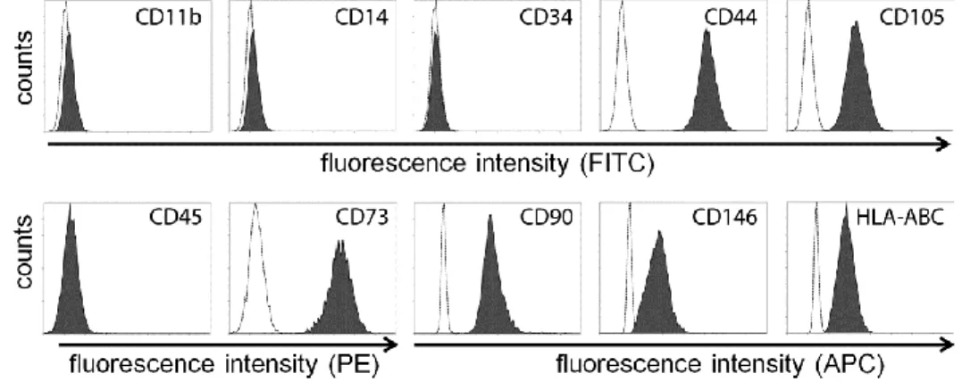
For the immune characterization of MSC by flow cytometry, cells were lifted by trypsin treatment and incubated for 45 minutes with the fluorophore- conjugated antibodies (diluted 1:100) as follows: CD73 (5’-nucleotidase), CD90 (Thy-1) and CD105 (endoglin), CD11b (integrin alpha M), CD14 (monocyte/macrophage marker), CD34 (hematopoietic stem cell marker) and CD45 (lymphocyte common antigen). Cells isolated with our method fulfilled the immunophenotypic criteria of the ISCT (Figure 1., Dominici et al., 2006).
3.1.3 Studies with hypoxia
For studies with hypoxia, cell cultures were performed in incubators (MCO -18M Sanyo) at 37°C with 5% CO2, humidified atmosphere and dedicated oxygen level (20 (atmospheric), 10, 5 or 1% O2), as established by replacement with nitrogen injections. In order to establish exact oxygen levels that the cells are exposed to during experimentation;
we monitored oxygen diffusion in culture media. An OxyLite monitor and probe were used
29
in 12-well culture plates containing 1ml of media (αMEM) per well to determine dissolved oxygen concentration (pO2) over 2 hours, in incubators set for 20%, 10%, 5% and 1% O2. After placing cell cultures in the respective incubator, full hypoxic levels in the media were reached within one hour and did not change thereafter, when plates were undisturbed (Figure 2).
Figure 1. MSC isolated from healthy individuals exhibit a common immune phenotype.
Specific surface antigen expressions were determined by flow cytometry based on the ISCT minimal criteria for multipotent mesenchymal stromal cells: CD105, CD73, CD 90 ≥ 95%
positive; and CD45, CD34, CD14, CD11b ≤ 2% positive. MSC also expressed HLA-ABC, CD 146, and CD 44.
3.1.4 Cell proliferation analysis
For proliferation assays, MSC were seeded into 12 well plates at 1000 cells/cm2 seeding density. At every time point, (2-3 days) cells were lifted with trypsin treatment and counted using Trypan blue exclusion dye (Gibco, Thermo Fisher Scientific, South Logan, USA) and a hemocytometer.
3.1.5 Cell cycle analysis
To determine the status in cell cycle, MSC were seeded into 75 cm2 culture flasks at 500 cells/cm2 seeding density, and incubated in standard culture medium with varying levels of hypoxia for 4 days, with media change at day 2. Then, cells were lifted by trypsin treatment and permeabilized with ice cold 70% ethanol overnight. After 45 minutes of
30
incubation with 50μg/ml propidium iodide solution (Roche, Indianapolis, IN, USA) containing 20ng/ml RNAse A (Qiagen, Valencia, CA, USA), followed by a washing step with PBS, samples were evaluated by flow cytometry and analyzed using ModFit LT software (Verity Software House). With this method, the amount of propidium iodide taken up by cells is proportionate to their DNA content.
Figure 2. Oxygen partial pressure in culture media placed in incubators with different levels of hypoxia. We monitored oxygen partial pressure in culture media after placement in the respective incubators in order to establish exact level and onset of hypoxia. Note that respective oxygen pressure is reached in the culture medium within one hour of placement in the respective incubator. Data represent the average of 3 measurements.
3.1.6 Osteogenic and adipogenic differentiation assays
For both osteogenesis and adipogenesis, 10,000 MSC/cm2 were cultured for 14 days in incubators with varying hypoxia levels (20, 10, 5 and 1% O2), with media changes every three days. Osteogenic medium consisted of standard culture medium supplemented with 0.2 mM ascorbic acid, 0.1 μM dexamethasone, and 20 mM β-glycerolphosphate.
Adipogenic medium contained standard culture medium with 0.5 mM isobutilmethylxantine, 50 μM indomethacin, and 0.5 μM dexamethasone. To quantify osteogenesis, alkaline phosphatase (ALP) activity was measured by lifting the cells with trypsin and lysing the cells for protein extraction using 1.5mM Tris-HCl solution containing 1.0 mM ZnCl2, 1.0mM MgCl2, and 1% Triton X-100 for 15 minutes. Lysates were then centrifuged at 12,000g for 10 minutes and incubated with p-
31
nitrophenylphosphate liquid substrate solution (Sigma-Aldrich, St. Louis, USA) for 30 minutes. Released p-nitrophenolate was determined spectrophotometrically at 405 nm.
Calcium precipitation was determined using Alizarin Red S indicator (ARS; Ricca Chemicals, USA). Cells were fixed with 10% v/v formalin solution for 15 minutes, washed once with PBS, and stained for 20 minutes with 1% w/v ARS over gentle shaking. Samples were then photographed for visual documentation and then incubated with 10% v/v acetic acid for 30 minutes, the cell layer scraped, and vortexed for 30 seconds, then centrifuged at 12,000 x g for 10 minutes. Optic density of the supernatants was measured at 405 nm. For both ALP and ARS, total protein concentration was determined with Coomassie staining and measured at 595 nm. For adipogenesis, cells were fixed with 10% v/v formalin for 15 minutes, rinsed once with PBS, and stained for 30 minutes with Oil Red O (Electron Microscopy Sciences, Hatfield, PA, USA). Cells were then washed three times with PBS, photographed and incubated with 4% Tween 20 (Affymetrix, Santa Clara, USA) in isopropanol for 5 minutes, in order to release the dye. Optic density of supernatants was then measured at 490 nm.
3.1.7 In vitro cell survival and apoptosis detection
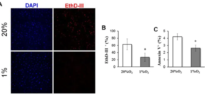
To evaluate survival of MSC in hypoxia and serum deprivation, we first incubated MSC (10,000 cells/cm2) in 12-well plates in standard culture medium and 1% O2, for varying amounts of time (0, 16, 48 or 96 hours). Then, culture media was changed to MEMα alone (without FBS) and all plates were transferred to 1% O2 for up to 12 days with no additional media changes. Every three days, cells were detached by trypsin treatment and counted using Trypan blue exclusion dye and a hemocytometer. To quantify the percentage of dead cells, MSC were cultured on glass cover slips (10,000 cells/cm2) and incubated for 48 hours in standard culture medium in 20% O2 (control) or 1% O2 (HP).
Then, medium was changed to serum free medium and plates were transferred to 1% O2 for 9 days with no additional media changes. Cover slips were then incubated in 4 μM Ethidium- homodimer III (EthD- III; Biotium, Hayward, USA) in PBS for 30 minutes and then mounted on slides with Vectashield Mounting Medium with 4',6-diamidino-2- Phenylindole dihydrochloride (DAPI, Vector Laboratories, Burlingame, USA). Images
32
were acquired and processed for large-field merge using a BZ-9000 (BIOREVO) fluorescence microscope (Keyence, Itasca, USA) and analyzed for automated counting with NIS-Elements BR software (Nikon). To quantify the percentage of apoptotic cells, MSC were cultured as described above for EthD-III, but in 6-well plates. At day 9, cells were lifted using trypsin and stained with phycoerythrin- Annexin V- Apoptosis Detection Kit I (BD Pharmingen, San Diego, USA) following manufacturer’s instructions and measured by flow cytometry.
3.1.8 In vivo retention study
In order to generate luciferase-expressing MSC, we used a third generation lentiviral vector with the general form pCCLc-MNDU3-Luciferase-PGK-eGFP-WPRE. We transduced the cells using protamine sulfate (20 μg/ml) and a quantity of virus equivalent to a multiplicity of infection of 20. Using this protocol, over 90% of cells were enhanced green fluorescent protein (eGFP) positive. For cell administration, immune compromised NOD/SCID-IL2Rγ-/- (NSG) mice were anesthetized using inhaled isoflurane and then injected in the medial hamstring muscles with the luciferase-expressing MSC in 20 μL of HyStem C (BioTime, Alameda, USA). To generate the standard curve, increasing number of cells were used (from 12,500 to 100,000 cells), while, for the retention studies, 200,000 cells were injected. For imaging, 100 μL of 20 mg/mL D-Luciferin Firefly (Perkin Elmer) were injected intraperitoneally into the animals, 10 minutes before bioluminescence detection via In Vivo Imaging System (IVIS) Spectrum imaging for 5 minutes exposure time. LivingImage software was used to quantify MSC’s bioluminescence (Perkin Elmer).
All animal procedures were performed as approved by the Institutional Animal Care and Use Committee.
3.1.9 Glucose and lactate measurements
Supernatants were collected every three days from MSC cultured under identical conditions to the in vitro survival assay (in 12-well plates) described above. Supernatants for glucose measurement were stored at -20°C and those for lactate at -80°C. Glucose and lactate concentrations of supernatants were determined using a Glucose
33
Colorimetric/Fluorometric Assay Kit (BioVision, Milpitas, USA) and a Lactate Colorimetric Assay Kit (BioVision), respectively, following their provided protocols.
3.1.10 Ultrahigh pressure liquid chromatography- quadrupole time-of-flight tandem mass spectrometry (UHPLC- QTOF-MS/MS)
After culture in either 20% or 1% O2 for 48 hours, MSC were lifted by trypsin treatment (Thermo Scientific) and stored at -80°C. Then, samples were processed for total lipid extraction and mass spectrometry analysis as described by Fiehn and Kind (2007).
After quenching the cells, 1 x 10⁶ dried cells were added to a 1.5 mL Eppendorf tube, placed on dry ice for 20 minutes to completely freeze and then thawed on ice. The freeze- thaw cycle was repeated twice. Then, 1 mL of pre-chilled (-20˚C) extraction solvent was added to the cells (acetonitrile: isopropanol: water 3: 3: 2 v/v/v) and the freeze-thaw cycle repeated two more times. Samples were then vortexed for 10 seconds, shaken for 5 minutes at 4˚C and centrifuged for 2 minutes at 14,000 x g. 500 μl of the supernatant was evaporated in a cold trap concentrator (Labconco Centrivap, Kansas City, USA) to complete dryness. Lipidomics data were acquired using UHPLC – QTOF MS/MS (Waters Acquity UPLC CSH C18 column). Chromatographic separation was followed by electrospray ionization (ESI) in both positive (Agilent 6530 QTOF MS, Agilent Technologies, Santa Clara, USA) and negative mode (Agilent 6550 QTOF MS); and QTOF-MS/MS. Data were analyzed in a four-stage process. First, raw data were processed in an untargeted (qualitative) manner by MassHunter Qual (v. B.05.00, Agilent Technologies) to find peaks in up to 300 chromatograms. Peak features were then imported into Mass Profiler Professional (Agilent Technologies) for peak alignments to seek which peaks are present in multiple chromatograms, using exclusion criteria by the minimum percentage of chromatograms (30%) in which these peaks are positively detected. Peaks were then manually collated and constrained within the MassHunter quantification software (v. B.05.01 Agilent Technologies) on the accurate mass precursor ion level, using the MS/MS information and the LipidBlast library to identify lipids with manual confirmation of adduct ions and spectral scoring accuracy. The following normalization steps were performed: ‘vector normalization’ in which the sum of all peak heights was calculated for