Proton Transport
Warren S. Rehm
I. Introduction 187 A. Proton Conductance in Water 188
II. Passive Proton Transport 189 A. Passive Transport via Carriers 189 B. Proton Conductance of Biological Membranes 190
C. Mechanisms for Proton Conductance 201
D. Proton-Cation Exchange 203 III. Active Proton Transport 204
A. Concept of Active Proton Transport 204
B. Postulated Mechanisms for H+ Production 206
C. Evidence for Electrogenicity of the H+ Mechanism 207 D. Model to Explain Electrogenicity of the H+ Mechanism 219
E. Role of ATP in Gastric Η+ Secretion 225
F. A Chemiosmotic Model for Gastric H+ Secretion 230
References 236
I. INTRODUCTION
The problem of proton transport in living tissues is often identified with the problem of acid production. However, production of acid may not involve the transport of protons per se. For example, Schilb and Brodsky [180] have found that the urinary bladder of the turtle can acidify its luminal fluid and have presented evidence [13] indicating that the mechanism does not involve the transport of protons but re
sults from the transport of HC03~ along with N a+ from the lumen to the blood side. This theory is somewhat similar to a theory originally proposed by Hogben [92] for the production of HC1 of gastric secretion (Section III, B).
* This work was supported by National Institutes of Health Grant 5 R01 EY00456 and National Science Foundation Grant GB-6927X.
187
On the basis of the H C 03 absorption theory the source of the H+ is the H20 in the lumen; the equilibrium
C 02 + H20 , H2C 03 , H++ H C 03"
is shifted to the right by the absorption of H C 03" , and the C 02 can be continually replenished by diffusion of C 02 from the tissue into the lumen. Although other workers [188-190] believe that an H+ secretory mechanism is present in the turtle's bladder, the mechanism proposed by Brodsky and colleagues has substantial experimental support and must be considered as a possible mechanism for acidification. It is therefore possible that acidification for a particular biological system may not involve the transport of protons. However, until more is known about acid secretory mechanisms, the problem of proton transport cannot be easily separated from that of acid production. Consequently in this review we are primarily concerned with the problem of proton transport within the framework of the problem of acid production.
Since other chapters in this book are concerned with mitochondria and other subcellular organelles, most of our attention is focused on the problem of proton transport in intact tissues.
The area of proton transport is a borderline area, and, in reviews of such areas, emphasis is inevitably placed on those aspects of the field of prime interest to the reviewer. In this review, emphasis is placed on the electrophysiological aspects of proton transport. There is some merit to this orientation, in the author's opinion, since most reviews in this area emphasize other aspects of the subject. An attempt is made to present a picture of the ideas forming the conceptual framework for experimentation rather than an overwhelming amount of detail. Con- siderable attention is directed to work that challenges seemingly well- established concepts. Owing to limitations of space, many important findings in contiguous areas have been omitted.
This chapter is subdivided into sections on passive and active proton transport. It is assumed that the reader has a reasonably good picture of these concepts, so we postpone a consideration of the problem of de- fining the types of transport until Section III. It is also assumed that the reader has an adequate grasp of certain experimental techniques such as the use of the 4-electrode method for studying ionic transport [144].
Before considering the problem in tissues, a brief account of proton transport in aqueous solutions is given.
A. Proton Conductance in Water
It is well known that the rate of diffusion of molecules is inversely related to their molecular weights (e.g. [195]). However, for the alkali
metals the rate of diffusion in aqueous solutions is greater the larger the atomic weight, and this has been universally interpreted [168] to mean that the effective water of hydration is inversely related to the diameter of the naked ion, so that the order of mobility is Li+ < N a+
< K+. On the basis of these considerations the mobility of the H+ should be very low, but it is found that the mobility is much greater than that of any other ion. A way out of this dilemma was suggested by Bernal and Fowler [8,9]. They postulated that the proton "jumps"
from one water cluster to the next, so that the conduction is mainly a result of proton jumps, a postulate similar to but not identical with the old Grotthaus concept [61]. Between jumps the proton plus its water of hydration would have a very low mobility. The OH" also has an abnormally high mobility, and it has been suggested that this is due to proton "jumps" from water molecules to adjacent OH" resulting in the donor becoming an OH" and the receptor, a water molecule. The reason for the higher mobility of H+ compared to the OH" is a function of the structure of water clusters (see Home [99] for a more detailed version).
More recently Eigen and DeMaeyer [65,66] have shown that the conductance of pure water and that of ice are approximately the same but that the mobility of the proton in ice is very much higher than in liquid water. In fact the movement of the proton in ice is so fast as to suggest quantum-mechanical tunneling of the proton from site to site.
It has been suggested many times (e.g. [68,71,83,212]) that structured water exists in biological membranes and the findings of Eigen and DeMaeyer raise the possibility that the proton mobility through the postulated structural water in membranes may be of a higher order of magnitude [24,28]. We shall use the symbol H+ to indicate the hydrogen ion plus its water of hydration regardless of the amount of hydration.
II. PASSIVE P R O T ON TRANSPORT A. Passive Transport via Carriers
It has been established since Overton's research in 1907 [134] that weak acids in their neutral form (a proton in combination with an anion) readily penetrate living membranes. This finding is well illustrated by the work of Collander [32,33] and of Visscher [209]. Visscher and col- leagues used a large number of pH indicators and showed that weak acids are concentrated in alkaline pancreatic juice and weak bases in acid gastric juice. An overwhelming mass of evidence indicates that most of these indicators move passively through living membranes as neutral
molecules. The high concentrations of weak acids and the low con- cencentrations of weak bases in pancreatic juice, and vice versa for gastric juice, are examples of the well-known trapping concept of Jacobs [103], According to this concept the permeability of membranes to the neutral form of molecules is in general very much greater than to their ionized form with the consequence that a weak acid may be " trapped "
in a compartment as a result of a pH gradient. As an example, for a weak base having a pK of 7.4, with a pH of interstitial fluid equal to 7.4, the total concentration in gastric juice withapH of 1.0 could theoretically be over a million times that in the interstitial fluid. In other words, the cation form of a weak base or the anion form of a weak acid could be transported by the trapping mechanism against their respective electro- chemical potential gradients (Section III, A). The energy source for the transport would be obtained from the H+ gradient between the two compartments.
The trapping concept has been made use of in the studies of Shore et a l [184], Jacobson et a l [105], and Moody [127] for measurement of blood flow through the gastric mucosa and by Jacobs and Stewart [104] and Berliner [7] for the transport of ammonia across living tissues.
Also the use of 5,5-dimethyl 2,4-oxazolidinedione (DMO) for the mea- surement of intracellular and intramitochondrial pH is based on the trapping theory [210]. Hogben et a l [96], Davenport [53], and Daven- port et al. [54] have shown that the rate of penetration of weak acids into the mucosal cells of the stomach from the lumen side is much greater when they are in their neutral form.
The main point to be made in this section is that protons can readily diffuse across living membranes in association with anions as neutral moieties. It is therefore possible by this mechanism to have a high rate of proton transport across the membrane when the proton conductance of the membrane is vanishingly small. In the next section we discuss the problem of proton conductance of membranes.
B. Proton Conductance of Biological Membranes
The problem of proton conductance is of importance not only for tissues that produce definitive acid or alkaline secretions but also, as we shall see later, for an understanding of the recent theories of mem- brane functions. In the first part of this section we examine the evidence for proton conductance of membranes and in the following section, discuss the possible mechanisms responsible for proton conductance.
The resistance of living membranes can be determined by applying current (/) and measuring the change in the transmembrane potential
difference (PD); the resistance equals the APD per unit current, and the conductance is the inverse of the resistance (dl/dV) [133]. With regard to the problem of the determination of the conductance of a given ion, attention is usually focused on its relative conductance, i.e., the conduc- tance as a fraction of the total conductance.
The relative ionic conductance of living membranes has been deter- mined classically by measuring the effects of changing the external concentrations of a given ion (by substitution of an impermeant ion) on the PD across the membrane. A plot of the PD versus the log of the concentration of the ion is analyzed on the basis of an appropriate equation such as the Goldman equation [80] or an adaptation of it
[91] and the relative ionic conductances assessed.
Parenthetically it should be pointed out that there may be a sub- stantial net movement of a given ion across a membrane when its partial ionic conductance is zero. For example, in a given cell there may be a neutral N a+- K+ exchange mechanism, and the relative N a+ conductance of its limiting membranes may be essentially zero.
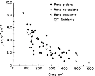
If the membrane of a given tissue is permeable (in the net transport of charge sense) to an ion, then changing its concentration should change the PD provided the substituted ion is impermeant. If the change in concentration does not change the PD, then this indicates that the rela- tive conductance of the membrane to this ion is essentially zero. It is well known that increasing the K+ concentration of the bathing fluid of axons [50], of striated muscle [90] of frog skin [203], and of gastric mucosa [82] produces a marked change in the transmembrane PD, indicating a high relative conductance to K+. In fact, it would appear that the total conductance of the membranes of many tissues could be accounted for on the assumption that the relative proton conductance is zero. It has been reported [81, 117] that changing the pH of the bathing fluid of striated muscle over a wide range did not result in any change in the resting membrane potential, indicating that the relative proton conductance of the muscle membrane is essentially zero. In our studies (see below) on the gastric mucosa the relative proton conduc- tance of the lumen-facing membrane (dog) and the submucosal-facing membrane (frog) appears to be very low.
On the basis of the above-cited work it would be tempting to extra- polate and assume that the relative proton conductance of living tissues is essentially zero. However, Carter et a l [23,24] have suggested that the relative proton conductance of the limiting membrane of rat striated muscle is substantially greater than that of K+. In studies with glass microelectrodes they report (a) that the H+ is in electrochemical equi- librium across the limiting muscle membrane and (b) that with
triple-barreled microelectrodes (a glass electrode, a reference electrode, and a current-sending electrode) changing the voltage across the muscle membrane by sending current (either inward or outward) re
sults in a change of H+ concentration within the muscle fiber to a new electrochemical equilibrium in less than 1 minute. These findings indi
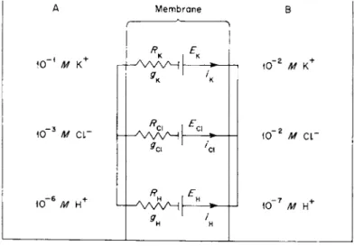
cate a very high relative ionic conductance of the muscle membrane to protons. The findings of Carter and colleagues are difficult to reconcile with other reports using glass microelectrodes and with those of Hodgkin and Horowicz [90] on the effect of changes in external ion concentration on the PD across the limiting membrane of striated muscle. This problem can be illustrated by analysis of the conceptual model shown in Fig. 1, where side A represents a small volume of fluid (comparable to the cell volume) containing 10"1 M K+, 10"3 MCI", and 10"6 Μ H+, and side Β represents a large volume (comparable to the extracellular fluid) containing ΙΟ"2 Μ K+, 10"2 Μ CI", and ΙΟ"7 Μ H+. The concentra
tions are chosen for convenience of analysis. The membrane contains three types of selective channels, one each for K+, CI", and H + , and is completely impermeable to all other ions. It is assumed that PDBA = Ei — RJi for each channel, where Et is the emf for the ith ion calculated from the Nernst equation and Ri9 the resistance of the fth ion; it is assumed that the resistance is constant (independent of the current strength). On the basis of this latter assumption, and recalling that con-
A Membrane Β
(
10" 1 Μ κ+ r ι£κ > Ι Ο "2 Μ 10" 1 Μ κ+ r Ι Ο "2 Μ
10~3 Μ c r H | * Ι Ο "2 Μ c r
10~3 Μ c r
«ο
Ι Ο "2 Μ c r
10~6 Μ H+ Λ 10"
7 Μ Η+
10~6 Μ H+
9*
H l , * Η
10" 7 Μ Η+
FIG. 1. Conceptual model to explain transient PD changes in response to step change in ion concentrations. Membrane permeable only to K+, Cl", and H+ via separate channels.
ductance is the inverse of resistance, it is easily shown that the PD across the membrane is given by
PD^ = ffn Eh +9kEk+ gC\EC\
9h +9k +9ci (1)
where PDBA is the PD across the membrane (positive when Β is positive to A); gH, gcu and gK are the H+, CI", and K+ conductances of the membrane; and EH, Ecl, and EK are the Nernst potentials for the ions, i.e., Et =(RT/zF)ln (CA/CB), where Λ, Γ, z, and F have their usual meaning and CA and CB the concentrations of a given ion in the two compartments (z = +1 for K+ and H+ and —1 for CI").
With the initial conditions all emf's would equal 60 mV (assuming RT/zF = 60 mV), and PDBAl would equal 60 mV. To illustrate the principle let us assume that gH =20 gK and gK=gCi and examine the situation when the fluid in compartment Β is changed stepwise to a fluid contain
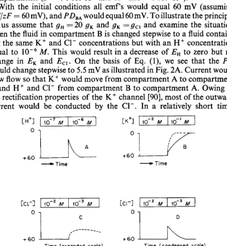
ing the same K+ and CI" concentrations but with an H+ concentration equal to 10"6 M. This would result in a decrease of EH to zero but no change in EK and Ecl. On the basis of Eq. (1), we see that the PD would change stepwise to 5.5 mV as illustrated in Fig. 2A. Current would now flow so that K+ would move from compartment A to compartment Β and H+ and CI" from compartment Β to compartment A. Owing to the rectification properties of the K+ channel [90], most of the outward current would be conducted by the CI". In a relatively short time,
[ H+] 0
h60
10 7 Μ 10 6 Μ 10- 2 Μ 10— \ Μ 0 π
+ 60
- Time »Time
— 2 - 3
10 Μ 10 Μ 0 η
+ 60
Time (expanded scale)
— 2 - 3
10 Μ 10 Μ
+ 60
Time (condensed scale)
FIG. 2. Bars at top give concentrations of ions in compartment Β of model of Fig. 1.
Predicted changes in PD (versus time) for concentration changes shown by bars. For details see text.
assuming no buffer in A, the H+ concentration in compartment A would be increased to approximately 10"5 Μ (pH 5) and electrochemical equi
librium reached with EH, EK9 and PDBA all essentially 60 mV. In the absence of buffer it would take a very small amount of H+ transport from Β to A to change the concentration from 10"6 to 10"5 M, so that the change in the K+ and Cl~ concentrations in compartment A would be very small. The K+ and CI" would poise the system, and, if we were not watching for a transient change in PD, we might easily overlook it.
A step change in compartment Β to a fluid with the same H+ and CI"
concentrations but with a K+ concentration of 10"1 Μ would result in an initial small step change in PD [APD = 2.7 mV from Eq. (1)] followed by an exponential-like change to a new level determined by the K+ gradient as illustrated by the solid line in Fig. 2B. Again assuming no buffer in the system, the PD would relatively rapidly reach its new steady state; on a condensed time scale (dashed line of Fig. 2B) the response of the PD would appear almost like a step change.
A step change in the Cl~ concentration in compartment Β in Fig. 1 from 10"2 Μ to ΙΟ"3 Μ would result in a small step change in PDBA
[(2.7 mV from Eq. (1)] followed by an exponential-like change in the PD to a temporary level shown in Fig. 2C ( H+, CI", and K+ would all move from compartment A to compartment Β of Fig. 1). On a con
densed time scale (Fig. 2D) the response of Fig. 2C would appear as a step change which would be followed (as the CI" concentration in A decreased to about 10" 4 M) by a gradual change in PD to approximately the original level as shown in Fig. 2D. This latter type response was that observed by Hodgkin and Horowicz [90] in frog striated muscle follow
ing a stepwise decrease of the CI" concentration in the bathing medium.
As Hodgkin and Horowicz indicate, the high concentration of Κ+ in the cell poises the system so that in the new steady state the K+ concentration in compartment A of Fig. 1 would be changed by only a small amount.
On the basis of the concept of Carter and colleagues, a situation similar to that for CI" might exist for H+, but, since the intracellular concentra
tion of H+ is smaller, the transient response to changes in H+ might be of much shorter duration and could be easily overlooked. The duration of the transient response to changes in the external ion concentrations would be a function of, among other factors, the amount of buffer present and also the thickness of the unstirred layers adjacent to the membrane.
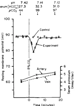
In rat striated muscle Carter and colleagues [19] found that sustained changes in the pH of blood resulted in a transient change in PD in the predicted direction (a transient increase in magnitude of the PD was found for an increase in pH of blood and a transient decrease for a
pH 7.42 7.14 7.12 Serum[HC0;]27.3 32.3 31.0
pC02 44 99 97
1 1 I
100 n ,
FIG. 3. Effect of hyperventilation induced by increased rate of mechanical respirator on potential difference (PD) of rat striated muscle membrane. Measurements of PD were made alternately with the same microelectrode on experimental and control rats lying side by side. pH, serum H C 03" and pC02 measurements were made on arterial blood. Serum K+
from femoral, arterial, and venous blood of leg on which PD was measured. Printed by permission from Campion et al. [19].
sustained decrease in the pH of blood, as illustrated in Fig. 3). However, these transient reponses lasted about 5 minutes.
Using the data from Conway [37] for the buffer content of striated muscle and assuming that a reasonable fraction of the buffer content is readily available to the continuous phase of the muscle cytoplasm, one would predict on the basis of the high proton conductance hypothesis a duration of the transient response of the order of minutes as found by Carter and colleagues. The rate of movement of H+ across the muscle membrane following a change in external ion concentrations would be a function of the Κ+, CI ~, and H+ conductances but, with gH > #K = # c i >
the rate of movement would be primarily a function of gK and gcl. Carter et al. [24a] realize that these findings are not compatible with the findings of Hodgkin and Horowicz. On the basis of the duration of the transient response resulting from changes in H+ concentration one would predict that the changes in external K+ (or for a change in
which the product of the K+ and Cl" concentrations was held constant) would yield a response like that shown in Fig. 2B with the duration of the response about 5 minutes. In sharp contrast to this prediction Hodgkin and Horowicz [90] found transient responses with durations of only a few seconds. Furthermore Hodgkin and Horowicz estimated the individual conductances of K+ and Cl~ and found that the sum of these conductances agreed reasonably well with the total conductance determined as dl/dV (from the data of Fatt and Katz [67]). On the basis of this latter agreement it is difficult to believe that the partial ionic conductance to any other ion would be many times the conductance of K+ and Cl".
Woodbury et al. [213,213a] in studies on the effect of changes of the pH of the fluid bathing in vitro frog muscles reported changes in PD in the direction predicted by the concept of Carter et al., but their analysis revealed that the ratio of proton to K+ mobility was only 100 (the ratio of the P's of the Goldman equation). The proton conductance would obviously be considerably smaller than the K+ conductance when the respective concentrations were taken into account. On the basis of Woodbury's work it would appear that the proton conductance might be finite but considerably less than that of K+ and Cl~. It should be mentioned that Hutter and Warner [100,101] reported for striated muscle that changes in external pH produced no change in PD or a change in the direction opposite to that predicted on the basis of a finite proton conductance.
Stephens [191] has taken the concepts of Carter et al. and applied them to the problem of the spread of excitation in excitable tissues.
According to Stephen's concept, one of the initial events in the process of the excitation of a previously resting region is the movement of H+ outward which results in a significant increase in the H+ concentration locally at the junction of the membrane and the external fluid; he assumes that this increase displaces C a2 + from fixed negative sites followed, in turn, by a marked increase in the N a+ conductance.
On the basis of the findings presented above, one might be tempted to conclude that for striated muscle the high proton conductance hypothesis is untenable. However, in the author's opinion more work is needed before the hypothesis can be rigorously excluded. It is possible that a conceptual model might be devised with the necessary ad hoc postulates that can reconcile, to some extent at least, the two sets of conflicting data. The postulates would involve (a) the presence of unstirred layers adjacent to the limiting membrane of the muscle; (b) the effect of buffers on the thickness of the unstirred layers; and (c) the availability of buffer to the unstirred layers in the cytoplasm.
A problem associated with that of proton conductance is whether the proton is in electrochemical equilibrium across the muscle membrane.
Conway and Fearon [40] first suggested that there was electrochemical equilibrium for the H+ and, as mentioned above, this concept has been revived by Carter et a l [24]. Carter et a l report that the pH inside muscle is approximately 6.0, i.e., the pH expected for electrochemical equilibrium. It should be pointed out that Caldwell [16,17] and also Kostynk and Sorokina [116] using glass microelectrodes found the pH of muscle to be about 7.0. However, Carter and colleagues [22-24]
argue that they did not obtain pH's consistent with electrochemical equilibrium until after making substantial improvements in their technique that included the use of very stringent requirements for the performance of their electrodes.
Waddell and Butler [210] and Butler et a l [15], using the DMO method, have concluded that the pH inside muscle is closer to 7.0, thus confirming the work of Fenn [69] with the C 02 method.
It is possible that both the findings of Butler et a l [15] and Carter et a l [23,24] are not incompatible. The glass microelectrode method would be expected to yield a measure of the pH of the continuous phase of the muscle cytoplasm while the DMO method would give an " aver- age pH." For the DMO method to measure the pH of the continuous phase, the postulate would have to be made that the pH of each muscle compartment is the same. Simple calculations show that, if there is a compartment that occupies about 10% of the volume of the muscle (Peachey [139] found that the sarcoplasmic reticulum was about 13%
of the muscle volume) with a pH of about 8, then the continuous phase could have a pH of 6 and the " DMO pH " would be about 7. Pertinent to this problem would be studies on the pH of sarcoplasmic reticulum.
I could not find any data on the pH of this compartment, but it is of interest to point out that there is evidence [1] showing that the pH in the mitochondrial matrix (inner aqueous compartment) during absorption of C a2 + is about 1.5 pH units greater than that of the bathing medium.
Since it is well known that the sarcoplasmic reticulum can transport C a2 + [211], it would not be surprising to find a high pH in this structure.
Obviously more work is needed in this area. It should be pointed out that the H+ could be transported outward against its electrochemical gradient and the H+ inside the muscle maintained at a lower concentra- tion than that demanded by electrochemical equilibrium by a neutral carrier exchange (see below) mechanism between N a+ and H+. The N a+ is maintained at low levels inside the cell by an active process, and the presence of a neutral carrier exchange between H+ and N a+ could account for the low H+ concentration in the cell; the energy for the
transport of H+ against its electrochemical potential gradient would be provided by the N a+ gradient from the outside to the inside.
Returning to the hypothesis of a high proton conductance of living membranes, it would be theoretically possible to test this hypothesis by applying a high H+ concentration to a tissue. A high concentration of H+ comparable to that of K+ (or other appropriate ions) should result in a sustained change in PD, and K+ could no longer poise the system. Most biological membranes would be irreversibly damaged by such procedures, and I doubt that many biologists would put much credence in such an experiment for tissues such as striated muscle.
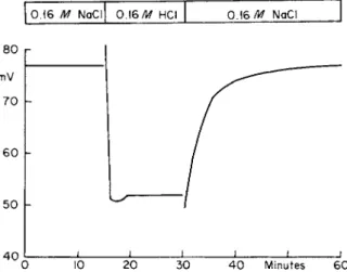
However, there is at least one tissue normally exposed to a high H+ concentration and that is the gastric mucosa. If in the resting gastric mucosa the lumen-facing membrane had a much higher conductance to protons than to other ions, then changing the lumen fluid from an isotonic NaCl solution to an isotonic HC1 solution should produce a very marked and sustained rise in PD (the serosal side becoming much more positive). These experiments have been done [161] and a typical one is shown in Fig. 4. Apart from a transient change, there is not a large sustained increase in PD when the bathing fluid is changed from isotonic NaCl to isotonic HC1. It should be added that great care was taken to make sure that a readily renewable saturated KC1 junction made con
tact between the mucosal fluid and the nonpolarizable electrode on that side. The transient is predicted since a liquid junction pontential would
0 . 1 6 Μ NaCl 0.16/tf HCI 0 . 1 6N a C l
8 0 mV 7 0
6 0
5 0 h
4 0 I ι i l I ι ι
0 10 20 30 4 0 Minutes 60
FIG. 4. Effect on PD of resting dog stomach of changing fluid bathing mucosal surface with intact blood supply. Fluid changed from 0.16 Μ NaCl to 0.16 Μ HCI and back to 0.16 Μ NaCl. Serosa is positive to mucosal side. Renewable saturated KC1 junction between electrode and fluid in contact with mucosa. Reprinted by permission from Rehm et al. [161].
be established between the NaCl previously equilibrated with the mucous coat (which has the characteristics of a weak gel) and the fresh HC1 solution. The time constant for diffusion across this coat to the outer gastric cells is about a minute, and we see that the PD reaches a steady state level in a few minutes [60]. These experiments indicate that the proton conductance of the lumen-facing membrane of the gastric mucosa in its resting state instead of being greater may actually be less than the N a+ conductance.
There is a substantial body of evidence (e.g. [94]) indicating that the CI" conductance of the mucosal membrane is greater than the sum of the other ionic conductances. For example, it was found that diluting a saline solution in contact with the resting mucosal surface of the dog's stomach results in an increase in the measured PD (the serosal side becomes more positive [163]). Davies and Ogston [58] obtained similar results in the in vitro frog stomach. It is apparent that for the mucosal cell surface of the resting dog stomach the order of conductances is 9c\ > # N a > 9h · The data indicate that the proton conductance of this surface is low, and hence for a situation in which the high proton conductance hypothesis is put to a stringent test it is disproved (see Section III, D for a discussion of the problem in the secreting stomach).
The mucosal surface of the stomach may be considered atypical since it has been evolved to withstand the onslaught of a highly acid solution.
On the other hand, experiments have been performed on the membrane facing the submucosal side in the in vitro frog gastric mucosa and no evidence of a high proton conductance was found [152,178,187]. These experiments were designed primarily to determine the relative H C 03"
conductance of the membrane facing the submucosa. We changed the H C 03" concentration from 25 to 5 mM and back again; we replaced
H C O 3" with SO4" (plus sucrose for the osmotic deficit). Experiments were performed in which the C 02 of the bathing medium was changed simultaneously with the change in the H C 03" and in which there was no change in the C 02 tension with the consequent reduction of the pH of about 0.7 unit. Experiments were also performed in which only the C 02 tension was changed (95% 02- 5 % C 02 to 99% 02- l % C02).
Experiments were done with both chloride and sulfate bathing media (the sulfate bathing media is chloride free), and a typical experiment is shown in Fig. 5. There is a diffusion barrier between the bathing fluid and the mucosal cell layer that delays the change in ionic composition at the cell border. However, the time constant for diffusion of ions across this barrier is about a minute [186]. This is demonstrated in the ex- periment on the left side of Fig. 5 in which potassium was increased from 4 to 20 mM (K+ replacing N a+) , and one can see that there was a rapid
FIG. 5. Effect of changing K+ (left graph) and H C 03~ (right graph) concentrations in fluid bathing nutrient (submucosal) side in vitro frog gastric mucosa [178a].
and dramatic drop in PD. Forte and Davies [74] also reported no effect on the PD from changes in nutrient H C 03" .
If there were a significant H C 03" conductance of the nutrient mem- brane, there should be a rapid change in PD just as in the case of the potassium. Similar experiments in which CI" was replaced with S 04"
resulted in a rapid change in PD (the conductance of the nutrient mem- brane appears to equal the sum of the K+ and CI" conductances; (see Harris and Edelman [82] and Spangler and Rehm [186]). These experi- ments, designed primarily to determine the relative HC03~ conductance of the nutrient membrane, also throw light on the OH" and H+ con- ductance of this membrane. It is really not possible to change the H C 03"
concentration in the bathing fluid without simultaneously changing the H+ and OH " gradients between cell and bathing fluid, assuming that the membranes are quite permeable to C 02 as such. For example, when the bathing medium is changed from 5% C02-25 mM H C 03" to 1%
C 02- 5 mM H C 03" , the pH of the bathing fluid would not change, but the cells have been previously equilibrated with 5 % C 02 and C 02 would rapidly diffuse out of the cells and in the cell interior the OH" concentra- tion would increase and the H+ concentration would decrease, resulting in an increase in the H+ gradient from bathing media to cell and an increase in the OH" gradient from cell to media. The direction of change in all three emf's (the H+, OH", and H C 03" ) would be the same, and, since there was essentially no change in the PD as a result of changes in the bathing media, it would appear that the sum of the relative con-
ductances of H+, OH", and H C 03" of the nutrient-facing membrane of the mucosal layer is essentially zero. However, on the basis of the considerations presented in Figs. 1 and 2, the possibility of high proton conductance cannot be rigorously excluded. But if there is a high proton conductance, the total transient response to a change in the pH of the bathing medium would have to be of a duration of no more than a few seconds since a change in pH of about 0.7 pH (25 mM H C 03" , 5%
C 02 to 5 mM HC03~, 5% C02) does not result in a change in PD In summary, it is not possible to reconcile easily the available data with the concept that the limiting membranes of striated muscle and gastric cells have a high relative proton conductance.
C. Mechanisms for Proton Conductance
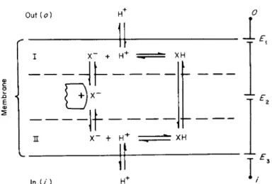
On the basis of the usual criteria for determining the relative ionic conductance of a membrane to a given ion, there are essentially two types of mechanisms that could account for what would appear to be a finite relative conductance. These are (1) ionic conductance per se and (3) a carrier ionic conductance. The carrier mechanism could be either passive or active. Passive carrier proton conductance would be a mech- anism in which the current was carried by protons from the bathing media into and out of a membrane but in which the current through the membrane was not carried by a proton as such. A model illustrating this latter mechanism is shown in Fig. 6. This model differs from a forced carrier exchange, a passive carrier exchange, or the simultaneous trans- port of a proton and an anion by a single carrier in that these latter mechanisms are neutral and would have zero conductance unless special ad hoc postulates are made. In the model there is no change in the affinity between the carrier and the H+. It is shown in the following that thePZ) across this membrane under equilibrium conditions would be given by the Nernst equation.
The PDoi would be given by [178].
PDoi E1+E2+E3 (2) where
II
(3)
(4)
(5)
Out {o)
ί ^
X " + Η Γ XH
— d h — -
X" + Η " XH
In (/)
I.
FIG. 6. Model to illustrate passive carrier proton conductance of cellular limiting membrane. X~ is the carrier and after combining with H+ moves as a neutral molecule from one side of membrane to the other side and moves as an ion in the opposite direction.
Ει is the Nernst potential for H+ between side I of membrane and outer aqueous solution, E2 for X " from side I to side II, and E3 for H+ from inner aqueous solution to side II of membrane.
and the nomenclature is easily understood from examination of Fig. 6.
Substituting Eqs. (3), (4), and (5) into (1), we have
rjJoi z/ 7m [ H- ]o[ X - ]I I[ H+]I I W
At equilibrium, [XH],=[XH]„ and hence [Χ"]„[Η+]„ = [Χ"]Ι[Η+]Ι so that
In the model the charged carrier would carry the current across the membrane. The model could be modified to one in which the carrier was neutral, and the carrier ion complex was charged with the current being carried by the complex.
Some of the work on thin lipid membranes is of interest in this con
nection. Finkelstein [70] found that weak-acid uncouplers of oxidative phosphorylation can increase the conductance of thin lipid membranes by several orders of magnitude, and in the high conductance state they appear to be selectively permeable to H+ (or OH"), i.e., the PD across the membrane is given by the Nernst equation. However, Finkelstein presents evidence indicating that the primary charge carrier is neither
H+ nor OH", but a negatively charged dimer formed between the un- dissociated and dissociated forms of the weak acid; the dimer carries the current not only within the lipid membrane but also across the boundaries between the lipid and aqeuous solutions. This type of mech
anism could be designated apparent proton conductance.
Andreoli and Troutman [3a] have found that for thin lipid mem
branes with the polyene antibiotic candicidin a tenfold K+ gradient (ΙΟ"3 Μ to 10"4 M) and a simultaneous tenfold H+ gradient in the opposite direction ( 1 0 "3M t o l 0 "4M ) across the membrane gives a PD of 58 mV with the orientation being determined by the H+ gradient indicating that the apparent H+ conductance is very much greater than that for K+. Since the concentration of candicidin in the bathing media is very small ( < 1 0 "6 Μ and identical in both aqueous phases), the most reasonable interpretation is that the conductance into and out of the membrane is by Η + but that the conductance across the membrane is by a complex between the H+ and the candicidin. This would be an ex
ample of passive carrier proton conductance. The problem of active carrier proton conductance is taken up in Section III, D.
D. Proton-Cation Exchange
Examples of proton-cation exchange come from the work of Teorell [197] in studies on the gastric mucosa and from Conway [36] and Roth
stein [170] in studies on yeast, and from Pitts and Alexander [140] and Berliner [7] in studies on the kidney. Teorell found that when HC1 solutions are placed in contact with the resting stomach H+ disappear and are replaced by a cation. These findings have been repeatedly con
firmed [6,11]; N a+ is the cation exchanged for H+. These findings are often quoted as a classic example of a proton-Na+ exchange, but they can also be explained on the basis of Hollander's postulate of the secre
tion of sodium bicarbonate [98] and hence the actual mechanism may not be a proton-Na+ exchange. The HC03~ would combine with the H+ to form C 02 which would diffuse away. However, the work of Cope et al [43] and of Code et al [31 ] in which it was shown that the unilateral flux of N a+ from the lumen contents to the blood was appreciable when the lumen fluid was neutral but dropped to essentially zero when the lumen fluid was acidified is evidence in support of a carrier-mediated exchange between H+ and N a+.
The finding of a concurrent movement of H+ and a cation in opposite directions does not mean that the process is dependent on a carrier- mediated exchange. In many tissues the postulation of a proton-cation exchange via a single carrier (whether active or passive) is based more on
a bookkeeping procedure to ensure that electroneutrality is not violated than on substantive evidence. In other words, the finding of a movement of H+ and cations in the opposite direction does not automatically provide us with an explanation of the mechanism. In subcellular organelles the PD across the subcellular membrane is not generally known with any certainty, and it is difficult to distinguish between passive and active processes.
III. ACTIVE PROTON TRANSPORT A. Concept of Active Proton Transport
The purpose of this section is to examine the mechanisms for active proton transport. Since most of the work in this field has been performed on the gastric mucosa, we confine our attention primarily to this tissue.
We present in outline form essentially all of the mechanisms that have been proposed to account for HC1 secretion. This approach does not lack generality since all of the postulated mechanisms for active proton transport in other tissues and subcellular organelles have their counter- part in the gastric mucosa. Because of the restrictions of electroneutrality the problem of the transport of a given ion cannot be readily dissociated from that of the transport of the companion ion (or ions), and in the case of the stomach the problem is primarily that of HC1 transport.
We consider first the problem of whether the CI" is actively transported by the gastric mucosa.
Rosenberg [169] and Ussing [201] have defined active transport as the transport of an ion against its electrochemical gradient. Once it has been established that a given ion is actively transported, it is usually assumed that a metabolic machine must be present that performs os- motic work at the expense of cellular chemical energy. However, an ion may be actively transported as the result of the establishment of a gradient of another ion that has an affinity for the same carrier. If the latter possibility is ruled out, then it seems likely that a metabolic machine providing energy must be present for the uphill transport of the ion. It has been known for many years that CI" is transported against a concentration gradient from about 110 mM in interstitial fluid to about 170 mM in gastric juice [4]. It has also been established that the serosal side of the stomach is positive to the mucosal side by about 60 mv for the dog [143] and about 30 mv for the in vitro frog stomach [46]. Hence it is clear that the CI" is transported up a concen- tration gradient and against the force of the electric field.
As there is no evidence of the presence of another ion gradient that could furnish the energy for the uphill transport of the CI", it would seem necessary to postulate the existence of a metabolic machine for Cl~ transport. However, it is theoretically possible that water drag may be responsible for active ion transport. Ussing [202, 203] has examined the problem of the effect of water drag on ion transport and found that it could theoretically play a partial role in the active transport of N a+ in the frog skin. In the case of the gastric mucosa there is excellent evi- dence supporting the osmotic gradient theory for H20 transport [59,78,
129,200], and it can be easily shown and is intuitively understandable that on the basis of this mechanism for H20 transport the Cl~ uphill transport could not be accounted for by water drag [153a]. Therefore, there must be some force other than the forces of diffusion, the electric field, and water drag acting on the CI". The only conceivable forces can be classified as forces between molecules, and there must be some type of carrier-mediated mechanism. By carrier I mean some substance that has an affinity for the ion being transported; the particular mechan- ism by which the complex moves from one side of the membrane to another [137] is not germane to the problem of whether carrier transport is involved. It also follows that there must be a cyclic change in the affinity between the carrier and the substance transported, otherwise there could not be a directional transport. Analysis of studies of unilateral fluxes of Cl~ across the mucosa by Hogben [95], Heinz and Durbin [84], Villegas [206, 208], and Forte [72] has provided further sub- stantial evidence for CI" carrier transport.
The main purpose of the above approach to the problem of ion trans- port is to find out whether it is necessary to postulate the presence of a machine furnishing metabolic energy for transport. It is clear that such is necessary for the CI". Most workers seem to believe that the preferred way to determine if an ion is actively transported is by the Ussing- Zerahn [204] short circuit technique. The short circuit technique has the advantage of determining whether an ion is passively transported. It also may reveal that ions other than those being studied are actively transported, i.e., when the short circuit current does not equal the alge- braic sum of the net transport of the ions under consideration. The finding that a given ion is actively transported by the short circuit technique does not ensure that the transport mechanism is potent enough to transport the ion under normal conditions. For example, in the case of gastric CI" transport we wish to know if the active mechanism is potent enough to transport the ion up the normal concentration gradient and against the normal potential difference of about 60 mV. To be sure, it is possible by analysis of unilateral flux ratios to obtain an
estimate of the potency of the transport mechanism under short circuit conditions, but the surest way is to determine the direction of its movement under normal open circuit conditions.
The problem of deciding whether or not there is active H+ transport is a problem of definition. It is not strictly correct to conclude that there is active H+ transport if we decide that the transport is not passive. Let us examine the possibility that the H+ is passively transported from an interstitial fluid with a pH of 7.4 to the lumen fluid with a pH of 0.8.
Then we would need a PD of about 400 mV to transport the H+, i.e., the H+ equilibrium emf would be about 400 mV and in order to trans- port passively the H+ the serosal side would have to be over 400 mV positive to the mucosal side. The PD across the stomach is about 60 mv or less, and microelectrode exploration has not revealed a potential difference much higher than this [21,205], so we may conclude that the H+ is not passively transported. Therefore, we might postulate active transport, but H+ production in the lumen may not arise from the transport of H+ of the interstitial fluid. The source of H+ may be the water in the lumen (Section HI, B). Regardless of the source of the H+, it is clear that H+ is not passively transported and there must be some kind of metabolic machine present that leads to the end result of a high H+ concentration (Section III, B).
We next present in outline form the mechanisms that have been postulated to explain HC1 production.
B. Postulated Mechanisms for H+ Production
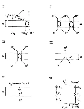
The postulated mechanisms are shown in Fig. 7. The scheme labeled I is the idea put forward by Hogben [92-94]. According to this scheme, C 02 moves across the membrane into the lumen and there combines with H20 to form H+ and HC03~ and the HC03~ is then exchanged for CI" by means of an active exchange mechanism, the net result being formation of HC1. Brodsky and Schilb's [13] theory of acidification by the turtle's urinary bladder is similar to Hogben's theory. Both theories postulate active transport of HC03~ and the derivation of the H+ from water in the lumen.
The theory of Bull and Gray [14] is similar to the theory of Hogben, only these authors postulate an acid such as pyruvic in place of the C 02. The acid diffuses into the lumen and the anion is replaced by Cl~ via an exchange mechanism.
The scheme labeled II is a scheme proposed by Conway et a l [41]
according to which isotonic potassium chloride is secreted and in which there is a forced exchange between K+ and H+ with the net result of HC1 production. Hirschowitz [89] has suggested a similar mechanism but with N a+ as the cation.
Scheme III illustrates what is referred to as a unitary process. Accord
ing to the scheme, a carrier picks up a CI" and H+ (or some precursor of H+) from the cytoplasm and liberates H+ and CI" into the lumen by a reaction on the luminal side of the membrane (there is no net transport of charge in the process). Schemes I and HI are neutral mechanisms and it is not mandatory for them to give rise to an emf; any PD arising from these mechanisms would be an indirect effect of the establishment of ion gradients.
Schemes IV and V represent electrogenic mechanisms for H+ pro
duction (Sections III, Β and III, F). In both of these latter schemes there is a net transport of charge at the site of the mechanisms. They would both possess emf's and the potency of the mechanisms would be pro
portional to the magnitudes of their emf's and inversely proportional to their resistances. According to Scheme IV, H+ would be transported into the lumen, the source of H+ would be hydrogen atoms donated by a hydrogen carrier from the cytoplasm, and a negative charge would be transported into the cytoplasm. Scheme V represents an electrogenic scheme suggested by Nielsen and Rosenberg [132] in which the H+ are not transported outward but the OH" move from the lumen and the H+ from the cytoplasm into the mechanism in the membrane.
Obviously for an electrogenic H+ scheme to function there must be parallel arrays of active CI" units in the membrane, and since there is a net transport of charge across the electrogenic H+ units, there must be a net transport of charge across the CI" mechanism, i.e., the CI" mech
anism must also be electrogenic. An equivalent circuit for H+ and CI"
secretion is shown in Fig. 7 (Scheme VI).
Because of the limitations of space, we will not examine the data pertinent to each of these postulated mechanisms. There are many obvious objections to a number of these schemes, and only the electro
genic concept has been vigorously exploited. Therefore in the next section we examine the main lines of evidence bearing on this latter concept (see Davies [55], and Heinz and Obrink [86], Forte [73] for a detailed analysis of the other schemes presented in Fig. 7).
C. Evidence for Electrogenicity of the H+ Mechanism
1. CHLORIDE TRANSPORT IN RESTING MUCOSA
In the resting stomach there is an active CI" mechanism tending to transport CI" from blood to lumen and also in some species an active N a+ mechanism tending to transport N a+ from lumen to blood
[11,48,49,113,114]. Since the active CI" mechanism is present in the
H+ CL"
HCO,'
Π
co2 HCO3~ cr
Μ Μ
>
cr B+ H"»
nr E Z
H20- »OH~+ H"*"
- 4 -
OH"
4-
Χ LILU men)
Β (BLOOD\
FIG. 7. Models I through V represent postulated mechanisms to explain acidification.
See text. Model V I is an equivalent circuit for electrogenic mechanisms for Η+ and Cl~
secretion; Ix represents externally applied current, ICi is Cl" current and equals net Cl"
transported in direction opposite to ICi ,and IH is H + current and equals net H+ trans
ported in direction of IH.
resting stomach, the simplest explanation would be that with the onset of secretion there is an activation of an Η+ mechanism in limbs parallel to the Cl" mechanism. In the equivalent circuit in Fig. 7 (VI), EH would be zero (and RH infinite) in the resting state; the return circuit would include parallel pathways for the other ions (primarily N a+ and K+) . Obviously the actual geometry of the stomach is more complex, but for our purposes this simple equivalent circuit is used here. For simplicity of presentation we ignore the small amount of cations other than H+ in gastric juice.
2. EFFECT OF EXTERNAL CURRENT ON H+ RATE
During secretion there would be electrical coupling between the CI"
mechanism and the H+ mechanism as illustrated in Fig. 7 (Scheme VI). Current would flow in the indicated direction, and the current flow in the CI" limb would be in the direction for the transport of CI" from blood to lumen and in the H+ limb for the transport of H+ into the lumen and for the transport of a negative charge (or its equivalent) into the cytoplasm. Application of an external current Ix in the direction of blood to lumen would obviously result in a decrease in the CI"
current and an increase in the H+ current, provided the parameters of the system are unchanged as a result of the external current. In other words, one would predict that current application from blood to lumen would result in an increase in H+ secretory rate and a concurrent de- crease in the Cl~ secretory rate.
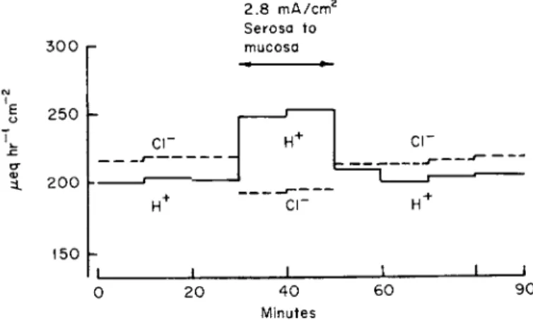
Figure 8 shows an experiment on the dog's stomach in which current was sent in this direction, and it can be seen that during the period of current application there was an increase in H+ rate and a decrease in CI"
rate, and that following the cessation of current the rates returned rapidly to their original control value [148]. Current sent in the opposite direc- tion would be predicted to have opposite effects. However, interpretation of these experiments for the dog stomach is rather involved because current sent in the opposite direction results in a change in the param- eters of the system; following the cessation of current there is a marked and sustained depression of the secretory rate; the rate does not return
2.8 mA/cm2 Serosa to
300 r mucosa
250 h
C I " C I "
3. 200
150
c r
20 40
Minutes 60 90
FIG. 8. Effect of applied current across flap of dog stomach with intact blood supply on H+ and Cl~ secretory rates; 2.8 mA/cm2 current sent for 20 minutes from serosa (blood side) to mucosa (lumen side). Reprinted by permission from Rehm [148].
rapidly to the control levels as it does when current is sent in the direction of that in Fig. 8.
Forte [73] has found for the frog stomach that current sent from the lumen to the serosal side decreases the H+ rate (confirming Crane et al [47]) and also increases the CI" rate just as predicted on the basis of the circuit in Fig. 7 (Scheme VI).
While the use of the Ussing-Zerahn [204] short circuit technique can- not be applied in a meaningful way to the dog mucosa with an intact blood flow [153] it is applicable to the in vitro stomach. It can be seen from the equivalent circuit of Fig. 7 (VI) that
- / x = / c , - /H (8) The sign of Ix is negative because the direction of current flow for short
circuiting is in the direction of lumen to serosa (blood side). Hogben [92,93] has found for the in vitro frog gastric mucosa that this equation holds to a good first approximation.
On the basis of these experiments on the in vivo dog stomach and on the in vitro frog stomach, it is apparent that the H+ rate and the CI" rate can be dissociated. This would be predicted on the basis of the theory that there are two separate arrays of electrogenic pumps, one array representing CI" units and the other parallel array representing H+ units. It is of interest to note that for the experiments on the in vivo dog stomach, in which current was sent in the direction to increase the H+ rate, the electrical energy capable of doing useful work is only a fraction of the minimum free energy necessary for the increased H+ production [144]. In other words, the externally applied current must increase the rates of the biochemical reactions responsible for H+ production.
The effect of current flow on the H+ and CI" rates would seem to constitute crucial evidence in favor of the author's theory of separate H+ and CI" mechanisms [145,146]. However, another explanation for the effect of applied current on the H+ and CI" secretory rates has been offered by Durbin [62]. He suggested that the increase in H+ rate may be a result of the current conducting H+ out of the lumen with a con- sequent reduction in H+ concentration at the site of its production that, in turn, would increase the H+ rate on the basis of the law of mass action. According to Durbin this could happen if there were a unitary process producing HCI, and hence the effect of current would not be crucial evidence for the electrogenic theory.
While a complete analysis of the implication of Durbin's explanation is beyond the scope of this review, it should be pointed out that on the
basis of the osmotic theory of water transport it does not necessarily follow that, since the current would conduct H+ away from the site of secretion, this would decrease the H+ concentration at the site of the mechanism. According to the osmotic theory of water transport the osmolarity of the fluid in the lumina of the tubules is slightly greater than that of interstitial fluid [78,129,200] and for our present purposes can be considered constant. Now if a unitary process produced H Q and all the current passing via the tubular cells was carried by CI" moving from tubular lumen to the interstitial fluid (the lumen side has a much higher conductance to CI" than to other ions), then the concentration of Η + in the tubular lumina would remain essentially constant. Without appropriate ad hoc postulates concerning the partial ion conductance of the tubular cells Durbin's argument would not be valid.
3. ABILITY OF INHERENT EMF's TO PRODUCE CURRENT
On the basis of the electrogenic theory, the emf of the CI" mech
anism should be able to deliver enough electrical current to account for the observed rates of HC1 secretion. Rehm and Hokin [160] deter
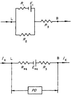
mined the ability of the emf's of the resting dog mucosa to deliver current in an external circuit. The gist of the method used is shown in Fig. 9. Current from an external source was sent in the direction indi
cated in this figure and at intervals the circuit was interrupted for a fraction of a second and the open circuit PD measured. It was found that with currents of the order of 1 mA cm"2 or less the open circuit PD remained relatively high under steady state conditions. However, when the externally applied current approached 5 mA cm"2, the steady state open circuit PD was reduced to low values. On the basis of these ex
periments it was concluded that the inherent emf's of the resting stomach could produce currents of the order of magnitude of those required, but under these conditions the PD across the stomach was definitely reduced from its resting value. Since that time it has been shown for the in vitro frog stomach [149] and also for the in vitro necturus stomach [130] that stimulating the stomach to secrete acid increases the potency of the CI" mechanism.
There is no real problem as far as the in vitro frog stomach is con
cerned, since it has been repeatedly shown that the short circuit currents have values of the order of the H+ rate [3,74,84,93,205]. We conclude that the inherent emf's can produce enough current to satisfy the re
quirements of the electrogenic concept.
![FIG. 5. Effect of changing K + (left graph) and H C 0 3 ~ (right graph) concentrations in fluid bathing nutrient (submucosal) side in vitro frog gastric mucosa [178a]](https://thumb-eu.123doks.com/thumbv2/9dokorg/1152374.82867/14.664.141.498.108.369/effect-changing-concentrations-bathing-nutrient-submucosal-gastric-mucosa.webp)