752
Glutamic Dehydrogenase
Ellen Schmidt
Glutamic dehydrogenase ( G I D H ) has been detected in m i c r o - o r g a n i s m s
1 _ 9 )
, higher p l a n t s
1 0
.
1 1
) and warm-blooded tissues 12-16). i
n
mammals the richest source is the liver. T h e liver of a healthy person contains 3 000—4 000 units *)/g. fresh w e i g h t1 7
) , f o l l o w e d by kidney cortex (600—800 units/g.), cerebral cortex, gastric mucosa, lymph nodes, lung, cerebral medulla and cerebellum (100—160 u n i t s / g . )
1 7
) . The concentration in muscle is very l o w
1 6
.
1 7
) . T h e enzyme is not detectable with the usual methods in non-nucleated erythrocytes
1 7
) or in normal s e r u m
1 8
.
1 9
) . A higher activity can occur in serum, mainly after liver cell d a m a g e
1 8
.
1 9
) .
Liver G I D H reacts with diphosphopyridine nucleotide ( D P N ) and triphosphopyridine nucleotide ( T P N )
l 4
.
1 5
. 2 0 - 2 4 ) . O f the four possible methods (two coenzymes, two reaction directions) of measur
ing the activity o f G I D H from warm-blooded animals, the measurement o f the rate o f D P N H oxidation is the most convenient (limitation: very narrow optimum concentration range for D P N H
2 3
. 25), see Fig. 1.). With human liver extract and under optimum conditions, the following activities were measured: compared with D P N H , 5 0 % activity was obtained with T P N H , 2 0 % with D P N and only 3 % with T P N .
Principle
Glutamic dehydrogenase ( G I D H ) catalyses the reaction:
(1) oc-Oxoglutarate + N H
4 +
+ D P N H L - ( + ) g l u t a m a t e + D P N + + H
2
0The equilibrium lies in favour o f amino acid formation. Oxidation o f D P N H is directly proportional to the reduction of the substrate and can be followed by the decrease in optical density (AE) at 340 or 366 mfji.
Definition according to Th. Bucher et al. (p. 33).
H. v. Euler, E. Alder and T. Steenhoff-Eriksen, Hoppe-Seylers Z. physiol. Chem. 248, 227 [1937]
E. Alder, V. Hellstrom, G. Gunther and H. v. Euler, Hoppe-Seylers Z. physiol. Chem. 255, 14.
[1938].
E. Adler, G. Gunther and / . E. Everett, Hoppe-Seylers Z. physiol. Chem. 255, 27 [1938].
S. Barban, J. Bacteriol. 68, 493 [1954].
B. Nisman, Bacteriol. Rev. 18, 16 [1954], B. A. Fry, Biochem. J. 60, 6 [1955].
J. T. Wachsman, J. biol. Chemistry 223, 19 [1956].
/. R. S. Fincham, Biochem. J. 65, 721 [1957].
H. Holier and S. Schneider, Biochem. Z. 329, 361 [1957].
M. Damodaran and K. R. Nair, Biochem. J. 32, 1064 [1938].
W. A. Bulen, Arch. Biochem. Biophysics 62, 173 [1956].
/. A. Olson and C. B. Anfinsen, J. biol. Chemistry 197, 67 [1952].
H. J. Strecker, Arch. Biochem. Biophysics 46, 128 [1953].
/. E. Snoke, J. biol. Chemistry 223, 271 [1956].
J. B. Solomon, Biochem. J. 66, 264 [1957].
Ch. D. Kochakian, B. R. Endahl and G. L. Endahl, Amer. J. Physiol. 197, 129 [1959].
E. Schmidt and F. W. Schmidt, Klin. Wschr. 38, 957 [I960].
U. Gerlach, Klin. Wschr. 35, 1144 [1957].
E. Schmidt, F. W. Schmidt and E. Wildhirt, Klin. Wschr. 36, 280 [1958].
C. De Duve, C. Pressman, R. Gianetto, R. Wattiaux a n d F . Appelmans, Biochem. J. 60, 604 [1955].
H. J. Strecker in S. P. Colowick and N. O. Kaplan: Methods in Enzymology. Academic Press, Inc.
N e w York 1955, Vol. II, p. 220.
M. Dixon and E. C. Webb: Enzymes. Longmans, Green and C o . , London, N e w York, Toronto 1958.
C. Frieden, J. biol. Chemistry 234, 809 [1959].
J. Struck, jr. and 1. W. Sizer, Arch. Biochem. Biophysics 86, 260 [I960].
E. Schmidt and F. W. Schmidt, unpublished.
II.2.C Glutamic Dehydrogenase 753
Optimum Conditions for Measurements
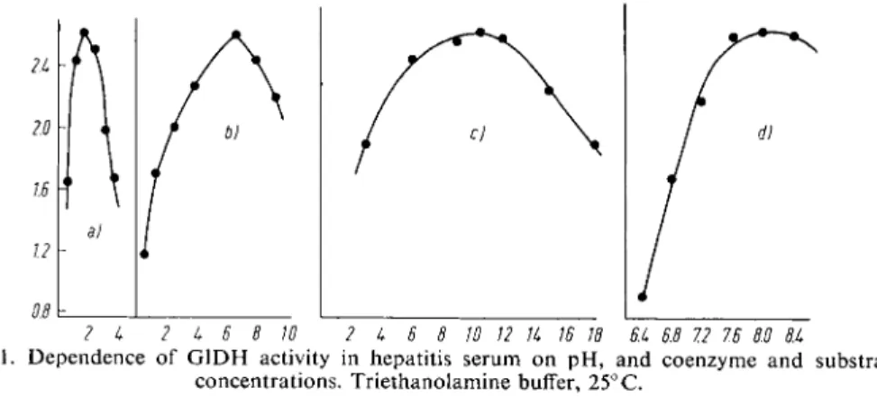
Figure 1 gives the relationship between enzyme activity in serum from patients with liver disease and substrate concentration, and p H .
2 k 2 k 6 8 10 2 k 6 8 10 12 Ik 16 18 6.k 6.8 7.2 7.6 60 8.k
Figure 1. Dependence o f G I D H activity in hepatitis serum o n p H , and coenzyme and substrate concentrations. Triethanolamine buffer, 25° C.
a) Effect o f varying D P N H with constant: a-oxoglutarate 6.5X 10~3 M, N H
4
+ 10.5X 10~2
M and p H 8.0
b) Effect o f varying a-oxoglutarate with constant: D P N H 1.5X 10⁻⁴ M, N H
4
+ 10.5xlO~2M and p H 8.0c) Effect o f varying N H
4 +
with constant: D P N H 1 . 5 x l 0
_
4 M , a-oxoglutarate 6.5X 10~
3
M and pH 8.0
d) Effect o f varying p H with constant: D P N H 1.5x10⁻⁴ M, a-oxoglutarate 6 . 5 x l 0 ~ 3 M , N H10.5X10-2
4
+Ordinate: Units/ml. serum
Abscissa: a) D P N H (X 10~4 M) - b) a-oxoglutarate ( x 10"3 M) - c) N H
4
+ (X 10~2 M) - d) p H The p H optimum depends on the buffer used and lies between 7.6 and 8 . 61 0 , 1 1 , 1 4 , 2 1 , 2 3 , 2 4 , 2 6 ) . T he
activity o f G I D H is primarily dependent o n the D P N H concentration, and secondly o n the con
centration of a-oxoglutarate and N H
4 +
.
Reagents
1. Triethanolamine hydrochloride 2. Sodium hydroxide, 2 N, A. R.
3. a-Oxoglutarate
commercial preparation, see p. 1024
4. Ammonium acetate, A. R.
5. Reduced diphosphopyridine nucleotide, DPNH
sodium salt, DPNH-Na2; commercial preparation, see p. 1011.
6. Ethylene-diamine-tetra-acetic acid, EDTA
disodium salt, E D T A - N a
2
H2
- 2 H2
0 , e.g. Titriplex HI*)Preparation of Solutions
I. Triethanolamine buffer (0.05 M; pH 8.0):
Dissolve ca. 930 mg. triethanolamine hydrochloride in ca. 80 ml. doubly distilled water, adjust pH to 8.0 with 1.58 ml. 2 N NaOH (glass electrode) and dilute to 100 ml.
*) e.g. from E. Merck, Darmstadt (Germany)
26) H. Beaufay, D. S. Bendall, P. Baudhuin and C. De Duve, Biochem. J. 73, 623 [1959).
754
Sectio C: Measurement of Enzyme ActivityII. Sodium a-oxoglutarate (ca. 0.4 M):
Dissolve 146 mg. a-oxoglutaric acid in ca. 1 ml. doubly distilled water, bring to pH 6.8 with several drops 2 N NaOH, and dilute to 2.5 ml.
III. Ammonium acetate (ca. 3 M):
Dissolve 2.35 g. ammonium acetate in 10 ml. doubly distilled water.
IV. Reduced diphosphopyridine nucleotide (ca. 0.01 M (3-DPNH):
Dissolve 15 ml. DPNH-Na2 in 1.5 ml. doubly distilled water.
V. Ethylene-diamine-tetra-acetate (ca. 0.26 M):
Dissolve 100 mg. EDTA-Na 2 H 2 • 2 H 2 0 in 1 ml. doubly distilled water.
Stability of the solutions
Store the D P N H and a-oxoglutarate solution, and the buffer at 0° to 4°C. The D P N H solution is stable for at least a week, the a-oxoglutarate solution for about four weeks if its p H is below 7.0.
The other solutions are stable indefinitely.
Procedure
Before commencing the measurements bring the daily requirements of buffer to 25° C in a thermostatically controlled water bath (pour, do not pipette!). Keep the DPNH and a-oxo
glutarate solutions in an ice bath.
Preferably use fresh serum. Haemolysis does not interfere with the assay because erythro
cytes contain no GIDH, however considerable haemolysis can increase the "preliminary reaction" (see below) resulting in the oxidation of too large a portion of the added DPNH.
Spectrophotometric m e a s u r e m e n t s
Wavelength: 366mu.; light path: 1 cm.; final volume: 2.0ml.; temperature: 25°C. A blank is unnecessary. Read against air or water.
Pipette into the cuvette:
1.34 ml. buffer (solution I) 0.03 ml. DPNH solution (IV) 0.02 ml. EDTA solution (V)
0.07 ml. ammonium acetate solution (III) 0.50 ml. serum.
Mix with a glass rod flattened at one end. Wait until the decrease in optical density stops (2 — 10 min.). This "preliminary reaction" is variable and is due to interaction of substrates and DPNH-linked dehydrogenases in the serum. If the optical density change exceeds 0.100 (corresponding to the oxidation of more than 0.06 [xmoles DPNH), then another 0.01 ml.
DPNH solution (IV) must be added.
Start the GIDH reaction by mixing in 0.04 ml. a-oxoglutarate solution (II).
Note the time (in sec.) taken for a decrease in optical density of 0.020 and continue readings until the total decrease in optical density is > 0.120. Average the time (in sec.) taken for each optical density change of 0.02 and use this average for the calculations.
The reactions is linear with time at least until the optical density has decreased by 0.100.
Premature retardation of the rate is usually due to lack of DPNH, and gradual acceleration
II.2.C Glutamic Dehydrogenase
755 to excess DPNH (refer to Fig. 1). The decrease in optical density remains linear with time even if other than optimum concentrations of a-oxoglutarate or
N H 44
"
are used. If the optical density decreases by less than 0.005 in 5 minutes, then measurement is discontinued;
>6000sec. (AE = 0.100) means <0.06 units GIDH/ml. or "normal". Dilution of the serum is not necessary even with high activities.
Calculations
According to Biicher e t a l .
2 7 )
a unit of enzyme activity for D P N and TPN-dependent dehydrogenases is the amount of enzyme contained in 1 ml., which at 25° C and with a light path of 1.0 cm, decreases the optical density of D P N H ( T P N H ) by 0.100 in 100 seconds at 366 mu,. A unit corresponds to the reduction of 1.09u,moles of substrate/ml./hour
2 1
\ Therefore with a reaction volume of 2 ml., a light path of 1 cm, A E = 0.020 and 0.5 ml. of serum added:
100 2.0 0.020 80 . , , X --- X .
f
.n
= = units/ml. serum sec. 0.5 0.100 sec.Stability of the Enzyme in the Serum Sample
Serum can be stored in a refrigerator ( < 7 ° C ) for at least 48 hours without loss of G I D H activity.
Sources of Error
On completion of the "preliminary reaction" no interference by other enzymes occurring in serum has been observed.
Effect of Exercise or Corticosteroid Therapy
A significant rise in the G I D H activity of serum from healthy people occurs with severe muscular exercise, the origin of which is still unknown
2 8
) . With corticosteroid therapy the G I D H activity in serum falls significantly
2 8
> as do all the other "key pathway" enzymes*) so far investigated. The enzyme is inhibited by sulphonylurea derivatives
2 9
).
Details for Measurements in Tissues
In liver the G I D H is located in the mitochondria
2 0
>
2 6
>
3
o - 3 2 ) . On cell fractionation it sediments with cytochrome oxidase, but leaks out into the surrounding medium. It can be measured within the mitochondria if the membrane is made permeable to coenzymes and substrates
2 5
*
3 3
). Therefore it follows that the extracting agent and the degree of mechanical disintegration of the mitochondria are of great importance for the quantitative measurement of the enzyme in tissues
2 5
.
3 4
) . After h o m o genizing human liver for 2 min. in 0.15 M NaCl in a Potter-Elvehjem glass homogenizer and immediately centrifuging, less than 10% of the G I D H activity is found in the supernatant. Even after standing for 3 hours only ca. 7 0 % of the total G I D H activity is found in the whole homogenate after disintegration as described. In the course of 24 — 72 hours this percentage rises to ca. 8 0 % in
*) " K e y pathway" enzymes are found in all types of cell. They represent the basic elements for respiration, glycolysis and general amino acid metabolism, (cf. Th. Biicher, E. Schmidt and F. W. Schmidt: Serum Patterns of " K e y Pathway" Enzymes. Lecture, 9th Middle East Medical Assembly, May 1959, American Univ., Beirut, Lebanon).
2
?) G. Beisenherz, H. J. Boltze, Th. Biicher, R. Czok, K. H. Garbade, E. Meyer-Arendt and G. Pflei
derer, Z. Naturforsch. 8b, 555 [1953].
28
> E. Schmidt and F. W. Schmidt, Vortrag Frankfurter Med. Ges. 4. 3. 1959.
2
9) K. Wallenfels and H. D. Siimm, Klin. Wschr. 35, 849' [1957].
3
0) G. H. Hogeboom and W. C. Schneider, J. biol. Chemistry 204, 233 [1953].
31
> G. S. Christie and / . D. Judah, Proc. Roy. Soc. [London] Ser. B 141, 420 [1953].
3 2
) C. Allard, G. de Lamiranda and A. Cantero, Exp. Cell Res. 13, 69 [1957].
33
) E. Schmidt, F. W. Schmidt and E. Wildhirt, Klin. Wschr. 37, 1221 [1959].
34) G. L. Endahl and Ch. D. Kochakian, Proc. Soc. exp. Biol. Med. 94, 192 [1957].
756
Section C : Measurement of Enzyme Activitythe whole homogenate and to ca. 6 0 % in the supernatant obtained immediately before measure
ment 25,33). jf doubly distilled water is used instead of NaCl solution then about 8 0 % of the G I D H
is detectable in the supernatant after standing for 2 hours 2 5, 3 4 ) .
Homogenization for 2 min. in an Ultra-Turrax (Janke and Kunkel & C o . , see p. 51) results in 9 2 — 9 7 % of the G I D H activity being found in the supernatant, and the total activity after standing for 24 hours 25). A determination in duplicate on liver requires 1 mg. fresh weight. Other tissues require other conditions of disintegration.
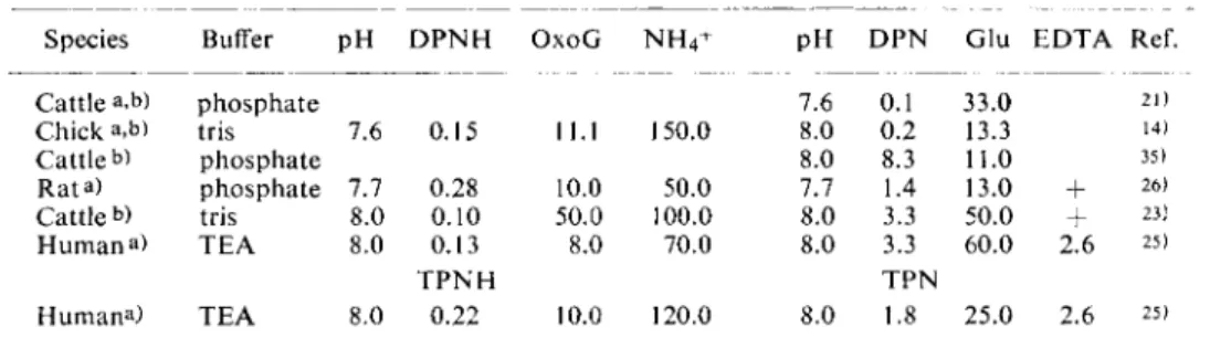
For assay conditions for human liver (2 min. extraction in 0.15 M N a C l in an Ultra-Turrax), see Table 1. These data are not valid for isolated liver mitochondria and in the same way the optimum substrate concentrations obtained for human serum or liver must not be assumed for other tissues from other species.
Table 1. Conditions for the determination of G I D H from liver
Species Buffer p H D P N H O x o G N H
4
+ PH D P N Glu E D T A Ref.Cattle a,b) phosphate 7.6 0.1 33.0 21)
Chick a,b) tris 7.6 0.15 11.1 150.0 8.0 0.2 13.3 14)
Cattle b) phosphate 8.0 8.3 11.0 35)
Rata) phosphate 7.7 0.28 10.0 50.0 7.7 1.4 13.0
+
26)Cattle b) tris 8.0 0.10 50.0 100.0 8.0 3.3 50.0
+
23)Human a) T E A 8.0 0.13 8.0 70.0 8.0 3.3 60.0 2.6 25)
T P N H T P N
Humana) T E A 8.0 0.22 10.0 120.0 8.0 1.8 25.0 2.6 25) All concentrations are given in [imoles/ml.
a) Crude or fractionated tissue extract b) Purified or crystalline enzyme preparation
Abbreviations: E D T A = ethylene-diamine-tetra-acetate
O x o G = a-oxoglutarate Tris = tris-hydroxymethyl-aminomethane buffer Glu = glutamate T E A = triethanolamine buffer
35) S. J. Adelstein and B. L. Vallee, J. biol. Chemistry 234, 824 [1959].