736
Lactic Dehydrogenase
U O. Warburg: Wasserstoffiibertragende Fermente. Dr. W. Saenger, Berlin 1948, p. 40.
2) F. Kubowitz and P. Ott, Biochem. Z. 314, 94 [1943].
Cf. A. Kornberg in S. P. Colowick and TV. O. Kaplan: Methods in Enzymology. Academic Press, N e w York 1955, Vol. I, p. 441.
G. Beisenherz, J. H. Boltze, T. Biicher, R. Czok, K. H. Garbade, E. Meyer-Arendt and G. Pflei
derer, Z. Naturforsch. 8b, 555 [1953].
Cf. J. B. Neilands in S. P. Colowick and TV. O. Kaplan: Methods in Enzymology. Academic Press, N e w York 1955, Vol. I, p. 449.
J. Szulmajster, M. Grunberg-Manago and C. Delavier-Klutekko, Bull. Soc. Chim. biol. 35, 1381 [1953].
Cf. M. Dixon in S. P. Colowick and N. O. Kaplan: Methods in Enzymology. Academic Press, N e w York 1955, Vol. I, p. 444.
F. Wroblewski, Scand. J. Clin. & Lab. Invest. 10, 230, Suppl 3 1 ; Int. Congr. clin. Chem. Stock
holm 1957.
9) P. G. Cabaud, F. Wroblewski and V. Ruggiero, Amer. J. clin. Pathol. 30, 234 [1958].
1Q
) H.-U. Bergmeyer and E. Bernt, unpublished.
11
) Th. Wieland and G. Pfleiderer Biochem. Z. 329, 112 [1957].
12) G. Pfleiderer and D. Jeckel, Biochem. Z. 329, 370 [1957].
!3) Th. Wieland, G. Pfleiderer, and F. Ortanderl, Biochem. Z. 331, 103 [1959].
14
) Th. Wieland, G. Pfleiderer, I. Haupt and W. Worner, Biochem. Z. 332, 1 [1959].
is) B. Hess, Klin. Wschr. 36, 985 [1958].
16) B. Hess, Ann. N . Y. Acad. Sci. 75, 292 [1958].
1
7
) B. Hess in W. H. Hauss and H. Losse: Struktur und Stoffwechsel des Herzmuskels. G. Thieme, Stuttgart 1959, p. 128.
18) B. Hess and S.-I. Walter, Klin. Wschr. 38, 1080 [I960].
19) E. S. Vesell and A. G. Beam, Proc. Soc. exp. Biol. Med. 94, 96 [1957].
20) E. S. Vesell and A. G. Beam, J. clin. Invest. 37, 672 [1958].
21) E. S. Vesell and A. G. Beam, Ann. N . Y. Acad. Sci. 75, 286 [1958].
22) B. R. Hill, Cancer Res. 16, 460 [1956].
23) F. W. Sayre and B. R. Hill, Proc. Soc. expl. Biol. Med. 96, 695 [1957].
24) B. R. Hill, Ann. N . Y. Acad. Sci. 75, 304 [1958].
25) R. J. Wieme and L. Demeulenaere, Acta gastro-ent. belg. 22, 69 [1959].
26) TV. O. Kaplan, M. M. Ciotti, M. Hamolsky and R. E. Bieber, Science [Washington] 131, 392 [I960].
27) R. Richterich, E. Gautier, W. Egli, K. Zuppinger and E. Rossi, Klin. Wschr. 39, 346 [1961].
Hans-Ulrich Bergmeyer, Erich Bernt and Benno Hess
Lactic dehydrogenase ( L D H ) , the "reduzierende Garungsferment" (Warburg
1
)), was first crystallized from rat muscle in 1943
2
>. The best k n o w n lactic dehydrogenases are the enzymes from skeletal
3
*
4
) and heart muscle
5
). Lactic dehydrogenases from bacteria
6
) and y e a s t
7
) are not linked to the pyridine nucleotide coenzymes.
In human organs the L D H activity decreases in the following orders) (related to g. fresh weight):
kidney > heart > skeletal muscle > pancreas > spleen > liver > lung > serum.
The activity of L D H of animal origin is measured spectrophotometrically
2
) (see equation 1). Measure
ments o f the activity by the colour reactions (e.g.
9
>) o f the 2,4-dinitrophenylhydrazone of the unreac- ted pyruvate are difficult, because the D P N H produced in the reaction also forms a hydrazone which absorbs in the same r e g i o n
1 0
) .
The lactic dehydrogenase activity of serum is composed o f several enzyme proteins with the same action and substrate specificity but of different origin. The lactic dehydrogenases in serum which originate from liver, heart, skeletal muscle, erythrocytes, tumours, etc. are not only different from one another, but themselves consist of several enzymatically active fractions which can be separated from each o t h e r
1 1 - 2 7
) . Such enzymes, which differ in their protein structure and therefore in the
II.2.a Lactic Dehydrogenase 737
optimum conditions for their action, but have the same specificity (in this case towards L-lactate), are termed " i s o z y m e s "
2 8
) or "isoenzymes"
2 9
*.
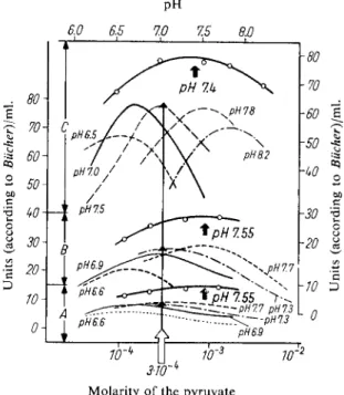
A method is described below which gives approximately optimum conditions for the measurement in serum of the L D H from heart and liver (Fig. 1).
Fig. 1. Determination of the optimum conditions for the measurement of L D H activity in serum.
25° C; 0.05 M phosphate buffer Section A : N o r m a l serum
Section B: Serum in hepatitis
Section C : Serum after myocardial infarction
Left hand and bottom ordinates (broken and thin-lined curves): Dependence of the activity on the pyruvate concentration with constant p H (which is given on each curve)
Right hand and upper ordinates (thick-lined curves — o — o — ) : Dependence of the activity on the pH with constant pyruvate (3 X 10~
4
M) (optimum activity)
Principle
Lactic dehydrogenase ( L D H ) catalyses the reaction:
(1) Lactate -f D P N + pyruvate + D P N H + H+
The equilibrium is far on the side of lactate and diphosphopyridine nucleotide ( D P N ) . The L D H activity is measured by the rate of consumption of pyruvate and reduced diphosphopyridine nucleo
tide ( D P N H ) ; the decrease of optical density at 340 or 366 m[i due to the oxidation o f D P N H is measured.
28
> C. L. Markert and F. Moller, Proc. nat. Acad. Sci. U S A 45, 753 [1959].
29
> F. Wroblewski and K. Gregory: Proc. 14th Internat. Congr. clin. Chem. Edinburgh, 1960, E. &
S. Livingstone Ltd., Edinburgh and London 1961, p. 62.
PH
6.0 6.5 7.0 7.5 8.0
r-J 1 1 1 1 —
3W
K
Molarity of the pyruvate
738
Section C: Measurement of Enzyme ActivityOptimum Conditions for Measurements
The most important characteristics for the assay of activity of the individual lactic dehydrogenases are their different substrate and pH optima. Measurements which are not carried out under optimum conditions naturally result in values for the activity which are too low. The optimum conditions for the measurement of the enzyme in serum after myocardial infarction, in liver damage, blood diseases and tumours have been more or less established
1 5
-24,30,31). The measurements shown in Fig. 1 were made with phosphate buffer at 2 5° C
3 2 )
. The optima of activity with variation of the pyruvate con
centration are strongly dependent on p H . A pyruvate concentration of 3 X 10
4
M at pH 7.5 gives nearly optimum activities in human serum for the enzymes from liver and heart. The temperature of the measurements is 25 C and the D P N H concentration is 1.3 X 10
4
M. The dependence of the L D H activity on the D P N H concentration is not shown in Fig. 1. D P N H over the range of about 5x 10 -
s
to 5X 10
4
M gives a wide activity optimum. With higher temperatures, for example, 37°C, higher substrate concentrations are required for optimum activity.
Reagents*)
1. Potassium dihydrogen phosphate, KH2PO4, A. R.
2. Dipotassium hydrogen phosphate, K2HPO4, A. R.
3. Sodium pyruvate
commercial preparation, see p. 1027.
4. Reduced diphosphopyridine nucleotide, DPNH
sodium salt, D P N H- N a 2 ; commercial preparation, see p. 1011.
Preparation of Solutions
I. Phosphate-pyruvate solution (0.05 M phosphate buffer pH 7.5; 3.1 x 1 0 -4
M pyruvate):
Dissolve 700 mg. K2HPO4, 90 mg. K H 2 P 0 4 and 3 mg. Na pyruvate in doubly distilled water and make up to 80 ml.
fl. Reduced diphosphopyridine nucleotide (ca. 8 x 10~~
3
M (3-DPNH):
Dissolve 10 mg. DPNH-Na2 in 1.5 ml. phosphate-pyruvate solution (I).
Stability of the solutions
Store all the solutions, stoppered, in a refrigerator at 0 —4°C. Prepare the D P N H solution freshly each week. Deterioration of the buffer is usually due to bacterial contamination, which can be preven
ted by addition of a few drops of chloroform.
Procedure
Use only fresh serum free from haemolysis.
Spectrophotometric m e a s u r e m e n t s
Wavelength: 340 or 366 mu.; light path: 1 cm.; final volume: 3.0 ml.; temperature 25°C (preferably use a constant temperature cuvette holder).
A control cuvette is not necessary. Before the assay bring the reaction mixture and the serum to 25°C (water bath).
*) Complete reagent kits are available commercially, see p. 1036.
30) R. E. Thiers and B. Vallee, Ann. N. Y. Acad. Sci.
75, 214 [1957].
31
) E. Schmidt and F. W. Schmidt, personal communication.
32) H.-U. Bergmeyer. Lecture, Kongr. f. Lab. Med., Berlin April 1961.
ILL a Lactic Dehydrogenase 739
Pipette successively into the cuvette:
2.85 ml. phosphate-pyruvate solution (I) 0.05 ml. DPNH solution (II)
0.10 ml. serum.
Mix, immediately start a stopwatch and read the optical density at minute intervals for 3 to 5 min.
The measured optical density difference AE/min. should not be greater than 0.020/min.;
otherwise dilute the serum 1:10 with the phosphate-pyruvate solution (I).
Calculations
According to Wroblewski and LaDue
33
) a unit is the amount o f L D H which changes the optical density of D P N H at 340 m^x by 0.001 in 1 min., in a 3 ml. assay mixture and at 24 — 27° C.
It therefore follows that with 0.1 ml. of serum
(2) (AE
3 4
o/min.) x 10000 = L D H units/ml. serum (3) ( A E3 6 6
/ m i n . ) x 18900 = L D H units/ml. serumObtain the mean of the measured AE/min. values and use these for the calculations. Depending on the type of serum the reaction curves are either linear or non-linear. In the latter case (the values for AE/min. decrease with time) use the mean of the first three readings.
N o r m a l v a l u e s See page 705.
Example
0.1 ml. of normal serum was analysed and the following optical densities were measured at 366 m^x (linear curve):
0 min. 0.402
A E = 0.015 1 min. 0.387
A E = 0.014 2 min. 0.373
A E = 0.016 3 min. 0.357
A E = 0.015 4 min. 0.342
Mean AE/min. - 0.015
0.015X 18900 = 284 units/ml. serum (according to Wroblewski) C o n v e r s i o n to other units
1. For D P N - l i n k e d dehydrogenases a unit according to Bucher et at.
4
) is the amount of enzyme con
tained in 1 ml. which changes the optical density of D P N H at 366 mpi by 0.100 in 100 sec. at 25° C and with a 1 cm. light path.
Therefore at 25° C :
1 unit (Bucher): A E
3 6 6
/ 1 0 0 sec. = 0.100 1 unit (Wroblewski): A E3 4 0
/ m i n . = 0.001 A E3 6 6
/ n i i n . = 0.060A E
3 4
o / m i n . = 0 . 1 1 3 for a 3 ml. assay mixture AE3
4o/min. = 0.0377 37.7 units (Wroblewski) = 1 unit (Bucher) 0.0265 units (Bucher) = 1 unit (Wroblewski)33) F. Wroblewski and J. S. LaDue, Proc. Soc. exp. Biol. Med. 90, 210 [1955].
740 Section C : Measurement of Enzyme Activity
2. According to Racker et a/. 34) a unit is the amount of enzyme which converts 1 u.mole of substrate/
min. at 25°C.
Therefore:
1 unit (Racker): 1 [xmole/min.
1 unit (Wroblewski): AE34o/min. = 0.001 (3 ml. assay mixture) as AE34o/min. = 0.001 corresponds to the conversion of 4.82X 1 0
-4
[jimoles of substrate/3 ml.
1 unit (Wroblewski) = 4.82X 1 0 ~
4
units (Racker) or
1 unit (Racker) = 2073 units (Wroblewski)
T o calculate directly from the measured values with 1 ml. serum:
Measurements at 340 my. :
(2) ( A E / m i n . )X 10000 = units (Wroblewski) (2 a) (AE/min.) X 265 = units (BUcher) (2b) (AE/min. X 4.82 = units (Racker) Measurements at 366 mu,:
(3) (AE/min.) X 18900 = units (Wroblewski) (3 a) (AE/min.) X 501 = units (BUcher) (3 b) (AE/min.) X 9.1 = units (Racker)
Sources of Error
Experience has shown that none o f the substances present in blood interfere in the assay. The con
centration of pyruvate in normal human serum is about 2 orders of magnitude lower than that of the assay mixture and therefore, contrary to several reports in the literature, it cannot affect the optimum concentration.
It is necessary to determine the optimum concentration of pyruvate (that of D P N H is less important) for serum L D H from other organs. For example, in carcinoma of the bronchus it is 2 X 10~
3
M
3
5 ) .
Stability of the Enzyme in the Serum Sample
The loss of activity after storage of serum at different temperatures has been measured by SUdhof et al.
36
). Storage at 4 ° C or in the frozen state results in ca. 15% loss of activity in 12 hours and 2 7 % in 24 hours. Similar values are obtained at r o o m temperature.
Details for Measurements in Tissues and Other Body Fluids
L D H is a cytoplasmic e n z y m e
3 7
) ; simple homogenization in a Potter-Elvehjem homogenizer (refer to p. 49) is sufficient to completely extract the enzyme. The activity is determined in the supernatant after centrifuging the homogenate at high speed. For measurements on, for example, liver punctures, 10 mg. fresh weight of tissue is sufficient
3 8
).
34
> /. Cooper, P. A. Srere, M. Tabachniek and E. Racker, Arch. Biochem. Biophysics 74, 306 [1958].
35) E. Schmidt and F. W. Schmidt, personal communication.
36) H. SUdhof and E. Wotzel, Klin. Wschr. 38, 1165 [I960].
37) Th. BUcher and P. Baum, Lecture, Dtsch. Kongr. f. arztl. Fortbildung, Berlin 1958.
38) E. Schmidt, F. W. Schmidt and E. Wildhirt, Klin. Wschr. 36, 172 [1958].
II.2.a Lactic Dehydrogenase 741
The optimum conditions for human serum given in Fig. 1 do not necessarily hold for sera from other species or for other organs and body fluids. The following concentrations are suitable for measure
ments on cerebrospinal fluid39)
:
8 x 1 0 - 4 to 2 X 1 0- 2 M D P N H ; 3 x l 0 ~ 4 to 1 X 10~3
M pyruvate;
pH 6.5 to 7.4 (25°C).
Differentiation of the Lactic Dehydrogenases in Serum Originating from Specific Organs
The individual types of L D H proteins can be separated from each other e l e c t r o p h o r e t i c a l l y
1 1 - 1 4 )
or chromatographically i5-i8,40,4i). Liver and skeletal muscle L D H occurring in serum can be deter
mined separately in the presence of L D H from heart, kidney and erythrocytes.
Principle
The L D H activity of a dialysed serum sample is assayed before and after treatment with D E A E - cellulose. Under defined conditions, the DEAE-cellulose adsorbs the heart muscle-type of L D H protein, while the liver-type of L D H protein remains in solution *). For the principle of the spectro
photometric assay, see p. 737.
Reagents
Additional to 1 —4 on page 738:
5. Sodium dihydrogen phosphate, NaF^PC^-LbO, A. R.
6. Disodium hydrogen phosphate, ISk^HPCV12 H2O, A. R.
7. Sodium chloride, A. R.
8. Diethylaminoethyl-cellulose, DEAE-cellulose
from Serva, Heidelberg, Germany**). Capacity: 0.6 mequiv./g.
Preparation of Solutions
Additional to I and II on page 738:
III. Phosphate buffer (2 x 10~2 M; pH 6.0):
Mix 12.3 ml. of a solution of 7.164 g. N a 2 H P 0 4 - 1 2 H 2 O/100 ml. doubly distilled water with 87.7 ml. of a solution of 2.76 g. N a H 2 P O 4 H 2 O / 1 0 0 ml. doubly distilled water and dilute to 1000 ml. with doubly distilled water.
IV. DEAE-cellulose suspension (ca. 10% w/v):
Suspend 5 g. DEAE-cellulose in 50 ml. phosphate buffer (solution III), disperse thorough
ly, allow to sediment for ca. 45 min. and decant from the coarse, sedimented material.
Repeat the sedimentation procedure three times. Wash the fine, similar-sized adsorbent
*) The method is also suitable for the differentiation of other enzymes and proteins apart from the L D H isoenzymes, providing that they are specifically adsorbed by DEAE-cellulose. For example, serum free from a-globulin can be produced by this method.
**) Or a complete reagent kit (see p. 1036) from C. F. Boehringer & Soehne G m b H , Mannheim, Germany. Other preparations have not been tried.
39) H. R. Tyler and L. Bromberger, J. nerv. ment. Dis. 130, 54 [I960].
40) B. Hess and S.-I. Walter, Klin. Wschr. 39, 213 [1961].
41) B. Hess and S.-I. Walter, Ann. N . Y. Acad. Sci. 94, 890 [1961].
742
Section C : Measurement of Enzyme Activityparticles with phosphate buffer (solution III) until the pH of the washings is constant (pH 6.0) and then resuspend in 50 ml. phosphate buffer (solution III). Add a few drops of toluene to prevent bacterial contamination and store at 0—4°C.
V. Phosphate-NaCl solution (2-10-1 M phosphate, 2 x 101 M NaCl; pH 6.0):
Mix 12.3 ml. of a solution of 7.164 g. N a 2 H P 0 4 - 1 2 H 2 O/100 ml. doubly distilled water with 87.7 ml. of a solution of 2.76 g. N a H 2 P O 4 H 2 O / 1 0 0 ml. doubly distilled water.
Dissolve 1.169 g. NaCl in this solution and make up to 100 ml.
Procedure
Preliminary treatment of the experimental material
Dialyse 3 ml. of serum or tissue extract for 2 hours against 1000 ml. phosphate buffer (solu
tion III). A small amount of protein precipitates out on dialysis (equal amounts of a- and P-globulins), but no LDH activity is lost. Use the contents of the dialysis sac for the fracti
onation without filtering.
Fractionation o n DEAE-cellulose
In principle, it is sufficient to adsorb the heart muscle-type LDH on the DEAE-cellulose and then to assay the activity in the supernatant after removal of the cellulose. If it is wished to check the method, the adsorbed LDH is eluted and the LDH activity of the eluate is also measured.
Adsorption:
Suspend the DEAE-cellulose evenly by shaking. Use pipettes with a wide tip (or broken tip).
Pipette into a conical centrifuge tube:
2 ml. dialysed serum
2 ml. DEAE-cellulose suspension (IV).
Mix with a thin glass rod, allow to stand for 10 min. with occasional stirring. Centrifuge at ca. 4000 g for 5 —10 min. and pour off the supernatant into a dry test tube.
Elution:
Add to the residue in the centrifuge tube ca. 3 ml. phosphate buffer (solution III),
mix and centrifuge. Discard the supernatant. Repeat the washing 2—3 times. This removes any of the liver-type LDH activity which may have been carried down with the cellulose.
Add sufficient
phosphate buffer-Nad solution (V)
to the sediment to make up to 4 ml., stir thoroughly and centrifuge for 3 min. at 3000 g.
Pour the supernatant (eluate) into a dry test tube.
To check the method by preparing a balance of the activity (but without obtaining the exact fraction of LDH adsorbed), the washing procedure can be omitted and the adsorbed LDH + any liver-type LDH carried down with the cellulose sediment can be eluted immediately with solution V.
Activity m e a s u r e m e n t s
As described above (p. 738).
II.2.a Lactic Dehydrogenase
743 Calculations
Use the formula o n p. 739 to calculate the activity in the dialysed serum, in the supernatant after adsorption and in the eluate. The activity is obtained in units/ml. of solution taken for the assay.
As 1 g. DEAE-cellulose binds 3 ml. of water, the "solute space" with 200 mg. cellulose in a final volume of 4 ml. is 4.0 — 0.6 = 3.4 ml.
The following formula gives the fraction of the L D H activity which is not adsorbed as a percentage of the total activity in the sample taken:
(4) % L D H
l i v e r
.t y P
e = 100 X = 100 X and the percentage o f L D H adsorbed is(5) % LDHjjeart muscle-type = 100 X ( l
2
) where A i = activity/ml. dialysed serum
A2 = activity/ml. supernatant
The result given by equation (5) includes the fraction of the liver-type L D H carried d o w n with the cellulose sediment and the result given by equation (4) is too low by this amount.
The activity of the heart muscle-type L D H can be calculated more exactly by A3 (activity/ml.) in the eluate of the washed cellulose sediment:
(6) % L D H
h e a r t m u 5 d e
- ,y P
e = 100 X = 100 XIf the values given by equations (4) and (6) are added and compared with 2 A i a deficit is obtained.
The amount of liver-type L D H carried down on the DEAE-cellulose can be determined by omitting the washings and eluting the cellulose sediment directly. Let the activity of this eluate be A
4
, then the total amount o f L D H on the cellulose sediment is:(7) % L D H
s e d i m e n t
= 100 X - - ^ = 100 X2 A i Ai
The difference between the values given by equations (7) and (6) is the percentage of liver-type L D H activity carried down by the cellulose. The sum of the fraction of L D H activity carried down with the cellulose, the liver-type L D H (according to equation (4)) and the heart muscle-type L D H (accord
ing to equation (6)) should be a 100. Any deviation gives the error of the method.
Sources of Error
1 g. DEAE-cellulose adsorbs about 150 mg. protein
4 2
), the exact amount depending on the capacity of the preparation. Therefore the cellulose suspension should be prepared according to the capacity.
The 0.12 mequiv. used in the assay should not be completely utilized. According t o
4
° ) 1 g. cellulose adsorbs about 250 L D H units*). A sufficiently sharp separation of the fractions is only obtained if the conditions of the method are strictly adhered to.
*) Defined according t o
3 4 42
) .> H. A. Sober and A. E. Peterson, J. Amer. chem. Soc. 76, 1711 [1954].