0139–3006 © 2018 Akadémiai Kiadó, Budapest DOI: 10.1556/066.2018.47.1.8
CHARACTERIZATION AND PROBIOTIC POTENTIALS OF LACTIC ACID BACTERIA ISOLATED FROM INGESTA OF SELECTED
RUMINANTS
E.I. NNAWUIHE, K. BANWO*, K. AND A.I. SANNI
Department of Microbiology, University of Ibadan, Oyo State. Nigeria (Received: 18 March 2017; accepted: 23 June 2017)
Studies were carried out to isolate and screen lactic acid bacteria with probiotic potentials and amylolytic activities from the ruminal ingesta of cow and goat. Ninety isolates obtained from the three abattoirs were divided into the following species: Lactobacillus plantarum, L. fermentum, L. pentosus, L. brevis, L. buchneri, L. collinoides, and Leuconostoc mesenteroides. The isolates were screened for probiotic potentials and amylolytic activities. Four isolates possessed probiotic potentials: L. plantarum CA3b, L. fermentum GA2d, L. plantarum GA1d, and L.
plantarum GA3e. Lactobacillus plantarum CA3b, L. fermentum GA2d, and L. plantarum GA3e survived pH 2.5 and 3.5 after 180 min. Lactobacillus plantarum GA1d had the highest tolerance of 82.44% and 71.03% to 0.3% and 1%
bile salts concentrations, respectively. Lactobacillus plantarum CA3b hydrolysed 3% starch with an average zone of clearance of 10.0 mm, while L. plantarum GA1d hydrolysed 2% with a zone of 9.5 mm. These two strains also possessed bacteriocin activities against E. coli, and were characterised based on their reactions to pH, temperature, and enzymes treatments. Ruminal ingesta of cow and goat harbour lactic acid bacteria possessing amylolytic and probiotic potentials, which can be exploited in the fermentation of feedstock, conferring health benefi ts and improved performance to these ruminants.
Keywords: ruminal ingesta, lactic acid bacteria, probiotics, amylolytic activity
Ruminants are mammals of the order Artiodactyla (DAVIS & MILNER, 2009). They have four chambered stomach made up of the reticululum, rumen, omasum, and abomasum (DEHORITY
& TIRABASSO, 2000; DAVIS & MILNER, 2009). Ruminant animals such as cow, sheep, goat, and deer, however, do not synthesize fi bre digesting enzymes, but they have formed a symbiotic relationship with ruminal microorganisms that do (DEHORITY & TIRABASSO, 2000).
The microbial quality of the ingesta (ingested food) depends to a large extent on the quality and composition of feed stuff (BELANCHE et al., 2012; UYENO et al., 2015). The microbial fl ora of the ingesta is a function of the nature, quality, and composition of the animals’ feed, which is dominated by complex polysaccharide and starch degrading microbiota. Lactic acid bacteria (LAB) among other bacteria and fungi found in the ingesta of ruminants have been isolated and documented (BELANCHE et al., 2012; UYENO et al., 2015).
These are added as bacteriocin producing cultures or by the addition of pure or semi-pure bacteriocin preparations. Several LAB have been isolated from intestinal tracts of ruminants.
These include species of the genera Lactococcus, Leuconostoc, Streptococcus, Pediococcus, Ruminococcus, and Lactobacillus (FRAGA et al., 2013; UYENO et al., 2015).
Lactic acid bacteria constitute an integral part of the healthy gastrointestinal (GI) micro- ecology and are involved in the host metabolism. A number of mechanisms work to prevent harmful bacteria from growing and attaching to the intestinal epithelium, examples include
* To whom correspondence should be addressed.
Phone: +234-8056100840; e-mail: kolabanwo@yahoo.com
production and secretion of antimicrobial agents such as bacteriocins and organic acids, and adherence via competition for the binding sites interfering with pathogens (REID et al., 2001;
WANG et al., 2010).
The aim of this study was to screen various isolates of LAB from ruminal ingesta of cow and goat for the possible exploitation as potential probiotics and starter culture(s) for feedstock of ruminant diet.
1. Materials and methods
1.1. Collection of samples
Samples were obtained from healthy cattle (cow and goat) from three selected abattoirs; the University of Ibadan Teaching and Research farm and Bodija and Moniya cattle markets in Ibadan, South West Nigeria. Three cows and three goats were sampled from each cattle farm at each sampling time. The sampling was carried out at fi ve different times from each farm.
Samples were collected immediately after evisceration from the recticulo-rumen with the assistance of a qualifi ed veterinarian assigned to each abattoir.
1.1.1. Isolation of LAB. The samples obtained from the cows and goats were homogenized separately. A total of 10 g per 100 ml (w/v) of the homogenized sample were stored in normal saline for 5 h. A 10-fold serial dilution was made using 10 ml stock sample into 90 ml distilled water. These were cultured for 48 h in de Man Rogosa Sharpe agar (MRS) using pour plate method under anaerobic conditions. They were further sub-cultured in MRS agar under anaerobic conditions until pure isolates were obtained. Pure cultures were maintained as stocks in MRS broth at –4 °C, with 15% glycerol.
1.2. Biochemical characterization of the LAB isolates
1.2.1. Carbohydrates fermentation. Biochemical characterization was carried out on the isolates by assimilation of carbohydrates using API 50CH strips and API 50CHL medium (Biomérieux, Marcy Etoile, France) according to the instructions of the manufacturer. The results from the assimilation profi les were identifi ed using the ApiwebTM software version 5.1.
1.3. Probiotic potentials of the selected LAB isolates
1.3.1. Resistance to low pH. The ability of the isolates to tolerate low pH was determined according to the method of VINDEROLA and REINHEIMER (2003). The mixture was enumerated for the presence of viable cells after 1, 2, and 3 h by measuring the optical density (OD) at 560 nm wavelength using a spectrophotometer (Jenway UV-Vis 5210, Essex, UK), and the results were compared with a control.
1.3.2. Bile tolerance and hydrophobicity tests. Bile salt tolerance and hydrophobicity were assayed by the method of VINDEROLA and REINHEIMER (2003). Hydrophobicity was calculated as the percentage decrease in the (OD600nm) of the initial aqueous bacterial suspension due to cells partitioning into a hydrocarbon layer. The percentage of cell surface hydrophobicity (%H) of the strain adhering to xylene was calculated using the equation:
H%=[(A0–A)/A0×100].
1.4. Starch hydrolysis
Surface dried plates of starch based MRS agar [1, 2, and 3% starch (w/v)] (SANNI et al., 2002) were streaked with 18 h old culture of LAB and incubated at 30 °C for 3 days. The plates were fl ooded with Gram`s iodine solution for 15 to 30 min and examined for clear zones around the growth for assessment of amylolytic activity.
1.5. Genotypic characterization of the selected amylolytic LAB isolates
The genomic DNA of the selected amylolytic LAB strains were extracted using the modifi ed lysozyme-heat lysis method. The DNA quantity and purity were determined using a spectrophotometer at absorbance reading of 260 nm (Nanodrop, Thermo Scientifi c, USA).
The partial 16S rRNA nucleotide sequence was amplifi ed by PCR using universal primers designed for lactic acid bacteria, forward was (5’ – GAG TTT GAT CCT GGC TCA G – 3’), while the reverse was (5’ – AGA AAG GAG GTG ATC CAG CC – 3’) (BANWO et al., 2012).
The PCR products were cleaned using spin columns and subsequently sequenced in a commercial laboratory (Beijing, China).
1.6. Antimicrobial activities of selected LAB isolates
The antibacterial activity of the selected isolates against indicator pathogens Listeria monocytogenes, Escherichia coli, Klebsiella pneumoniae, and Pseudomonas aeruginosa were determined. The isolates were grown in MRS broth (pH 6.5) inoculated with 1% of an overnight culture and incubated at 37 °C for 18–24 h. After incubation, cells were removed from the growth medium by centrifugation (6000×g for 20 min, 4 °C). The antimicrobial spectrum of the inhibitory substances from LAB was determined using the agar well diffusion method (AWD) and observed for zones of inhibition (SCHILLINGER & LÜCKE, 1989).
1.6.1. Screening for bacteriocin producing isolates. The cell-free supernatants of LAB were adjusted to pH 6.0–6.5 using 5 N NaOH to exclude the antimicrobial effect of organic acids. Inhibitory activity of hydrogen peroxide was eliminated by the addition of catalase at a fi nal concentration of 1.0 mg ml–1. The catalase-treated samples were incubated for 2 h at 37 °C. After incubation, the treated and neutralized cell-free supernatants were then tested for antagonistic activity against indicator bacteria by the AWD (SCHILLINGER & LÜCKE, 1989).
1.6.2. Characterization of bacteriocin. The bacteriocins were subjected to treatment at different temperatures, pH, and enzymes. The enzymes were added to 1 ml of neutralized cell free culture supernatant to a fi nal concentration of 2.0 mg ml–1 in all cases. The tubes were incubated at 37 °C for 1 h and the residual bacteriocin activities were determined by measuring the zones of inhibition on agar plates (BANWO et al., 2013). Untreated cell free supernatants were used as control.
1.7. Statistical analyses
Each experiment described in this research study was done in replicates and data obtained from the study were analysed using descriptive statistics and analysis of variance. The differences among means were separated using Duncan’s multiple range test. Signifi cant differences were accepted at P>0.05. The statistical software used was SPSS 16.5 for Windows 7.
2. Results and discussion
2.1. Characterization of LAB isolates using API 50CHL Kit
Ninety LAB isolates were biochemically characterized into the following species of Lactobacillus plantarum (32), L. fermentum (25), L. pentosus (15), L. brevis (4), L. buchneri (4), L. collinoides (3), and Leuconostoc mesenteroides (7) (data not shown). The assessment of biochemical and physiological properties of lactic acid bacteria maybe a good method for phenotypic characterization, but it is not adequate (MOHAMMED et al., 2011; BANWO et al., 2012). There were differences in the species of LAB isolates from each ruminant in a particular sampling site, which agreed with the report of BELANCHE and co-workers (2012), that the different species isolated from the rumen (recticulo-rumen) is largely dependent on nutrition and health status of the ruminant. Selection of the isolates was based on the desirable properties of probiotic potentials and amylolytic activities.
2.2. Resistance to low pH
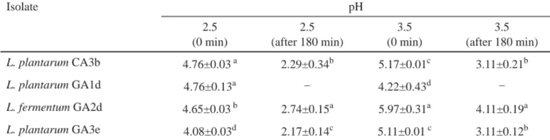
The results of colony forming units (CFU ml–1) are shown in Table 1. In this study, it was observed that some strains tolerated pH 2.5 and 3.5 for three hours, which is in agreement with previous reports of probiotic LAB (BANWO et al., 2013). Some authors proposed that strains intended for probiotic purposes should be screened according to their tolerance to pH 2.5 in an HCl-acidifi ed culture medium for 3 hours (WANG et al., 2010; MOHAMMED et al., 2011).
Table 1. Survival count of selected LAB strains on exposure to pH 3.5 and 2.5 (log10 CFU ml–1)
Isolate pH 2.5
(0 min)
2.5 (after 180 min)
3.5 (0 min)
3.5 (after 180 min)
L. plantarum CA3b 4.76±0.03 a 2.29±0.34b 5.17±0.01c 3.11±0.21b
L. plantarum GA1d 4.76±0.13a – 4.22±0.43d –
L. fermentum GA2d 4.65±0.03 b 2.74±0.15a 5.97±0.31a 4.11±0.19a
L. plantarum GA3e 4.08±0.03d 2.17±0.14c 5.11±0.01 c 3.11±0.12b
Values are means of three independent experiments (mean ± SD)
–: No visible growth/loss of viability; C: cow; G: goat; A: animal; 1: University of Ibadan Teaching and Research farm; 2: Bodija cattle farm; 3: Moniya cattle farm; b;d;e: sampling periods
Values with the same letters in superscript along a column are not signifi cantly different (P>0.05).
2.3. Bile salt tolerance
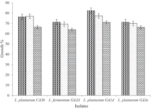
The isolates from this study grew at 0.3% bile salt concentration, however, tolerance varied at higher concentrations of 0.5% and 1.0% (Fig. 1). Though the bile concentrations in the gastrointestinal tract vary, the mean concentration is 0.3% w/v (VINDEROLA & REINHEIMER 2003). Interestingly, the strains stable at low pH were also bile salt tolerant. This is similar to the result obtained by WANG and co-workers (2010), who reported that the strains of L.
buchneri, L. brevis, and L. plantarum were resistant to 0.3% bile salt at pH 4.0.
Fig. 1. Percentage bile salt tolerance of selected LAB strains to 0.3%, 0.5%, and 1.0% bile salt concentrations. C:
cow; G: goat; A: animal; 1: University of Ibadan Teaching and Research farm; 2: Bodija cattle farm; 3: Moniya cattle farm; a,b,c,d,e: sampling periods.
Bars represent the mean and standard deviations of three independent determinations : 0.30%; : 0.50%; :1.00%
2.4. Bacterial adhesion to hydrocarbon assay (BATH) and starch hydrolysis activities of the strains
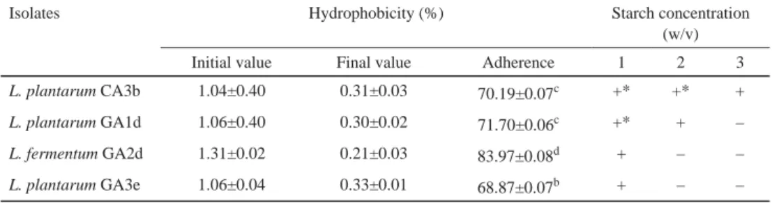
The selected LAB isolates were screened for their cell surface hydrophobicity as shown in Table 2. The gastrointestinal tract, especially the small intestine, is a dynamic environment, and the fl ow of digesta washes out any bacterium unable to counter the fl ow either by rapidly multiplying or by attaching itself to intestinal surfaces (MOHAMMED et al., 2011; GITIKA &
TIWARI, 2013; UYENO et al., 2015). Amylase is an enzyme, which breaks down starch into simple sugars. Consequently, only few lactic acid bacteria possess the ability to hydrolyse starch to produce amylase. The selected isolates demonstrated amylase activity by showing clear zones of hydrolysis on starch based MRS agar plates. Only one isolate, L. plantarum CA3b displayed the ability to hydrolyse 3% starch concentration (Table 2). Very few amylolytic lactic acid bacteria (ALAB) are reported from traditional fermentation of food and feedstock (SANNI et al., 2002; REDDY et al., 2008). Potential probiotic LAB able to hydrolyse starch will be an added advantage in the fermentation of feedstock with added health benefi t. The two isolates that possessed amylolytic potentials were identifi ed using 16S rRNA gene sequencing as L. plantarum CA3b and L. plantarum GA1d with accession numbers MF099794 and MF099795, respectively.
Table 2. Cell surface hydrophobicity and starch hydrolysis assay of the selected LAB strains
Isolates Hydrophobicity (%) Starch concentration
(w/v)
Initial value Final value Adherence 1 2 3
L. plantarum CA3b 1.04±0.40 0.31±0.03 70.19±0.07c +* +* +
L. plantarum GA1d 1.06±0.40 0.30±0.02 71.70±0.06c +* + –
L. fermentum GA2d 1.31±0.02 0.21±0.03 83.97±0.08d + – –
L. plantarum GA3e 1.06±0.04 0.33±0.01 68.87±0.07b + – –
Values are expressed as the mean ± standard deviation of three independent determinations
Values with the same letters in superscript along a column are not signifi cantly different (P>0.0.5). C: cow; G: goat;
A: animal; 1: University of Ibadan Teaching and Research farm; 2: Bodija cattle farm; 3: Moniya cattle farm; b;d;e:
sampling periods
–: No visible amylolytic activity (no visible zone of clearance); +: Amylolytic activity; +*: Strong amylolytic activity. Experiments were carried out at 37 ºC over 5 days
2.5. Antimicrobial activities, production and characterization of crude bacteriocin
Crude cell free culture supernatants of strains were tested against common pathogens as indicator microorganisms (Table 3). After neutralization of the cell free culture supernatants, four strains demonstrated antagonistic activities against E. coli. As previously reported, the antagonistic activities of LAB towards pathogens can be attributed to the production of bactericidal substances like bacteriocins, organic acids, and hydrogen peroxide (TALPUR et al., 2012; FRAGA et al., 2013).
Table 3. Zones of inhibition (mm) of antimicrobial action of cell free culture supernatant obtained from the LAB strains against indicator microorganisms before and after neutralization
Isolates ZI before neutralization
Indicator microorganisms
ZI after neutralization Escherichia
coli
Listeria monocytogenes
Klebsiella pneumoniae
Pseudomonas aeruginosa
Escherichia coli L. plantarum CA3b 25.0±0.08a 24.4±0.09b 22.4±0.11a 19.0±0.12a 18.0±0.08b L. plantarum GA1d 24.0±0.09b 21.3±0.11c 16.3±0.20d 17.0±0.21c 18.3±0.08b L. fermentum GA2d 21.1±0.10c 16.4±0.21d 18.0±0.12b 18.0±0.20b 19.7±0.06a L. plantarum GA3e 23.1±0.10c 22.2±0.11c 17.1±0.15c 18.0±0.20b – Activity expressed as average zone of inhibition (mm). (P>0.0.5), C: cow; G: goat; A: animal; 1: University of Ibadan Teaching and Research farm; 2: Bodija cattle farm; 3: Moniya cattle farm; b;d;e: sampling periods. Each test was carried out at 30 ºC for 18–24 hours. Values with the same letters in superscript along a column are not signifi cantly different (P>0.0.5).
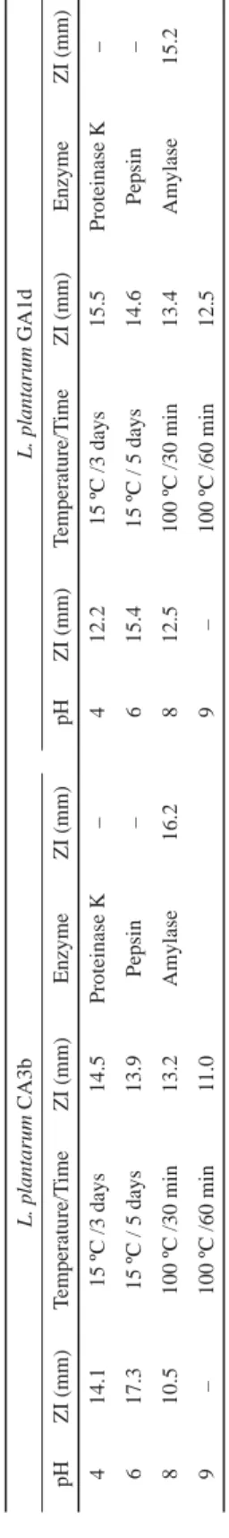
Tables 4 show the effect of pH, temperature, and enzyme treatments on bacteriocins produced by the amylolytic strains L. plantarum CA3b and L. plantarum GA1d against E.
coli. The activity of the bacteriocins produced was not signifi cantly affected by pH 4.0 to 8.0, but activity was highest at pH 6.0, while there was no activity at pH 9.0. Maximum activity was observed at pH 6.0, suggesting that compounds other than acids inhibited growth (BANWO et al., 2013).
Table 4. Effect of pH, temperature, and enzyme treatments on bacteriocins produced by L. plantarum CA3band L. plantarum GA1d against E. coli L. plantarum CA3bL. plantarum GA1d pHZI (mm)Temperature/TimeZI (mm)EnzymeZI (mm)pHZI (mm)Temperature/TimeZI (mm)EnzymeZI (mm) 414.115 ºC /3 days14.5Proteinase K–412.215 ºC /3 days15.5Proteinase K– 617.315 ºC / 5 days13.9Pepsin–615.415 ºC / 5 days14.6Pepsin– 810.5100 ºC /30 min13.2Amylase16.2812.5100 ºC /30 min13.4Amylase15.2 9–100 ºC /60 min11.09–100 ºC /60 min12.5 –: No activity; C: cow; G: goat; A: animal; 1: University of Ibadan Teaching and Research farm; 2: Bodija cattle farm; 3: Moniya cattle farm; b;d: sampling periods. Values are expressed as average zone of inhibition. Each test was carried out at 37 ºC for 18–24 h
The bacteriocins were stable at 15 ºC and 100 ºC temperature for 30 and 60 min, but activity was lost at 121 ºC for 15 min. This supports the fi ndings of TODOROV and co-workers (2011), they observed that bacteriocin GP1 produced by Lb. rhamnosus had a remarkable stability over heat treatment even at the autoclaving temperature for 20 min. Heat stability of bacteriocins produced by lactic acid bacteria at 100 °C is important as a food biopreservative.
Complete inactivation was observed after treatment of the cell-free supernatant with pepsin and proteinase K, which confi rmed the proteinaceous nature of the active agent. Amylase treatment did not result in inactivation. This implies that carbohydrate moiety if existing in the culture supernatant is not signifi cantly responsible for the inhibitory activity. Similar results were recorded by TODOROV and co-workers (2011) for bacteriocins produced by L. plantarum ST13BR. They observed that only proteolytic enzymes neutralized the activities of the bacteriocins.
3. Conclusions
The strains identifi ed as Lactobacillus plantarum CA3b and Lactobacillus plantarum GA1d in this study produced bacteriocins and possessed amylolytic activities together, which is a very rare quality in the literature of lactic acid bacteria. Nature and composition of feedstock and other ecological factors could be associated with this factor. The lactic acid bacteria isolated possessed the generally regarded as safe status, which enable them to be employed as potential starter cultures for the fermentation of feedstock for enhanced performance.
*
The authors appreciate Prof. Yin Li and Mr. Liangtan Miao of the Centre of Excellence for Biotechnology, Institute of Microbiology, Chinese Academy of Sciences for the molecular characterization of the two strains that possess amylolytic activities.
References
BANWO, K., SANNI, A.I., TAN, H. & TIAN, Y. (2012): Phenotypic and genotypic characterization of LAB isolated from Nigerian traditional fermented foods. Food Biotechnol., 26, 124–142.
BANWO, K., SANNI, A. & TAN, H. (2013): Functional properties of Pediococcus species isolated from traditional fermented cereal gruel and milk in Nigeria. Food Biotechnol., 27(1), 14–38.
BELANCHE, A., DOREAU, M., EDWARDS, J.E., MOORBY, J.M., PINLOCHE, E. & NEWBOLD, C.J. (2012): Shifts in the rumen microbiota due to the type of carbohydrate and level of protein ingested by dairy cattle are associated with changes in rumen fermentation. J. Nutr., 142(9), 1684–1692.
DAVIS, C.D. & MILNER, J.A. (2009): Gastrointestinal microfl ora, food components and colon cancer prevention. J.
Nutr. Biochem., 20, 743–752.
DEHORITY, B.A. & TIRABASSO, P.A. (2000): Antibiosis between ruminal bacteria and ruminal fungi. Appl. Environ.
Microb., 66, 2921–2927.
FRAGA M.I., PERELMUTER, K.I., VALENCIA, M.J., CAJARVILLE, C. & ZUNINO, P.J. (2013): Characterization of the ruminal bacterial microbiota of a grazing bovine through classical and culture independent methods. Veterinaria, 49, 40–55.
GITIKA, S. & TIWARI, S.K. (2013): Probiotic potential of Lactic acid bacteria strain LD/4. Int. J. Pharm. Life Sci., 4(10), 3000–3006.
MOHAMMED, B., NAOUAL, J. & ABDELAZIZ, B. (2011): Probiotic potential of Lactobacillus strains isolated from known popular traditional Moroccan milk. Br. Microbiol. Res. J., 1(4), 79–94.
REDDY, G., ALTAF, MD., NAVEENA, B.J. VENKATESHWAR, M., & VIJAY KUMAR, E. (2008): Amylolytic bacterial lactic acid fermentation – A review. Biotechnol. Adv., 26(1), 22–34.
REID, G., CUPERUS, P.L., BRUCE, A.W., VAN DER MEL, H.C., TOMZEK, L., KHOURY, A.H. & BUSSCHES, H.J. (2001):
Comparison of contact angles and adhesion to hexadecane of urogenital, dairy, and poultry lactobacilli: effect of serial culture passages. Appl. Environ. Microb., 58, 1549–1553.
SANNI, A.I., MORLON–GUYOT, J. & GUYOT, J.P. (2002): New effi cient amylase-producing strain of Lactobacillus fermentum isolated from different Nigerian traditional fermented foods. Int. J. Food Microbiol., 72, 53–62.
SCHILLINGER, U. & LÜCKE F. (1989): Antibacterial activity of Lactobacillus sake isolated from meat. Appl. Environ.
Microb., 55(8), 1901–1906.
TALPUR, A.D., MEMON, A.J., KHAN, M.I., IKHWANUDDIN, M., DANISH, M.M., DANIEL, M. & ABOLMUNAFI, A.B. (2012):
Inhibition of pathogens by lactic acid bacteria and application as water additive multi isolates in early stages larviculture of P. pelagicus (Linnaeus, 1758). J. Anim. Plant Sci., 22(1), 54–64.
TODOROV, S.D., RACHMAN, C., FOURRIER, A., DICKS, L., VAN REENEW, C., PRÉVOST, H. & DOUSSET, X. (2011):
Characterization of a bacteriocin produced by Lactobacillus sakei R1333 isolated from smoked salmon.
Anaerobe, 17(1), 23–31.
UYENO, Y., SHIGEMORI, S. & SHIMOSATO, T. (2015): Effect of probiotics/prebiotics on cattle health and productivity.
Microbes Environ., 30(2), 126–132.
VINDEROLA, C.G. & REINHEIMER, J.A. (2003): Lactic acid starter and probiotic bacteria: a comparative in vitro study of probiotic characteristics and biological barrier resistance. Food Res. Int., 36, 895–904.
WANG, C.Y., LIN, P.R., NG, C.C. & SHYU, Y.T. (2010): Probiotic properties of Lactobacillus strains isolated from the feces of breast-fed infants and Taiwanese pickled cabbage. Anaerobe, 166, 578–585.