https://doi.org/10.1007/s42977-021-00091-3 ORIGINAL PAPER
Interactions between probiotic and oral pathogenic strains
Noémi N. Gönczi1 · Orsolya Strang1 · Zoltán Bagi1 · Gábor Rákhely1,2 · Kornél L. Kovács1,3
Received: 19 August 2020 / Accepted: 19 May 2021
© Akadémiai Kiadó Zrt. 2021
Abstract
More than 6 billion bacteria and other microorganisms live in the adult oral cavity. As a result of any deleterious effect on this community, some microorganisms will survive better than others, which may trigger pathogenic processes like caries, halitosis, gingivitis or periodontitis. Oral dysbiosis is among the most frequent human health hazards globally. Quality of life of patients deteriorates notably, while treatments are often unpleasant, expensive and irreversible, e.g. tooth loss. In the experiments reported here, we investigated the individual interactions between 8 pathogenic and 8 probiotic strains and a commercially available probiotic product. Almost all pathogens, namely Fusobacterium nucleatum, Porphyromonas gingivalis, Aggregatibacter actinomycetemcomitans, Streptococcus mutans, Streptococcus oralis, Streptococcus gordonii, Enterococcus faecalis and Prevotella buccae are pathogens frequently occurring in the oral cavity. The used probiotic strains were Lactobacillus plantarum, Lactobacillus rhamnosus, Lactobacillus casei, Lactobacillus acidophilus, Lactoba- cillus delbrueckii, Bifidobacterium thermophilum and two Streptococcus dentisani isolates. Using a modified agar diffusion method, we investigated capability of the probiotic bacteria to prevent the growth of the pathogenic ones in order to identify candidates for future therapeutic treatments. The results indicated successful bacteriocin production, i.e. growth inhibition, against every pathogenic bacterium by at least 5 probiotic strains.
Keywords Oral diseases · Dysbiosis · Bacteriocin · Modified agar diffusion test
Introduction
Human oral cavity is a moist environment, having relatively stable temperature, ranging between 34 and 36 °C, and pH that is close to neutral (Marcotte and Lavoie 1998). Thanks to these favourable conditions, the microbiome of the mouth is very rich and diverse. In general, at least 6 billion bacteria live in the oral cavity of every human, consisting of more than 700 different species (Metwalli et al. 2013). Moreover, other microorganisms, i.e. fungi, mycoplasma, protozoa and viruses, also populate the main entrance of our body (How et al. 2016). The microorganisms tend to form biofilms to prevent microbial washout (Berger et al. 2018).
The primary colonizers of the dental biofilm are Strepto- coccus species (Dige et al. 2009; Brennan and Garrett 2019).
The oral streptococci are Gram-positive, facultative anaer- obes, which initiate the formation of dental plaque (Wang and Kuramitsu 2005). Streptococcus mutans, a member of the oral streptococci, was described in 1924 as the causative agent of the dental caries (Clarke 1924; How et al. 2016). S.
mutans has multiple cariogenic effects. It can produce large amounts of extracellular polysaccharides (EPS) and organic
* Noémi N. Gönczi
nikolett.noemi.gonczi@gmail.com Orsolya Strang
strang@bio.u-szeged.hu Zoltán Bagi
bagi.zoltan@bio.u-szeged.hu Gábor Rákhely
rakhely@brc.hu Kornél L. Kovács
kovacs.kornel@bio.u-szeged.hu
1 Department of Biotechnology, University of Szeged, Közép fasor 52., Szeged 6726, Hungary
2 Institute of Biophysics, Biological Research Centre, Temesvári krt. 62., Szeged 6726, Hungary
3 Department of Oral Biology and Experimental Dental Research, University of Szeged, Tisza Lajos körút 64-66., Szeged 6726, Hungary
acids and tolerates low pH (Lemos et al. 2019). The EPS- matrix provides protective environment for the other micro- organisms. Elevated concentrations of acids demineralize the enamel and cause tooth decay (Hata and Mayanagi 2003;
Lemos and Burne 2008; Forssten et al. 2010; Bowen 2016).
The other two Streptoccus strains used in the experiments were S. oralis and S. gordonii. These bacteria do not cause tooth decay directly like S. mutans does. However, they pro- duce EPS as well and play key role as primary colonization (Banas et al. 2007).
After primary colonizers attach to the surface success- fully, a bridge bacterium, called Fusobacterium nucleatum binds to them and provides a link between the early, Gram- positive and the late, mostly Gram-negative bacteria (Park et al. 2016; Brennan and Garrett 2019). F. nucleatum is a Gram-negative (Han 2015), obligate anaerobic bacterium (How et al. 2016) with a tapered rod shape (Brennan and Garrett 2019). This bacterium expresses numerous adhesins, and the elongated shape helps to establish connection with many microorganisms (Brennan and Garrett 2019). This is the reason why F. nucleatum is one of the key players in dental plaque formation. Moreover, F. nucleatum was iden- tified as a volatile sulphur compound (VSC) producer bac- terium. The VSCs, such as methyl mercaptan and hydrogen sulphide, can cause halitosis, which affects 2.4% of the adult population in industrialized societies (Krespi et al. 2006).
Periodontal diseases affect 10–15% of the adult popula- tion worldwide (Petersen and Ogawa 2012). Although the latest models show that periodontal diseases are linked to a dysbiotic community rather than to a single bacterium, strong correlation was found between the periodontal dis- eases and Porphyromonas gingivalis (Hajishengallis and Lamont 2014; How et al. 2016; Ebbers et al. 2018). P. gin- givalis is a Gram-negative, anaerobic bacterium (How et al.
2016), which requires heme or hemin and vitamin K for growth (Bostanci and Belibasakis 2012). The main habitat of P. gingivalis is the deep subgingival pocket where the level of sugars is low; therefore, this bacterium ferments amino acids as energy source (Bostanci and Belibasakis 2012).
The main end-products of the fermentation are propionate, n-butyrate and acetate (Holt et al. 1999). The virulence fac- tors of this bacterium could penetrate the gingival tissue, cause inflammation and inhibit the regeneration (Kato et al.
2014; How et al. 2016).
A special type of periodontitis is the localized aggres- sive periodontitis (LAP), which appears frequently in juve- nile people and primarily affects the first molars. The main symptom is fast bone destruction (Miller et al. 2018). This disease is rare, affects only 1% of the population worldwide, whereas the pathogenic agent—namely Aggregatibacter actinomycetemcomitans—is present in at least one-third of the oral normal population (Zambon 1985). A. actinomycet- emcomitans is a Gram-negative bacterium (Fine et al. 2015)
and for a long time it was regarded as a late colonizer, but recent experiments showed that this bacterium colonizes the cleaned tooth surface in 6 h (Fine et al. 2010). As a slow-growing bacterium, A. actinomycetemcomitans has to choose an alternative survival method; hence, this bacterium uses lactate as carbon source (Brown and Whiteley 2007).
During infection, A. actinomycetemcomitans first colonizes the supragingiva. In the next step, it integrates and survives in the biofilm’s environment. Then, it migrates below the gum, where the bacterium suppresses the immune system of the host (Fine et al. 2019). The virulence factors of A.
actinomycetemcomitans are able to modulate inflammatory response, cause tissue destruction and prevent from healing.
It induces a quick course (Raja et al. 2014).
Prevotella buccae is a Gram-negative anaerobic bacte- rium (Cobo et al. 2017). Correlation has been found between this bacterium and periodontitis associated with advanced caries (Borsanelli et al. 2017; Praveen et al. 2018).
Most of the above-mentioned bacteria are normal mem- bers of the human oral cavity, dysbiosis occurs when their abundances increase in the community (Wilkins et al. 2003;
Do et al. 2009; Raja et al. 2014; Dadon et al. 2017; Brennan and Garrett 2019). Enterococcus faecalis, on the contrary, can be found in the oral cavity, but it is not indigenous to the normal oral flora (Distel et al. 2002). This Gram-positive facultative anaerobic bacterium is well known as a noso- comial infective agent (Anderson et al. 2016). It is often detected in patients who have post-treatment apical or mar- ginal periodontitis (Et and Mpa 2014), or primary or persis- tent endodontic infections (Karayasheva and Radeva 2017).
There are three factors, which help E. faecalis to become a successful survivor. The antibiotic resistance level of this bacterium is very high (Distel et al. 2002); it has a strong biofilm forming potential and powerful capability to attach to the host cells (Anderson et al. 2016).
Metchnikoff was one of the first who defined dysbiosis and probiotics. He thought that there were toxin-producer bacteria in the gut, which cause diseases and shorten the lifetime, but when we supply our body with useful bacteria, then the number of harmful bacteria will decrease, and con- sequently, life expectancy and quality of life can improve (Anukam and Reid 2007; Andrade et al. 2012). Following Metchnikoff, we have a substantial amount of information on the probiotics in the gut. Pan et al. examined the probi- otic effect of L. casei on rheumatoid arthritis in rat model and they found that probiotics ease the symptoms in sick animals (Pan et al. 2019). Kim et al. made a comprehen- sive research on probiotics affecting neuronal system disor- ders, like autism, Alzheimer’s disease, Parkinson’s disease, depression or stress (Kim et al. 2018). The results have been very impressive, but the precise mechanism is not yet dis- closed. Firstly, probiotic bacteria compete for binding site and nutrients with other microorganisms. Secondly, they can
degrade toxins and produce antimicrobial substances, like short-chain fatty acids, hydrogen peroxide, nitric oxide and bacteriocins (Dobson et al. 2012). Finally, they are able to induce local or systemic immune modulation (Jakubovics and Palmer Jr. 2013). Of course, in most cases, one pro- biotic strain does not have all three effects; therefore, they are mostly combined for efficient treatment (Jakubovics and Palmer Jr. 2013).
Bacteriocins are small, bacterially produced, ribosomally synthesized peptides which are effective against other bac- teria. There are numerous broad- and narrow-spectrum bacteriocins identified, which affect targeted pathogens without hurting the normal microflora. Bacteriocins have many benefits like their potency, low toxicity and the avail- ability of both broad and narrow-spectrum peptides (Cotter et al. 2013). Lactic acid bacteria (LAB) such as lactobacilli, bifidobacteria, non-pathogenic Escherichia coli and bacilli are used to prepare probiotic products (Dobson et al. 2012).
Several bacteriocins have been characterized from the pro- biotic strains employed in the present study (Barefoot and Klaenhammer 1984; Müller and Radler 1993; Simova et al.
2008; Martinez et al. 2013; da Silva Sabo et al. 2014; Jeong and Moon 2015; Ullah et al. 2017; Zhou and Zhang 2018;
Gaspar et al. 2018). The main goal of our study was to test probiotic bacteria (such as lactobacilli, bifidobacteria and particularly Streptococcus dentisani, a recently recognized probiotic bacterium) against selected oral pathogenic strains.
Materials and methods
Strains and mediaDuring the experiments, the following pathogenic strains were used: Fusobacterium nucleatum, Porphyromonas gingivalis, Streptococcus mutans, Streptococcus gordo- nii, Streptococcus oralis, Aggregatibacter actinomycetem- comitans, Prevotella buccae, Enterococcus faecalis. The applied probiotic strains were: Lactobacillus acidophilus, Lactobacillus casei, Lactobacillus rhamnosus, Lactobacil- lus plantarum, Lactobacillus delbrueckii, two Streptococ- cus dentisani strains (DSMZ 27088, 27089) and the mixed probiotic product, Florabalance Plus (Goodwill Pharma Ltd.) which is commercially available, including the fol- lowing strains: Bifidobacterium infantis, Bifidobacterium longum, Bifidobacterium bifidum, Lactobacillus rhamno- sus, Lactobacillus bulgaricus, Lactobacillus plantarum, Lactobacillus helveticus and Lactobacillus lactis. Strains were provided by University of Szeged, Institute of Clini- cal Microbiology, except L. delbrueckii and L. casei, which were from the microbial strain collection of Univer- sity of Szeged, Department of Biotechnology. The two S.
dentisani were purchased from DSMZ. Strains were stored at − 80 °C with 50 v/v% glycerine.
Two media were used for propagation of the strains—
the ATCC 2722 (2722) and a modified DSMZ 58 (58).
ATCC 2722 had the following composition: tryptic soy broth (see below) 30.00 g, yeast extract 5.00 g L-cysteine HCl 0.50 g, hemin stock (5%) (see below) 1.00 ml, vita- min K1 stock (1 mg/ml) 1.00 ml, in 1000.00 ml distilled water. The modified DSMZ 58 had the following com- position: hemin stock (5%) (see below) 1.00 ml, tryptic soy broth 15.00 g, casein peptone (tryptic digest) 1.50 g, yeast extract 5.00 g, meat extract 5.00 g, glucose 8.75 g, K2HPO4 0.75 g, MgSO4 × 7 H2O 0.20 g, MnSO4 × H2O 0.05 g, Tween 80 1.00 ml, NaCl 2.5 g, L-cysteine HCl 0.50 g, salt solution (see below) 40.00 ml, in 960.00 ml distilled water. The composition of the hemin stock (5%) solution was: hemin 0.25 g, distilled water 50.00 ml. The tryptic soy broth had the following components: casein peptone 15.00 g, soymeal peptone 3.00 g, glucose 2.50 g, NaCl 5.00 g, K2HPO4 2.50 g in 30 g. The salt solution used in modified DSMZ 58 had the following composition:
CaCl2 × 2 H2O 0.25 g, MgSO4 × 7 H2O 0.50 g, K2HPO4 1.00 g, KH2PO4 1.00 g, NaHCO3 10.00 g, NaCl 2.00 g, in 1000.00 ml of distilled water.
Media were prepared for solution, agar plate and top agar. 20 ml solution was filled in hypovial bottles. Each bottle was sealed with butyl rubber stopper and an alu- minium cap and was flushed with nitrogen gas for 10 min to establish anaerobic environment. For making the agar plates, 1.5% agar was added to the solution and for the top agar 2% agar was added to the solution.
Modified agar diffusion test
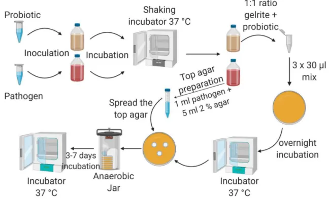
The basic agar disc-diffusion method has been developed in 1940 (Balouiri et al. 2016). In this, well-known proce- dure plates are inoculated with a single test microorgan- ism in a lawn. Then, paper discs, containing the test com- pounds/microbes, are placed on the agar surface. During incubation, the test compound, a potential antimicrobial agent, diffuses into the agar plate and inhibits the growth of the test microorganism. In the end, the zones of inhibi- tion are measured (Balouiri et al. 2016). In our case, the plates were inoculated with probiotic microbes, and the top agar with the pathogenic strain was spread onto it.
During the incubation, the antimicrobial agent produced by the probiotic culture, diffuses into the top agar and in optimal case, interferes with the growth of the pathogen, which appears as an inhibition zone in the top agar. The experimental workflow consisted of 5 steps (Fig. 1): inoc- ulation, preparation, top agar formation, incubation and evaluation.
Experimental set‑up Inoculation
Pure single colonies of test bacteria were inoculated into 20 ml of anaerobic medium (2722 or 58) one day before application, except Pr. buccae, which needs two days to grow up. The anaerobic, sealed bottles were placed on a shaking incubator (37 °C, 160 rpm) overnight.
Preparation
The fresh probiotic cultures (OD600 was between 0.5–0.7 / cm in case of medium #2722 and between 0.6 and 0.8 /cm in case of medium #58) were mixed with gelrite (Sigma- Aldrich) in the volumetric ratio of 1:1. Gelrite is a trans- parent biopolymer, which forms a gel in the presence of cations at room temperature (Shungu et al. 1983). The gel- rite solute ion is made from 0.04 g of gelrite powder and 10.00 ml of triply distilled water and sterilized by passing through a 0.2 µm pore size syringe filter. Each drop had
30 µl volume; one drop contained 15 µl gelrite and 15 µl of fresh probiotic culture. Three drops were placed on each Petri dish. After the drops solidified on the plates, they were placed to 37 °C in an incubator overnight. The initial cell concentration (living cell/ml) of the used probiotic cultures during preparation was determined with CFU (colony forming unit).
Top agar layer
For the top agar, 5 ml media (2722 or 58) containing 2% agar were mixed with 1 ml of pathogenic culture (OD600 was between 0.5 and 1.0 /cm, which is about 1–4 × 10⁸ cell/ml in case of medium #2722 and between 0.5 and 1.2 /cm, which is about 3–6 × 10⁸ cell/ml in case of medium #58) a sterile centrifuge tube and were spread on the agar plates having the probiotic spots. Control plates were prepared without the probiotic strains, only with top agar containing the pathogenic bacteria.
Fig. 1 The workflow of the experiments. The cultures were inocu- lated a day prior application (except Pr. buccae). First, the test pro- biotic strain (0.5–1 ml) was inoculated to the medium (20 ml) and was placed on a shaking incubator (37 °C, 160 rpm) overnight. Next day, the plates were prepared. The probiotic culture was mixed with gelrite in the ratio of 1:1, and then, 3 drops of mixture were placed on the plate. One drop had 30 µl volume (15 µl probiotic culture and 15 µl gelrite). Simultaneously with the preparation, CFU from the used probiotic culture was made to determine the living cell concen- tration of the culture (the figure does not show this step). The plates
were incubated overnight (37 °C). Test pathogen was inoculated on the medium and incubated overnight on a shaking incubator (37 °C, 160 rpm). Next day, the top agar layer was poured on the prepared plates (plates with probiotic drops, after the overnight incubation).
During the top agar preparation, 1 ml pathogenic culture and 5 ml 2% agar were used; it was stirred in a 15 ml sterile Falcon tube, and poured on the prepared plate. Finally, test plates were placed in an anaerobic jar and put into an incubator (37 °C) for 3–7 days. After incubation, plates were evaluated; the zones of inhibition were meas- ured and averaged
Incubation
The plates were placed into anaerobic jars (Thermo Fisher Scientific) with an anaerobic atmosphere generation bag (Thermo Fisher Scientific), and they were put into an incu- bator at 37 °C. The incubation lasted from 3 to 7 days.
Evaluation
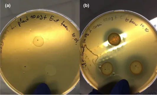
The zone of inhibition is the area where no growth is visible to the unaided eye (Fig. 2). The distance from the edge of the probiotic drop to the distant edge of the zone was meas- ured. Three probiotic drops were placed on each plate; the size of zones was measured and averaged. Zones less than 1 mm were recorded as 0.5 mm; complete inhibition reached 15-mm-wide zones under these experimental conditions.
Statistical analysis
In the figures, we show the data’s mean values, with their standard deviations. Statistical analyses were performed with SigmaPlot 14.0 software (Systat Software Inc., Erkeath, Germany) using one-way analysis of variance (ANOVA) followed by Duncan’s multiple range test (DMRT) at 5%
significance level.
Results and discussion
Two experimental series were studied on two growth media:
DSMZ 58 and ATCC 2722. The total number of the indi- vidual plate tests using medium 58 and 2722 was 63 and 70, respectively (Table 1). In all cases, we had at least three averaged readings.
We can evaluate the results from two aspects, i.e. the sensitivity of the pathogens and the effectiveness of the probiotics.
The percentages of plates displaying inhibition zone over total number of plates indicate the sensitivity of pathogens (Table). In this context, a pathogen is considered sensitive if > 80% of the tested plates showed inhibition zone. The number of effective probiotic strains is proportional to the non-specific sensitivity of the pathogenic strain in question (Table 2).
According to the data, the most sensitive bacteria were Pr. buccae, P. gingivalis and S. oralis, while the most resist- ant was E. faecalis, since E. faecalis is a multidrug-resistant bacterium (Kouidhi et al. 2011). F. nucleatum, A. actinomy- cetemcomitans, S. oralis and Pr. buccae were inhibited by all probiotic strains, while E. faecalis was inhibited only by
Fig. 2 a Pathogenic growth in the top agar layer without inhibition zone b Pathogenic growth in the top agar layer with inhibition zone
Table 1 Effects of the media
Two types of media were used, ATCC 2722 and DSMZ 58. The
“plates grown” means the plates where pathogens were grown suc- cessfully. In regular case 72 types of experiments (8 pathogens and 9 probiotics mean 72 interactions (8*9)) were prepared on plate #2722 and on #58 as well. P. gingivalis could not grow in two cases on plate
#2722, so it means only 70 interactions, P. gingivalis with L. casei and P. gingivalis with Florabalance Plus interactions were not inves- tigated. In medium #58, P. gingivalis could not grow, which means 9 interactions (P. gingivalis and the 9 probiotics) could not be inves- tigated. “Successful interaction” indicates plates, where both visible growth of the pathogenic strain and an inhibition zone were observed.
“Average zone” was referred to the average size of all inhibition in mm
Medium Plates grown Successful
interaction (%) Average zone (mm)
2722 70 33 47.1 1.9 ± 1.6
58 63 53 84.1 8.8 ± 4.8
L. rhamnosus, L. plantarum, L. acidophilus, L. casei and the Florabalance Plus.
During preparation, the initial cell number of the probi- otic cultures was determined before using them with CFU.
During the evaluation, we used these CFU data to compare the effectiveness of the different probiotic bacteria (not all data showed).
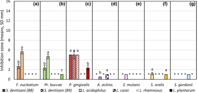
In Fig. 3, we present the results of plates #2722 (not all data showed). The maximum value was under 6 mm. S.
dentisani (89) was the most effective. Camelo-Castilo et al.
isolated two novel strains, which could inhibit the growth of S. mutans and named them S. dentisani (Camelo-Cas- tillo et al. 2014). Similar results were reported in a sepa- rate study, where S. dentisani inhibited the growth of S.
mutans and killed F. nucleatum (López-López et al. 2017).
Others added A. actinomycetemcomitans to the list of oral pathogens inhibited by S. dentisani (Conrads et al. 2019).
Our results corroborate these observations.
In Fig. 4, the results are presented in case of 1–2.5 × 10⁸ living cell/ml initial concentration of the pro- biotic cultures on plates #58. Results of L. plantarum can be compared on Fig. 3 to Fig. 4. On plate #2722 and #58 L. plantarum caused inhibition in case of Pr. buccae and S. oralis as well and the initial probiotic concentration was similar (1.5–5 × 10⁸ cell/ml vs. 1–2.5 × 10⁸ cell/ml).
But the difference is well marked between the two media types. On #2722 L. plantarum caused only 1 mm zones in
Table 2 Sensitivity of the pathogens
F. n.—F. nucleatum, P. g.—P. gingivalis, A. a.—A. actinomycetemcomitans, S. m.—S. mutans, S. o.—S.
oralis, S. g.—S. gordonii, E. f.—E. faecalis, Pr. b.—Pr. buccae. In regular case, all pathogenic strain had 18 interactions (interactions with 9 probiotics on the plates #2722, and another 9 on plates #58). “Plates grown” means the interactions where the pathogens could grow in the top agar. P. gingivalis could not grow in medium #58 and was not able to grow in two cases in medium #2722. “Successful interaction”
indicates, where both visible growth of the pathogenic strain and an inhibition zone were observed. “Sen- sitivity” is the percentage of “Plates grown” and “Successful interaction”. Pr. buccae, P. gingivalis and S.
oralis were the most sensitive, and E. faecalis was the most resistant in this the experimental arrangement.
Outstanding sensitive strains are italicized, and insensitive is marked with bold in Table 2
F. n P. g A. a S. m S. o S. g E. f Pr. b
Plates grown 18 7 18 18 18 18 18 18
Successful interaction 11 6 14 9 15 10 5 16
Sensitivity (%) 61.1 85.7 77.7 50 83.3 55.5 27.2 88.8
(a) (b) (c) (d) (e) (f) (g)
Fig. 3 Effectiveness of the probiotics on plate #2722 in case of 1.5–5 × 10⁸ cell/ml initial probiotic concentration. Orange marks F. nucleatum a, light green is Pr. buccae b, dark red is P. gingivalis c, purple is A. actinomycetemcomitans d, lavender is S. mutans e, gold is S. oralis f, and light blue is S. gordonii g. Trellis pattern- filled column marks inhibition zones caused by S. dentisani (DSMZ 27088) strain, downward diagonal stripes pattern is S. dentisani
(DSMZ 27089), zigzag pattern is L. acidophilus, checker board pat- tern is L. casei, upward diagonal stripes pattern is L. rhamnosus, and solid-filled columns marks inhibitions caused by L. plantarum.
Whiskers on the columns mark the standard deviations. Different lower-case letters indicate statistical differences among treatments (n = 3, p ≤ 0.05), *means data not shown. F(A) (1, 4) = 40,500; F(B)(2, 6) = 46,500; F(C)(3, 8) = 16,000; F(D)(1, 4) = > 1e40; F(F)(1, 4) = 1000
case of Pr. buccae and S. oralis. On the contrary, on #58 L. plantarum caused complete inhibition in both cases.
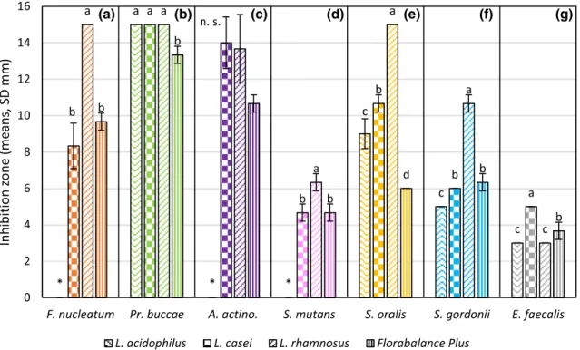
Figure 5 shows the result of plates #58 in case of 8.7 × 10⁸–3 × 10⁹ cell/ml initial concentration of the used probiotic cultures. The effectiveness of few different probi- otics can be compared, like L. acidophilus, L. casei, L. rham- nosus and the Florabalance Plus. L. rhamnosus was the most effective against F. nucleatum and the pathogenic Strepto- coccus strains. L. casei was the most effective against A.
actinomycetemcomitans and E. faecalis. Pr. buccae was the most sensitive pathogenic bacterium, which was inhibited completely by L. acidophilus, L. casei and L. rhamnosus. S.
mutans and E. faecalis were the most resistant as can be seen in Fig. 5. According to other studies, L. casei and L. rhamno- sus could inhibit the growth of S. mutans, and L. rhamnosus and L. plantarum were effective against A. actinomycet- emcomitans and P. gingivalis (Badet and Thebaud 2008).
These findings are in line with our results. Florabalance Plus was quite effective against all of the test pathogens. Flora- balance Plus could be a good basis for an effective probiotic mixture against oral pathogens. An improved Florabalance Plus could also be developed into a product family targeted against oral pathogens predominating specific dysbiotic microbiota causing various oral diseases.
We can speculate about the mechanism of the probiotic inhibition as follows. Three mechanisms could be consid- ered: 1. competition for binding site and nutrients, 2. produc- tion of antimicrobial agents, bacteriocins and 3. alerting the response of the host immune system. The experiments were done in vitro, which precludes the last possibility. Under the employed experimental arrangements, the probiotic bacteria were fixed in gelrite and the pathogens were grown in the top agar. Consequently, there was no chance for competi- tion between the two bacteria on the plate. Therefore, the most likely option to explain the observed inhibitory effect invokes the antimicrobial substances, which could freely dif- fuse in the agar surrounding the probiotic drop and reach the pathogens to inhibit the growth of the pathogens. The size of the inhibition zone should be proportional to the amount and biological activity of the excreted bacteriocins.
We noted substantial differences between the two growth media used to cultivate the bacteria. This points at the need for thoroughly defined, standardized experi- mental conditions to be maintained in such studies in order to allow a straightforward comparison of the data.
In our case, medium #58 gave clearer and larger inhibition zones than medium #2722 did. The exceptions were the two S. dentisani strains where there was no pronounced
(a) (b) (c) (d) (e) (f) (g)
Fig. 4 Effectiveness of the probiotics on plate #58 in case of 1–2.5 × 10⁸ cell/ml initial probiotic concentration. Orange marks F.
nucleatum a, light green is Pr. buccae b, purple is A. actinomycetem- comitans c, lavender is S. mutans d, gold is S. oralis e, light blue is S. gordonii f, and grey is E. faecalis g. Sphere pattern-filled column marks inhibition zone caused by L. delbrueckii and solid-filled col-
umn marks the effect of L. plantarum. Whiskers on the columns mark the standard deviations. Different lower-case letters indicate statisti- cal differences among treatments (n = 3, p ≤ 0.05), *means data not shown, n.s. means not significant. F(B)(1, 4) = 1000; F(E)(1, 4) = 1000;
F(F)(1, 4) = 1,250
difference in either medium, and apparently S. dentisani performed better medium #2722 than on #58.
A major difference between the two media is in the Tween-80, which is absent from medium 2722. Tween-80, also known as polyoxyethylene (20) sorbitan monooleate, is a non-ionic surfactant and emulsifier, which is often used in foods, cosmetics and pharmacology as additive.
Tween-80 has been found to have beneficial effect on lac- tobacilli growth. Some Lactobacillus species need deter- gents like Tween-80 or Tween-20 which supports growth as fatty acid source (Reitermayer et al. 2018). It is relevant to recall that under adequate conditions Tween-80 elevated bacteriocin production of L. cremoris by about fourfold relative to the control medium (Huot and Petitdemange 1996). The detergent Tween-80 also promotes bacteriocin detachment from the probiotic cell wall (Md Sidek et al.
2018; Reitermayer et al. 2018).
The other important difference between the two media is in their glucose content, which is one fourth in medium
#2722 compared to #58. The high sugar content could result in high acid production. Acids are antimicrobial agents themselves, which may supplement the bacteriocin effect. According to a new study, acids have other effects as well, more recently, a facilitated release of bacteriocins
from the L. plantarum cells at lower pH (De Giani et al.
2019).
In the future studies, composite mixtures of probiotics should be tested to elicit comprehensive probiotic effect in the complex oral microbial community. Specific interactions among the various strains may alter the effective biological activity of probiotic mixture preparations (Jeong and Moon 2015; Conrads et al. 2019).
Conclusions for future biology
In the tests of individual probiotic candidates, at least 5 probiotic strains effectively inhibited the growth of each selected pathogenic strains. Successful inhibitions were also observed in case of the multidrug-resistant E. faecalis and the acid-tolerating S. mutans. It is noteworthy that substan- tial differences were observed between probiotic-pathogenic pairs depending on the composition of the growth medium.
This emphasized the importance of using standardized con- ditions in these experiments. The findings corroborate that a rational management of oral pathogens by properly selected, well-defined, synthetic probiotic communities is feasible.
In this study, we tested potential probiotic bacterial strains
(a) (b) (c) (d) (e) (f) (g)
Fig. 5 Effectiveness of the probiotics on plate #58 in case of 8.7 × 10⁸–3 × 10⁹ cell/ml initial probiotic concentration. Orange marks F. nucleatum a, light green is Pr. buccae b, purple is A. actinomyce- temcomitans c, lavender is S. mutans d, gold is S. oralis e, light blue is S. gordonii f, and grey is E. faecalis g. Zigzag pattern-filled column marks inhibition caused by L. acidophilus strain checker board pat- tern is L. casei, upward diagonal stripes pattern is L. rhamnosus and
vertical stripes pattern column marks the effect of the Florabalance Plus. Whiskers on the columns mark the standard deviations. Dif- ferent lower-case letters indicate statistical differences among treat- ments (n = 3, p ≤ 0.05), *means data not shown, n.s. means not sig- nificant. F(A)(2, 6) = 42,000; F(B)(3, 8) = 25,000; F(C)(2, 6) = 3500;
F(D)(2, 6) = 8,333; F(E)(3, 8) = 127,000; F(F)(3, 8) = 113,333; F(G)(3, 8) = 32,000
for their ability of controlling the growth of oral pathogens and established an experimental set-up and collected useful information for further development of efficient probiotic composite microbial communities. However, we need more experiments in the future, including clinical experiments.
Our results indicate that the probiotics can inhibit the growth of the studied pathogenic strains, so we could use them for future therapeutic treatments in odontology.
Acknowledgements We thank University of Szeged, Institute of Clini- cal Microbiology and Goodwill Pharma Ltd. for providing strains and the probiotic preparation for this study.
Author Contributions NG, with the help from OS, designed and per- formed the experiments and contributed to the evaluation of the data.
KLK conceived the project and participated in its design. NG, OS, ZB and KLK drafted the manuscript. All the authors have read and approved the final manuscript.
Funding This study has been supported in part by the Hungarian National Research, Development and Innovation Fund projects GINOP- 2.3.2-15-2016-00011, GINOP-2.2.1-15-2017-00081, GINOP-2.2.1-15- 2017-00033 and EFOP-3.6.2-16-2017-00010. ZB received support from the Hungarian NKFIH fund FK123902.
Declarations
Conflict of interest The authors declare that the research was con- ducted in the absence of any commercial or financial relationships that could be construed as a potential conflict of interest.
References
Anderson AC, Jonas D, Huber I, Karygianni L, Wölber J, Hellwig E, Arweiler N, Vach K, Wittmer A, Al-Ahmad A (2016) Enterococ- cus faecalis from food, clinical specimens, and oral sites: Preva- lence of virulence factors in association with biofilm formation.
Front Microbiol. https:// doi. org/ 10. 3389/ fmicb. 2015. 01534 Andrade E, dos Santos Pires AC, Soares M, Jan G, de Carvalho AF
(2012) Probiotics in dairy fermented products. Probiotics. https://
doi. org/ 10. 5772/ 51939
Anukam KC, Reid G (2007) Probiotics: 100 years (1907-2007) after Elie Metchnikoff ’s observation. Commun Curr Reserach Educ Top Trends Appl Microbiol 466–474
Badet C, Thebaud NB (2008) Ecology of lactobacilli in the oral cavity:
a review of literature. Open Microbiol J 2:38–48. https:// doi. org/
10. 2174/ 18742 85800 80201 0038
Balouiri M, Sadiki M, Ibnsouda SK (2016) Methods for in vitro evalu- ating antimicrobial activity: a review. J Pharm Anal 6:71–79 Banas JA, Fountain TL, Mazurkiewicz JE, Sun K, Margaret Vickerman
M (2007) Streptococcus mutans glucan-binding protein-A affects Streptococcus gordonii biofilm architecture. FEMS Microbiol Lett 267:80–88. https:// doi. org/ 10. 1111/j. 1574- 6968. 2006. 00557.x Barefoot SF, Klaenhammer TR (1984) Purification and characteriza-
tion of the Lactobacillus acidophilus bacteriocin lactacin B. Anti- microb Agents Chemother 26:328–334. https:// doi. org/ 10. 1128/
AAC. 26.3. 328
Berger D, Rakhamimova A, Pollack A, Loewy Z (2018) Oral biofilms:
development, control, and analysis. High-Throughput 7:1–8.
https:// doi. org/ 10. 3390/ ht703 0024
Borsanelli AC, Gaetti-Jardim E, Schweitzer CM, Viora L, Busin V, Riggio MP, Dutra IS (2017) Black-pigmented anaerobic bacteria associated with ovine periodontitis. Vet Microbiol 203:271–274.
https:// doi. org/ 10. 1016/j. vetmic. 2017. 03. 032
Bostanci N, Belibasakis GN (2012) Porphyromonas gingivalis: an inva- sive and evasive opportunistic oral pathogen. FEMS Microbiol Lett 333:1–9
Bowen WH (2016) Dental caries—not just holes in teeth! A perspec- tive. Mol Oral Microbiol 31:228–233
Brennan CA, Garrett WS (2019) Fusobacterium nucleatum—symbiont, opportunist and oncobacterium. Nat Rev Microbiol 17:156–166 Brown SA, Whiteley M (2007) A novel exclusion mechanism for
carbon resource partitioning in Aggregatibacter actinomycetem- comitans. J Bacteriol 189:6407–6414. https:// doi. org/ 10. 1128/ JB.
00554- 07
Camelo-Castillo A, Benítez-Páez A, Belda-Ferre P, Cabrera-Rubio R, Mira A (2014) Streptococcus dentisani sp. nov., a novel member of the mitis group. Int J Syst Evol Microbiol 64:60–65. https://
doi. org/ 10. 1099/ ijs.0. 054098-0
Clarke JK (1924) On the bacterial factor in the etiology of dental caries.
Br J Exp Pathol 5:141
Cobo F, Rodríguez-Granger J, Sampedro A, Navarro-Marí JM (2017) Infected breast cyst due to Prevotella buccae resistant to metro- nidazole. Anaerobe 48:177–178. https:// doi. org/ 10. 1016/j. anaer obe. 2017. 08. 015
Conrads G, Westenberger J, Lürkens M, Abdelbary MMH (2019) Iso- lation and bacteriocin-related typing of Streptococcus dentisani.
Front Cell Infect Microbiol 9:1–11. https:// doi. org/ 10. 3389/ fcimb.
2019. 00110
Cotter PD, Ross RP, Hill C (2013) Bacteriocins-a viable alternative to antibiotics? Nat Rev Microbiol 11:95–105
da Silva-Sabo S, Vitolo M, González JMD, de Oliveira RPS (2014) Overview of Lactobacillus plantarum as a promising bacteriocin producer among lactic acid bacteria. Food Res Int 64:527–536 Dadon Z, Cohen A, Szterenlicht YM, Assous MV, Barzilay Y, Raveh-
Brawer D, Yinnon AM, Munter G (2017) Spondylodiskitis and endocarditis due to Streptococcus gordonii. Ann Clin Microbiol Antimicrob 16:4–7. https:// doi. org/ 10. 1186/ s12941- 017- 0243-8 De Giani A, Bovio F, Forcella M, Fusi P, Sello G, Di Gennaro P (2019)
Identification of a bacteriocin-like compound from Lactobacillus plantarum with antimicrobial activity and effects on normal and cancerogenic human intestinal cells. AMB Express. https:// doi.
org/ 10. 1186/ s13568- 019- 0813-6
Dige I, Nyengaard JR, Kilian M, Nyvad B (2009) Application of ste- reological principles for quantification of bacteria in intact dental biofilms. Oral Microbiol Immunol 24:69–75. https:// doi. org/ 10.
1111/j. 1399- 302X. 2008. 00482.x
Distel JW, Hatton JF, Gillespie MJ (2002) Biofilm formation in medi- cated root canals. J Endod 28:689–693. https:// doi. org/ 10. 1097/
00004 770- 20021 0000- 00003
Do T, Jolley KA, Maiden CJ, Gilbert SC, Clark D, Wade WG, Beighton D (2009) Population structure of Streptococcus oralis. Microbi- ology 155:2593–2602. https:// doi. org/ 10. 1099/ mic.0. 027284-0 Dobson A, Cotter PD, Paul Ross R, Hill C (2012) Bacteriocin produc-
tion: a probiotic trait? Appl Environ Microbiol 78:1–6
Ebbers M, Lübcke PM, Volzke J, Kriebel K, Hieke C, Engelmann R, Lang H, Kreikemeyer B, Müller-Hilke B (2018) Interplay between P. gingivalis, F. nucleatum and A. actinomycetemcomitans in murine alveolar bone loss, arthritis onset and progression. Sci Rep 8:1–10. https:// doi. org/ 10. 1038/ s41598- 018- 33129-z Et P, Mpa M (2014) JBR J Interdiscip Enterococcus faecalis Oral Infect
3:1–5
Fine DH, Markowitz K, Furgang D, Velliyagounder K (2010) Aggre- gatibacter actinomycetemcomitans as an early colonizer of oral tissues: Epithelium as a reservoir? J Clin Microbiol 48:4464–
4473. https:// doi. org/ 10. 1128/ JCM. 00964- 10
Fine DH, Patil AG, Velusamy SK (2019) Aggregatibacter actino- mycetemcomitans (Aa) under the Radar: Myths and misunder- standings of AA and its role in aggressive periodontitis. Front Immunol 10
Fine HD, Karched M, Furgang D, Sampathkumar V, Velusamy S, Godboley D (2015) Colonization and presistence of labeled and foreign strains of Aa inoculated into the mouths of rhesus monkeys. J Oral Biol 136:554–561. https:// doi. org/ 10. 1016/j.
ygyno. 2014. 12. 035. Pharm acolo gic
Forssten SD, Björklund M, Ouwehand AC (2010) Streptococcus mutans, caries and simulation models. Nutrients 2:290–298 Gaspar C, Donders GG, Palmeira-de-Oliveira R, Queiroz JA, Tomaz
C, Martinez-de-Oliveira J, Palmeira-de-Oliveira A (2018) Bacteriocin production of the probiotic Lactobacillus acido- philus KS400. AMB Express 8:153. https:// doi. org/ 10. 1186/
s13568- 018- 0679-z
Hajishengallis G, Lamont RJ (2014) Breaking bad: manipulation of the host response by Porphyromonas gingivalis. Eur J Immunol 44:328–338. https:// doi. org/ 10. 1002/ eji. 20134 4202
Han YW (2015) Fusobacterium nucleatum: a commensal-turned pathogen. Curr Opin Microbiol 23:141–147
Hata S, Mayanagi H (2003) Acid diffusion through extracellular polysaccharides produced by various mutants of Streptococcus mutans. Arch Oral Biol 48:431–438. https:// doi. org/ 10. 1016/
S0003- 9969(03) 00032-3
Holt S, Kesavalu L, Walker S, Genco CA (1999) Virulence factors of P.gingivalis. Periodontol 2000(20):168–238
How KY, Song KP, Chan KG (2016) Porphyromonas gingivalis:
an overview of periodontopathic pathogen below the gum line.
Front Microbiol 7:1–14
Huot E, Petitdemange H (1996) Tween 80 effect on bacteriocin syn- thesis by Lactococcus. Lett Appl Microbiol 22:307–310 Jakubovics NS, Palmer RJ Jr (2013) Oral Microbial ecology cur-
rent research and new perspectives. Caister Academic Press, Norfolk, UK
Jeong YJ, Moon GS (2015) Antilisterial bacteriocin from Lactobacil- lus rhamnosus CJNU 0519 presenting a narrow antimicrobial spectrum. Korean J Food Sci Anim Resour 35:137–142. https://
doi. org/ 10. 5851/ kosfa. 2015. 35.1. 137
Karayasheva D, Radeva E (2017) Importance of enterococci (Entero- coccus faecalis) for dental medicine ? Microbiological charac- terization, prevalence and resistance. Int J Sci Res 6:1970–1973.
https:// doi. org/ 10. 21275/ art20 175821
Kato H, Taguchi Y, Tominaga K, Umeda M, Tanaka A (2014) Por- phyromonas gingivalis LPS inhibits osteoblastic differentiation and promotes pro-inflammatory cytokine production in human periodontal ligament stem cells. Arch Oral Biol 59:167–175.
https:// doi. org/ 10. 1016/j. archo ralbio. 2013. 11. 008
Kim N, Yun M, Oh YJ, Choi HJ (2018) Mind-altering with the gut:
modulation of the gut-brain axis with probiotics. J Microbiol 56:172–182. https:// doi. org/ 10. 1007/ s12275- 018- 8032-4 Kouidhi B, Zmantar T, Mahdouani K, Hentati H, Bakhrouf A (2011)
Antibiotic resistance and adhesion properties of oral Entero- cocci associated to dental caries. BMC Microbiol. https:// doi.
org/ 10. 1186/ 1471- 2180- 11- 155
Krespi YP, Shrime MG, Kacker A (2006) The relationship between oral malodor and volatile sulfur compound-producing bacteria.
Otolaryngol Head Neck Surg 135:671–676
Lemos JA, Burne RA (2008) A model of efficiency: stress tolerance by Streptococcus mutans. Microbiology 154:3247–3255 Lemos JA, Palmer SR, Zeng L, Wen ZT, Kajfasz JK, Freires IA,
Abranches J, Brady LJ (2019) The biology of Streptococcus mutans. Microbiol Spectr 7:1–18. https:// doi. org/ 10. 1128/ micro biols pec. gpp3- 0051- 2018
López-López A, Camelo-Castillo A, Ferrer MD, Simon-Soro Á, Mira A (2017) Health-associated niche inhabitants as oral probiotics:
the case of Streptococcus dentisani. Front Microbiol 8:1–3.
https:// doi. org/ 10. 3389/ fmicb. 2017. 00379
Marcotte H, Lavoie MC (1998) Oral microbial ecology and the role of salivary immunoglobulin A. Microbiol Mol Biol Rev 62:71–109
Martinez FAC, Balciunas EM, Converti A, Cotter PD, de Souza Oliveira RP (2013) Bacteriocin production by Bifidobacterium spp. A Rev Biotechnol Adv 31:482–488. https:// doi. org/ 10.
1016/j. biote chadv. 2013. 01. 010
Md Sidek NL, Halim M, Tan JS, Abbasiliasi S, Mustafa S, Ariff AB (2018) Stability of bacteriocin-like inhibitory substance (BLIS) produced by Pediococcus acidilactici kp10 at different extreme conditions. Biomed Res Int. https:// doi. org/ 10. 1155/
2018/ 59734 84
Metwalli KH, Khan SA, Krom BP, Jabra-Rizk MA (2013) Strep- tococcus mutans, Candida albicans, and the human mouth: a sticky situation. PLoS Pathog. https:// doi. org/ 10. 1371/ journ al.
ppat. 10036 16
Miller K, Treloar T, Guelmann M, Rody WJ, Shaddox LM (2018) Clinical characteristics of localized aggressive periodontitis in primary dentition. J Clin Pediatr Dent 42:95–102. https:// doi.
org/ 10. 17796/ 1053- 4628- 42.2.3
Müller E, Radler F (1993) Caseicin, a bacteriocin from Lactobacillus casei. Folia Microbiol (praha) 38:441–446. https:// doi. org/ 10.
1007/ BF028 14392
Pan H, Guo R, Ju Y, Wang Q, Zhu J, Xie Y, Zheng Y, Li T, Liu Z, Lu L, Li F, Tong B, Xiao L, Xu X, Leung EL-H, Li R, Yang H, Wang J, Zhou H, Jia H, Liu L (2019) A single bacterium restores the microbiome dysbiosis to protect bones from destruction in a rat model of rheumatoid arthritis. Microbiome 7:1–11. https://
doi. org/ 10. 1186/ s40168- 019- 0719-1
Park J, Shokeen B, Haake SK, Lux R (2016) Characterization of Fusobacterium nucleatum ATCC 23726 adhesins involved in strain-specific attachment to Porphyromonas gingivalis. Int J Oral Sci 8:138–144. https:// doi. org/ 10. 1038/ ijos. 2016. 27 Petersen PE, Ogawa H (2012) The global burden of periodontal dis-
ease: Towards integration with chronic disease prevention and control. Periodontology 2000 60(1):15–39. https:// doi. org/ 10.
1111/j. 1600- 0757. 2011. 00425.x
Praveen T, Kotrashetti VS, Pattanshetty S, Hosmani J V, Babji D, Ingalagi P (2018) Detection of different Prevotella species from deep dentinal caries of primary teeth—a culture and biochemi- cal study. J Adv Clin Res Insights 5:65–68. https:// doi. org/ 10.
15713/ ins. jcri. 213
Raja M, Ummer F, Dhivakar CP (2014) Aggregatibacter actinomyce- temcomitans—a tooth killer. J Clin Diagn Res 8:13–17 Reitermayer D, Kafka TA, Lenz CA, Vogel RF (2018) Interrelation
between Tween and the membrane properties and high pressure tolerance of Lactobacillus plantarum. BMC Microbiol 18:1–14.
https:// doi. org/ 10. 1186/ s12866- 018- 1203-y
Shungu D, Valiant M, Tutlane V (1983) GELRITE as an agar substitute in bacteriological media. Appl Environ Microbiol 46:840–845
Simova ED, Beshkova DM, Angelov MP, Dimitrov ZP (2008) Bac- teriocin production by strain Lactobacillus delbrueckii ssp.
bulgaricus BB18 during continuous prefermentation of yogurt starter culture and subsequent batch coagulation of milk. J Ind Microbiol Biotechnol 35:559–567. https:// doi. org/ 10. 1007/
s10295- 008- 0317-x
Ullah N, Wang X, Wu J, Guo Y, Ge H, Li T, Khan S, Li Z, Feng X (2017) Purification and primary characterization of a novel bacteriocin, LiN333, from Lactobacillus casei, an isolate from a Chinese fermented food. LWT Food Sci Technol 84:867–875.
https:// doi. org/ 10. 1016/j. lwt. 2017. 04. 056
Wang B, Kuramitsu HK (2005) Interactions between oral bacteria:
Inhib Soc 71:354–362. https:// doi. org/ 10. 1128/ AEM. 71.1. 354
Wilkins JC, Beighton D, Homer KA (2003) Effect of acidic pH on expression of surface-associated proteins of Streptococcus ora- lis. Appl Environ Microbiol 69:5290–5296. https:// doi. org/ 10.
1128/ AEM. 69.9. 5290- 5296. 2003
Zambon JJ (1985) Actinobacillus actinomycetemcomitans in human periodontal disease
Zhou B, Zhang D (2018) Antibacterial effects of bacteriocins isolated from Lactobacillus rhamnosus (ATCC 53103) in a rabbit model of knee implant infection. Exp Ther Med 15:2985–2989. https://
doi. org/ 10. 3892/ etm. 2018. 5790