P H Y S I O L O G Y
Reduced MC4R signaling alters nociceptive thresholds associated with red hair
Kathleen C. Robinson1*, Lajos V. Kemény1*, Gillian L. Fell1†, Andrea L. Hermann1,2, Jennifer Allouche1, Weihua Ding3, Ajay Yekkirala4, Jennifer J. Hsiao1, Mack Y. Su1, Nicholas Theodosakis1, Gabor Kozak5,6, Yuichi Takeuchi5,7,8,9, Shiqian Shen3, Antal Berenyi5,8,10,11, Jianren Mao3, Clifford J. Woolf4, David E. Fisher1‡
Humans and mice with natural red hair have elevated basal pain thresholds and an increased sensitivity to opioid analgesics. We investigated the mechanisms responsible for higher nociceptive thresholds in red-haired mice resulting from a loss of melanocortin 1 receptor (MC1R) function and found that the increased thresholds are melanocyte dependent but melanin independent. MC1R loss of function decreases melanocytic proopiomelanocortin transcription and systemic melanocyte-stimulating hormone (MSH) levels in the plasma of red-haired (Mc1re/e) mice. Decreased peripheral -MSH derepresses the central opioid tone mediated by the opioid receptor OPRM1, resulting in increased nociceptive thresholds. We identified MC4R as the MSH-responsive receptor that opposes OPRM1 signaling and the periaqueductal gray area in the brainstem as a central area of opioid/melanocortin an- tagonism. This work highlights the physiologic role of melanocytic MC1R and circulating melanocortins in the regulation of nociception and provides a mechanistic framework for altered opioid signaling and pain sensitivity in red-haired individuals.
INTRODUCTION
Humans and mice with red hair exhibit altered pain thresholds, in- creased nonopioid analgesic requirements, and enhanced responses to opioid analgesics (1–6). Red hair in both species is caused by loss- of-function variant alleles of the melanocortin 1 receptor (MC1R), a Gs-coupled receptor expressed on melanocytes, the pigment- producing cells of the skin (7). MC1R mutant red-haired mice are less sensitive to noxious thermal, mechanical, and chemical stimuli (4). However, the mechanism of this altered nociception has not been determined, prompting us to examine the mechanistic con- nection between MC1R and the modulation of nociception.
MC1R function is suppressed in the Mc1re/e mouse strain because of a frameshift mutation resulting in a premature stop codon. This strain (referred to herein as red-haired mice) has yellow/red hair and recapitulates features of red-haired humans including synthesis of red/blond pheomelanin pigment, inability to tan following UV
exposure, and increased ultraviolet (UV)–associated skin cancer risk (8, 9).
RESULTS
To initially assess nociceptive thresholds in Mc1re/e mice in a blind- ed fashion and to test the role of pigment (versus red-haired genetic background) in nociception, we crossed Mc1re/e mice with an albino strain containing a loss-of-function mutation in the tyrosinase gene (Tyrc/c). In these mice, melanocyte numbers are not altered, but they are unpigmented (fig. S1). Double mutant Mc1re/e;Tyrc/c mice are visually indistinguishable from Tyrc/c mice, as both have white hair.
After “unblinding” the measurements, mice homozygous for the red hair allele (Mc1re/e) exhibited significantly higher nociceptive thresh- olds for both pressure and temperature relative to Mc1rE/E wild-type (WT) mice in the albino genetic background (Tyrc/c) (Fig. 1, A and B). Similarly, increased nociceptive thresholds were observed for Mc1re/e mice compared with Mc1rE/E mice in the non-albino Tyr+/+
C57BL/6J background (Fig. 1, C and D), in agreement with previ- ous observations (4). This finding suggests a pigment-independent role of MC1R in the regulation of nociception.
To determine whether the increased nociceptive thresholds in red- haired mice are caused by MC1R loss of function within melanocytes or in other cell types, we compared three genetically matched (C57BL/6J) mouse models that differ in melanocyte numbers (summarized in table S1). Mice with increased numbers of epidermal melanocytes (K14-SCF) exhibited significantly lower nociceptive thresholds (Fig. 1, E and F), while mice lacking melanocytes (Mitf mi-wh/mi-wh) displayed higher nociceptive thresholds compared with WT mice (Fig. 1, G and H). These findings suggest that the number of epidermal mela- nocytes (independent of MC1R function) can modulate nociceptive thresholds. Next, we crossed red-haired mice with Mitf mi-wh/mi-wh
mice. Because Mitf mi-wh/mi-wh mice completely lack melanocytes, this comparison between “red-haired” Mc1re/e;Mitf mi-wh/mi-wh and
“black-haired” Mc1rE/E;Mitf mi-wh/mi-wh groups tests potential MC1R
1Cutaneous Biology Research Center, Department of Dermatology and MGH Cancer Center, Massachusetts General Hospital, Harvard Medical School, Boston, MA 02129, USA. 2Doctoral School of Clinical Medicine, University of Szeged, Szeged 6720, Hungary.
3MGH Center for Translational Pain Research, Department of Anesthesia, Critical Care and Pain Medicine, Massachusetts General Hospital, Harvard Medical School, Boston, MA 02129, USA. 4FM Kirby Neurobiology Center, Boston Children’s Hospital, and Department of Neurobiology, Harvard Medical School, Boston, MA 02115, USA.
5MTA-SZTE ‘Momentum’ Oscillatory Neuronal Networks Research Group, Department of Physiology, Interdisciplinary Excellence Centre, University of Szeged, Szeged H-6720, Hungary. 6University Neurology Hospital and Hertie Institute for Clinical Brain Research, Tübingen, Germany 7Department of Neuropharmacology, Graduate School of Pharmaceutical Sciences, Nagoya City University, Nagoya 467-8603, Japan.
8Neurocybernetics Excellence Center, University of Szeged, 10 Dom sqr, Szeged 6720, Hungary. 9Department of Physiology, Osaka City University Graduate School of Medicine, 1-4-3, Asahimachi, Abeno-ku, Osaka 545-8585, Japan. 10Neuroscience Institute, New York University, New York City, NY 10016, USA. 11HCEMM-USZ Magne- totherapeutics Research Group, University of Szeged, 10 Dom sqr, Szeged 6720, Hungary.
*These authors contributed equally to this work.
†Present address: Department of Surgery, Brigham and Women’s Hospital, 75 Francis Street, Boston, MA 02115, USA.
‡Corresponding author. Email: dfisher3@mgh.harvard.edu
Copyright © 2021 The Authors, some rights reserved;
exclusive licensee American Association for the Advancement of Science. No claim to original U.S. Government Works. Distributed under a Creative Commons Attribution NonCommercial License 4.0 (CC BY-NC).
on May 11, 2021http://advances.sciencemag.org/Downloaded from
function in nonmelanocytic cells. As shown in Fig. 1 (G and H), genetic absence of melanocytes ablated the ability of MC1R to influ- ence nociceptive thresholds, suggesting that altered thresholds are dependent on MC1R function in melanocytes rather than in other cell types.
One potential modulator of the elevated nociceptive threshold in red-haired mice is -endorphin, a posttranslational cleavage product of proopiomelanocortin (POMC), which is expressed in melano- cytes (10–12). POMC is induced by adenosine 3′,5′-monophosphate (cAMP) in other cell types (13, 14); thus, low cAMP levels in Mc1r
C57BL/6J (1 1)
Mitf
mi-wh/mi-wh
(8)
Mc1r
e/e;Mitf
mi-wh/mi-wh
(14) 0
2 4 6
Threshold pressure (g)
P = 0.0013 P = 0.0385
N.S.
Mechanical nociception D
-Endorphin level in plasma
Mc1r e/e (20)
Mitf mi-wh
(6) C57BL/6J (5)K14-SCF (8) 0
100 200 300 400 500
pg/ml
P < 0.0001 P = 0.0023
P < 0.0001
C
E F
A B
Parental shScr-clone4shScr-clone5shScr-clone8shScr-clone1
1
shMC1R-hairpin1-clone1shMC1R-hairpin5-clone2shMC1R-hairpin3-clone1 1
shMC1R-hairpin5-clone1 0.000 4
0.005 0.010 0.015
Pomc/18S RNA *** *** *** ***
MC1R regulates Pomc
G H
0 2 4 8 12 24 0.000
0.002 0.004 0.006 0.008 0.010
Pomc/18S RNA
P = 0.0103
Time (hours) -MSH regulates Pomc
K14-SCF (1 1)
K14-SCF;
Mc1r e/e (9) 0.000
0.005 0.010 0.015 0.020
Pomc expression
P = 0.069 Epidermal Pomc mRNA I
Mechanical nociception
C57BL/6J (17)K14-SCF (17 0 )
2 4 6
Threshold pressure (g)
P = 0.005
Thermal nociception
C57BL/6J (26)K14-SCF (22) 0
5 10 15
Latency to response (s) P = 0.0094
C57BL/6J (1 1)
Mitf
mi-wh/mi-wh
(7)
Mc1r
e/e;Mitf
mi-wh/mi-wh
(15) 0
5 10 15 20 25
Latency to response (s)
P < 0.0001 P < 0.0001
N.S.
Thermal nociception Mechanical nociception
Mc1r WT (13)
Mc1r e/e (12) 0
2 4 6
Albino (Tyrc/c)
Threshold pressure (g)
P = 0.0019
Thermal nociception
Mc1r WT (13)
Mc1r e/e (12) 0
5 10 15 20
Latency to response (s)
Albino (Tyrc/c) P = 0.0038
Mc1r WT (203)
Mc1r e/e (130) 0
5 10 15 20
Latency to response (s)
Thermal nociception P < 0.0001
Mc1r WT (92)
Mc1r e/e (29) 0
2 4 6
Threshold pressure (g)
Mechanical nociception P < 0.0001
J K L
Fig. 1. Nociceptive thresholds and POMC production are affected by melanocyte status and MC1R signaling. Loss of function of MC1R increases mechanical and thermal nociceptive thresholds in C57BL/6J Tyrc/c albino mice (A and B) and in non-albino C57BL/6J mice (C and D). Nociceptive thresholds are decreased in K14-SCF strain compared with C57BL/6J mice measured by both von Frey assay (E) and hot plate assay (F). Mc1re/e;Mitf mi-wh/mi-wh double mutants had increased thresholds compared with C57BL/6J, but similar to Mitf mi-wh/mi-wh, by von Frey (G) and hot plate assays (H). (I) Levels of -endorphin in blood plasma were assayed by radioimmunoassay, sug- gesting MC1R-dependent melanocytic regulation of POMC. (J) shMc1r in the mouse melanocytic Melan-C cell line decreases Pomc mRNA expression. ***P < 0.001 com- pared with all of the shControl clones and the parental line. (K) The MC1R ligand -MSH (2 M) increases Pomc production in Melan-C cells. (L) Pomc expression is decreased in the epidermal skin of red K14-SCF;Mc1re/e mice compared with black K14-SCF;Mc1rWT mice. Graphs show means ± SEM; numbers in parentheses = n per group. Data shown in (C) and (D) represent compilations of baseline thresholds from all black and red-haired male mice in the C57BL/6J background used in the study.
Results in (E) and (F) are a compilation of two and three independent experiments, respectively. Each group in (J) and (K) consists of three biological replicates. N.S., not significant.
on May 11, 2021http://advances.sciencemag.org/Downloaded from
mutant melanocytes may affect POMC expression. Red-haired (Mc1re/e) mice had significantly lower plasma levels of -endorphin than ge- netically matched black (C57BL/6J WT) mice (Fig. 1I). However, this difference did not suggest that changes in -endorphin mediate altered nociceptive thresholds because the direction of change is opposite from the phenotypic change, since opioid signaling pro- motes rather than diminishes analgesia. Moreover, plasma levels of
-endorphin were also inversely associated with nociceptive thresh- olds in K14-SCF mice (higher melanocyte numbers and lower noci- ceptive thresholds) and Mitf mi-wh/mi-wh mice (absent melanocytes and higher nociceptive thresholds). Collectively, these data demon- strated a consistent pattern in which melanocyte numbers (K14-SCF, Mitf mi-wh/mi-wh) and function (Mc1rE/E, Mc1re/e) were inversely cor- related with nociceptive thresholds despite being directly related to circulating -endorphin levels.
To test for a direct role of melanocytes in modulating POMC expression downstream of MC1R, we knocked down Mc1r mRNA (Fig. 1J) or stimulated MC1R with –melanocyte-stimulating hor- mone (-MSH) (Fig. 1K) in the mouse melanocyte line Melan-C (15) and observed decreased and increased Pomc mRNA produc- tion, respectively. Furthermore, isolated epidermal RNA from black (K14-SCF;Mc1rE/E) and red-haired (K14-SCF;Mc1re/e) mice revealed reduced Pomc expression in epidermal skin of mice harboring loss of function in MC1R (Fig. 1L), similar to the lower plasma levels of circulating -endorphin in the red-haired background (Fig. 1I, top).
Pomc expression did not show statistically significant reduction in the adrenal gland and in the pituitary gland of Mc1re/e mice (fig. S2), suggesting that the plasma level alterations of POMC in this genetic background are mainly due to the decrease in melanocytic Pomc production caused by loss of Mc1r function.
To determine whether the elevated nociceptive thresholds were a result of compensation for or adaptation to the lower -endorphin levels, we used two genetic models: mice carrying homozygous POMC mutation that expresses all melanocortin peptides but lacks the terminal -endorphin sequence (“-endorphin knockout”) and a strain deficient in the -endorphin receptor gene, -opioid receptor (Oprm1−/−). We observed no measurable effect of -endorphin knockout on the nociceptive threshold differences in black versus red-haired mice (Fig. 2, A and B), suggesting that -endorphin ex- pression does not account for the altered nociceptive thresholds, as anticipated because its levels were inversely related to phenotypes.
Oprm1 deletion had no effect on nociceptive thresholds in black mice, in agreement with previous observations (16, 17) and sup- porting the hypothesis that basal nociceptive thresholds in WT C57BL/6 mice are unaffected by endogenous opioids (18, 19). How- ever, the absence of OPRM1 abolished the elevated thresholds in red-haired mice (Fig. 2, C and D). Similarly, naloxone, a broad opi- oid receptor antagonist (20), and cyprodime, an antagonist specific for the -opioid receptor (21), both significantly reduced nocicep- tive thresholds in red-haired mice to levels in black mice or to even lower levels at high doses (Fig. 2, E and F). A decrease below WT levels was observed only with pharmacologic antagonists and may reflect a difference between acute blockade and chronic absence of signaling.
These findings suggest that the elevated nociceptive thresholds in red-haired mice are mediated by, and dependent on, -endorphin–
independent opioid receptor signaling. We also observed a decrease in nociceptive thresholds when Mitf mi-wh/mi-wh mice (devoid of me- lanocytes) were treated with naloxone (Fig. 2G), consistent with a
common endogenous opioid-dependent mechanism for Mitf mi-wh/mi-wh
and red-haired Mc1re/e mice.
We reasoned that higher -opioid receptor signaling in the pres- ence of low plasma levels of -endorphin could be explained by up-regulation of another endogenous opioid, an adaptation of the
-opioid receptor, or reduction of a pathway that antagonizes opi- oid signaling. Direct measurements of the circulating opioid ligands dynorphin, enkephalin, and endomorphin revealed no significant differences between red-haired and black mice (Fig. 3, A to C). Giv- en previously published data (2) on differences between red-haired and black mice in their response to pentazocine, a -opioid receptor agonist, we chose to further examine the receptor agonist dynor- phin. We crossed a dynorphin knockout strain with red-haired and black mice but did not observe measurable effects on nociceptive thresholds in red-haired or black genetic backgrounds (Fig. 3, D and E). These data suggest that elevation of an endogenous opioid is unlikely to be responsible for the increased nociceptive threshold in the red hair background.
Pharmacologic manipulation of the melanocortin pathway an- tagonizes certain effects of morphine (22, 23), leading us to examine plasma levels of MSH (melanocortin agonist), which, like -endorphin, is also encoded within POMC. We observed that plasma -MSH levels varied significantly across mice of different pigmentation pheno- types, paralleling -endorphin levels as anticipated given their com- mon POMC precursor, with higher -MSH levels in mice with more melanocytes or WT MC1R/black pigmentation (Fig. 4A) and lower in mice with fewer melanocytes or red-haired genetic background.
Plasma -MSH levels therefore vary in a manner that is consistent with and might be responsible for the observed nociceptive pheno- types, unlike -endorphin that is inversely related. Given that
-MSH [or its larger parent peptide adrenocorticotropic hormone (ACTH)] represents the only known melanocortin ligand, whereas
-endorphin is one of multiple endogenous ligands capable of acti- vating -opioid receptor signaling, a decrease in both -MSH and
-endorphin might result in a net increase in opioid signaling sus- tained by non–POMC-derived opioid ligands. To functionally evaluate whether proportionally low -MSH may contribute to the elevated nociceptive thresholds in the red hair background, we first rescued low endogenous -MSH levels in red-haired mice pharmacologically.
We observed that an -MSH peptide mimic, melanotan II, de- creased nociceptive thresholds in a dose-dependent fashion in male red-haired Mc1re/e mice but not in black Mc1rE/E mice (Fig. 4B).
Second, given the sex-dependent role of Mc1r in mediating phar- macologic -opioid analgesia (2), we repeated these experiments in female red-haired Mc1re/e mice and observed the same patterns as in male red-haired Mc1re/e mice (fig. S3, A to D). These results sug- gest that loss of function in MC1R signaling results in increased nociceptive thresholds because of melanocortin deficiency in a sex- independent (likely -opioid–independent) manner. As the MSH mimic-induced rescue phenotype occurred in red-haired mice car- rying MC1R deficiency (as a result of an Mc1r frameshift mutation), it suggested that an MSH receptor other than MC1R is the likeliest mediator of the altered nociception (and opioid antagonism).
Another melanocortin receptor, MC4R, and its inhibition have been implicated in pharmacologic analgesia and neuropathic pain (23, 24). We initially tested the peptide SHU 9119, which antagoniz- es MC4R and MC3R while being an agonist for MC1R and MC5R (25). SHU 9119 produced analgesia when injected into male black mice (Fig. 4C), mimicking the effect of the Mc1re/e genotype. Similarly,
on May 11, 2021http://advances.sciencemag.org/Downloaded from
in K14-SCF;Mc1re/e mice, which have levels of POMC products (fig.
S3) and nociceptive thresholds similar to black WT mice (Fig. 4C) but lack MC1R function, injection of SHU 9119 increased nocicep- tive thresholds. These functional results suggested that the analgesic effects of SHU 9119 are Mc1r independent and likely due to ligand
effects on MC4R or MC3R, consistent with the possibility that MC4R or MC3R signaling may balance opioid signaling to modulate noci- ceptive thresholds.
To explore whether MC4R plays a functional role in regulating nociceptive thresholds, we used Mc4r null mice and observed that Thermal nociception
10−3 10−2 10−1 100 101 102 0
5 10 15 20
Cyprodime (log, mg/kg)
Latency to response (s)
Mc1r WT Mc1r e/e Mechanical nociception
Mc1rWT(10) Mc1re/e(5) 0
2 4 6
Threshold pressure (g)
Saline
Naloxone (10 mg/kg) P = 0.0011 N.S.
A
C
E F
0 20 60
0 5 10 15 20
Naloxone (mg/kg)
Latency to response (s)
Thermal nociception in Mitfmi-wh/mi-wh
P = 0.0026 P = 0.0029 G
Mechanical nociception
C57BL/6J (20)-end
−/− (10) Mc1r
e/e (10) Mc1r
e/e; -end
−/− (10) 0
1 2 3 4 5
Threshold pressure (g) N.S.N.S. Thermal nociception
C57BL/6J (25)-end
−/− (13) Mc1r
e/e (10) Mc1r
e/e; -end
−/− (10) 0
5 10 15 20
Latency to response (s)
N.S.
N.S.
C57BL/6J (6)Oprm1
−/− (7) Mc1r
e/e (7)
Mc1r
e/e;Oprm1
−/−(12) 0
1 2 3 4 5
Threshold pressure (g)
Mechanical nociception P < 0.0001 N.S.
C57BL/6J (6)Oprm1
−/− (10) Mc1r
e/e (7)
Mc1r
e/e;Oprm1
−/− (12) 0
5 10 15 20
Latency to response (s)
Thermal nociception P < 0.0001 N.S.
D B
Fig. 2. Elevated nociceptive thresholds in red-haired mice depend on opioid signaling but not -endorphin. -Endorphin deletion has no effect on mechanical (A) or thermal (B) nociceptive thresholds in either black or red-haired mice (compilations of two independent experiments). Deletion of the -opioid receptor did not affect nociceptive thresholds in black mice but abolished the increased thresholds in red-haired mice in both mechanical (C) and thermal (D) nociception assays. (E) Nalox- one treatment (10 mg/kg, intraperitoneally) abolished the increased nociceptive threshold of red mice. (F) Cyprodime treatment showed a dose-dependent effect on thermal nociception in red-haired, but not black, mice (for Mc1rWT, n = 11, except n = 10 for 0.0064 mg/kg; for Mc1re/e, n = 9, except n = 7 for 9.6 mg/kg). (G) Naloxone treatment also decreased nociceptive thresholds in Mitf mi-wh/mi-wh homozygous mice (n = 8 per group). For (A) to (D), the investigator performing the experiment was blinded to the -endorphin or OPRM1 status of each mouse. Graphs show means ± SEM; numbers in parentheses = n per group.
on May 11, 2021http://advances.sciencemag.org/Downloaded from
they exhibit elevated nociceptive thresholds (Fig. 4, D and E). The absence of MC4R (on a genetically black Mc1rE/E background) also resulted in sensitivity to opioid antagonism (Fig. 4F), resembling red-haired Mc1re/e mice and suggesting that the nociceptive thresh- old may be determined by a balance between OPRM1 and MC4R signaling. We also repeated this experiment using naloxonazine, a specific antagonist for the -opioid receptor, and found that phar- macologic inhibition of OPRM1 is sufficient to restore nociceptive thresholds in both Mc4r null and Mc1re/e mice (Fig. 4G). Further- more, melanocortin agonist treatment reduced the elevated noci- ceptive thresholds in red-haired mice (Fig. 4B) but had no effect on nociception in Mc4r null mice (Fig. 4H), suggesting that MC4R is the key melanocortin receptor upon which MSH acts as a ligand for nociceptive threshold reduction.
We hypothesized that if MSH deficiency in Mc1re/e mice is act- ing through MC4R, then Mc1re/e;MC4R−/− mice should have similar nociception to Mc1re/e and MC4R null mice. When we generated Mc1re/e;MC4R−/− mice, we found that Mc1re/e;MC4R−/− mice have similar nociception to Mc1re/e and MC4R−/− mice (Fig. 4I), suggest- ing that there is a similar underlying mechanism responsible for the elevated nociceptive thresholds in these genetic mouse models.
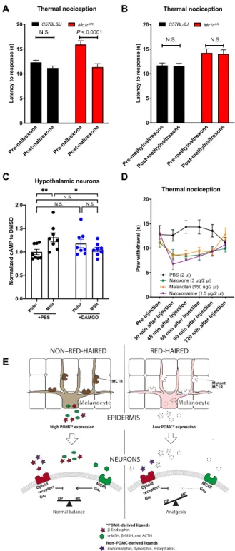
Next, we asked whether the site of the opioid/melanocortin an- tagonism modulating nociception is central or peripheral. The no- ciceptive difference observed between black and red-haired mice was diminished after peripheral (intraperitoneal) administration of naltrexone, an opioid receptor antagonist that is able to cross the blood-brain barrier (BBB) (Fig. 5A). However, peripheral adminis- tration of methylated naltrexone, a BBB-impermeable opioid antagonist,
did not diminish the nociceptive differences between black and red- haired mice (Fig. 5B), suggesting minimal peripheral influence. This suggests that the relative increase in opioid signaling in red-haired mice occurs centrally, not peripherally, and highlights the impor- tance of -MSH in balancing opioid receptor–mediated regulation of central nociception.
Previous studies have demonstrated a role for cAMP signaling in modulating opioid analgesia (26). We therefore measured the im- pact of antagonism between melanocortin and opioid signaling on cAMP content in rat primary hypothalamic neurons (RPHNs). We observed that the melanocortin agonist [Nle4,D-Phe7]--MSH in- creased cAMP content, whereas the opioid agonist morphine sig- nificantly inhibited melanocortin-induced cAMP elevation (Fig. 5C), consistent with previous observations (27). These data suggest that melanocortin and opioid signaling may antagonize each other in a cell-autonomous manner.
Costaining MC4R-GFP (green fluorescent protein) neurons with OPRM1 in mice has suggested the existence of neurons expressing both receptors in the periaqueductal gray area (PAG) (28). First, we measured the mRNA levels of opioid receptors in the PAG in differ- ent genetic backgrounds and found no significant alterations in the opioid receptors in the PAG in Mc1re/e and Mc4r−/ mice (fig. S5).
We next investigated the potential role of the PAG in modulating nociception and found that local antagonism of opioid receptors by naloxone, by the -opioid receptor–specific naloxonazine (29), or agonism of melanocortin receptors (independently of Mc1r) in PAG-cannulated Mc1re/e mice significantly decreases nociceptive thresholds (Fig. 5D). To further investigate the localization of MC4R Dynorphin level in blood
Mc1r WT (10)
Mc1r e/e (9) 0
20 40 60 80
pg/ml
N.S.
Enkephalin level in blood
0 100 200 300 400
Mc1r WT (9)
Mc1r e/e (4)
pg/ml
N.S.
Endomorphin level in blood
Mc1r WT (7)
Mc1r e/e (7) 0
50 100 150
pg/ml
N.S.
A B C
D E Thermal nociception
Mc1r WT (17)
Dyn−/− (9) Mc1r
e/e (7) Dyn−/−;Mc1r
e/e (6) 0
5 10 15 20
Latency to response (s)
N.S.
Mechanical nociception N.S.
Mc1r WT (17)
Dyn−/−
(9) Mc1r
e/e (7) Dyn−/−
;Mc1r e/e (6) 0
2 4 6
Threshold pressure (g) N.S.N.S.
Fig. 3. Low levels of -endorphin are not compensated by other endogenous opioids. (A to C) Plasma levels of dynorphin, enkephalin, and endomorphin are un- changed in red versus black mice. (D and E) Loss of the dynorphin gene (Dyn−/−) has no effect on nociceptive thresholds in red or black mice. Graphs show means ± SEM;
numbers in parentheses = n per group.
on May 11, 2021http://advances.sciencemag.org/Downloaded from
relative to -opioid receptors in the central nervous system (CNS), we analyzed publicly available transcriptomic data (“brain atlas”) from six postmortem human brains (30) and identified multiple ad- ditional regions showing enrichment for both MC4R and OPRM1 (fig. S6), some of which have been associated with the regulation of pain (31–34). We also investigated the possible correlation between MC4R and OPRM1 across brain regions and patients and found a slight positive correlation (figs. S7 and S8) in the posterior group of nuclei; however, these analyses are limited by the small number of available datapoints. Corroborating these human data, we stained
for both OPRM1 and MC4R proteins in rat brains and found costain- ing in several regions corresponding to the human brain regions where we observed coexpression of MC4R and OPRM1 mRNAs (fig. S9). It should be noted that MC4R antibodies have certain lim- itations. We tested multiple commercially available antibodies and used an antibody that showed the best signal-to-noise ratio in the arcuate nucleus (fig. S10). These results are consistent with the pos- sibility that melanocortin and opioid signaling may antagonize each other in a cell-autonomous manner. However, it is also possi- ble that other indirect or non–cell-autonomous mechanisms may
Thermal nociception
Mc1r WT Mc1r e/e 0
5 10 15 20
Latency to response (s)
Saline
Melanotan II (0.01 mg/kg) Melanotan II (0.5 mg/kg)
P = 0.0004 P = 0.0001 N.S.
N.S.
B C
A
Mc1r WT (14) K14-SCF;
Mc1r e/e (6) 0
5 10 15 20
Latency to response (s)
Thermal nociception
Pre-SHU 9119 Post-SHU 9119 P = 0.0005 P = 0.023
N.S.
-MSH level in plasma
Mc1r e/e (17)
Mitf mi-wh (16)
C57BL/6J (21)K14-SCF (28) 0
50 100 150 200
pg/ml
P = 0.0098 P = 0.0003 P = 0.0323
D E F
H
Mechanical nociception
C57BL/6J (1 1)
Mc4r −/− (9) 0
2 4 6
Threshold pressure (g)
P = 0.0021 Thermal nociception
C57BL/6J (1 2)
Mc4r −/− (9) 0
5 10 15 20
Latency to response (s)
P = 0.0075
Thermal nociception
C57BL/6J (8) Mc4r −/− (9)
Mc1r e/e (4) 0
5 10 15 20
Latency to response (s)
Saline
Naloxone (10 mg/kg) P = 0.0046
P < 0.0001
N.S.
P < 0.0001
P < 0.0001
Thermal nociception
C57BL/6J (12)
Mc4r −/− (9) 0
5 10 15 20
Latency to response (s)
Saline
Melanotan II (0.5 mg/kg) P = 0.0068
P = 0.0028 N.S.
N.S.
C57Bl/6J ( n = 10)
Mc4r
−/−(n = 9) Mc1r
e/e (n = 21) 0
5 10 15
Latency to response (s)
Thermal nociception N.S. 0.0002 0.0035
PBSNaloxonazine 5 mg/kg
G
Mc4r wt/wt
Mc4r −/−
Mc4r wt/wt
Mc4r −/−
0 5 10 15
Latency to response (s)
Thermal nociception
Mc1rwt/wt Mc1re/e N.S.
P = 0.0208
I
Fig. 4. MC4R signaling regulates nociception. -MSH in blood plasma was found to be lowest in red-haired mice and Mitf mi-wh/mi-wh mice but elevated in K14-SCF mice (A), suggesting that melanocyte number and function are positively associated with plasma -MSH level. Treatment with a melanocortin agonist, melanotan II (0.5 mg/kg), reduced thermal nociceptive thresholds in red-haired mice (B) (n = 10 per group, except n = 9 for Mc1re/e saline), while nociceptive thresholds are increased by the mela- nocortin antagonist SHU 9119 (1 g/kg) in both black and K14-SCF red-haired mice (C). (D and E) Elevated redhead nociceptive thresholds are mimicked in Mc4r null mice in both mechanical and thermal nociception assays. These elevated thresholds are sensitive to naloxone (F) and naloxonazine (G) but, unlike in red-haired Mc1re/e mice (B), cannot be decreased by melanocortin rescue with melanotan II (H). (I) Elevated nociceptive thresholds are not restored by genetic deletion of Mc4r in Mc1re/e back- ground (n = 10, 9, 20, and 6 per group). Graphs show means ± SEM; numbers in parentheses = n per group.
on May 11, 2021http://advances.sciencemag.org/Downloaded from
additionally contribute to increased opioid tone in red-haired Mc1re/e mice. For example, heterodimerization of G protein–coupled receptors can affect receptor function (35) and OPRM1 has been previously shown to interact with MC3R (36); therefore, it is possible that for- mation of different heterodimers might also contribute to the in- creased opioid tone. However, Mc1re/e;MC4R−/− mice have similar nociceptive thresholds to Mc1re/e mice (Fig. 4I), suggesting that ex- pression of MC4R is not required for maintenance of increased no- ciceptive thresholds in this background. Therefore, the possible involvement of OPRM1/MC4R heterodimerization is unlikely to be solely responsible for the elevated nociceptive thresholds in the red- haired genetic backgrounds.
The data presented here suggest that elevated nociceptive thresh- olds found in the red-haired genetic background arise from a reduc- tion in -MSH levels caused by decreased POMC production in melanocytes, resulting in diminished MC4R signaling. Lower MC4R signaling, in turn, decreases its antagonism of opioid signaling within the CNS, which, despite diminished -endorphin production, ex- hibits no discernible differences in other endogenous opioid ligands in the red hair background. Collectively, this produces a net mela- nocortin deficiency relative to opioid signaling, which alters the bal- ance in favor of -opioid receptor–induced analgesia within the red hair background (illustrated in Fig. 5E).
DISCUSSION
Our observations regarding high nociceptive thresholds in red-haired Mc1re/e mice match previous findings of genotype-driven and sex- i ndependent human and mouse nociceptive patterns (4). Also, it should be noted that despite the occasional minor variability in baseline nociceptive measurements (for example, Fig. 4, E and F), we consistently observed a significant difference between black and red-haired mice, suggesting that the differences are much more ro- bust than the minor fluctuations caused by other environmental/
technical factors. We provide mechanistic insights for these differ- ential nociceptive thresholds by identifying the role of MC1R variant (red-hair) loss of function in altering the balance of physiologic an- tagonism between OPRM1 and MC4R through decreased plasma MSH. However, other mechanisms could also contribute to the ob- served differences. Heterodimerization of OPRM1 with melanocortin receptors (possibly MC3) (36) or additional sex-dependent mecha- nisms could also contribute to the nociceptive differences observed in melanocortin-deficient mice. Alternatively, melanocortin signaling may directly influence nociception by modifying basal OPRM1 re- ceptor activities such as ion channel activities, G protein coupling, or cAMP inhibition. These possibilities warrant future investigations.
A recent study suggests that only a few polymorphisms in the MC1R coding region predict a differential sensitivity to pain (37). It is possible that although these variants would result in decreased MC1R function (as evidenced by red hair), the loss of function may not impair POMC production in these patients or POMC levels might be somehow compensated through other mechanisms. How- ever, it is challenging to study POMC levels in human populations that can significantly differ in UV shielding due to varying levels of pigmentation and that are exposed to various levels of UV radia- tion; therefore, the effect of MC1R coding variants on melanocytic POMC expression requires further in-depth examination. It is also possible that in humans, MC1R might regulate POMC in nonmela- nocytes as well, which also warrants further studies.
B
C A
Pre-methylnaltrexonePost-methylnaltrexonePre-methylnaltrexonePost-methylnaltrexone 0
5 10 15 20
Latency to response (s)
Thermal nociception
N.S. N.S.
C57BL/6J Mc1r e/e
Pre-naltrexonePost-naltrexone Pre-naltrexonePost-naltrexone 0
5 10 15 20
Latency to response (s)
Thermal nociception P < 0.0001 C57BL6/J Mc1r e/e N.S.
D
E
Opioid receptors
Gαi
GαMC4Rs cAMP
α-MSH, β-MSH, and ACTH β-Endorphin OP MC
*POMC-derived ligands
Non–POMC-derived ligands Endomorphin, dynorphin, enkephalins
Opioid receptors
Gαi
GαMC4Rs cAMP
OP MC
EPIDERMIS
NEURONS
NON–RED-HAIRED RED-HAIRED
High POMC* expression Low POMC* expression
MC1R Mutant
MC1R
Melanocyte
Normal balance Analgesia
Melanocyte
Water MSH Water MSH
0.0 0.5 1.0 1.5 2.0
Normalized cAMP to DMSO
Hypothalamic neurons
+DAMGO +PBS
N.S.
N.S.
N.S.
Pre-injection
30 min after injection45 min after injection60 min after injection90 min after injection120 min after injection 0
5 10 15 20
Paw withdrawal (s)
PBS (2 µl) Naloxone (3 µg/2 µl) Melanotan (150 ng/2 µl) Naloxonazine (1.5 µg/2 µl) Thermal nociception
Fig. 5. Increased central opioid tone regulates basal nociception in red-haired mice. Blood-brain barrier (BBB)–impermeable methylnaltrexone (10 mg/kg) can- not reverse increased nociception in red Mc1re/e mice when injected peripherally (A) (n = 20 for all groups), whereas BBB-permeable naltrexone (10 mg/kg) can (B) (n = 20 per group), suggesting the involvement of CNS regions in the increased noci- ception. (C) In line with the coexpression of MC4R and OPRM1, melanocortin and opioid signaling can antagonistically modulate cAMP levels in primary rat hypo- thalamic neurons. DMSO, dimethyl sulfoxide. (D) Local administration of naloxone, naloxonazine, or melanotan to the periaqueductal gray area (PAG) reduces noci- ceptive thresholds in red Mc1re/e mice (n = 9 using the same mice for all treat- ments). (E) Summary of the antagonistic opioid-melanocortin model in black and red-haired contexts. Graphs show means ± SEM; numbers in parentheses = n per group.
on May 11, 2021http://advances.sciencemag.org/Downloaded from
Our study does not distinguish between transcript level–reducing variants and MC1R activity variants, and it is possible that those variants might differentially regulate POMC in mouse and human melanocytes, as hypothesized by Zorina-Lichtenwalter et al. (37).
However, we have demonstrated that the number of melanocytes also regulates basal nociception, suggesting that besides the loss of MC1R function (as in Mc1re/e mice), the absolute number of me- lanocytes is also an important determinant in nociception. The question of whether the MC1R level altering variations that are associated with differential nociception in humans would also phe- nocopy the MC1R function loss in mouse models remains to be explored.
POMC induction causes -endorphin–mediated analgesia after UV exposure (38), whereas in the absence of UV exposure, a reduction in POMC results in melanocortin deficiency–mediated analgesia, as reported here. That both an increase and decrease in POMC can increase analgesia reflects the circumstance wherein a single gene encodes different peptides that activate two opposing pathways. The net effect of POMC depends on the balance of signaling between these two pathways and appears to be affected by differences in basal activities and the presence of other (non–POMC-derived) endoge- nous ligands. In the red hair background, the decrease in MC4R ligands with low POMC is proportionally large (as MC4R has no known ligands beyond POMC-derived MSH/ACTH) relative to -opioid receptor ligands where dynorphin, endomorphin, and enkephalin are unchanged (illustrated in Fig. 5E).
While our study focuses on the red hair phenotype, the underly- ing melanocortin/opioid signaling balance may also account for pain variations among non–red-haired individuals. For example, we ob- served that Mc4r null mice exhibit high nociceptive thresholds like Mc1re/e mice, but are not red haired, reflecting an alternative means to alter the melanocortin-opioid balance. Individuals with MC4R polymorphisms may also have elevated pain thresholds and altered sensitivities to analgesics similar to those reported in red-haired in- dividuals (4). This study has revealed previously unknown and unexpected insights into the molecular and signaling determinants of basal nociceptive thresholds.
MATERIALS AND METHODS
MiceAll experiments were performed with male mice at least 8 weeks of age, with the exception that female mice were used in the experiments of fig. S3. Mice were bred on site. All experiments were performed with approval from the Massachusetts General Hospital Institu- tional Animal Care and Use Committee. All mice used were on the C57BL/6J background or had been backcrossed to the C57BL/6J background at least 10 generations, except for the Mc4r−/− strain, which was developed on the 129S background and backcrossed with C57BL/6J for only one generation. However, Mc4r−/− mice were visually indistinguishable from mice on the C57BL/6J background, making blind experiments possible. Mc1re/e, Tyrc/c, -endorphin−/−, Dynorphin−/−, Mc4r−/−, and Oprm1−/− mice were obtained from The Jackson Laboratory (39, 40, 16, 41–43). The K14-SCF strain was a gift from T. Kunisada (44), whereas Mitf mi-wh/mi-wh mice were a gift from N. Jenkins (45). Mice were used for single experiments, except the same groups of mice were used for measuring thermal and me- chanical nociception and for the effects of different pharmacological compounds.
Drugs
Naloxone hydrochloride (N7758), naltrexone hydrochloride (N3136), and melanotan II acetate salt (M8693) were purchased from Sigma- Aldrich; SHU 9119 (NC9447656), cyprodime hydrochloride (NC9079878), naloxonazine hydrochloride (059110), and DAMGO (11711) were from Thermo Fisher Scientific; and methylnaltrexone bromide (787933) was from McKesson. Morphine was obtained from Patterson Veterinary #078924699. All drug administrations to mice were by intraperitoneal injection.
Nociceptive testing
The hot plate assay was performed as originally described (46) on a 52°C hot plate with a maximum cutoff time of 20 s, except for the experiments using Mitf mi-wh/mi-wh, where a cutoff of 30 s was used to enable detecting additive effects of the incorporation of the Mc1re/e allele. The von Frey assay was performed as described previously (47). Where possible, the investigator was blinded to the genotypes of the mice.
PAG cannulation
For cannulation, mice underwent isoflurane anesthesia. Cannulas were positioned to target the ventral lateral PAG (coordinates used bregma −4.5 mm and lateral 0.5 mm) with a custom-made infusion system (P1 Technologies, VA, USA). Custom-made guide cannulas were cut at 2.2-mm length. For intra-PAG injections, an internal can- nula of 2.5-mm length was used to administer phosphate-buffered saline (PBS), naloxone, or melanotan under isoflurane anesthesia in 2-l volume. Nociceptive thresholds were then measured using the hot plate assay. The same animals were used to test all pharmaco- logic agents for this experiment.
mRNA enrichment analysis
Enrichment analyses of MC4R and OPRM1 in human postmortem brain transcriptomic profiles were performed as described previously (48). Briefly, transcriptomic profiles of six postmortem human brains were downloaded from the Allen Human Brain Atlas (30) in May 2017. To identify brain regions enriched for OPRM1 and MC4R, 190 regions that had transcriptomic profiles from at least four pa- tients were used. We averaged MC4R and OPRM1 expression per region per patient and performed z transformation. The obtained z values indicate the difference in number of SDs for expression in an individual brain region compared with the average expression across all regions. For OPRM1 and MC4R, we considered a z value of ≥1 to be enriched.
Epidermal RNA isolation
Black K14-SCF;Mc1rE/E and red K14-SCF;Mc1re/e mice were eutha- nized by cervical dislocation. Epidermis was isolated from the ears of the animals after dispase digestion for 3 hours at 37°C. Two ears from each mouse were pooled.
Cell culture
Melan-C cells were grown in Ham’s F-10 medium supplemented with 10% fetal bovine serum and 1% penicillin/streptomycin. Cells were serum-starved for various lengths of time before adding 2 M
-MSH (Phoenix Pharmaceuticals Inc., #043-01), and then RNA was harvested from all samples at the same time 25 hours after ini- tiation of serum starvation. RNA was isolated with RNeasy Mini Kit (74104, Qiagen, NE).
on May 11, 2021http://advances.sciencemag.org/Downloaded from
MC1R knockdown and single clone isolation
Lentivirus was generated in 293T cells. Cells were transfected using 250 ng of vesicular stomatitis virus glycoprotein, 1250 ng of the plasmid PsPAX2, and 1250 ng of luciferase short hairpin RNA (shRNA) (control) or Mc1r shRNA. Melan-C cells were plated the day before transduction.
After transduction with lentivirus, plates were centrifuged at 3000 rpm for 30 min at 37°C. Transduced cells were selected with 1:1000 pu- romycin (Thermo Fisher Scientific, catalog no. NC9138068). Single-cell clones were picked up using Scienceware 3.2-mm-diameter cloning discs (Sigma-Aldrich). Mc1r shRNA sequences: shMc1r1, CATC- CCTTCCTGATCTCCATT; shMc1r2, CGTCACTTTCTTTCTAG- CCAT; shMc1r3, CCTGATGGTAAGTGTCAGCAT; shMc1r4, CGTGCTGGAGACTACTATCAT; shMc1r5, GAGAATGTGCT- GGTTGTGATA.
Primers and qRT-PCR
For quantitative reverse transcription polymerase chain reactions (qRT-PCRs), the forward and reverse primers for Pomc were AA- GAGGCTAGAGGTCATCAG and AGAACGCCATCATCAAGAAC, for OPRM1 CAGGGCTTGTCCTTGTAAGAAA and GACTCGG- TAGGCTGTAACTGA, for OPRD ATGGCCTCATGCTACTGCG and CACCAGCGTCCAGACGATG, for OPRK TCCCCAACTG- GGCAGAATC and GACAGCGGTGATGATAACAGG, for 18S AGGTTCTGGCCAACGGTCTAG and CCCTCTATGGGCTC- GAATTTT, for Actb GGCTGTATTCCCCTCCATCG and CCA- GTTGGTAACAATGCCATGT, and for Hprt1 TCAGTCAACGGG GGACATAAA and GGGGCTGTACTGCTTAACCAG. KAPA SYBR FAST One-Step qRT-PCR (KK4651, Kapa Biosystems) was used for qRT-PCR assays. For quantifying Pomc mRNA expression, Pomc levels in ear epidermal samples were normalized to the aver- age of 18S, Atcb, and Hprt1 to minimize variation.
For measuring levels of opioid receptors in the PAG, micro- punches were obtained from freshly isolated PAG tissues. After cer- vical dislocation, brains were removed in ice-cold PBS. Micropunches (1.5 mm wide) were then obtained, and RNA was isolated as previ- ously described (49).
cAMP measurements
RPHNs (Lonza, R-HTH-507) were seeded in poly-d-lysine– and laminin-coated wells at a density of 50,000 cells per well. RPHNs were cultured in primary neuron growth medium (Lonza, CC-3256) containing growth supplements (Lonza, CC-4462). Medium was changed every 48 to 96 hours. On day 11 after seeding cells, serum starvation was performed for 30 min, and then cells were treated first with either 1 M DAMGO or PBS for 7.5 min and then with either 1 M [Nle4,D-Phe7]--MSH (M8764, Sigma-Aldrich) or wa- ter (vehicle control) for 10 min. Cells were incubated at 37°C during treatments. Ten minutes after [Nle4,D-Phe7]--MSH or water treat- ment, cells were lysed and processed for cAMP measurements with cAMP-GLO Assay (V1501, Promega, WI, USA) according to the manufacturer’s instructions.
Radioimmunoassays
Plasma levels of -MSH (RK-043-01), -endorphin (RK-022-06), enkephalin-leucine (RK-024-21), endomorphin-1 (RK-044-10), and dynorphin (RK-021-03) were measured using radioimmunoas- say kits (Phoenix Pharmaceuticals Inc., CA, USA) as described pre- viously (38). The specificities of the kits were determined by the manufacturer.
Histology
All histology experiments were performed in accordance with European Union guidelines (2003/65/CE) and were approved by the Ethical Committee for Animal Research at the Albert Szent-Györgyi Medical and Pharmaceutical Center of the University of Szeged.
MC4R and OPRM1 double immunohistochemistry
Three male Long-Evans rats (450 to 600 g) were anesthetized with 4 to 5% isoflurane followed by urethane (1.5 g/kg) injection (intraperi- toneally) and then transcardially perfused with physiological saline followed by 4% paraformaldehyde and 0.2% picric acid in 0.1 M phosphate buffer (pH 7.2 to 7.3). After removal, brains were post- fixed overnight, embedded in 4% agarose, coronally sectioned at 40-m thickness using a vibrating blade microtome, and harvested in 0.1 M PBS. The following staining procedure was performed at room tem- perature. All incubations were followed by washing with 0.1 M PBS containing 0.3% Triton X-100 (PBS-X). Sections were incubated successively with 10% normal donkey serum (NDS) in PBS-X for 30 min, a mixture of primary antibodies rabbit anti-MC4R at 1:250 (Alomone Labs, catalog no. AMR-024, RRID:AB_2039980) and guinea pig anti-OPRM1 at 1:500 (Abcam, catalog no. ab64746, RRID:AB_
1141103) (50) in PBS-X containing 1% NDS (PBS-XD) for 48 hours, and then a mixture of secondary antibodies Alexa Fluor 555–conjugated donkey anti-rabbit immunoglobulin G (IgG) (2 g/ml) (Thermo Fisher Scientific, catalog no. A-31572) and Alexa Fluor 647–conjugated donkey anti–guinea pig IgG (2 g/ml) (EMD Millipore, catalog no.
AP193SA6) in PBS-XD for 2 hours. Sections were then counterstained with a fluoro-Nissl solution (Thermo Fisher Scientific, catalog no.
N-21479), mounted on gelatin-coated glass slides, and coverslipped with 50% glycerol and 2.5% triethylene diamine in 0.1 M PBS. Cor- pus callosum, caudate putamen, and arcuate nucleus were analyzed as double and single negative controls.
Confocal microscopy
Eight-bit images of stained sections were acquired using a Zeiss LSM880 scanning confocal microscope. Low-magnification images of 10-m optical thickness were acquired using a Plan-Apochromat 20×/0.8 M27 objective lens (Carl Zeiss) with 2.06-s pixel time and four times frame average at 512 × 512 resolution. High-magnification images of 1.2-m optical thickness were acquired using an alpha Plan-Apochromat 63×/1.46 Oil Korr M27 objective lens with 4×
digital zoom, 4.12-s pixel time, and 16 times frame average at 512 × 512 resolution. Z-stack acquisitions were conducted over 10- to 20-m ranges at 1-m steps, each step of which was at the same setting as it is on the 63× objective lens. The power of the lasers was 0.4 to 0.7 mW, which did not induce obvious fading. Acquired raw image files were opened and only linearly processed using a Carl Zeiss software (ZEN Digital Imaging for Light Microscopy, RRID:SCR_
013672), and each image was exported as an eight-bit jpeg file for display.
Statistics
For pairwise comparisons in the nociceptive assays and serum peptide measurements, two-tailed t tests were used. For the cAMP experiment and MSH time course experiment, one-way analysis of variance (ANOVA) was used with Dunnett’s multiple comparison test. For Mc1r knockdown experiments, one-way ANOVA with Tukey’s multiple comparison test was used. For all other experi- ments, two-way ANOVA with Sidak’s multiple comparison test was used. Grubbs test with P = 0.05 was used to identify and remove
on May 11, 2021http://advances.sciencemag.org/Downloaded from