SEMMELWEIS EGYETEM DOKTORI ISKOLA
Ph.D. értekezések
2407.
HADJADJ LEILA
A vérkeringési rendszer normális és kóros működésének mechanizmusai című program
Programvezető: Dr. Benyó Zoltán, egyetemi tanár Témavezető: Dr. Várbíró Szabolcs, egyetemi docens
Effect of hyperandrogenism and vitamin D deficiency on metabolic parameters and coronary artery function
in female rats
PhD thesis
Leila Hadjadj MD
Doctoral School of Basic and Translational Medicine Semmelweis University
Supervisor:
Szabolcs Várbíró, MD, Ph.D, Med. Habil.
Official reviewers:
Zsolt Nagy MD, Ph.D Gergely Gősi MD, Ph.D
Head of the Final Examination Committee:
István Pénzes MD, Ph.D, DSc
Members of the Final Examination Committee:
Ádám László MD, Ph.D, Med. Habil.
Péter Studinger MD, Ph.D
Budapest
202
1
CONTENTS
Abbreviations ... 3
1 Introduction ... 7
1.1 Background ... 7
1.2 Polycystic ovarian syndrome ... 8
1.2.1 Definition ... 8
1.2.2 Diagnosis ... 9
1.2.3 Pathophysiology of PCOS ... 11
1.3 Etiology of PCOS ... 17
1.3.1 Metabolic syndrome ... 18
1.3.2 Insulin resistance ... 20
1.3.2.1 Definition and diagnosis ... 20
1.3.2.2 Metabolic effects of insulin resistance in PCOS ... 22
1.3.2.1 The effects of insulin resistance on the female reproductive system ... 25
1.3.2.2 Hemodynamic consequences of insulin resistance ... 27
1.3.2.3 Molecular effects of insulin resistance in PCOS ... 29
1.3.3 Cardiovascular disease ... 30
1.3.3.1 The role of endothelial dysfunction ... 31
1.4 Vitamin D ... 33
1.4.1 Vitamin D and cardiovascular complications ... 35
1.4.2 Vitamin D and insulin resistance ... 36
1.4.3 Vitamin D and female fertility ... 38
1.5 Evaluation of rodent models for PCOS ... 39
1.6 Connections between the vascular effects of PCOS and vitamin D supply ... 41
2 Aim of the study ... 43
3 Materials and methods ... 44
3.1 Chemicals ... 44
3.2 Animals ... 44
3.3 Chronic treatment ... 45
3.4 Oral glucose tolerance test and homeostatic assessment for insulin resistance ... 46
3.5 Sexual steroid, leptin, and vitamin D plasma levels ... 46
3.6 Vaginal smear examination and ovarian morphology ... 47 3.7 Transthoracic echocardiography and invasive arterial blood pressure measurement . 48
2
3.8 Pressure arteriography of coronary arterioles ... 49
3.9 Biomechanical calculations ... 50
3.10 Histology ... 51
3.11 Immuno-histochemistry of coronary arterioles ... 52
3.12 Statistical analysis ... 52
4 Results ... 53
4.1 Bodyweight, heart weight, and body mass gain ratio ... 53
4.2 Serum hormone and leptin levels ... 53
4.1 Ovarian morphology and estrus cycle ... 55
4.2 Transthoracic echocardiography and blood pressure results ... 58
4.3 Geometry of the coronary arteriole ... 59
4.4 Control of coronary arteriolar smooth muscle tone ... 61
4.1 Elasticity of the coronary arteriolar wall ... 63
4.2 Elastica staining... 65
4.3 OGTT: plasma glucose, insulin and HOMA IR levels... 67
4.4 Insulin-induced relaxation in coronary arterioles ... 69
4.5 Insulin and vitamin D receptor density in coronary arteriolar tissue ... 71
5 Discussion ... 73
5.1 Phenotypical and cardiometabolic changes after transdermal testosterone treatment with and without vitamin D deficiency ... 73
5.2 Biomechanical and pharmacological changes of the coronary arterioles... 76
5.3 Insulin resistance of the coronary arterioles ... 79
6 Conclusions ... 83
7 Summary ... 86
8 Összefoglalás ... 87
9 References ... 88
10 Publications ... 109
11 Acknowledgements ... 111
3
Abbreviations
Acs –Cross-sectional area of the arteriole AE – Androgen excess
Akt – Protein kinase B, serine/threonine-specific protein kinase AMH – Anti-Müllerian hormone
ANOVA – Analysis of variance Ca free – Calcium-free solution CV risk – Cardiovascular risk cJUN – Transcription factor JUN DHT – 5-dihidrotestosterone
DHEAS – Dehydroepiandrosterone sulphate dP – Intraluminal pressure change in the arteriole
dRo – Change in a vessel’s outer radius due to intraluminal pressure change Einc – Incremental elastic modulus of the arteriole
eNOS – Endothelial nitric oxide synthase ET-1 – Endothelin 1
FFA – Free fatty acid
FSH – Follicle-stimulating hormone GnRH – Gonadotropin-releasing hormone h – Wall thickness of the arteriole
HDL-C – High-density lipoprotein cholesterol
HOMA IR – Homeostatic assessment for insulin resistance
4 HSD – Hydroxysteroid dehydrogenase ICAM 1 – Intercellular adhesion molecule 1 IGF-1 – Insulin-like growth factor 1
IGFBP-1 – Insulin-like growth factor 1 binding protein IGT – Impaired glucose tolerance
IR – Insulin resistance
IRS-1 – Insulin receptor substrate 1
IVSD(d) – Thickness of interventricular septum measured in a cross-sectional image at the level of the papillary muscles in diastole
LDL-C – Low-density lipoprotein cholesterol LH – Luteinizing hormone
LogEinc – Incremental elastic modulus of the vessel wall LVAd – Left ventricular area in diastole
LVAs – Left ventricular area in systole
LVIDd – Left ventricular internal diameter in diastole LVIDs – Left ventricular internal diameter in systole LVLd – Left ventricular length in diastole
LVLs – Left ventricular length in systole MAPK – Mitogen-activated protein kinase MetS – Metabolic syndrome
nKR – Normal Krebs solution NO – Nitric oxide
OGTT – Oral glucose tolerance test
5 P – Intraluminal pressure
P450c17alpha - 17-alpha hydroxylase PCOS – Polycystic ovarian syndrome PI3-K – Phosphatidylinositol 3-kinase
RAde – Radius of the arteriole if adenosine was added to the organ chamber Ri – Inner radius of the arteriole
RIns – Radius of the arteriole if insulin was added to the organ chamber Ro – Outer radius of the arteriole
Ru46619 – Radius of the arteriole if thromboxane A2 agonist was added to the organ chamber
RXR – Retinoid X receptor SEM - Standard error of the mean SHBG – Sex hormone-binding globulins T – Testosterone
T1D – Type 1 diabetes mellitus T2D – Type 2 diabetes mellitus
TAde – Adenosin-induced relaxation of the arteriole TFull – Full contraction of the arteriole
Tg stress – Tangential wall stress of the arteriole Tins – Insulin-induced relaxation of the arteriole TnKR – Spontaneous (myogenic) tone of the arteriole U46619 – Potent thromboxane A2 agonist
VCAM-1 – Vascular cell adhesion molecule 1
6 VD – Vitamin D
VDD – Vitamin deficiency VDR – Vitamin D receptor
7
1 Introduction
1.1 Background
Polycystic ovary syndrome (PCOS) is reported to be the most common endocrine disorder affecting women of reproductive age. According to the National Institutes of Health, the prevalence of PCOS is estimated to be 8–12%, but some recent screenings suggest even larger numbers [1].
In addition to androgen excess (AE), oligomenorrhea, infertility, and insulin resistance (IR), PCOS is frequently accompanied by a higher prevalence of metabolic disorders and cardiovascular risk factors. Different PCOS phenotypes differ significantly in their cardiometabolic risk profile depending on the presence and severity of AE. Although PCOS diagnosis and care have improved significantly during the last three decades, longer-time-scale data regarding cardiovascular morbidity, mortality, and the possible beneficial effects of different interventions are still missing.
Although the pathophysiology of PCOS is not fully understood, possible connections between cardiometabolic complications, IR, genetic polymorphisms, and environmental factors have been identified recently. According to some estimations, 30–40% of the world’s population is reported to have IR, and the proportion is even greater (75%) among PCOS women [2]. Normally, female gender has a positive impact on IR, body fat composition, and cardiometabolic risk. This is supported by the fact that estrogen replacement after menopause has beneficial effects on insulin sensitivity and keeps cardiovascular risk low [3]. A higher-than-normal serum concentration of sexual steroids of the opposite gender increases the prevalence of coronary disease, stroke, and hypertension, as proven by data from individuals that underwent female-to-male sex- change interventions [4].
Vitamin D deficiency (VDD) affects approximately 30–50% of the world’s population. Exposure to sunlight or dietary supplementation (fortified foods and dietary
8
supplements, which are widely accepted in the United States) could help achieve sufficient vitamin D (VD) serum levels. Recently, an inverse relationship was found between VD serum levels and the occurrence of coronary heart disease. Previous studies reported that hyperinsulinemia, VDD, and AE coexist in PCOS women [5, 6], and the prevalence of VDD in women with AE is predicted to be 67–85% worldwide.
1.2 Polycystic ovarian syndrome
1.2.1 Definition
The first description of PCOS dates back to 1721, when Vallisneri described a young, moderately obese, and infertile woman with “two larger than normal ovaries, bumpy, shiny, just like pigeon eggs” [7]. In 1935, Stein and Leventhal presented the cases of seven fertile reproductive-aged women with similar endocrine conditions and ovarian morphology. They identified infertility, amenorrhea, hirsutism, enlarged ovaries with multiple cysts, and thickened ovarian tunica as leading symptoms and proposed PCOS as a name for this condition. They also began to perform wedge biopsies on their patients, which was demonstrated to be an effective treatment; later on, 90% reported normal menstruation cycles and 65% became pregnant [8].
During the last two decades, the definition of PCOS has changed and improved.
The first commonly used diagnostic criteria were proposed in 1990 by the National Institutes of Health. This definition regarded oligo-anovulation and any signs of AE (either biochemical or clinical) as sufficient for diagnosis only if other endocrine disorders had already been excluded as the underlying cause. In 2003, the European Society of Human Reproduction and Embryology and the American Society of Reproductive Medicine organized a consensus workshop to refine and reconsider the definition of PCOS. The results were summarized in the Rotterdam criteria, which introduced ultrasound diagnosis of polycystic ovaries as a new provision. It also redefined the terms of AE, changing its description to an excess of androgen activity.
9
Although oligo-anovulation remained a lead criterion, two of the three above-mentioned conditions were sufficient for diagnosis [9].
As the Rotterdam criteria broadened the spectrum of patients and focused more on women with AE, it was widely criticized. Therefore, in 2006, the Androgen Excess PCOS Society discussed the topic again and proposed the currently accepted definition.
Today, an excess of androgen activity, oligo-anovulation and/or polycystic ovaries, and exclusion of other diseases or conditions that could cause an excess of androgen activity are necessary to diagnose PCOS [10]. Based on these criteria, four different phenotypes of PCOS have been described [11, 12]:
The hyperandrogenic form with oligo-anovulation and polycystic ovarian morphology, which is the “classic” PCOS phenotype.
The hyperandrogenic form with oligo-anovulation without relevant signs of polycystic ovarian morphology.
Hyperandrogenism with polycystic ovarian morphology but no relevant ovulation disturbances.
Oligo-anovulation with polycystic ovarian morphology.
1.2.2 Diagnosis
Signs of AE should be judged based upon the presence of hirsutism and elevated levels of circulating androgens, mainly testosterone (T) and its active metabolite, 5- dihidrotestosterone (DHT). In cases of hirsutism, excessive terminal (coarse) hair is found in a male pattern. This differs from hypertrichosis, in which there is no typical pattern and excessive growth of hair follicles all over the body. High levels of circulating androgen enhance cytokine and specific growth factor production and trigger the conversion of T to DHT by 5α-reductase in the pilosebaceous unit, leading less visible vellus hair to turn into terminal hair [12]. The modified Ferriman-Gallwey scale is used for clinical evaluation [13]. In addition, hirsutism shows racial differences;
Caucasian or African women develop it much more frequently than East Asian women [14, 15].
10
Analysis of circulating androgen levels requires sensitive methods, such as liquid chromatography and tandem mass spectrometry, immunochemiluminescence, or radioimmunoassay [16]. One standard method is to use high-quality T and steroid- binding globulin (SHBG) assays, which provide the total T and calculated free T.
Ideally, these tests should be performed in the follicular phase, and reference hormone values should be obtained from women with regular menses [17, 18]. Another candidate hormone for AE measurement is dehydroepiandrosterone, which well represents adrenal hyperandrogenism. Although dehydroepiandrosterone itself is a weak androgen, it serves as a substrate for further androgen synthesis. The serum level of its precursor, dehydroepiandrosterone sulphate (DHEAS), is higher in PCOS women and is associated with elevated prevalence of IR [19]. According to some recent data, 75% of the adult PCOS female population has increased levels of all three biomarkers (T, androstenedione, and DHEAS), but serum T levels are the most frequently tested overall [20].
Anti-Müllerian hormone (AMH) is not an androgen, but a member of the transforming growth factor-β superfamily, which recently became of interest in relation to PCOS. Preantral or small antral follicle release of AMH clearly correlates with their number as well as the secretory activity of granulosa cells. Above all, AMH acts in opposition to follicle-stimulating hormone (FSH) in the ovarian tissue and shifts steroid synthesis towards androgenesis [21, 22]. Neurons that secrete gonadotropin-releasing hormone (GnRH) dispose AMH receptors on their surface. If the serum level of AMH is high, both GnRH neuronal firing and the pulsatility of gonadotropin secretion are more intense, which increases the secretion of luteinizing hormone (LH) and results in AE [23]. Although the level of AMH is relatively stable during the menstrual cycle, it is limited as a biomarker of PCOS. AMH secretion is age-dependent, reaching its peak in the individual’s early 20s and declining slowly until menopause [24]. In this context, age-specific normal values of AMH levels are probably needed.
Oligomenorrhea is present in 85% of the whole PCOS population [10]. It is not always easy to determine the right diagnosis; on one hand, chronic anovulation could occur among normal fertile females, but on the other hand, some PCOS women could have normal ovulatory patterns. According to the criteria, oligo-anovulation leads menstrual cycles to last longer than 35 days and occur fewer than eight times a year
11
[25]. Low serum progesterone levels (lower than 3–4 ng/ml) from the midluteal phase of the cycle could help to prove the diagnosis [26].
Previously, polycystic ovarian morphology was defined after ultrasound examination based upon the presence of 10 or more follicles measuring 2–8 mm in diameter. Typically, follicles are arranged around the dense core of the stroma or are randomly scattered in the enlarged stroma [27]. Later on, this definition was changed due to modifications made by the Rotterdam criteria. The current definition requires the presence of 12 or more follicles measuring 2–9 mm in diameter in each ovary and/or increased ovarian volume (>10 ml) in at least one ovary. Furthermore, smaller follicle size (diameter of 2–5 mm) correlates strongly with serum T, DHT, and DHEAS levels and the severity of IR [28]. As PCOS may alter ovarian aging, some studies indicate the importance of adolescent ultrasound examinations. Early enlargement of the ovaries could be detected right after menarche, which provides the opportunity to implement early goal-oriented therapy [29]. Multiple follicles can often be detected at this age, whilst total ovarian volume correlates better with PCOS prevalence in adulthood. Other studies focusing on the number of follicles suggested raising the threshold from 12 to 25 and argued that PCOS morphology is commonly found in normal women without AE and oligo-anovulation [30].
1.2.3 Pathophysiology of PCOS
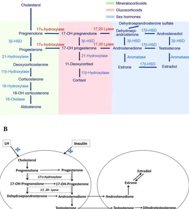
The underlying pathomechanism of PCOS is still under debate. Androgen overproduction, which is due to disturbed gonadotropin, FSH, and LH secretion, is the cornerstone of PCOS. Under normal circumstances, female androgen production takes place in the adrenal glands and the ovaries. Figures 1A and 1B show the different steps of adrenal and ovarian sex hormone genesis. Circulating levels of androgens could be the result of direct secretion or enzymatic conversion of 17-ketosteroids into androstenedione, mainly in the liver, skin, and adipose tissue. In the adrenal glands, androgen production is regulated by autocrine and paracrine signaling, whilst the control of the hypothalamic–pituitary axis dominates in the ovaries.
12
Figure 1 Different steps of adrenal and ovarian sex hormone genesis. A. Adrenal steroid hormone genesis in a human. Different types of steroid hormones are labelled with different colors. The first step is the conversion of cholesterol into pregnenolone, which is catalyzed by the 17α-hydroxylase/17,20-lyase enzyme. Androstenedione is the substrate of 17β-hydroxysteroid dehydrogenase and aromatase that leads to the formation of T and estrone. This figure is taken from the article
“17α-hydroxylase/17,20-lyase deficiency in congenital adrenal hyperplasia: A case
A
B
13
report” [31]. B. Sexual steroid hormone biosynthesis in an ovary. In the ovarian theca cells, androgen formation is stimulated by LH and modulated by cytochrome P450c17, which engages in both 17-hydroxylase and 17,20-lyase activities. The formed androstenedione is taken up by granulosa cells and helps the formation of estrogens.
Androstenedione in theca cells is converted to T, which leads to hyperandrogenism.
Insulin enhances androgen synthesis in the theca cells. This figure is taken from the article “Association of polycystic ovary syndrome with metabolic syndrome and gestational diabetes: Aggravated complication of pregnancy” [32]. HSD, hydroxysteroid dehydrogenase; LH, luteinizing hormone.
In PCOS, ovarian androgen synthesis is the main cause of AE, but adrenal androgen production is reported to be elevated in 30–50% of PCOS women.
Additionally, these women show enhanced 17-ketosteroid responses to adrenocorticotropic hormone [7, 12].
Androgens play a key role in the early gonadotropin independent phase of the menstrual cycle by initiating the growth of primordial follicles and recruiting small preantral follicles. A similar mechanism can be observed in girls during premenarche, when adrenal androgen synthesis helps induce gonadarche and menarche. Moreover, girls with “hyperpuberty” are at higher risk for subsequent development of PCOS [33, 34].
During the normal follicular phase of the menstrual cycle, LH triggers the androgenic precursor output of the ovarian theca cells and FSH regulates their conversion into estrogens (estradiol) at the granulosa cells. This hormonal balance promotes normal folliculogenesis in fertile females.
The main feature of PCOS is follicular arrest, which is provoked by an imbalance of LH, FSH, and sexual steroids. Increased follicular activation, a higher proportion of primordial follicles, and a corresponding increase in activated growing (primary) follicles are also typical features. Activated follicles show a lower rate of atresia and fail to mature. These characteristics of PCOS could be explained by the observed high
“steady state” LH serum levels and lack of FSH peaks during the follicular phase. As a
14
consequence, theca cell hyperplasia occurs as well. At the level of the hypothalamic–
pituitary axis, gonadotropin release increases and GnRH pulse secretion occurs at a rapid frequency, which keeps LH levels high [35].
In 1993, Crawley summarized possible theories for the development of AE [7, 36]:
The top-down theory claims that the hypothalamic–pituitary axis is responsible for AE due to central dysregulation of GnRH firing, which leads to LH overproduction. The end effect is follicular theca cell hyperplasia and aromatase upon activation. In rats and mice, imbalance of GnRH secretion resulted in changes at the level of LH and FSH synthesis [37].
The bottom-up hypothesis presumes the failure of adrenal sexual steroid synthesis. In this case, failure of peripheral androstenedione conversion is responsible for pituitary LH overproduction and, thus, increased ovarian androgen synthesis [36].
The androgen theory summarizes different explanations for the first occurrence of AE. According to the theory, upon sudden peripheral (ovarian, adrenal) androgen overproduction or exposure, PCOS is induced.
The so-called “fetal AE” theory supports the notion of early encounter with high serum androgen levels that provoke the onset of PCOS in adulthood. This explanation is also supported by some non-primate models that successfully produced phenotypical changes close to those seen in humans affected by PCOS [38]. Other explanations focus on the dysfunction of specific enzymes, like 11β-hydroxysteroid dehydrogenase.
It has two subtypes (type 1 and 2) that coordinate to inactivate and reactivate adrenal glucocorticoid. Activation of subtype 2 triggers 11β- hydroxy androstenedione, 11β-hydroxytestosterone, and 11β- hydroxyprogesterone production [39]. Dysfunction of cytochrome P450c17a also leads to disturbed androgen synthesis, as it is responsible for both 17α-hydroxylase and 17-20-desmolase activities in the ovarian and adrenal cells. As a single gene encodes this cytochrome P450 enzyme, mutation or dysfunction could be responsible for AE [40].
15
The insulin theory proposes IR as a provoking factor for AE. The major actions of insulin are mediated via three different pathways. Its metabolic effects are regulated by the phosphatidyl-inositol 3-kinase (PI3-K) pathway, whilst the mitogenic effects of insulin are coordinated by mitogen-activated protein kinases (MAPKs). The protein kinase C pathway is responsible for phospholipase C activation, which enhances second messengers in several tissues. In ovarian theca cells, LH increases steroidogenesis via the cyclic adenosine monophosphate–protein kinase A pathway. Insulin could trigger the accumulation of cyclic adenosine monophosphate through the PI3-K and protein kinase C cascade and increases LH secretion, which could prove the existence of cross-talk between LH and insulin signaling in ovarian tissue [41].
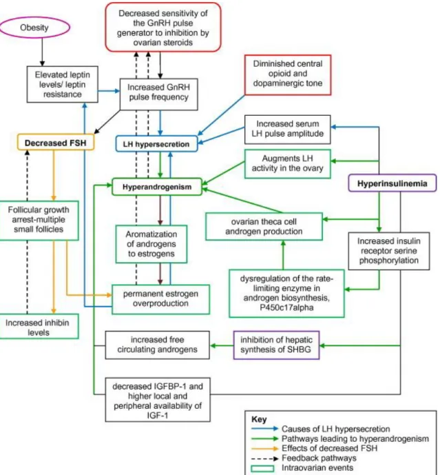
Figure 2 shows the complex hormonal disturbances that affect the development of PCOS.
16
Figure 2. Endocrine disturbances in the background of PCOS. Decreased sensitivity of the GnRH pulse generator and increased GnRH firing frequency provoke LH hyper- and FSH hyposecretion and therefore lead to hyperandrogenism and ovarian arrest.
Moreover, hyperinsulinemia results in further LH production and increases the level of circulating androgens by inhibiting the production of SHBG. Abbreviations: GnRH, gonadotropin-releasing hormone; LH, luteinizing hormone, FSH, follicle-stimulating hormone; SHBG, sex hormone-binding globulin; IGF-1, insulin-like growth factor 1;
IGFBP-1, insulin-like growth factor 1 binding protein; P450c17alpha, 17-alpha hydroxylase. This figure is adopted from the article “Polycystic ovary syndrome and impact on health” [42].
17
1.3 Etiology of PCOS
Although the Rotterdam criteria suggest that PCOS is a well-characterized endocrine disorder, its symptoms are in fact highly variable and heterogeneous.
Genetics, environmental background, nutrition, and ethnicity are important factors affecting its development. PCOS has the same prevalence among Caucasian women from the United States, Spain, and Australia (6–9%), but Southeastern Asian women—
especially Chinese women (2.2%) have the lowest prevalence in the world [1]. The Hispanic PCOS population in the United States shows higher rates of hirsutism, metabolic syndrome (MetS), and obesity compared to Caucasians [43].
Lifestyle factors and eating habits also play an important role in the development of PCOS. The Western diet contains many processed foods, which are rich in advanced glycation end products. These glycotoxins enhance intracellular signal transduction, trigger pro-inflammatory gene transcription, macrophage activation, cytokine (e.g., interleukins 1, 6, and 8), chemokine release, and reactive oxygen species production (i.e., the Maillard reaction) [44, 45]. Additionally, the circulating level of glycotoxins is well correlated with central obesity, waist-to-hip ratio, and IR in PCOS women [44, 46].
Obesity is a common health problem that affects 50–80% of patients [47], strongly influencing their reproductive function and cardiometabolic health [43].
Approximately 75–80% of PCOS women suffer from IR, a significantly higher proportion than in the age- and body mass index-matched female population [48]. Non- alcoholic fatty liver disease is believed to be one of the major consequences of IR, and it shows a clear correlation with AE. Its estimated prevalence may exceed 40% among obese PCOS women [49].
PCOS is associated with a significant increase in risk factors such as cardiovascular disease, atherosclerosis, obesity, MetS, dyslipidemia, type 2 diabetes mellitus (T2D), and IR [12, 50]. Cognitive or mood disorders, like depression and anxiety, are more frequent among PCOS women compared to the healthy female population [51].
18
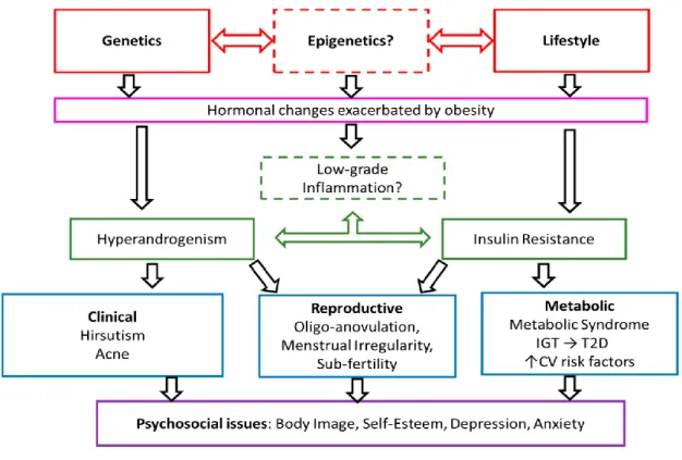
Figure 3 shows the complex etiology of PCOS.
Figure 3. Complex etiology of PCOS. Genetics, epigenetics, and lifestyle could promote the development of hyperandrogenism, obesity, IR, and chronic inflammation.
Hyperandrogenism and IR are responsible for further clinical, metabolic, reproductive and psychosocial consequences of these endocrine disturbances. Abbreviations: IGT, impaired glucose tolerance; T2D, type 2 diabetes mellitus; CV risk factors, cardiovascular risk factors. This figure is adopted from the article “The genetics of polycystic ovary syndrome: An overview of candidate gene systematic reviews and genome-wide association studies” [52].
1.3.1 Metabolic syndrome
The key elements of MetS are AE and IR, which join together all the necessary components of the syndrome. According to the definition utilized by the World Health
19
Organization, MetS is characterized by hyperglycemia (fasting glucose levels of 5.6 mmol/L or above), central obesity (increased waist circumference according to population- and country-specific definitions), elevated arterial blood pressure (130/85 mmHg or above), high total triglyceride (1.7 mmol/L or above), and a low high- density lipoprotein cholesterol (HDL-C) level (less than 1.29 mmol/L in women) [53, 54].
The prevalence of MetS is two to four times higher in the PCOS population than among healthy individuals. The presence of central obesity has a high impact on MetS prevalence [53]. If central obesity is combined with AE, the prevalence reaches approximately 29%, and if it is combined with polycystic morphology and AE, it reaches 35% [55]. The prevalence of IR is also around 30% in PCOS females, and it increases to 45–50% if centripetal obesity is present.
Higher triglyceride, lower HDL-C, and higher low-density lipoprotein cholesterol (LDL-C) levels are more common in PCOS patients than in the non-PCOS population [56]. Recently, some new biomarkers have been introduced to improve evaluation. For example, lipoprotein is a predictor of early-onset cardiovascular disease in PCOS women, even if they have normal lipid profiles [57]. In addition, reduced efflux capacity from macrophages, an indicator of HDL-C function, was decreased in the PCOS population [58].
Several rodent and non-primate models support the notion that fetal or adolescent exposure to androgens (mainly T or DHT) could provoke MetS [59, 60]. Furthermore, in a mouse model of letrozole-induced PCOS, antiandrogen therapy ameliorated symptoms of MetS and reduced body weight and adipocyte size [61]. Enlargement of adipocytes (hypertrophic obesity) is typical in PCOS patients. Apparently, this hypertrophy is due to disturbed lipolysis, reduced lipoprotein lipase activity, and altered storage capacity of adipocytes [62].
The most promising results for MetS were achieved if both AE and IR were treated at the same time. Flutamide is an androgen receptor antagonist that, together with metformin, has been reported to reduce plasma androgen indices and improve ovarian function. Metformin therapy induced hepatic estrogen receptor α expression,
20
while combination with flutamide decreased intestinal androgen receptor expression and increased estrogen receptor α expression. Hepatic and intestinal insulin and insulin receptor signaling were also improved by this “double” therapy [63].
Fasting blood sugar levels and dyslipidemia (characterized by a high triglyceride level and low HDL-C ratio) were found to be the strongest indicators of MetS onset. A study conducted in India reported that dyslipidemia was present in 90% of adolescent PCOS girls. Interestingly, more than 20% of these girls had already developed glucose intolerance, which indicates early onset of dysfunctional glucose metabolism and insulin sensitivity [64]. Insulin sensitivity and circulating SHBG levels also show a strong correlation, as IR is often associated with reduced SHBG levels and elevation of free androgens.
The development of MetS could be associated with failure of central regulation and energy metabolism. The role of decreased sympathetic tone has already been described in rodent models of neonatal androgenization, leptin resistance, and DHT- induced female AE. Central hyperleptinemia with peripheral leptin resistance (unresponsiveness to its anorexigenic effects) is often detected in MetS cases. In addition, energy intake is regulated by the firing activity of specialized fuel-sensing neurons of the hypothalamus. The hypothalamic expression of proopiomelanocortin neurons is found to be diminished in hyperandrogenic female mice. Moreover, chronic AE increases the adrenocorticotropic hormone sensitivity of proopiomelanocortin cell bodies and decreases fiber projection intensity, leading to increased energy intake and obesity. Reduced levels of α-melanocyte-stimulating hormone are also detected in AE cases [65].
1.3.2 Insulin resistance
1.3.2.1 Definition and diagnosis
Under normal circumstances, insulin regulates glucose homeostasis by enhancing glucose uptake in the target tissues (mainly adipocytes and skeletal and myocardial
21
muscle cells), decreasing hepatic glucose production, and modulating free fatty acid (FFA) release by lipolysis. The classic definition of IR is characterized by damage of all three pathways, which results in impaired glucose intake and metabolism with a compensatory increase in beta-cell insulin production and hyperinsulinemia. In the long term, hyperglycemia and hyperinsulinemia promote chronic inflammation and oxidative stress, which contribute to the development of arterial hypertension, dyslipidemia, visceral adiposity, endothelial dysfunction, a prothrombotic state, and T2D.
Among patients diagnosed with PCOS based on the Rotterdam criteria, 70% have impaired glucose tolerance or T2D, which are mainly diagnosed in their fourth decade of their life. Some recent data show that the prevalence of IR is almost the same among adolescents and young adults suffering from PCOS [64, 66], which suggests early onset of dysfunctional insulin signaling. Patients with AE and obesity are at the highest risk, although the lean PCOS phenotype seems to play no protective role in the development of dysglycemia. Elevated basal insulin secretion with insufficient insulin secretory responses to a glucose load are the main characteristics of IR in PCOS patients.
Furthermore, in most cases, IR is selective and affects the metabolic—but not the mitogenic—signaling pathways [53, 67].
The hyperinsulinemic–euglycemic glucose clamp technique is the gold-standard method to assess metabolic IR in vivo. The clamp technique enables quantitative evaluation of the effect of insulin on whole-body glucose uptake, while euglycemia is maintained with the administration of different insulin concentrations and adjusted amounts of glucose infusion [68]. Insulin-mediated glucose disposal is used as an indicator of insulin sensitivity. It could be defined under steady-state conditions in which the amount of infused glucose equals the amount of glucose taken up by the peripheral tissues. Hepatic glucose production and renal elimination can be measured with the help of isotopic-labelled glucose infusion through the clamp [66].
Sequential multiple insulin dose euglycemic clamps provide the opportunity to investigate the concentration-dependent saturable actions of insulin in vivo. On one hand, any type of alteration in insulin receptor binding or phosphorylation could be assessed by insulin sensitivity, which is evaluated based on the concentration required for a half-maximal insulin response. On the other hand, the maximal biological effect of
22
insulin is defined by insulin responsiveness, which reflects post-receptor events such as glucose transporter translocation. Interestingly, there is no glucose load or dose- dependent difference in insulin sensitivity values between lean and obese PCOS women. This finding suggests that both phenotypes act similarly to compensate for sudden glucose uptake. In addition, basal endogenous glucose production and half- maximal insulin response are disturbed only in obese PCOS women, which may support the notion of a possible connection point between impaired lipid metabolism and IR [69].
The clamp technique could be used to measure insulin clearance, which is strongly related to insulin receptor activation. In hyperinsulinemia cases, longer elimination time and clearance of insulin should be expected. Interestingly, there is no significant difference between hyperinsulinemic PCOS patients and healthy individuals in terms of posthepatic insulin clearance. Nevertheless, the C-peptide ratio was elevated in PCOS patients, which suggests hepatic dysregulation of insulin extraction [70].
Another method for measuring whole-body insulin sensitivity in subjects without diabetes is the frequently sampled intravenous glucose tolerance test with minimal model analysis. The minimal model allows for evaluation of insulin sensitivity (i.e., the sensitivity index), which correlates well with glucose-uptake-stimulated insulin action and suppressed glucose production [71].
Although these techniques offer correct and complex results regarding whole- body insulin sensitivity, the oral glucose tolerance test (OGTT) and fasting glucose and insulin level measurements are still the most commonly used clinical techniques. OGTT values correlate well with the results for the euglycemic glucose clamp and frequently sampled intravenous glucose tolerance test, but they are insensitive to large changes in insulin sensitivity and inaccurate for evaluation of β-cell dysfunction [66].
1.3.2.2 Metabolic effects of insulin resistance in PCOS
The metabolic effects of IR are mainly detected in skeletal muscle and adipose tissue. Skeletal muscle tissue is responsible for primary peripheral glucose uptake,
23
which exceeds 70–85% of the whole-body glucose uptake under normal circumstances.
If this capacity reaches its maximum, de novo lipogenesis takes place in the liver, which leads to an increased level of circulating FFAs. In the adipose tissue, failure of insulin signaling could provoke lipolysis, which also serves as a source of FFAs. Consequently, chronic inflammation and ectopic fat deposition occur, leading to vascular and metabolic complications.
In skeletal muscle, insulin receptor activation is followed by transmembrane translocation of glucose transporter 4 to promote glucose uptake. Recent studies found that reduced transcription of the β subunit can be detected in PCOS skeletal muscle cells. Although this mechanism provokes IR, it has some protective effects as well. If hyperinsulinemia persists, lower expression of the insulin receptor subunit would help to maintain the euglycemic state [72]. Additionally, glucose transporter 4 translocation problems could be associated with insulin downstream signaling failure. Some in vitro studies conducted with PCOS myotubes revealed that insulin receptor substrate 2- associated phosphatidyl-inositol 3-kinase (PI3-K) activity is decreased [73]. Moreover, essential changes in skeletal muscle morphology and reduced whole-body insulin sensitivity were found in a female rat model of T- and DHT-induced PCOS. These changes included a lower amount of insulin-sensitive fibers, a higher amount of less insulin-sensitive muscle fibers, reduction of capillary density, glycogen synthase dysfunction, and lower glucose transporter 4 protein expression [60].
Insulin resistance is responsible for specific changes in lipid homeostasis in PCOS cases. Insulin-induced suppression of lipid oxidation is reduced in PCOS females.
Interestingly, no significant changes were detected between PCOS and healthy women regarding peripheral lipid uptake [73]. Furthermore, IR has selective effects on adipose tissue in different locations; adipocytes isolated from the subcutaneous fat of lean PCOS patients are reported to have decreased insulin and catecholamine-stimulated lipolysis capacity and bigger cell size, whilst adipocytes from visceral fatty tissue show increased lipid oxidation rate. A higher rate of visceral lipolysis can elevate the FFA concentration of portal blood flow and promote the development of hepatic IR in PCOS [66]. Not only lipolysis but also the endocrine function of adipocytes might play a role in insulin sensitivity. Adiponectins are adipocyte-derived hormones that positively affect whole-body insulin sensitivity, pancreatic β-cell viability, and secretory function
24
[74]. Leptin, known as the prototypical adipokine, is one protein that plays a regulatory role at the hypothalamic–pituitary axis. Infertility is one of the characteristics of congenital leptin deficiency, as leptin affects LH and GnRH secretion. In PCOS, low leptin serum levels are usually detected in association with hyperinsulinemia and AE [75]. In addition, a reduction in the adiponectin receptors of theca cells in PCOS ovaries (compared with normal ones) is frequently detected [76].
Fat accumulation may have an effect on hepatic glycogen synthase activity and pancreatic β-cell function. Non-alcoholic fatty liver disease and impaired glycogen synthase activity show a significant correlation with the prevalence of obesity in PCOS patients [49]. A high FFA concentration related to obesity has a negative impact on pancreatic β-cell insulin secretion and calcium channel distribution, which match well with the fat accumulation within the islets. In addition, intrapancreatic fat depots elevate the local amount of FFAs and trigger chronic inflammation and cytokine production [66]. In PCOS-related IR, chronic pancreatic islet inflammation (insulitis) could occur, which promotes β-cell dysfunction or apoptosis later on. Hyperinsulinemia and β-cell hyperplasia are the first consequences of insulitis and increased insulin demand due to IR, but some recent data suggest that mitochondrial dysfunction has a role in the pathophysiology of β-cell secretory failure [77]. Glucolipotoxicity could enhance the activation of reactive oxygen and nitrogen species, which leads to mitochondrial dysfunction and inhibition of the electron transport chain and, in turn, results in reduced energy production [44].
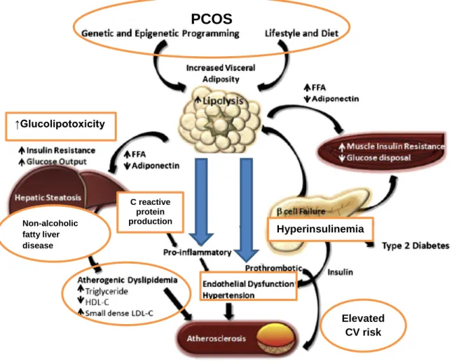
The complex consequences of impaired lipid metabolism are shown in Figure 4.
25
Figure 4. The effects of disturbed lipid metabolism on insulin resistance and cardiovascular complications in PCOS patients. In PCOS, impaired lipolysis results in free fatty acid overproduction and decreased adiponectin release. It also promotes pro-inflammation and thrombosis as well as IR of the liver and muscle cells.
Atherogenic dyslipidemia due to hepatic failure, endothelial dysfunction, hypertension, and atherosclerosis are the end results. Abbreviations: FFA, free fatty acids; LDL-C, low-density lipoprotein cholesterol; CV risk, cardiovascular risk. This figure is adopted and modified from the article “Metabolic syndrome” [78].
1.3.2.1 The effects of insulin resistance on the female reproductive system
Insulin regulates several signaling mechanisms that are necessary to maintain normal function of the female reproductive system. In PCOS, hyperinsulinemia
Non-alcoholic fatty liver disease
Hyperinsulinemia
Elevated CV risk
↑Glucolipotoxicity
PCOS
T
C reactive protein production
26
dysregulates this physiological balance of signaling pathways, leading to AE and selective IR of the ovaries.
At the hypothalamus–hypophysis–adrenal axis, insulin triggers GnRH and LH secretion pulsatility and frequency by potentiating GnRH gene transcription. Insulin may increase hypothalamic corticotropin-releasing hormone secretion and, possibly, sensitize the adrenal cortex to adrenocorticotropic hormone stimulation. At the site of the adrenal glands, insulin promotes androgen secretion and consecutive AE.
The most important mitogenic effect of insulin is that it indirectly diminishes SHBG synthesis, which causes higher free androgen serum levels in PCOS patients. Not only hyperinsulinemia but also elevated concentrations of glucose and fructose are important indirect factors for SHBG decrease, as both monosaccharides downregulate hepatocyte protein synthesis by reducing hepatic nuclear factor 4-α activity [66, 79].
In ovarian tissue, theca cells and granulosa cells present insulin receptors and insulin-like growth factor-1 (IGF-1) receptors on their surfaces. The action of insulin was tested by direct administration of antibodies against both receptors, which led to a decrease in ovarian steroidogenesis [80]. Further investigations found that insulin modulates the activity of steroidogenic acute regulatory protein, which is a molecule involved in the transportation of cholesterol to the cholesterol side-chain cleavage enzyme and the rate-limiting enzyme of ovarian steroid synthesis. In addition to enhancing cholesterol side-chain cleavage enzyme activation, insulin increases the expression of 7-α-hydroxylase/17,20-lyase, 3-β-hydroxysteroid dehydrogenase, and aromatase [79]. It was recently reported that inositol phosphoglycans, which are second messengers in the metabolic signal transduction pathway of classic insulin receptor signaling, might have similar effects on the enzyme cascade of steroid genesis [79]. In this regard, hyperinsulinemia strongly shifts the balance towards overexpression of androgen-producing enzymes. Moreover, the hyperinsulinemic state represses IGF-1 binding protein synthesis, which increases the amount of available free IGF-1 and leads to further enhancement of ovarian insulin signaling. Together, these complex mechanisms lead to ovarian selective insulin resistance, which means that the insulin sensitivity of the ovarian tissue is unchanged or enhanced, although metabolic IR is present in all other insulin-sensitive tissues [81].
27
1.3.2.2 Hemodynamic consequences of insulin resistance
In addition to its above-mentioned actions, insulin has direct hemodynamic effects. Insulin improves regional blood flow, increases capillary recruitment, and aids peripheral vasodilatation, which leads to better glucose uptake of the peripheral tissue.
This vascular action of insulin is mediated by nitric oxide (NO) derived by the endothelial cells of the vasculature. In order to enhance NO production, insulin must cross the endothelial barrier. Although it is not well understood yet, insulin probably reaches the endothelial cell by receptor-mediated internalization. Some studies of obese and T2D patients show evidence that damage of the endothelial barrier or longer transport time through the capillary bed interferes with insulin-induced local blood flow [82, 83].
According to recent findings, dysregulation of endothelial nitric oxide synthase (eNOS) is one of the key elements of IR. Moreover, glucose and lipid toxicity promote the production of reactive oxygen and nitrogen species, which reduce the available NO concentration [84, 85]. Chronic inflammation and a prothrombotic state are additional consequences of the activation of reactive oxygen species, which increases the local level of vascular and intercellular adhesion molecules (VCAM and ICAM), C-reactive protein, tumor necrosis factor α, interleukin 6, and endothelin-1 (ET-1) [83]. In addition, chronic inflammation triggers the migration and proliferation of vascular smooth muscle cells, which shifts vascular reactivity towards vasoconstriction in the absence of a sufficient amount of NO [86]. To determine whether the metabolic or mitogen insulin- signaling pathway coordinates NO release, endothelial cells and vascular smooth muscle cells harvested from healthy coronary arterioles were cultured in high-glucose, fatty acid, and insulin-containing media. Interestingly, mitogen signaling, including MAPK/ERC activation, was preserved or slightly augmented, whilst the metabolic pathway (PI3-K signaling) was reduced [86]. These results may prove that the metabolic effects of IR are responsible for the disadvantageous changes in arterial reactivity and that elevated CV risk is an indirect metabolic complication.
Regarding PCOS, several studies supported the theory of compromised arterial reactivity; however, it is not easy to distinguish between the impacts of IR and AE. In DHT-induced rodent models of PCOS, insulin-induced relaxation of the aorta was
28
significantly reduced [87]. Recent human studies showed that PCOS with or without the clinical features of IR and MetS elevates the level of reactive oxygen species, endoplasmic reticulum stress markers (GRP78, sXBP1, ATF6), and leukocyte rolling flux. [88]. Furthermore, a recent publication reported that 12 weeks of treatment with metformin improved endothelial function and significantly decreased the levels of ICAM-1, E-selectin, interleukin 6, and tumor necrosis factor α in PCOS patients [89].
Additionally, coronary artery and aortic calcification were increased in women with PCOS compared with healthy individuals, and age and body mass index were both determining factors for coronary artery calcification [90].
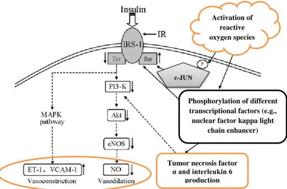
Figure 5 demonstrates a possible mechanism of vascular IR.
Figure 5. The effect of insulin resistance on vascular reactivity. Normally, insulin signaling leads to vasodilatation by PI3-K signaling and NO production. If the amount of reactive oxygen species is increased, downregulation of PI3-K signaling and upregulation of MAPK signaling are observed. As an end effect, NO production is decreased and vasoconstriction occurs. Abbreviations: IRS-1, insulin receptor substrate 1; MAPK, mitogen-activated kinase; ET-1, endothelin-1; VCAM-1, vascular adhesion molecule 1; PI3-K, phosphatidyl-inositol 3-kinase; Akt, protein kinase B; eNOS, endothelial nitric oxide synthase; NO, nitric oxide; eNOS, endothelial nitric oxide synthase; c-JUN, c-Jun N-terminal kinases. Adopted and modified from the article
Phosphorylation of different transcriptional factors (e.g., nuclear factor kappa light
chain enhancer) Activation of
reactive oxygen species
Tumor necrosis factor α and interleukin 6
production c-JUN
29
“Tectorigenin attenuates palmitate-induced endothelial insulin resistance via targeting ROS-associated inflammation and IRS-1 pathway” [91].
1.3.2.3 Molecular effects of insulin resistance in PCOS
In addition to its well-known effects on glucose homeostasis, insulin affects lipid and protein metabolism, cell growth, and differentiation. Insulin signaling is mediated by insulin receptors, which are heterotetramers of α- and β-subunits. The extracellular α-subunit is responsible for ligand binding, whilst the β-subunit is activated by autophosphorylation. The cytoplasmic β-subunits have intrinsic protein tyrosine kinase activity. The classic insulin receptor shares substantial structural homology with the IGF-1 receptor and insulin-related receptor. Hence, insulin could induce autophosphorylation of different receptors. It was recently revealed that this autophosphorylation depends on insulin concentration [92] and that the physiological serum concentration promotes activation of the classic receptor.
The activated insulin receptor initiates tyrosine phosphorylation of intracellular substrates, like different insulin receptor substrates (IRS-1–4). Glucose uptake and metabolic effects are mediated by P-I3K, which then phosphorylates membrane phospholipids and phosphatidylinositol 4,5-bisphosphate, resulting in activation of the serine/threonine protein kinases (Akt) and atypical protein kinase C. These signaling pathways are responsible for transmembrane translocation of glucose transporter 4 or indirect activation of glycogen synthase. Protein synthesis and cellular nutrient sensing are also regulated by the PI3-K pathway via the mammalian target of rapamycin. The so-called mitogen pathway is regulated by the cascade of serine/threonine kinases and the enhancement of different MAPK kinases [93].
Because the metabolic and mitogen effects of insulin are regulated differently, impairments caused by them could develop separately. Metabolic IR is the cornerstone of PCOS. According to several studies, protein kinase C-mediated serine phosphorylation of the insulin receptor, serine phosphorylation of insulin receptor substrate 1, and increased insulin-independent serine phosphorylation are the most
30
important steps in the pathogenesis of metabolic IR in PCOS [67]. Presumably, downstream signaling pathways might be intact as Akt, protein kinase C, and B activation were unchanged in PCOS adipocytes [94]. However, a few studies suspected indirect regulation problems with Akt signaling [73, 93]. Increased extrinsic insulin- independent serine phosphorylation of the classic insulin receptors seems to be specific to PCOS, as it is not detectable in other IR-related diseases (i.e., obesity or T2D) [66].
In addition to the above-mentioned defects of insulin signaling, some data suggest that mitochondrial dysfunction of skeletal muscle cells plays a further role in PCOS [95].
1.3.3 Cardiovascular disease
The multiple metabolic complications caused by PCOS increase patients’ CV risk.
Women with hyperandrogenic PCOS have a relative risk of coronary heart disease at least two times higher than that of healthy controls [96, 97]. The rate of coronary artery calcification, an indicator of subclinical cardiovascular disease, was found to be present 40% of fertile-aged PCOS females (aged under 40) [98].
The prevalence of hypertension, which is one of the earliest consequences of atherosclerosis and reduced compliance of the vessel wall, was reported to be present in 65% of the premenopausal PCOS population. This proportion is even larger if the clinical picture involves IR, AE, and obesity or if patients are already in menopause [99].
Altered intima–media thickness of the vessel is considered to be a noninvasive marker of coronary and cerebrovascular events [100]. Premature atherosclerosis and higher intima–media thickness values (compared to healthy individuals) are frequently present in premenopausal PCOS women [100, 101]. This early stage of atherosclerosis in PCOS is often associated with elevated LDL serum levels or obesity [97].
Further cardiovascular abnormalities of PCOS patients include decreased cardiac systolic flow velocity, diastolic dysfunction, increased vascular stiffness, and endothelial dysfunction. Conventional echocardiography is still the easiest noninvasive
31
way to evaluate left ventricular function in PCOS patients, but the results are highly dependent on image quality, the assumption of left ventricular geometry, and the experience of the investigator. Some studies found evidence of a connection between early left ventricular impairment when mitral inflow deceleration time, isovolumetric relaxation time, and diastolic function were altered in the PCOS group [102]. Systolic blood flow velocity was also found to be decreased in PCOS, with an inverse correlation between systolic outflow parameters and fasting insulin levels [103].
Chronic activation of the sympathetic nervous system could lead to vascular dysfunction and hypertension. In PCOS, activation of the sympathetic nervous system and IR is a complex mechanism that is related to obesity. In PCOS patients, impairment of the sympathetic nervous system was demonstrated by an exaggerated systolic blood pressure response to exercise and delayed heart rate recovery [104]. Another study demonstrated that a high density of catecholaminergic nerve fibers is present in the ovaries of women with PCOS, which correlates with skeletal muscle and adipose tissue results [66].
1.3.3.1 The role of endothelial dysfunction
In PCOS, endothelial dysfunction is the source of further cardiovascular complications, and its severity is proportional to the level of androgens and IR.
Hyperinsulinemia-induced endothelial dysfunction and hypertrophic remodeling occur due to its effects on vascular endothelial and smooth muscle cells as well as ET-1 production. ET-1 release is reported to be significantly increased in PCOS females, and this is also a sign of oxidative stress and altered vascular compliance [105]. Disturbed insulin-stimulated eNOS activation and NO release appear to negatively influence capillary network expansion, resulting in impaired microcirculation and blood flow regulation in metabolically active tissues.
Not only hyperinsulinemia but also an elevated level of circulating FFAs and chronic inflammation could enhance endothelial impairment by altering activation of the renin–angiotensin–aldosterone axis. The vascular effects of angiotensin II are mediated by two G-protein coupled receptor subtypes (subtype 1 and 2) with different
32
effects. Both receptors are expressed on the endothelial surface, but receptor subtype 1 is responsible for the well-known vasoconstrictor effects. PCOS rodent models revealed that reduced endothelial NO bioavailability, impaired insulin signaling, and IR are promoted by subtype 1 activation [106, 107]. Some recent clinical trials were able to prove the beneficial effects of selective angiotensin receptor subtype 1 blocking agents in PCOS females, such as improved cardiometabolic risk due to increased insulin sensitivity and prevention of T2D [107]. The effects mediated by angiotensin receptor subtype 2 include vasodilation and augmented insulin-mediated glucose disposal. These facts suggest a possible imbalance between the two receptor signaling mechanisms in PCOS, which could be indirectly improved by inhibition of angiotensin receptor subtype 1 [108].
Hormonal modulation failure of vascular reactivity plays an important role in impaired endothelial function. The two main effects of estrogens are eNOS phosphorylation due to activation of the PI3-K/Akt pathway and increased eNOS mRNA expression. In transgenic mice, the activation of estrogen receptor α was responsible for eNOS protein production [109]. In addition, estrogen regulates vascular endothelial growth factor production (via estrogen receptor α) in coronary circulation.
These findings align with the flow-mediated vasodilatation results of healthy, premenopausal, and estrogen-deficient postmenopausal women [110].
Regarding the effects of androgens, the tissue-specific distribution of two key enzymes, aromatase and 5-α-reductase, must be taken into consideration. DHT is pure androgen, but T could be converted into estrogen and activate estrogen receptor α.
Furthermore, T has complex vascular effects: it could relax coronary arteries by opening the large-conductance, calcium-activated potassium channels and up-regulating cyclooxygenase-2 activity, which in turn leads to increased prostacyclin formation [109]. The vascular effects of T may be gender-dependent, as flow-mediated vasodilatation was reduced in female-to-male transsexuals after administration of high concentrations of androgen. Animal studies of sexual transition suggest that T has a negative effect on eNOS-mediated vascular responses [111]. Androgens can also induce vasoconstriction by modulating the production of ET-1 or enhancing the production of arachidonic acid intermediates, including thromboxane A2 and 20- hydroxyeicosatetraenoic acid. In female-to-male transsexuals and in postmenopausal
33
women, AE correlates well with ET-1 levels and subclinical cardiovascular risk [109, 112].
It was recently revealed that the levels of adipocytokines, such as visfatin, vascular endothelial growth factor, and matrix metalloprotease, can be used as specific indicators of endothelial dysfunction. Visfatin has insulin-mimicking effects and lowers blood sugar values. According to recent data, lower concentrations were found in PCOS females compared to controls with a similar body mass index. Increased vascular endothelial growth factor production is an indicator of chronic mild inflammation and is related to impaired vascular reactivity. Increased metalloprotease 9 concentrations predict excessive CV risk and extracellular matrix remodeling [113].
1.4 Vitamin D
VD is a fat-soluble vitamin that also acts as a steroid hormone. As a steroid hormone, VD is involved in mineral homeostasis, has immunomodulatory effects, and seems to have a protective role against MetS and cardiovascular disease.
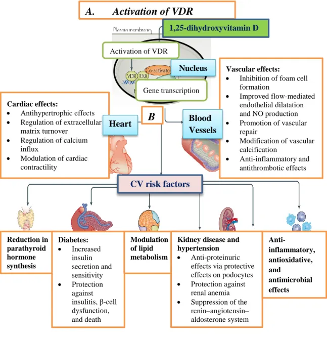
VD is produced non-enzymatically under the skin upon exposure to ultraviolet sunlight, but dietary sources are important for a sufficient supply. Cytochrome p450 enzymes, namely CYP2R1, CYP27B1, CYP24A1, and CYP27B1, are involved in the metabolism of VD, which takes place in the liver and kidney. According to some estimations, more than 200 genes, most of which are responsible for cellular proliferation, differentiation, apoptosis, and angiogenesis, are directly or indirectly controlled by 1,25-hydroxyvitamin D. VD receptors (VDR) are reported to be present in brain, prostate, breast, colon, and pancreas tissue as well as in immune cells. Normal serum levels of VD are considered to be within the range of 30–100 ng/ml. Figure 6 shows the specific effects of VDR activation in different tissues.
VD insufficiency is a comorbidity that affects approximately 50% of the whole global population [114]. Very low levels of VD (<20 ng/ml) are reported to be present
34
in 67–85% of PCOS women. VDD could influence PCOS through hormonal and gene modulation, resulting in infertility, metabolic syndrome, and IR [115].
Figure 6. Effects of VDR activation in the human body, in special regard to cardiovascular risk factors. A. VDR activation in target cells. In the nucleus, VDR and retinoid X receptor form a heteromer, which promotes the transcription of several genes. B. VD-mediated effects in the heart and blood vessels. VD promotes vascular repair, improves flow-mediated endothelial relaxation, and modulates cardiac contractility. Further organ-specific positive effects on cardiovascular risk factors are shown below. Abbreviations: VDR, vitamin D receptor; RXR, retinoid X receptor; NO,
Cardiac effects:
Antihypertrophic effects
Regulation of extracellular matrix turnover
Regulation of calcium influx
Modulation of cardiac contractility
Reduction in parathyroid hormone synthesis
Diabetes:
Increased insulin secretion and sensitivity
Protection against insulitis, β-cell dysfunction, and death
Modulation of lipid metabolism
Vascular effects:
Inhibition of foam cell formation
Improved flow-mediated endothelial dilatation and NO production
Promotion of vascular repair
Modification of vascular calcification
Anti-inflammatory and antithrombotic effects
Kidney disease and hypertension
Anti-proteinuric effects via protective effects on podocytes
Protection against renal anemia
Suppression of the renin–angiotensin–
aldosterone system
Anti-
inflammatory, antioxidative, and
antimicrobial effects
A. Activation of VDR
B
.
1,25-dihydroxyvitamin D
CV risk factors
Heart Blood
Vessels Nucleus Gene transcription Activation of VDR