Themed Section: Pharmacology of the Gasotransmitters
REVIEW
Measurement of NO in biological samples
C Csonka1,2, T Páli3, P Bencsik1,2, A Görbe1,2, P Ferdinandy2,4and T Csont1,2
1Cardiovascular Research Group,Department of Biochemistry,University of Szeged,Szeged, Hungary,2Pharmahungary Group,Szeged, Hungary,3Institute of Biophysics,Biological Research Centre,Hungarian Academy of Sciences,Szeged, Hungary, and4Department of Pharmacology and Pharmacotherapy,Semmelweis University,Budapest, Hungary
Correspondence
Csaba Csonka, Cardiovascular Research Group, Department of Biochemistry, University of Szeged, Dóm tér 9, H-6720 Szeged, Hungary. E-mail:
csonka.csaba@med.u-szeged.hu
---
Received 30 January 2014 Revised 16 June 2014 Accepted 25 June 2014
Although the physiological regulatory function of the gasotransmitter NO (a diatomic free radical) was discovered decades ago, NO is still in the frontline research in biomedicine. NO has been implicated in a variety of physiological and pathological processes; therefore, pharmacological modulation of NO levels in various tissues may have significant therapeutic value. NO is generated by NOS in most of cell types and by non-enzymatic reactions. Measurement of NO is technically difficult due to its rapid chemical reactions with a wide range of molecules, such as, for example, free radicals, metals, thiols, etc. Therefore, there are still several contradictory findings on the role of NO in different biological processes. In this review, we briefly discuss the major techniques suitable for measurement of NO (electron paramagnetic resonance, electrochemistry, fluorometry) and its derivatives in biological samples (nitrite/nitrate, NOS, cGMP, nitrosothiols) and discuss the advantages and disadvantages of each method. We conclude that to obtain a meaningful insight into the role of NO and NO modulator compounds in physiological or pathological processes, concomitant assessment of NO synthesis, NO content, as well as molecular targets and reaction products of NO is recommended.
LINKED ARTICLES
This article is part of a themed section on Pharmacology of the Gasotransmitters. To view the other articles in this section visit http://dx.doi.org/10.1111/bph.2015.172.issue-6
Abbreviations
DAC, o-phenylenediamine cyanine; DAF, diaminofluorescein; DAMBO, diamino-boron-dipyrromethene; DAN, diaminonaphthalene; DAR, o-diaminorhodamine; DETC, N,N-diethyl-dithiocarmabate; DTC, dithiocarbamate; EPR, electron paramagnetic resonance spectroscopy; ESR, electron spin resonance spectroscopy; MGD,
N-methyl-D-glucamine dithiocarmabate; RSNO, S-nitrosothiols; sGC, soluble GC
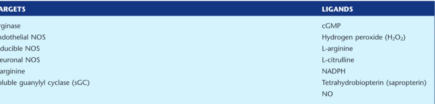
Table of Links
TARGETS LIGANDS
Arginase cGMP
Endothelial NOS Hydrogen peroxide (H2O2)
Inducible NOS L-arginine
Neuronal NOS L-citrulline
L-arginine NADPH
Soluble guanylyl cyclase (sGC) Tetrahydrobiopterin (sapropterin)
NO
This Table lists key protein targets and ligands in this document, which are hyperlinked to corresponding entries in http://
www.guidetopharmacology.org, the common portal for data from the IUPHAR/BPS Guide to PHARMACOLOGY (Pawsonet al., 2014) and are permanently archived in the Concise Guide to PHARMACOLOGY 2013/14 (Alexanderet al., 2013).
Introduction
Although the physiological regulatory function of the gas- otransmitter NO was discovered decades ago, NO is still in the frontline research as shown by the continuously increasing number of annual hits for ‘nitric oxide’ in the PubMed data- base. The increasing interest in NO in biomedical research is due to several facts: (i) NO is an ubiquitous free radial mol- ecule found in most of cells of all tissues intracellularly as well as in the extracellular fluids; (ii) NO is involved in a variety of physiological and pathological processes; and (iii) utilization of gaseous NO and some NO donor molecules for human therapy has entered into clinical therapy and the develop- ment of further NO-related therapies are promising (see for review, Pacheret al., 2007). However, the chemical reactions of NO with other free radicals and various small and macro- molecules have raised many new questions regarding the involvement of NO in different cellular or intercellular/
interorgan signalling pathways. Moreover, measurement of NO is technically difficult, due to its rapid chemical reactions with a wide range of biomolecules and its very short half-life of approximately a few seconds. Therefore, there are still contradictory findings on the role of NO in several biological processes as reviewed previously by Ferdinandy and Schulz (2003) and Schulz et al. (2004). Correct application of the different NO measurement techniques are essential to gain more knowledge on the physiology and pathology of NO.
In this review, we describe the major techniques used most frequently in the literature for measurement of NO in biological samples and discuss the advantages and disadvan- tages of each method.
Synthesis and major metabolic pathways of NO
NO is a diatomic hydrophobic gas that can permeate various cellular membranes and other hydrophobic structures, and thus it has a high diffusion capacity in a physiological envi- ronment. NO can be produced in biological systems by both enzymatic and non-enzymatic reactions. In mammals, NO is biosynthesized endogenously by various isoforms of
NOS using the substrates L-arginine and molecular oxygen (Figure 1). NOS catalyses the oxidation of the terminal guani- dino nitrogen of L-arginine to produce NO and L-citrulline.
Several cofactors are required for the reaction including NADPH, flavin adenine dinucleotide, flavin mononucleotide, haem, tetrahydrobiopterin and calmodulin. Insufficient availability of L-arginine and some of the cofactors (i.e.
tetrahydrobiopterin), as well as S-glutathionylation of certain NOS isoforms may lead to reduced NO formation and uncoupling of NOS resulting in superoxide production (Forstermann and Li, 2011; Zweieret al., 2011). Three distinct isoforms of NOS have been described in mammals: neuronal NOS (nNOS, NOS-1), endothelial NOS (eNOS, NOS-3) and inducible NOS (iNOS, NOS-2) (Knowleset al., 1989; Mayer et al., 1989; Mulschet al., 1989; Palacioset al., 1989; Palmer and Moncada, 1989; Stuehret al., 1989; Moncadaet al., 1997;
Forstermann and Sessa, 2012). Moreover, the existence of a putative mitochondrial NOS (mtNOS) has also been sug- gested (Zaobornyj and Ghafourifar, 2012). The constitutively expressed isoforms nNOS and eNOS are Ca2+-dependent, whereas iNOS is Ca2+-independent (Moncada et al., 1991).
The biological effects of NO and the activities of NOS isoen- zymes are further regulated by compartmentalization (Villanueva and Giulivi, 2010) as well as transcriptional, post-
Figure 1
Major metabolic pathways of NO.
transcriptional and post-translational modulations such as for instance phosphorylation, S-nitrosation, interaction with modulatory proteins (e.g. calmodulin, HSP90, caveolin, etc.), dimerization, inhibition by endogenous methyl-arginines, etc. (as reviewed in detail elsewhere, Zhou and Zhu, 2009;
Pautzet al., 2010; Qian and Fulton, 2013).
It has been clarified that NOS-independent reduction of dietary or endogenous sources of nitrate and nitrite are important contributors for the production of NO in mamma- lian tissues (Lundberg et al., 2008; Rassaf et al., 2014) (Figure 1). Nitrate found in significant amounts in certain vegetables (e.g. lettuce, spinach and beetroot, etc.), needs to be initially reduced to nitrite by nitrate reductase enzymes of bacteria in the gastrointestinal tract. Nitrite – also found in certain dietary sources – can be reduced to NO by several pathways and conditions including low pH, ascorbic acid, haemoglobin, myoglobin, polyphenols and xanthine oxi- doreductase (Lundberg et al., 2008; Rassafet al., 2014). The formation of NO by these pathways may become especially important during hypoxia when pH becomes acidic and oxygen-dependent NOS activities are limited.
NO has a short biological half-life (estimated to be a few seconds) due to its rapid reaction with a variety of molecules (Figure 1). Although the chemistry of NO is well-established in a test tube, the exact biochemistry of NO is still far from clear (Hillet al., 2010). The biologically relevant reactions of NO have been reviewed elsewhere in more detail (Gow, 2006;
Bryan and Grisham, 2007; Habib and Ali, 2011; Tennyson and Lippard, 2011). The major pathway for the metabolism of NO is its oxidation to nitrite and nitrate eventually fol- lowed by their urinary excretion. NO in the presence of molecular oxygen is oxidized to nitrogen-dioxide (NO2), which by reacting with another NO molecule forms N2O3, an intermedier that participates in nitrosation reactions. N2O3
may be decomposed to nitrite and a one-electron reduction of NO2may also lead to nitrite formation. Nitrite has a half- life of a few minutes in the circulation as it can be further oxidized to the more stable nitrate by certain oxyhaemopro- teins such as oxygenated haemoglobin or myoglobin. Alter- natively, NO may directly react with oxyhaemoproteins to form nitrate (Bryan and Grisham, 2007). In addition to its reaction with oxygen, NO rapidly reacts with superoxide to yield peroxynitrite, a short-lived oxidant, nitrating and nitro- sating agent (Pacheret al., 2007; Radi, 2013).
Important molecular targets of NO are transition metal ions. NO binds to transition metal ions to form nitrosyl-metal ion complexes. The nitrosyl-Fe2+adduct – such as in haem – is particularly stable, as the binding of the nitrosyl ligand to Fe2+
is very strong. Relevant examples of proteins in which the formation of nitrosyl-metal complexes affect biological func- tion include soluble GC (sGC, see later), haemoglobin, cyto- chromes, etc (Toledo and Augusto, 2012). NO – following oxidation to N2O3– plays an important role in the formation of S-nitrosothiols (RSNOs) via S-nitrosation of small molecu- lar weight thiols and thiol-containing proteins (Broniowska and Hogg, 2012). This type of reaction is often incorrectly referred to as ‘S-nitrosylation’ (i.e. direct addition of NO to a reactant) in the literature [for more details on NO chemistry and terminology see a recent review by Heinrich et al., (2013)]. Nevertheless, NO may also react directly with thiyl radicals formed after oxidation of thiols to produce RSNOs.
Enzyme-dependent and -independent S-nitrosation, transn- itrosation and denitrosation are potential post-translational modifications that may regulate biological function of several proteins (Limaet al., 2010; Stamler and Hess, 2010; Gould et al., 2013; Maronet al., 2013). RSNOs may play an impor- tant role in endogenous transport and storage of NO as well as in NO-related cell signalling.
Analytical tools for the assessment of NO: what to consider
before selection?
In general, NO measurement techniques can be classified as direct (the target of measurement is NO itself) and indirect methods. Most of the NO measurement techniques in the literature are indirect ones that are measurements of NOS activities, activation of molecular targets of NO, such as GC-derived cGMP, or products of reactions of NO, such as RSNOs or nitrite/nitrate. The major analytical tools for the detection of these analytes are spectroscopic or electrochemi- cal methods. The spectroscopic methods include colorimetric, fluorometry, luminometry and electron spin resonance spec- troscopy (ESR). These analytical tools have been extensively reviewed previously (Hetrick and Schoenfisch, 2009; Coneski and Schoenfisch, 2012). The sensitivity and specificity of these techniques for NO varies a lot, and they cannot provide an insight into thein situNO levels in biological systems. Direct NO measurement techniques that are more specific for NO such as, for example, ESR afterin vivoorex vivospin trapping or NO-specific biosensors are less frequently used.
Choosing the most appropriate method for measurement of NO in biological systems is not easy. Therefore, we suggest consideration of two additional aspects before planning experiments. Firstly, for the measurement of NO, a variety of different commercial products are available on the market, most of which can be purchased in ready-to-use formats.
However, these kits and/or instruments that operate with different background principles are developed for various sci- entific, industrial and environmental application purposes.
Secondly, the specificity and cross-reactivity of the NO sensors with NO derivatives (e.g. reactive nitrogen species such as peroxynitrite) and other non-NO-related molecules (e.g. reactive oxygen species) still remain a major challenge in NO research (Rodriguez-Rodriguez and Simonsen, 2012;
Woolleyet al., 2013). This is mainly due to the small size of the NO molecule and its complex chemical nature in biologi- cal systems. Therefore, before scientific use, careful consid- eration of various aspects including background assay principle, specificity, sensitivity, advantages, disadvantages and financials (see Table 1) is strongly recommended to choose the most proper analytical technique(s), which best fits the study objectives. As an aid to finding the most appro- priate method(s) for measurement of NO in a particular study, a detailed questionnaire can be constructed and answered as suggested by Wardman (2007). Moreover, we suggest the use of NO donors for positive control and inhibi- tors of NO formation as negative controls to complete the data obtained either by direct or indirect assays to understand the exact role of NO in biological systems.
In the present review, we focus on the most important possibilities to determine NO or its derivatives in biological matrices. We discuss background assay principles, specifici- ties, sensitivities, advantages/disadvantages and possible limitations.
Direct methods to estimate NO
Electron paramagnetic resonance spectroscopy
As NO is a free radical, hence of paramagnetic nature, elec- tron paramagnetic resonance spectroscopy (EPR; also referred to as ESR) is considered to be the most appropriate tool for the direct detection of NO. The main advantage of EPR compared with other NO detection techniques is that it only detects paramagnetic molecules, and the EPR spectrum is a unique fingerprint of the chemical and electronic structure around the unpaired electron. EPR measures the transitions induced between the Zeeman levels of a paramagnetic molecule via its interaction with a static magnetic field and an oscillating electromagnetic field, most commonly in the X microwave
frequency band, around 9–9.5 GHz. To take an EPR spectrum, the microwave frequency is held constant and the static mag- netic field is swept (despite its name) across the desired range.
The equation for resonance absorption isΔE =hv=gβB, in which h is Planck’s constant;νis the frequency of the micro- waves;βis the Bohr magneton, a physical constant; B is the external magnetic field; andgis theg-value, or the Zeeman splitting factor (Weil and Bolton, 2007). The g-value is a characteristics of the paramagnetic molecule [in case of NO in liquidg=2.035, (Hogg, 2010)]. A further specific property of EPR spectra originates from the coupling of the electron spin with the surrounding nuclear magnetic spins, measured by the so-called hyperfine coupling constant, A, in mT.
The paramagnetic NO can be measured directly by EPR irrespective of the optical appearance of the sample; however, due to the rapid relaxation of its excited electron spin state to the ground state (Mapleset al., 1991) and the high reactivity of NO, spin-trapping techniques have been developed. Spin traps are compounds that interact with the less stable radi- cals, producing a more stable adduct, which can be then detected by EPR (Janzen, 1984; Tosakiet al., 1996; Berliner and Fujii, 2004; Villamena and Zweier, 2004).
Table 1
Specificity and major advantages and disadvantages of techniques of NO assessment with a brief summary
Technique Sensitivity Advantages Disadvantages
EPR
NO spin trapping followed by spectrometry in magnetic field
0.05–0.4 nMa Direct, considered as a ‘stand alone’
assay; the most specific method, due to NO-specific spin traps
Semiquantitative; expensive, not particularly common instrumentation; complex evaluation requires significant expertise Electrochemistry
amperometry or voltammetry using NO-specific electrode
0.3–10 nMb Direct; continuous; real-time, portable; no chemical contamination of the sample
Difficult to calibrate; influenced by temperature and ambient electrical noise;
uncertain specificity (NO-specific membrane or layer); sensitive to electrode tip position Fluorometry
spectrometry or imaging of fluorophore-labelled NO
0.6–8 nMc Direct; sensitive (detection limit in nM range); can be two- or three-dimensional
Uncertain specificity (NO-specific fluorescent dyes); semiquantitative
Griess assay
diazotization assay measures nitrite by photometry
500 nMd Cheap, fast; commercially available ready-to-use kits
Indirect, indicative of NO oxidative products;
only measures nitrite (nitrate should be reduced); interferes with dietary and environmental nitrite and nitrate NOS activity
biochemical enzyme activity assay
n.a. Measures enzymatic production of NO; sensitive; specific
Indirect; non-enzymatic NO formation is not considered; measures active NOS protein under optimizedin vitroconditions, does not reflectin situNO synthesis
RSNO
detection of nitrosated proteins/peptides
n.a. Important NO target besides cGMP;
represents downstream NO signalling
Indirect; represents downstream NO signalling; does not reflect actual NO levels
cGMP assays
measurement of cGMP level
n.a. Most known cellular target of NO is sGC resulting in cGMP formation;
represents downstream NO signalling
Indirect, assesses effect of NO on sGC;
represents downstream NO signalling; does not consider NO-independent cGMP formation
NO donors and NOS inhibitors
n.a. Use of them completes other assays;
helps to understand the role of NO
Alone does not reflect NO production
aMülschet al., 1992; Khooet al., 2004; Nedeianuet al., 2004.
bGriveauet al., 2013.
cZhanget al., 2014.
dSunet al., 2003. n.a., not applicable.
As natural spin traps, the iron centre of haemoglobin and other haem-proteins may interact with NO with high affinity forming nitrosyl derivatives, which are paramagnetic and exhibit a characteristic EPR spectrum (Greenberget al., 1990;
Henryet al., 1991; Eriksson, 1994; Katayamaet al., 2001). The spectra of the haemoglobin-NO is sensitive to many factors such as tertiary and quaternary structures of the protein, concentration of O2, pH, degree of hydration, temperature, etc. (Sanches, 1988; Hall and Buettner, 1996). The detection limit of haemoglobin-NO has been reported to be∼200 nM, but the basal level of NO in blood appears to be below this limit (Piknovaet al., 2005).
There are several classes of chemical spin traps for the detection of NO like the (i) nitroxide spin traps (Arroyo and Forray, 1991a; Arroyo and Kohno, 1991b) including nitrones (e.g. DMPO, 5,5-dimethyl-pyrroline-N-oxide and its related spin trap, DEPMPO, 5-diethoxyphosphoryl-5-methyl-1- pyrroline-N-oxide; PBN, α-phenyl-N-tert-butyl-nitrone; and POBN,α(4-pyridyl-1-oxide)-N-tert-butyl-nitrone) and nitroso compounds (e.g. MNP, 2-methyl-2-nitrosopropane and DBNBs, 3,5-dibromo-4-nitrosobenzene); (ii) the NO cheletro- pic traps (double carbon-centred biradical equivalents; Korth et al., 1992); and (iii) the nitronyl nitroxides (e.g. carboxy- PTIO, [2-(4-carboxyphenyl)-4,4,5,5-tetramethylimidazoline- 1-oxide] (Katayamaet al., 2001; Hawkins and Davies, 2014).
However, the spin traps mentioned earlier have several limi- tations, such as instability at certain pH and low sensitivity or selectivity (Katayamaet al., 2001; Hawkins and Davies, 2014).
The efficiency of NO spin trapping can be sharply increased using various transition metal complexes (see, e.g.
Archer, 1993; Henryet al., 1993). Thus, the most popular and most extensively used NO spin traps are the iron (II) dithi- ocarbamate (DTC) type of complexes (Kleschyovet al., 2007;
Hong et al., 2009; Hogg, 2010). Iron complexed with N,N- diethyl-dithiocarmabate [DETC, (Mordvintcev et al., 1991;
Mülsch et al., 1992)] is hydrophobic, whereas iron com- plexed with N-methyl-D-glucamine dithiocarmabate (MGD;
Komarovet al., 1993) is hydrophilic; hence, these iron com- plexes trap NO in different environments. One of the major advantages of using these iron complexes as spin traps is that they react with NO extremely rapidly, that is NO binds to dithiocarbamate complexes with a rate constant of 1–5 × 10⁸M−1·s−1(Hogg, 2010). Fe2+(DTC)2are suitable spin traps for NO in vivoand in real-time measurements because the rate constant of the formation of NO-Fe2+(DTC)2adduct is much larger than that with other NO spin traps (Vaninet al., 2000;
Nagano and Yoshimura, 2002), and also because of the high solubility of NO in membranes (Nedeianuet al., 2004).
Whereas DETC is soluble in aqueous media, it is ferrous and the mononitrosyl ferrous complexes precipitate at neutral pH (Csontet al., 2003). Therefore, when injected in animals, it is recommended to inject Fe2+ separately from DETC. The reagents are inexpensive, commercially available and are typically usedin vivo[100 mM DETC, 20 mM Fe(II), usually at a ratio of 1 iron : 5 ligand (Berliner and Fujii, 2004)]. The Fe2+(DETC)2is distributed throughout the body, as observed by the detection of the NO-Fe2+(DETC)2 com- plexes in different organsin vitroandin vivoin animal models [see, e.g. Csontet al. (1998); Fejeret al. (2005); for a review, see Honget al. (2009)], while Fe2+(MGD)2is suitable forex vivo spin trapping in different tissue samples and cell cultures
(Csonka et al., 1999; Radak et al., 1999; Csontet al., 2010), including human tissue samples (Radaket al., 1999).
NO interacts with high affinity with these Fe2+(DTC)2
complexes forming stable nitrosyl iron-dithiocarbamate com- plexes [according to Vanin et al. (2000) the predominant binding of iron to dithiocarbamate ligands takes place only after its binding to NO, as nitrosylated iron manifests much higher affinity for these ligands that for any non-thiol com- pounds (Vanin and Poltorakov, 2009)]. At ambient tempera- ture, the solution of NO-Fe2+(DTC)2 is characterized by the isotropic EPR signal at ag-value of 2.035 and triplet hyperfine structure (not shown) and a spectrum with axial symmetry in the frozen state (Figure 2A). The triplet (three-line) splitting originates from the hyperfine interaction of an unpaired elec- tron with the14N nucleus of the NO ligand [solution spectra are characterized in Nedeianu and Pali (2002)]. DETC pen-
g = 2.004
× 0.1
Figure 2
X-band EPR spectra of NO-Fe2+(DETC)2 complex in left ventricular tissue samples of rat hearts. Trace A: dotted line, positive control (NO donor sodium nitroprusside), with gain reduced by a factor of 10 compared with curves B–D; solid line, sum of Lorentzians fitted to the NO signal. Trace B: background spectrum, Cu2+(DETC)2. Trace C:
negative control (NOS inhibitor NG-nitro-L-arginine). Trace D:
increased NO production in nitroglycerine-tolerant animals. The solid lines on trace D are the fitted spectrum from trace A, but scaled down to match the+1 and−1 hyperfine lines, upper and lower solid lines, respectively, of the NO spectral component of the spectrum.
The +1, 0, −1 numbers indicate the hyperfine lines of NO-Fe2+(DETC)2triplet (perpendicular orientation). The+3/2,+1/2,
−1/2, −3/2 numbers indicate the hyperfine lines of Cu2+(DETC)2
(perpendicular orientation). Sample temperature was 160 K, central field and scan range are 335.6 and 34.0 mT respectively. Theg = 2.004 position of theg-value was determined using ag standard.
Figure was modified with permission from Csontet al. (1998).
etrates the cell wall and binds not only the free intracellular Fe2+, but also endogenous Cu2+ forming a Cu2+(DETC)2
complex, characterized by a four-line EPR spectrum that over- laps with the NO-Fe2+(DTC)2 spectrum at low temperature (Suzukiet al., 1997; see also Figure 2B).
Specimens in a quartz tube with a small sample volume (<100μL) are measurable in an X-band EPR spectrometer.
However, larger biological samples (e.g. tissues, organs and live animals) cannot be measured with a conventional X-band spectrometer due to the high dielectric loss of water at such frequencies, and the small size of the EPR cavity resonator. EPR spectroscopy at lower frequencies, the L-band (0.4–1.6 GHz) or S-band (1.6–4 GHz), can be used forin vivo EPR imaging (Nagano and Yoshimura, 2002; Fujii and Berliner, 2004). As the NO-Fe2+(DTC)2complexes are still reac- tive free radicals, freezing of the samples until and during measurement is required. Measurements are done typically below 200 K. The detection limit of NO by the DTC-based spin trapping EPR method is 0.05 nM in biological samples (Mülschet al., 1992; Khooet al., 2004). We found the detec- tion limit for NO released from an NO donor to be 0.4 nM in solution under anaerobic conditions (Nedeianuet al., 2004).
However, precise and absolute quantification of EPR detec- tion of NO in biological samples is not reliable due to an uneven distribution of the spin trap complex Fe2+(DTC)2 in the aqueous and the lipid phase of biological samples (Ferdinandyet al., 1997). In addition, as the concentration of the Fe2+(DTC)2complexes cannot be predicted from the con- centration of Fe2+and DTC administered, and as the trapping efficiency of the spin trap in the given environment is not known, absolute quantitation of NO is not a realistic objec- tive in practice. Hence, in the vast majority of studies, relative changes in the NO level are reported as NO signal intensity in arbitrary units.
Typical EPR spectra are shown in Figure 2 for NO trapped with Fe2+(DETC)2(traces A, C and D), together with the most common compromising signal Cu2+(DETC)2(trace B) and the spectra of control samples. The EPR spectrum is proportional with the first derivative of the microwave absorption (Y-axis, measured in arbitrary units) as function of the magnetic field (B, X-axis). The second integral of the EPR spectrum (the area under the absorption spectrum) is proportional to the number of spins present in the active volume of the resona- tor. Therefore, in this case, the second integrals can be used for relative quantitation of NO. In Csont et al. (1998) for instance, analysis of NO content was performed with double integration of all spectra, after subtracting the background signal of Cu2+(DETC)2. However, in most studies, the back- ground signal also changes significantly from sample to sample, even if the same test tube is used for the sample and the background signals. As the second integral approach is very sensitive to the baseline, an alternative approach is fol- lowed: the use of a positive control, which is the same tissue, but loaded with an NO donor and the spin trap. A positive control yields an NO triplet that has much higher intensity than the background signal, hence, it can be analysed and fitted easily [this technique was used in Fejeret al. (2005)]. A fit of a simulated NO signal to the positive control is also shown in Figure 2 trace A, solid line. A simple approach to evaluate NO spectra is to measure the peak-to-peak amplitude of the+1 and/or the−1 NO peak and compare them with that
of a positive control signal (Radaket al., 1999; Csontet al., 2010).
In summary, EPR measurement of NO spin-trapped with Fe2+(DTC)2 provides a very sensitive and one of the most specific methods for direct detection of NO bothin vivoand in vitro. The EPR spectrum is characteristic for the nitrosyl- adduct and it can be recorded under various conditions;
however, the limitation of this method is that complex evalu- ation of the results requires significant expertise.
Electrochemical assays using microelectrodes specific for NO
There are a lot of different electrochemical assays available for NO measurement. They all integrate the advantages of elec- trochemistry: small size, continuous (oftenin situ) and fast measurements, no chemical contamination of the samples (thus samples can be used for other assays). However, large differences may occur among the different electrodes in selec- tivity. Selectivity is controlled by the voltage applied between the electrodes (∼860 mV) and the NO-selective layer around or on the surface of the electrode.
The majority of electrodes on the market dedicated to measure NO uses micro ion electrodes measuring simply nitrite and/or nitrate. For the advantage and disadvantage of nitrite and nitrate measurement versus direct NO measure- ment, see succeeding sections. Compared with the classical Griess spectrophotometric assay, which is the gold standard of nitrite/nitrate determination, use of electrodes has several advantages: (i) requires smaller amount of samples; (ii) wide range of samples can be used including turbid, opaque or even non-liquid samples for example cell cultures, tissues and tissue homogenates; (iii) per sample cost savings over Griess;
(iv) avoid interferences during measurements from other components for example NADPH and antioxidants; and (v) minimize sample preparation steps.
To date, among the several electrochemical techniques that have been shown to be useful for the direct measurement of NO, amperometric detection of NO is the most popular technique sensitive enough to detect relevant concentrations of NO in real-time andin vivo(Serpe and Zhang, 2007; Davies and Zhang, 2008; Yapet al., 2013). Generally, this technique involves applying a fixed (poise) voltage potential to a working electrode versus a reference electrode, and monitor- ing the redox current produced by the oxidation of NO (Serpe and Zhang, 2007). This technique has proven to be very useful for NO detection due to its fast response time of less than a few seconds, and its acceptable sensitivity. In amper- ometry, both electrodes are encased within a protective Faraday-shielded stainless steel sleeve. The tip of the sleeve is covered with an NO-selective membrane, and the sleeve itself contains electrolyte (Davies and Zhang, 2008). NO diffuses across the gas-permeable/NO-selective membrane (e.g. nitro- cellulose, chroloprene, silicon, Teflon, graphene, Nafion, cel- lulose acetate and polycarbazole) and ultimately is oxidized at the working electrode surface producing a redox current (Serpe and Zhang, 2007; Wang and Hu, 2009). The selectivity of the membranes for NO is still debatable, and after NO diffuses through the membrane, there is no more qualitative control on the analytical signal. The amount of NO oxidized is proportional to the current flow between the working and reference electrodes, which is measured by an NO meter. The
redox current generated by the oxidation of NO in biological systems is extremely small, typically in the range of 1–10 pA corresponding to an approximately 10−8–10−9M concentra- tion range (Table 1).
In addition to amperometry, voltammetry techniques may provide an alternative electrochemical approach to detect NO; however, they are less frequently used. These techniques typically employ a classical three-electrode con- figuration consisting of a working electrode, reference elec- trode and a counter electrode.
Besides the classic Clark-type electrodes mentioned earlier, where selectivity is insured by a passive NO-selective membrane or layer around the electrode, a new generation of NO electrodes have been recently developed (Wang and Hu, 2009; Bediouiet al., 2010; Jianget al., 2013; Yapet al., 2013).
Instead of membranes believed to ensure NO-selectivity, an NO-specific active scavenger layer is electropolymerized onto the surface of a glassy carbon electrode that can chemically react directly with NO. Among these materials, the electropo- lymerized film of metalloporphyrins has been used most extensively (Diabet al., 2003; Liet al., 2009). Electric signals form only after the direct chemical link between NO and the electropolymerized layers of the electrode; thus, electrocata- lytic oxidation of NO enhance NO specificity of the electrode significantly. Parallel with advancements in nanotechnology (e.g. Santoset al., 2013), recently there has been significant progress in the development of these electrodes. Hence, they have great potential as a future technology suitable for direct NO measurements in biological samples.
Even if electrochemical techniques offer great promise for the measurement of NO production, some disadvantages should be considered. The tip of the electrode is used to detect the signal; therefore, the location of the electrode is critical as small changes in its position can dramatically influ- ence spatial information. Theoretically, this problem can be eliminated using ultramicroelectrode sensor arrays in which each electrode, or groups of electrodes, are individually addressable, and used for mapping the analyte to achieve spatio–temporal analysis (Quintonet al., 2011; Griveau and Bedioui, 2013). As NO dissolves well in lipids, the distance of the electrode from different membranes may also influence signal intensity. The diameter of the electrodes, which varies from the micrometre to the millimetre range, also influences the sensitivity of the electrode as the surface of the electrode (determined by diameter and length) is proportional to the NO signal detected. To get reliable results using electrochemi- cal assays, the preparation of standard solutions to calibrate the electrode is also a critical step in the measurement, and all the following options are rather problematic: (i) application of gaseous NO to prepare a standard solution (concentration can be calculated from the dissolving coefficient, and deoxy- genation of the standard is important to prevent oxidation of NO); (ii) known concentration of NO donors; or (iii) NO produced by chemical reactions (Serpe and Zhang, 2007).
Temperature may affect sensitivity of the electrode by influ- encing the partial pressure of dissolved NO, the permeability of the coatings and the conductivities of various sensor com- ponents. Therefore, a careful temperature control is recom- mended during calibration and experiments. External electrical noise sources (e.g. magnetic stirrers, fluorescent lights, MRI machines, electric motors, computers, pumps and
other electrical instruments) may couple into the sensor signal path electromagnetically and impose undesirable signals on the output record. Because NO signal intensity is in the pA range, it is important to ground and shield the system properly (Serpe and Zhang, 2007).
Fluorometry
Fluorescence assays are sensitive techniques for detection of NO. The assay principle is that NO and/or its oxidative derivatives react with a non-fluorescent compound forming a fluorescent product. Fluorescent compounds (frequently called probes or dyes) greatly vary in their specificity for NO and applicability in biological systems. In order to detect the spatial and temporal dimensions of NO generation and accu- mulation in living cells, the fluorescent compounds must pass several strict criteria. The probe should be small, cell or membrane permeable, non-toxic, water soluble, photostable, have excitation/emission wavelengths in the visible range to minimize damage of biological samples, well-separated excitation/emission wavelengths, possesses a large extinction coefficient and quantum yield, and have a large signal-to- noise ratio and linear response. In addition, it is highly desir- able that the molecular probe is selective for NO among competing reactive nitrogen species. If oxidized forms of NO such as nitrite and nitrate, and also reactive oxygen species, such as superoxide, hydrogen peroxide, and peroxynitrite, do not react with the probes to give a fluorescent product, then in the absence of NO fluorescent dye is not formed.
Once the fluorescent dye is formed, it can be detected by any instrument suitable for detection of fluorescence, including flow cytometers, microscopes, fluorescent microplate readers and fluorometers. Fluorescence microscopy can provide two- or even three-dimensional imaging, and high spatial–
temporal resolution; therefore, in biological samples, it is the most informative detection technique despite limited quantification.
There are two main classes of different fluorescent probes specifically designed for NO measurement: organic-based and metal-based sensors. In both instances, the goal is to alter the fluorescent properties of the probe by specific reaction with NO. Organic probes employ fluorophores with pendant func- tional groups that serve to quench their fluorescence until restored by a specific reaction with NO. Metal-based probes take advantage of the high reactivity of NO with transition metals to form metal-nitrosyl complexes (McQuade and Lippard, 2010b).
The first fluorescent probe developed for the measure- ment of NO was diaminonaphthalene (DAN) (Ji and Hollocher, 1988). However, Ji and Hollocher (1988) suggested that chemical nitrosation of DAN requires O2, therefore, DAN directly measures oxidative derivatives of NO (N2O3). Later, Miskoet al. developed an assay using DAN to detect nitrite in biological samples (Misko et al., 1993). Another popular organic-based fluorescein probe, diaminofluorescein (DAF), was developed more than 15 years ago (Kojima et al., 1998a,b). DAF is essentially non-fluorescent until it reacts with NO (or more precisely N2O3) to form a fluorescent ben- zotriazole. Besides specificity, cell membrane permeability was improved by developing its diacetate derivative, which
passively diffuses across cellular membranes. Once inside cells, it is deacetylated by intracellular esterases to become membrane-impermeable. These probes have been extensively used for estimation of NO production in a variety of biologi- cal samples. However, it turned out that DAF-2 can be enzy- matically converted into a variety of highly fluorescent derivatives both intra- and extracellularly, of which only a minor part appeared NO-dependent (Roychowdhury et al., 2002). Thus, DAF-fluorescence does not necessarily indicate NO·production; therefore, it is not generally accepted as an NO-specific probe. The presence or absence of oxygen (i.e. in ischaemia or reperfusion) can also influence fluorescence through formation of N2O3(Rumeret al., 2012). Despite these limitations, further applications of DAF were developed, that is a high-throughput technique based on DAF-fluorescence capable of detecting RSNOs (Doctor et al., 2005). Other frequently used organic NO fluorophores are o- diaminorhodamines (DARs; typically DAR-2 and DAR-4M AM), o-diamino-boron-dipyrromethenes (DAMBOs; typically DAMBO and DAMBO-CO2Et), and o-phenylenediamine cya- nines (DACs) (Nagano, 2009; Zhang et al., 2014). The cur- rently used organic fluorescent probes are reviewed in (Nagano, 2009; 2010; Woolleyet al., 2013).
As organic probes are not suitable for direct detection of NO, metal-based probes were also developed. As transition metals can reversibly form bonds with NO, many researchers have focused on metal–ligand constructs where the ligand contains a fluorophore. (McQuade and Lippard, 2010a). To date, NO sensing has been accomplished using a wide array of metal ions, including Co(II), Fe(II), Ru(II), Rh(II) and Cu(II) (Honget al., 2009; Huet al., 2011). One of the most promis- ing of metal-based sensors for direct, cellular detection of NO is a copper-based fluorescent probe CuFL (Limet al., 2006;
McQuade and Lippard, 2010b).
Although, the use of fluorescent techniques is a pro- mising tool to detect NO, it has several disadvantages. The optimal dilution buffer and working concentration of the fluorescent dye, and optimal loading concentration, time and temperature are needed to be determined empirically, which can vary among different labs. Moreover, quantification of NO is nearly impossible. It is desirable to use the lowest dye concentration yielding fluorescence signals with adequate signal-to-noise ratios. Moreover, close excitation/emission wavelengths, cytotoxicity, strong auto-fluorescence, small extinction coefficient and insufficient solubility in neutral buffers, pH dependence of the fluorescence intensity are mentioned as further disadvantages of this method. Some further limitations still exist to affect sensitive imaging of NO, such as poor photostability (e.g. DAFs, DACs and transition metal complex probes), small changes of fluorescence quantum yield after reaction with NO (e.g. DACs and transi- tion metal complex probes), fast leakage from cells (e.g. DAFs and DARs) and possible fluorescence interference from bio- logical matrix (e.g. DAFs, DAMBOs and DARs) (Zhanget al., 2014).
A great deal of improvement has been made to fluorescent NO probes in recent years, mostly in the area of sensitivity and selectivity (e.g. Nagano, 2010; Rodriguez-Rodriguez and Simonsen, 2012; Woolley et al., 2013; Alam et al., 2014;
Zhang et al., 2014). Although these improvements have allowed for more accurate measurements of NO, the specific-
ity of fluorophores for NO is still a very important issue and needs to be further validated in biological systems.
Indirect methods to estimate NO Determination of nitrate/nitrite
Due to its short half-life, the direct measurement of NO is extremely challenging in a complex biological environment.
As NO is rapidly metabolized to nitrite and nitrate in the presence of oxygen (Figure 1), the determination of both nitrite and nitrate (termed NOx) is commonly used to esti- mate total NO production. A number of analytical techniques have been developed to determine nitrite and nitrate in bio- logical samples including the Griess colorimetric assay, fluorometry, flow or sequential injection analysis with visible absorbance, chemiluminescence and electrochemical detec- tion (Moorcroft et al., 2001; Bryan and Grisham, 2007).
Chromatographic methods including GC–MS, capillary elec- trophoresis, and HPLC have also been developed using a variety of detection systems.
A major general disadvantage of nitrite/nitrate determi- nation to estimate tissue NO level is that dietary intake of nitrite (e.g. cured meat) and nitrate (e.g. vegetables) are rather significant, which markedly influences plasma NOx level (Rassaf et al., 2014). In addition, environmental conditions such as nitrite/nitrate pollution in chemical reagents, cell culture media, and even plastic labware may interfere with nitrite/nitrate analysis.
Nitrite and nitrate can be measured from a variety of biological fluids. A relatively simple, long-known colorimet- ric assay based on the Griess reaction is probably used most extensively for assaying NOx. The Griess reaction – also called diazotization assay – is based on the conversion of nitrite to a purple-coloured azo-dye that can be spectrophotometrically assayed at a wavelength of∼540 nm. The most widely used reagents required for the reaction are sulphanilamide and N-naphthyl-ethylenediamine (Moorcroft et al., 2001; Sun et al., 2003; Bryan and Grisham, 2007; Hetrick and Schoenfisch, 2009). Advantages of this method include a strong literature background, numerous commercially avail- able reagent kits and wide availability of infrastructure.
However, prior to this reaction, nitrate needs to be reduced either chemically or enzymatically to nitrite in order to deter- mine NOx. Chemical reduction is not specific for the nitrate–
nitrite conversion and nitrite may be reduced further to NO thereby leading to underestimation of nitrite (Sun et al., 2003). Enzymatic reduction of nitrate by nitrate reductase requires NADPH; however, excess NADPH interferes with the subsequent Griess reaction and thus limits sensitivity of the assay. Moreover, the high protein content of cell lysates and plasma may interfere with the nitrate reduction or the Griess reaction; therefore, deproteinization of samples is highly rec- ommended. As the detection limit is ∼0.5μM of nitrite/
nitrate, this assay is not suitable for measuring physiological amounts of NO (Sunet al., 2003; Hetrick and Schoenfisch, 2009).
The other most widely used approach for the analysis of nitrite and nitrate is based on electrochemical detection. For general advantages and disadvantages of the electrochemical approach, see previous sections.
Estimation of NO formation by measuring NOS activity
Measurement of NOS activity may provide an alternative solution for investigating NO metabolism bothin vivoandin vitro(Figure 1). NOS activity can be determined by measuring either L-citrulline or the NO metabolites nitrite/nitrate. The classical citrulline assay is based on the conversion of3H- or
14C-labelled L-arginine to L-citrulline, where after removal of unreacted L-arginine with resin, radioactivity of the end- product L-citrulline is proportional to NOS activity. Radioac- tivity is usually measured by liquid scintillation counting.
This method is suitable for distinguishing Ca2+-dependent and Ca2+-independent NOS activities. It is relatively sensitive (Hevel and Marletta, 1994) and after improvement by Giraldez and Zweier (1998) it became suitable for detection of low levels of NOS activity found in certain tissues (e.g. heart).
NOS enzyme activity measured in vitro conditions strongly depends on the availability of exogenously admin- istered cofactors such as tetrahydrobiopterin (also known as sapropterin) and NADPH. A radiochemical HPLC-based cit- rulline assay was developed for the measurement of NOS activity in intact tissue samples (de Bonoet al., 2007; Crabtree et al., 2009a,b), thereby avoiding the influence of the meas- urements by exogenously administered cofactors.
Other commonly used assays for measurement of NOS activity are based on detection of nitrite/nitrate generated from NO. These assays are less specific, but more simple and cost effective compared with the citrulline assay, and are also suitable for high-throughput analysis. Another method to improve specificity and sensitivity of NOS activity assays based on nitrate/nitrite determination involves a GC–MS based measurement of15N-labelled nitrite/nitrate converted from L-[guanidino-15N]-arginine-derived15NO (Tsikas, 2008;
Shin and Fung, 2011).
NOS activity assays indicate the amount of enzymatically active NOS protein in a biological system under optimizedin vitroconditions; however, they do not necessarily reflectin vivo NO production and actual NO concentration. Care should be taken to avoid interference by other enzymes affecting L-arginine metabolism (e.g. arginase) and non- enzymatic NO formation should be also considered. Specific- ity of the assays should be ensured by application of inhibitors of NOS isoforms in parallel measurements.
Detection of RSNOs in biological systems
Protein RSNOs derived from NO and/or its derivatives (Figure 1) play an important role in NO signalling in both physiological and pathological conditions. Therefore, measure- ment of RSNOs contributes to the understanding of NO bio- chemistry; however, it does not directly reflect actual NO levels.
To date, no universal direct method exists to identify protein RSNOs specifically. The most commonly used methods for the detection of S-nitrosated proteins are (i) Saville reaction (simplest and least expensive) in which mercury replaces a nitrosyl from a thiol group to form nitrite followed by Griess assay (Saville, 1958); (ii) biotin switch (suitable to detect low μM concentrations of biotinylated proteins without requiring special instrumentation) (Jaffrey and Snyder, 2001; Forrester et al., 2009); and (iii) chemiluminesence-based assays (Basu et al., 2008). A DAF-
fluorescence-based high-throughput technique measuring RSNOs in the low nanomolar range was also described (Doctoret al., 2005). Mass spectrometry detection became the gold standard for directly studying low-level RSNOs in a physiologically relevant context (Barglow et al., 2011);
however, it is expensive and requires 15N-labelled internal standards (Gowet al., 2007). The methods mentioned earlier suffer from lengthy preparative protocols and selectivity issues. Most recently, triarylphosphines and cyclization reac- tions with phosphines have been addressed to be the mile- stones of direct detection of RSNOs; however, these methods require further improvements (Bechtold and King, 2012).
Estimation of NO formation by measuring cGMP
In biological systems, the most known target of NO is sGC that is responsible for the synthesis of cGMP (Figure 1). The con- nection between NO and cGMP in most of the tissues is so close that cGMP is frequently used as a surrogate for the rate of NO synthesis bothin vitroandin vivo(Tsikas, 2008); however, the relationship between tissue NO and cGMP level in the heart tissue seems more complicated (Csontet al., 1998).
There are several available methods to measure cGMP in a variety of biological samples including tissue lysate, blood, urine and culture supernatants. RIAs are commercially avail- able, with high specificity and sensitivity (pM range) (Steiner et al., 1972; Evgenovet al., 2004). The method is based on the competitive binding of cGMP in the sample and a radioiodi- nated derivative of cGMP ([125I] cGMP) to a highly specific antibody. After separation of antibody-bound cGMP from free cGMP,125I is determined by aγ-counter.
A similar immune-detection approach applying non- radioactive reagents is used in commercially availableELISA
assays with colorimetric detection of optical densities by a plate reader. Another method for cGMP quantification is liquid chromatography coupled to tandem mass spectrom- etry (LC-MS) characterized by complicated sample prepara- tion and high sensitivity and selectivity (Lorenzetti et al., 2007; Martens-Lobenhofferet al., 2010).
All these methods mentioned earlier are expensive and labour intensive. Moreover, these are end point assays, not suitable for real-time measurements. However, real-time monitoring of cGMP based on homogenous time-resolved fluorescence has been recently described (van Mastbergen et al., 2012). In addition, NO reporter assays were also devel- oped allowing real-time detection of NO synthesis within living cells (Wunderet al., 2005; 2007).
Although determination of cGMP is frequently used to demonstrate NO production in biological systems, cGMP level is modified by several other factors including activities of par- ticulate GC and phosphodiesterases, sampling time, subcellular localization and NO-independent regulation of sGC (Lucas et al., 2000; Bender and Beavo, 2006; Franciset al., 2010).
Conclusions
A wide range of analytical methods are available for the detection of NO in biological samples; however, each method has certain advantages and limitations. Therefore, the use of
direct NO detection methods as first choice should be consid- ered. In addition, a series of methods to follow NO synthesis, NO content, molecular targets and reaction products of NO are recommended to get meaningful insights into the role of NO in a certain physiological or pathological process. Moreover, application of NO donors and NOS inhibitors to provide appropriate positive and negative controls is also recom- mended to overcome limitations of individual methodologies.
Acknowledgements
We thank partial financial support from the Hungarian National Science Fund (OTKA K101633, OTKA ANN 107803, OKTA K109737, PD106001), TÁMOP-4.2.2.A-11/1/KONV- 2012-0035. F. P. holds a Szentágothai Professorship of the National Excellence Program of Hungary (TÁMOP 4.2.4.A/2- 11-1-2012-0001). This paper was supported by the János Bolyai Research Scholarship of the Hungarian Academy of Sciences.
Author contributions
C. C. did the study concept and design; wrote the following sections: ‘Fluorometry’, ‘Electrochemical assays using micro- electrodes specific for NO’, ‘Electron paramagnetic resonance spectroscopy’, and ‘Analytical tools for the assessment of NO:
what to consider before selection?’, Table 1, Figure 1; and revised the paper. T. P. wrote the sections ‘Electron paramag- netic resonance spectroscopy’ and Figure 2. P. B. wrote the sections ‘Estimation of NO formation by measuring NOS activ- ity’ and ‘Detection of RSNOs in biological systems’. A. G. wrote the section ‘Estimation of NO formation by measuring cGMP’.
P. F. wrote the ‘Introduction’, and consulted, proofread and edited the paper to improve the language. T. C. did the study concept and design; wrote the following sections: ‘Synthesis and major metabolic pathways of NO’, ‘Determination of nitrate/nitrite’, ‘Analytical tools for the assessment of NO: what to consider before selection?’, ‘Conclusion’ and ‘Summary’;
edited and revised the paper; and did the final approval.
Conflict of interest
None.
References
Alam R, Mistri T, Mondal P, Das D, Mandal SK, Khuda-Bukhsh AR et al. (2014). A novel copper(II) complex as a nitric oxide turn-on fluorosensor: intracellular applications and DFT calculation. Dalton Trans 43: 2566–2576.
Alexander SPH, Benson HE, Faccenda E, Pawson AJ, Sharman JL, Spedding Met al. (2013). The Concise Guide to PHARMACOLOGY 2013/14:Enzymes. Br J Pharmacol 170: 1797–1867.
Archer S (1993). Measurement of nitric oxide in biological models.
FASEB J 7: 349–360.
Arroyo CM, Forray C (1991a). Activation of cyclic GMP formation in mouse neuroblastoma cells by a labile nitroxyl radical. An electron paramagnetic resonance/spin trapping study. Eur J Pharmacol 208: 157–161.
Arroyo CM, Kohno M (1991b). Difficulties encountered in the detection of nitric oxide (NO) by spin trapping techniques. A cautionary note. Free Radic Res Commun 14: 145–155.
Barglow KT, Knutson CG, Wishnok JS, Tannenbaum SR, Marletta MA (2011). Site-specific and redox-controlled S-nitrosation of thioredoxin. Proc Natl Acad Sci U S A 108: E600–E606.
Basu S, Wang X, Gladwin MT, Kim-Shapiro DB (2008).
Chemiluminescent detection of S-nitrosated proteins: comparison of tri-iodide, copper/CO/cysteine, and modified copper/cysteine methods. Methods Enzymol 440: 137–156.
Bechtold E, King SB (2012). Chemical methods for the direct detection and labeling of S-nitrosothiols. Antioxid Redox Signal 17:
981–991.
Bedioui F, Quinton D, Griveau S, Nyokong T (2010). Designing molecular materials and strategies for the electrochemical detection of nitric oxide, superoxide and peroxynitrite in biological systems.
Phys Chem Chem Phys 12: 9976–9988.
Bender AT, Beavo JA (2006). Cyclic nucleotide phosphodiesterases:
molecular regulation to clinical use. Pharmacol Rev 58: 488–520.
Berliner LJ, Fujii H (2004).In vivospin trapping of nitric oxide.
Antioxid Redox Signal 6: 649–656.
de Bono JP, Warrick N, Bendall JK, Channon KM, Alp NJ (2007).
Radiochemical HPLC detection of arginine metabolism:
measurement of nitric oxide synthesis and arginase activity in vascular tissue. Nitric Oxide 16: 1–9.
Broniowska KA, Hogg N (2012). The chemical biology of S-nitrosothiols. Antioxid Redox Signal 17: 969–980.
Bryan NS, Grisham MB (2007). Methods to detect nitric oxide and its metabolites in biological samples. Free Radic Biol Med 43:
645–657.
Coneski PN, Schoenfisch MH (2012). Nitric oxide release: part III.
Measurement and reporting. Chem Soc Rev 41: 3753–3758.
Crabtree MJ, Tatham AL, Al-Wakeel Y, Warrick N, Hale AB, Cai S et al. (2009a). Quantitative regulation of intracellular endothelial nitric-oxide synthase (eNOS) coupling by both
tetrahydrobiopterin-eNOS stoichiometry and biopterin redox status:
insights from cells with tet-regulated GTP cyclohydrolase I expression. J Biol Chem 284: 1136–1144.
Crabtree MJ, Tatham AL, Hale AB, Alp NJ, Channon KM (2009b).
Critical role for tetrahydrobiopterin recycling by dihydrofolate reductase in regulation of endothelial nitric-oxide synthase coupling: relative importance of thede novobiopterin synthesis versus salvage pathways. J Biol Chem 284: 28128–28136.
Csonka C, Szilvassy Z, Fulop F, Pali T, Blasig IE, Tosaki Aet al.
(1999). Classic preconditioning decreases the harmful accumulation of nitric oxide during ischemia and reperfusion in rat hearts.
Circulation 100: 2260–2266.
Csont T, Pali T, Szilvassy Z, Ferdinandy P (1998). Lack of correlation between myocardial nitric oxide and cyclic guanosine monophosphate content in both nitrate-tolerant and -nontolerant rats. Biochem Pharmacol 56: 1139–1144.
Csont T, Csonka C, Kovacs P, Jancso G, Ferdinandy P (2003).
Capsaicin-sensitive sensory neurons regulate myocardial nitric oxide and cGMP signaling. Eur J Pharmacol 476: 107–113.
Csont T, Gorbe A, Bereczki E, Szunyog A, Aypar E, Toth MEet al.
(2010). Biglycan protects cardiomyocytes against
hypoxia/reoxygenation injury: role of nitric oxide. J Mol Cell Cardiol 48: 649–652.
Davies IR, Zhang X (2008). Nitric oxide selective electrodes.
Methods Enzymol 436: 63–95.
Diab N, Oni J, Schulte A, Radtke I, Blochl A, Schuhmann W (2003).
Pyrrole functionalised metalloporphyrins as electrocatalysts for the oxidation of nitric oxide. Talanta 61: 43–51.
Doctor A, Platt R, Sheram ML, Eischeid A, McMahon T, Maxey T et al. (2005). Hemoglobin conformation couples erythrocyte S-nitrosothiol content to O2 gradients. Proc Natl Acad Sci U S A 102: 5709–5714.
Eriksson LE (1994). Binding of nitric oxide to intact human erythrocytes as monitored by electron paramagnetic resonance.
Biochem Biophys Res Commun 203: 176–181.
Evgenov OV, Ichinose F, Evgenov NV, Gnoth MJ, Falkowski GE, Chang Yet al. (2004). Soluble guanylate cyclase activator reverses acute pulmonary hypertension and augments the pulmonary vasodilator response to inhaled nitric oxide in awake lambs.
Circulation 110: 2253–2259.
Fejer G, Szalay K, Gyory I, Fejes M, Kusz E, Nedieanu Set al. (2005).
Adenovirus infection dramatically augments
lipopolysaccharide-induced TNF production and sensitizes to lethal shock. J Immunol 175: 1498–1506.
Ferdinandy P, Schulz R (2003). Nitric oxide, superoxide, and peroxynitrite in myocardial ischaemia-reperfusion injury and preconditioning. Br J Pharmacol 138: 532–543.
Ferdinandy P, Szilvassy Z, Horvath LI, Csont T, Csonka C, Nagy E et al. (1997). Loss of pacing-induced preconditioning in rat hearts:
role of nitric oxide and cholesterol-enriched diet. J Mol Cell Cardiol 29: 3321–3333.
Forrester MT, Foster MW, Benhar M, Stamler JS (2009). Detection of protein S-nitrosylation with the biotin-switch technique. Free Radic Biol Med 46: 119–126.
Forstermann U, Li H (2011). Therapeutic effect of enhancing endothelial nitric oxide synthase (eNOS) expression and preventing eNOS uncoupling. Br J Pharmacol 164: 213–223.
Forstermann U, Sessa WC (2012). Nitric oxide synthases: regulation and function. Eur Heart J 33: 829–837, 837a–837d.
Francis SH, Busch JL, Corbin JD, Sibley D (2010). cGMP-dependent protein kinases and cGMP phosphodiesterases in nitric oxide and cGMP action. Pharmacol Rev 62: 525–563.
Fujii H, Berliner LJ (2004). Detection of bioradicals byin vivo L-band electron spin resonance spectrometry. NMR Biomed 17:
311–318.
Giraldez RR, Zweier JL (1998). An improved assay for measurement of nitric oxide synthase activity in biological tissues. Anal Biochem 261: 29–35.
Gould N, Doulias PT, Tenopoulou M, Raju K, Ischiropoulos H (2013). Regulation of protein function and signaling by reversible cysteine S-nitrosylation. J Biol Chem 288: 26473–26479.
Gow A, Doctor A, Mannick J, Gaston B (2007). S-nitrosothiol measurements in biological systems. J Chromatogr B Analyt Technol Biomed Life Sci 851: 140–151.
Gow AJ (2006). The biological chemistry of nitric oxide as it pertains to the extrapulmonary effects of inhaled nitric oxide. Proc Am Thorac Soc 3: 150–152.
Greenberg SS, Wilcox DE, Rubanyi GM (1990). Endothelium- derived relaxing factor released from canine femoral artery by acetylcholine cannot be identified as free nitric oxide by electron paramagnetic resonance spectroscopy. Circ Res 67: 1446–1452.
Griveau S, Bedioui F (2013). Overview of significant examples of electrochemical sensor arrays designed for detection of nitric oxide and relevant species in a biological environment. Anal Bioanal Chem 405: 3475–3488.
Habib S, Ali A (2011). Biochemistry of nitric oxide. Indian J Clin Biochem 26: 3–17.
Hall DM, Buettner GR (1996).In vivospin trapping of nitric oxide by heme: electron paramagnetic resonance detectionex vivo.
Methods Enzymol 268: 188–192.
Hawkins CL, Davies MJ (2014). Detection and characterisation of radicals in biological materials using EPR methodology. Biochim Biophys Acta 1840: 708–721.
Heinrich TA, da Silva RS, Miranda KM, Switzer CH, Wink DA, Fukuto JM (2013). Biological nitric oxide signalling: chemistry and terminology. Br J Pharmacol 169: 1417–1429.
Henry Y, Ducrocq C, Drapier JC, Servent D, Pellat C, Guissani A (1991). Nitric oxide, a biological effector. Electron paramagnetic resonance detection of nitrosyl–iron–protein complexes in whole cells. Eur Biophys J 20: 1–15.
Henry Y, Lepoivre M, Drapier JC, Ducrocq C, Boucher JL, Guissani A (1993). EPR characterization of molecular targets for NO in mammalian cells and organelles. FASEB J 7: 1124–1134.
Hetrick EM, Schoenfisch MH (2009). Analytical chemistry of nitric oxide. Annu Rev Anal Chem (Palo Alto Calif) 2: 409–433.
Hevel JM, Marletta MA (1994). Nitric-oxide synthase assays.
Methods Enzymol 233: 250–258.
Hill BG, Dranka BP, Bailey SM, Lancaster JR Jr, Darley-Usmar VM (2010). What part of NO don’t you understand? Some answers to the cardinal questions in nitric oxide biology. J Biol Chem 285:
19699–19704.
Hogg N (2010). Detection of nitric oxide by electron paramagnetic resonance spectroscopy. Free Radic Biol Med 49: 122–129.
Hong H, Sun J, Cai W (2009). Multimodality imaging of nitric oxide and nitric oxide synthases. Free Radic Biol Med 47: 684–698.
Hu X, Wang J, Zhu X, Dong D, Zhang X, Wu Set al. (2011). A copper(II) rhodamine complex with a tripodal ligand as a highly selective fluorescence imaging agent for nitric oxide. Chem Commun (Camb) 47: 11507–11509.
Jaffrey SR, Snyder SH (2001). The biotin switch method for the detection of S-nitrosylated proteins. Sci STKE 2001: pl1.
Janzen EG (1984). Spin trapping. Methods Enzymol 105: 188–198.
Ji XB, Hollocher TC (1988). Mechanism for nitrosation of 2,3-diaminonaphthalene byEscherichia coli: enzymatic production of NO followed by O2-dependent chemical nitrosation. Appl Environ Microbiol 54: 1791–1794.
Jiang S, Cheng R, Wang X, Xue T, Liu Y, Nel Aet al. (2013).
Real-time electrical detection of nitric oxide in biological systems with sub-nanomolar sensitivity. Nat Commun 4: 2225.
Katayama Y, Soh N, Maeda M (2001). A new strategy for the design of molecular probes for investigating endogenous nitric oxide using an EPR or fluorescent technique. Chemphyschem 2: 655–661.
Khoo JP, Alp NJ, Bendall JK, Kawashima S, Yokoyama M, Zhang YH et al. (2004). EPR quantification of vascular nitric oxide production in genetically modified mouse models. Nitric Oxide 10: 156–161.
Kleschyov AL, Wenzel P, Munzel T (2007). Electron paramagnetic resonance (EPR) spin trapping of biological nitric oxide.
J Chromatogr B Analyt Technol Biomed Life Sci 851: 12–20.
Knowles RG, Palacios M, Palmer RM, Moncada S (1989). Formation of nitric oxide from L-arginine in the central nervous system: a transduction mechanism for stimulation of the soluble guanylate cyclase. Proc Natl Acad Sci U S A 86: 5159–5162.
Kojima H, Nakatsubo N, Kikuchi K, Kawahara S, Kirino Y, Nagoshi H et al. (1998a). Detection and imaging of nitric oxide with novel fluo- rescent indicators: diaminofluoresceins. Anal Chem 70: 2446–2453.
Kojima H, Sakurai K, Kikuchi K, Kawahara S, Kirino Y, Nagoshi H et al. (1998b). Development of a fluorescent indicator for nitric oxide based on the fluorescein chromophore. Chem Pharm Bull (Tokyo) 46: 373–375.
Komarov A, Mattson D, Jones MM, Singh PK, Lai CS (1993).In vivo spin trapping of nitric oxide in mice. Biochem Biophys Res Commun 195: 1191–1198.
Korth H-G, Ingold KU, Sustmann R, de Groot H, Sies H (1992).
Tetramethyl-Ortho-quinodimethane, first member of a family of custom-tailored cheletropic spin traps for nitric oxide. Angew Chem Int Ed Engl 31: 891–893.
Li CZ, Alwarappan S, Zhang W, Scafa N, Zhang X (2009). Metallo protoporphyrin functionalized microelectrodes for electrocatalytic sensing of nitric oxide. Am J Biomed Sci 1: 274–282.
Lim MH, Xu D, Lippard SJ (2006). Visualization of nitric oxide in living cells by a copper-based fluorescent probe. Nat Chem Biol 2:
375–380.
Lima B, Forrester MT, Hess DT, Stamler JS (2010). S-nitrosylation in cardiovascular signaling. Circ Res 106: 633–646.
Lorenzetti R, Lilla S, Donato JL, de Nucci G (2007). Simultaneous quantification of GMP, AMP, cyclic GMP and cyclic AMP by liquid chromatography coupled to tandem mass spectrometry.
J Chromatogr B Analyt Technol Biomed Life Sci 859: 37–41.
Lucas KA, Pitari GM, Kazerounian S, Ruiz-Stewart I, Park J, Schulz S et al. (2000). Guanylyl cyclases and signaling by cyclic GMP.
Pharmacol Rev 52: 375–414.
Lundberg JO, Weitzberg E, Gladwin MT (2008). The nitrate-nitrite- nitric oxide pathway in physiology and therapeutics. Nat Rev Drug Discov 7: 156–167.
Maples KR, Sandstrom T, Su YF, Henderson RF (1991). The nitric oxide/heme protein complex as a biologic marker of exposure to nitrogen dioxide in humans, rats, andin vitromodels. Am J Respir Cell Mol Biol 4: 538–543.
Maron BA, Tang SS, Loscalzo J (2013). S-nitrosothiols and the S-nitrosoproteome of the cardiovascular system. Antioxid Redox Signal 18: 270–287.
Martens-Lobenhoffer J, Dautz C, Bode-Boger SM (2010). Improved method for the determination of cyclic guanosine monophosphate (cGMP) in human plasma by LC-MS/MS. J Chromatogr B Analyt Technol Biomed Life Sci 878: 487–491.
van Mastbergen J, Jolas T, Allegra L, Page CP (2012). The mechanism of action of doxofylline is unrelated to HDAC inhibition, PDE inhibition or adenosine receptor antagonism. Pulm Pharmacol Ther 25: 55–61.
Mayer B, Schmidt K, Humbert P, Bohme E (1989). Biosynthesis of endothelium-derived relaxing factor: a cytosolic enzyme in porcine aortic endothelial cells Ca2+-dependently converts L-arginine into an activator of soluble guanylyl cyclase. Biochem Biophys Res Commun 164: 678–685.
McQuade LE, Lippard SJ (2010a). Fluorescence-based nitric oxide sensing by Cu(II) complexes that can be trapped in living cells.
Inorg Chem 49: 7464–7471.
McQuade LE, Lippard SJ (2010b). Fluorescent probes to investigate nitric oxide and other reactive nitrogen species in biology (truncated form: fluorescent probes of reactive nitrogen species).
Curr Opin Chem Biol 14: 43–49.
Misko TP, Schilling RJ, Salvemini D, Moore WM, Currie MG (1993).
A fluorometric assay for the measurement of nitrite in biological samples. Anal Biochem 214: 11–16.
Moncada S, Palmer RM, Higgs EA (1991). Nitric oxide: physiology, pathophysiology, and pharmacology. Pharmacol Rev 43: 109–142.
Moncada S, Higgs A, Furchgott R (1997). International Union of Pharmacology Nomenclature in Nitric Oxide Research. Pharmacol Rev 49: 137–142.
Moorcroft MJ, Davis J, Compton RG (2001). Detection and determination of nitrate and nitrite: a review. Talanta 54: 785–803.
Mordvintcev P, Mulsch A, Busse R, Vanin A (1991). On-line detec- tion of nitric oxide formation in liquid aqueous phase by electron paramagnetic resonance spectroscopy. Anal Biochem 199: 142–146.
Mulsch A, Bassenge E, Busse R (1989). Nitric oxide synthesis in endothelial cytosol: evidence for a calcium-dependent and a calcium-independent mechanism. Naunyn Schmiedebergs Arch Pharmacol 340 (6 Pt 2): 767–770.
Mülsch A, Mordvintcev P, Vanin A (1992). Quantification of nitric oxide in biological samples by electron spin resonance
spectroscopy. Neuroprotocols 1: 165–173.
Nagano T (2009). Bioimaging probes for reactive oxygen species and reactive nitrogen species. J Clin Biochem Nutr 45: 111–124.
Nagano T (2010). Development of fluorescent probes for bioimaging applications. Proc Jpn Acad Ser B Phys Biol Sci 86: 837–847.
Nagano T, Yoshimura T (2002). Bioimaging of nitric oxide. Chem Rev 102: 1235–1270.
Nedeianu S, Pali T (2002). EPR spectroscopy of common nitric oxide – spin trap complexes. Cell Mol Biol Lett 7: 142–143.
Nedeianu S, Pali T, Marsh D (2004). Membrane penetration of nitric oxide and its donor S-nitroso-N-acetylpenicillamine: a spin-label electron paramagnetic resonance spectroscopic study.
Biochim Biophys Acta 1661: 135–143.
Pacher P, Beckman JS, Liaudet L (2007). Nitric oxide and peroxynitrite in health and disease. Physiol Rev 87: 315–424.
Palacios M, Knowles RG, Palmer RM, Moncada S (1989). Nitric oxide from L-arginine stimulates the soluble guanylate cyclase in adrenal glands. Biochem Biophys Res Commun 165: 802–809.
Palmer RM, Moncada S (1989). A novel citrulline-forming enzyme implicated in the formation of nitric oxide by vascular endothelial cells. Biochem Biophys Res Commun 158: 348–352.
Pautz A, Art J, Hahn S, Nowag S, Voss C, Kleinert H (2010).
Regulation of the expression of inducible nitric oxide synthase.
Nitric Oxide 23: 75–93.
Pawson AJ, Sharman JL, Benson HE, Faccenda E, Alexander SP, Buneman OPet al.; NC-IUPHAR (2014). The IUPHAR/BPS Guide to PHARMACOLOGY: an expert-driven knowledgebase of drug targets and their ligands. Nucl Acids Res 42 (Database Issue): D1098–D1106.
Piknova B, Gladwin MT, Schechter AN, Hogg N (2005). Electron paramagnetic resonance analysis of nitrosylhemoglobin in humans during NO inhalation. J Biol Chem 280: 40583–40588.
Qian J, Fulton D (2013). Post-translational regulation of endothelial nitric oxide synthase in vascular endothelium. Front Physiol 4: 347.