Review
Green Silver and Gold Nanoparticles: Biological Synthesis Approaches and Potentials for Biomedical Applications
Andrea Rónavári1 , Nóra Igaz2 , Dóra I. Adamecz2, Bettina Szerencsés3, Csaba Molnar4, Zoltán Kónya1,5 , Ilona Pfeiffer3and Monika Kiricsi2,*
Citation: Rónavári, A.; Igaz, N.;
Adamecz, D.I.; Szerencsés, B.; Molnar, C.; Kónya, Z.; Pfeiffer, I.; Kiricsi, M.
Green Silver and Gold Nanoparticles:
Biological Synthesis Approaches and Potentials for Biomedical Applications.
Molecules2021,26, 844. https://
doi.org/10.3390/molecules26040844
Academic Editor: Priyanka Singh Received: 31 December 2020 Accepted: 2 February 2021 Published: 5 February 2021
Publisher’s Note:MDPI stays neutral with regard to jurisdictional claims in published maps and institutional affil- iations.
Copyright: © 2021 by the authors.
Licensee MDPI, Basel, Switzerland.
This article is an open access article distributed under the terms and conditions of the Creative Commons Attribution (CC BY) license (https://
creativecommons.org/licenses/by/
4.0/).
1 Department of Applied and Environmental Chemistry, University of Szeged, Rerrich Béla tér 1., H-6720 Szeged, Hungary; ronavari@chem.u-szeged.hu (A.R.); konya@chem.u-szeged.hu (Z.K.)
2 Department of Biochemistry and Molecular Biology and Doctoral School of Biology, University of Szeged, Közép fasor 52., H-6726 Szeged, Hungary; noraigaz@gmail.com (N.I.); doraadamecz@gmail.com (D.I.A.)
3 Department of Microbiology and Doctoral School of Biology, University of Szeged, Közép fasor 52., H-6726 Szeged, Hungary; betti414@gmail.com (B.S.); pfeiffer@bio.u-szeged.hu (I.P.)
4 Broad Institute of MIT and Harvard, Cambridge, 415 Main St, Cambridge, MA 02142, USA;
mcsaba@broadinstitute.org
5 MTA-SZTE Reaction Kinetics and Surface Chemistry Research Group, Rerrich Béla tér 1., H-6720 Szeged, Hungary
* Correspondence: kiricsim@bio.u-szeged.hu or kiricsim@gmail.com
Abstract: The nanomaterial industry generates gigantic quantities of metal-based nanomaterials for various technological and biomedical applications; however, concomitantly, it places a massive burden on the environment by utilizing toxic chemicals for the production process and leaving hazardous waste materials behind. Moreover, the employed, often unpleasant chemicals can affect the biocompatibility of the generated particles and severely restrict their application possibilities. On these grounds, green synthetic approaches have emerged, offering eco-friendly, sustainable, nature- derived alternative production methods, thus attenuating the ecological footprint of the nanomaterial industry. In the last decade, a plethora of biological materials has been tested to probe their suitability for nanomaterial synthesis. Although most of these approaches were successful, a large body of evidence indicates that the green material or entity used for the production would substantially define the physical and chemical properties and as a consequence, the biological activities of the obtained nanomaterials. The present review provides a comprehensive collection of the most recent green methodologies, surveys the major nanoparticle characterization techniques and screens the effects triggered by the obtained nanomaterials in various living systems to give an impression on the biomedical potential of green synthesized silver and gold nanoparticles.
Keywords:green synthesis; silver nanoparticle; gold nanoparticle; nanoparticle characterization;
antimicrobial activity; toxicity
1. Introduction
Owing to a number of revolutionary developments in nanobiotechnology, the synthe- sis methods of various nanomaterials seem uncomplicated and straightforward and enable the construction of literally any type and structured nanoparticle designed and tailored to essentially every possible application let it be in industry, technology or medicine. Metal nanoparticles represent a major class of nanomaterials, where singular physicochemical characteristics yield an ideal platform for the exploitation of such nanomaterials (mainly of silver and gold nanoparticles) for electronics, optics, household items, catalysis, and for various biomedical applications as well [1]. Together with the widespread utilization, the exponentially growing need for nanomaterials and the industrial scale production of these nanomaterials, some concerns have emerged mainly from environment-conscious and eco-sensitive individuals, including numerous researchers [2]. These originate from the fact that nanoparticle production places an enormous burden on the environment, since
Molecules2021,26, 844. https://doi.org/10.3390/molecules26040844 https://www.mdpi.com/journal/molecules
conventional synthetic approaches often require the administration of toxic chemical enti- ties during the production process, which may cause harmful reactions in the environment and possibly in animal and human health; moreover, such unpleasant chemicals might critically restrict the application possibilities and the biocompatibility of the generated particles [2]. Thus, the pressing demand for metal nanoparticles must be accompanied with eco-friendly, cheap and novel synthesis approaches in order to minimize or com- pletely avoid the administration of dangerous chemicals and at the same time diminish the accumulation of hazardous wastes. Safer production alternatives applying gentle solvents, environment-friendly reducing or stabilizing materials or mild experimental con- ditions, or even involving the application of biological materials—such as plant extracts or biomolecules of plants, or bacteria, fungi or their lysates—are called green approaches [3,4].
These strategies, although currently in an early phase—thus without substantial and reli- able experimental background information or know-how—rapidly gather ground owing to their low environmental footprint, easy methodology and low costs. In the following chapters, we summarize the available primary experimental data on nanoparticles synthe- sized by means of biological entities, the characterization techniques suggested to describe properly the physicochemical properties of the obtained particles and review the different biological activities exhibited by green synthesized nanomaterials, highlighting the major differences in nanoparticle performance in various biological host systems.
2. Synthesis
2.1. Synthesis of Silver and Gold Nanoparticles by Microorganisms
Since traditional physical or chemical methods of metal nanoparticle synthesis have obvious limitations and disadvantages, green chemical processes have emerged as a new direction in the chemical industry about two decades ago [5]. Ever since, these biologi- cally inspired green syntheses have attracted considerable attention, offering a promising alternative for maintaining economy while protecting the environment. Over the years, a number of innovative, sustainable synthesis methods have been developed to produce metal nanoparticles using mild experimental conditions (such as ambient pressure, pH and temperature), and a great variety of different non-toxic reducing-capping agents and solvents. In these processes, living organisms, cellular extracts or cell-free growth media of biological agents such as bacteria, fungi, yeasts, viruses, algae or plants are employed as green reaction milieu supplying the reducing as well as the capping agents for nanoparticle formation. These biological entities have been considered as biological “nano-factories”
(see Table1) [6]. Biological synthesis protocols offer a clean, highly tunable, and environ- mentally benign method for producing nanoparticles with a broad range of sizes, shapes, physical, chemical and biological properties and compositions. The so-formed nanopar- ticles have a huge advantage over conventionally produced materials: they are more environmentally friendly as compared to the materials covering their surface and are also originally natural, thus biocompatible.
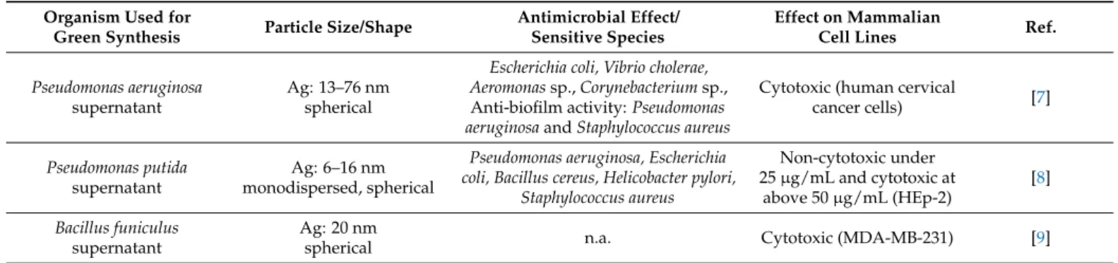
Table 1.Characteristics and biological activities of microbe-mediated silver and gold nanoparticles.
Organism Used for
Green Synthesis Particle Size/Shape Antimicrobial Effect/
Sensitive Species
Effect on Mammalian
Cell Lines Ref.
Pseudomonas aeruginosa supernatant
Ag: 13–76 nm spherical
Escherichia coli, Vibrio cholerae, Aeromonassp.,Corynebacteriumsp., Anti-biofilm activity:Pseudomonas aeruginosaandStaphylococcus aureus
Cytotoxic (human cervical
cancer cells) [7]
Pseudomonas putida supernatant
Ag: 6–16 nm monodispersed, spherical
Pseudomonas aeruginosa, Escherichia coli, Bacillus cereus, Helicobacter pylori,
Staphylococcus aureus
Non-cytotoxic under 25µg/mL and cytotoxic at
above 50µg/mL (HEp-2)
[8]
Bacillus funiculus supernatant
Ag: 20 nm
spherical n.a. Cytotoxic (MDA-MB-231) [9]
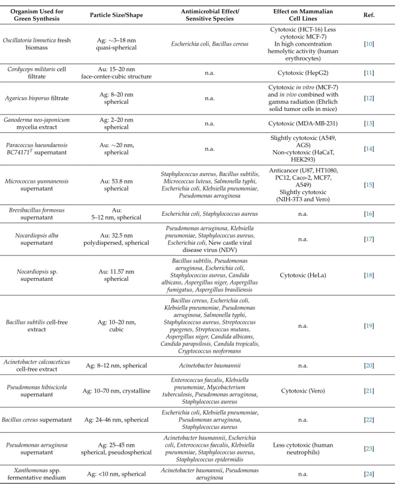
Table 1.Cont.
Organism Used for
Green Synthesis Particle Size/Shape Antimicrobial Effect/
Sensitive Species
Effect on Mammalian
Cell Lines Ref.
Oscillatoria limneticafresh biomass
Ag:∼3–18 nm
quasi-spherical Escherichia coli, Bacillus cereus
Cytotoxic (HCT-16) Less cytotoxic MCF-7) In high concentration hemolytic activity (human
erythrocytes)
[10]
Cordyceps militariscell filtrate
Au: 15–20 nm
face-center-cubic structure n.a. Cytotoxic (HepG2) [11]
Agaricus bisporusfiltrate Ag: 8–20 nm
spherical n.a.
Cytotoxicin vitro(MCF-7) andin vivocombined with gamma radiation (Ehrlich solid tumor cells in mice)
[12]
Ganoderma neo-japonicum mycelia extract
Ag: 2–20 nm
spherical n.a. Cytotoxic (MDA-MB-231) [13]
Paracoccus haeundaensis BC74171Tsupernatant
Au:∼20 nm,
spherical n.a.
Slightly cytotoxic (A549, AGS)
Non-cytotoxic (HaCaT, HEK293)
[14]
Micrococcus yunnanensis supernatant
Au: 53.8 nm spherical
Staphylococcus aureus, Bacillus subtilis, Micrococcus luteus, Salmonella typhi, Escherichia coli, Klebsiella pneumoniae,
Pseudomonas aeruginosa
Anticancer (U87, HT1080, PC12, Caco-2, MCF7,
A549) Slightly cytotoxic (NIH-3T3 and Vero)
[15]
Brevibacillus formosus supernatant
Au:
5–12 nm, spherical Escherichia coli, Staphylococcus aureus n.a. [16]
Nocardiopsis alba supernatant
Au: 32.5 nm polydispersed, spherical
Pseudomonas aeruginosa, Klebsiella pneumoniae, Staphylococcus aureus, Escherichia coli,New castle viral
disease virus (NDV)
n.a. [17]
Nocardiopsissp.
supernatant
Au: 11.57 nm spherical
Bacillus subtilis, Pseudomonas aeruginosa, Escherichia coli, Staphylococcus aureus, Candida albicans, Aspergillus niger, Aspergillus
fumigatus, Aspergillus brasiliensis
Cytotoxic (HeLa) [18]
Bacillus subtiliscell-free extract
Ag: 10–20 nm, cubic
Bacillus cereus, Escherichia coli, Klebsiella pneumoniae, Pseudomonas
aeruginosa, Salmonella typhi, Staphylococcus aureus, Streptococcus
pyogenes, Streptococcus mutans, Aspergillus niger, Candida albicans, Candida parapsilosis, Candida tropicalis,
Cryptococcus neoformans
n.a. [19]
Acinetobacter calcoaceticus
cell-free extract Ag: 8–12 nm, spherical Acinetobacter baumannii n.a. [20]
Pseudomonas hibiscicola
supernatant Ag: 10–70 nm, crystalline
Enterococcus faecalis,Klebsiella pneumoniae,Mycobacterium tuberculosis,Pseudomonas aeruginosa,
Staphylococcus aureus
Cytotoxic (Vero) [21]
Bacillus cereussupernatant Ag: 24–46 nm, spherical
Escherichia coli,Klebsiella pneumoniae, Pseudomonas aeruginosa,
Staphylococcus aureus
n.a. [22]
Pseudomonas aeruginosa supernatant
Ag: 25–45 nm spherical, pseudospherical
Acinetobacter baumannii,Escherichia coli,Enterococcus faecalis,Klebsiella pneumoniae,Staphylococcus aureus,
Staphylococcus epidermidis
Less cytotoxic (human
neutrophils) [23]
Xanthomonasspp.
fermentative medium Ag: <10 nm, spherical Acinetobacter baumannii,Pseudomonas
aeruginosa n.a. [24]
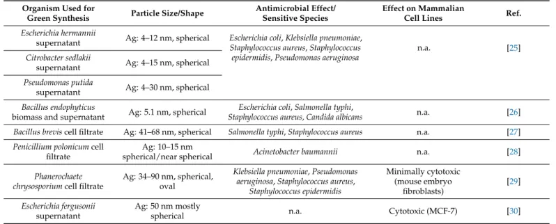
Table 1.Cont.
Organism Used for
Green Synthesis Particle Size/Shape Antimicrobial Effect/
Sensitive Species
Effect on Mammalian
Cell Lines Ref.
Escherichia hermannii
supernatant Ag: 4–12 nm, spherical Escherichia coli,Klebsiella pneumoniae, Staphylococcus aureus,Staphylococcus epidermidis,Pseudomonas aeruginosa
n.a. [25]
Citrobacter sedlakii
supernatant Ag: 4–15 nm, spherical Pseudomonas putida
supernatant Ag: 4–30 nm, spherical Bacillus endophyticus
biomass and supernatant Ag: 5.1 nm, spherical Escherichia coli,Salmonella typhi,
Staphylococcus aureus, Candida albicans n.a. [26]
Bacillus breviscell filtrate Ag: 41–68 nm, spherical Salmonella typhi,Staphylococcus aureus n.a. [27]
Penicillium polonicumcell filtrate
Ag: 10–15 nm
spherical/near spherical Acinetobacter baumannii n.a. [28]
Phanerochaete chrysosporiumcell filtrate
Ag: 34–90 nm, spherical, oval
Klebsiella pneumoniae,Pseudomonas aeruginosa,Staphylococcus aureus,
Staphylococcus epidermidis
Minimally cytotoxic (mouse embryo
fibroblasts)
[29]
Escherichia fergusonii supernatant
Ag: 50 nm mostly
spherical n.a. Cytotoxic (MCF-7) [30]
Non-cancerous cells: HaCaT (human keratinocyte cell line), 3T3-L1 (murine pre-adipocytes), HDF (human dermal fibroblasts), human lymphocytes, HUVEC (human endothelial cell line), ECV304 (human endothelial cell line), PBMC (human peripheral blood mononuclear cells), hSSCs (human neonatal skin stromal cells), L929 (murine fibroblast cell line), primary human fibroblast, NIH-3T3 (murine fibroblast cell line), rat splenocytes, HEK-293 (human embryonic kidney). Cancerous cells: B16 (murine melanoma cell line), HCT-116 (colon cancer cell line), A549 (human lung adenocarcinoma cancer cell line), MCF-7 (human breast adenocarcinoma cell line), AGS (human adenocarcinoma cell line), H1299 (human non-small lung carcinoma cancer cell line), VCaP (human prostate cancer cell line), BxPC-3 (human pancreas cancer cell line) MDA-MB-231 (human breast adenocarcinoma cell line), HCT116 (human colorectal carcinoma cell line), SH-SY5Y (human neuroblastoma cell line), COLO205 (human colon adenocarcinoma), HeLa (human cervical adenocarcinoma cell line), Hep2 (human carcinoma cell line), A2780 (ovarian carcinoma cancer cell line), HT115 (colon cancer cell line), HT29 (human colorectal adenocarcinoma cell line), melanoma cells, EAC (Ehrlich Ascites Carcinoma), Jurkat cells (immortalized line of human T lymphocytes).
Recently, various microorganisms, mainly bacteria and fungi, have been engaged to produce different metal nanoparticles, such as silver, gold, silver–gold alloy, iron, copper, zinc, palladium and titanium nanomaterials [31,32]. The earliest studies in the research area pointed out that microorganisms have always had a direct or indirect interaction with inorganic materials via geochemical biological processes, originating essentially from the beginning of life; therefore, microorganism-assisted particle synthesis should be regarded as a viable green option and shall be exploited even further. The synthesis of NPs via microbes is a bottom-up approach where nanoparticles are formed as a part of a defense mechanism- based detoxification, as a fundamental survival procedure involving oxidation/reduction of metal ions, generating phosphate, carbonate and sulfide metal forms, or volatilization of metal ions [33]. These processes are carried out by biomolecules, such as various proteins, enzymes, carbohydrates, sugars etc., of the microorganism; however, the exact events of the nanoparticle synthesis have not been fully elucidated yet [34]. The difficulty of identifying the precise mechanism and the active components responsible for the generation of nanoparticles lies in the fact that each kind of microorganism interacts in a different way with a particular metal ion and that the morphology, size and surface properties of the nanoparticles formed are greatly influenced by numerous other factors (mainly environmental conditions such as pH, pressure, temperature), not simply by the biological ingredients of the applied organism [35].
Certain metals, such as silver, are well known for their toxic effects; however, some silver-resistant bacteria can accumulate metals on/in their cell wall. This phenomenon is responsible for the idea of the first pioneering silver nanoparticle synthesis using a silver- resistant bacteriumPseudomonas stutzeri[36]. Samadi and co-workers demonstrated similar results obtained viaProteus mirabilisbacteria [37]. They also showed that a change in the culturing parameters can massively influence the formation of nanoparticles. Although this conclusion seemed quite unpleasant, it also offered the possibility of modulating
nanoparticle features by varying the experimental conditions and shifting the particle syntheses into a more favorable outcome.
Nanoparticles can be generated by microbes either intra- or extracellularly [38]. The intracellular mechanisms involve three main steps called trapping, reduction and stabiliza- tion. They rely primarily on the transport of the metal ions into the microbial cell wall. The method involves electrostatic interactions between the negatively charged cell wall and the positively charged metal ions. Then, enzymes residing within the cell wall reduce the toxic metals to harmless nanoparticles, and subsequently, these particles diffuse through the cell wall. Several reports suggested that metal NPs, such as silver and gold, can be easily and readily biosynthesized intracellularly. For example, by usingPseudomonasand Bacillusstrains, small, monodispersed gold nanoparticles were produced [39]. Nair et al.
successfully extended this synthesis method to prepare silver, gold and silver-gold alloy nanoparticles [40]. It was also proposed that the formation of nanoparticles using certain yeast strains could carry the greatest potential for nanoparticle manipulation, especially in case of maneuvering nanoparticle shape and size, by controlling culture parameters such as growth and other cellular activities [41].
As for the extracellular nanoparticle synthesis, metal ions on the surface of the cells are converted to metal nanoparticles by microbial enzymes, generally by nitrate reductase or hydroquinone-mediated redox reactions [42]. Successful extracellular biosynthesis of silver and gold nanoparticles was achieved usingAspergillus,FusariumandRhodopseudomonas strains [43–45]. Moreover, Lengke et al. reported that during extracellular synthesis using the cyanobacteriumPlectonema boryanum, the size and shape of the formed nanoparticles could be controlled very simply, simply by varying the external temperatures [46]. It is noteworthy that the recovery of metals from the environment by their adsorption onto bacteria also results in bioreduction, yielding metal nanoparticles [47].
In addition to the above described examples, several strains, such asPseudomonas fluo- rescens,Geobacillus stearothermophilusandStaphylococcus epidermidishave been successfully applied for the bioproduction of spherical gold nanoparticles in the size range of 5 and 90 nm [48]. Shape selectivity was again observed upon varying the culture conditions.
Gold and silver nanoparticles in various shapes (such as spherical and triangular) have been synthesized using algal strains such asPadina gymnosporaandEcklonia cava. One such study showed that the astaxanthin-containing green algaChlorella vulgariscan also be applied for gold nanoparticle synthesis [49]. Based on this finding, we utilizedPhaffia rhodozyma(perfect stateXanthophyllomyces dendrorhous), a basidiomycetous red yeast with high astaxanthin content for microbe-assisted nanoparticle synthesis [50]. The cell-free extract ofP. rhodozymaprovided almost monodisperse, well-separated and spherical silver and gold nanoparticles with a narrow size distribution (see Table1) [5].
Numerous yeasts, fungal and actinomycete strains, and even viruses, have been utilized to assemble gold nanoparticles to form microstructures [48]. Nevertheless, a high number of studies highlighted that among microorganisms, fungi-mediated syntheses hold major advantages over bacteria-, algae- or virus-assisted approaches. They justified this rationale as metal ion conversion to nanoparticles by means of fungal cells offers the easiest and most straightforward procedures to control nanoparticle size, shape and achieve monodispersity. As an example, fungi and yeast strains were used to demonstrate that by varying the pH and temperature during culturing, the size and shape of gold particles can be perfectly adjusted, and that decreasing the pH results in nanoplate formation instead of nanoparticles [45].
Despite the huge potential of using microorganisms for nanomaterial production, there are some limitations which should be considered before use. In fact, using biological agents for the synthesis of silver and gold NPs is preferable over chemical methods due to the simple, eco-friendly approach and also for minimizing the application of harmful chemical solvents and reagents. However, after carefully examining the above-presented microbe-assisted syntheses and the applied biological entities, it can be concluded that despite these approaches being relatively straightforward and favorable, they require
specific and rather tedious preparations and multistep processes such as culture isolation, maintenance or growth and inoculum standardization. Moreover, on the surface of the obtained particles, multicomponent residuals from microorganisms can accumulate, which would not only define the physical, chemical and biological characteristics of the obtained nanomaterials and their fate in the presence of living systems, but would also trigger potential immunological reactions after entering the organism. For this reason, the synthesis of metal particles using plants or any part of plants came forth and were later more and more prioritized. These reactions tend to be faster than those performed by microorganisms, are more cost effective and are relatively easy to scale-up for the generation of larger amounts of nanoparticles.
2.2. Synthesis of Silver and Gold Nanoparticles by Plants
As discussed above, nanoparticle synthesis using microorganisms is often rather slow, as the availability and maintenance of the various species used in the process is difficult and expensive; moreover, their application on a large scale is fairly restricted [51].
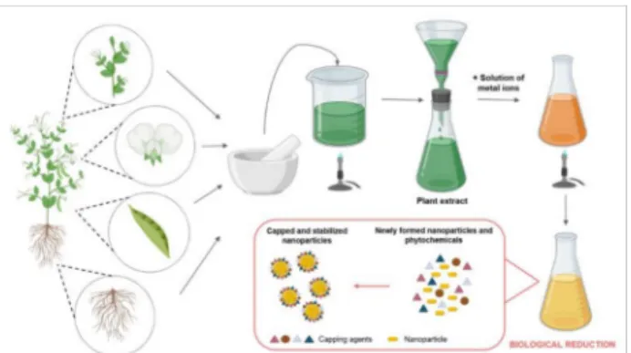
On the other hand, plant-mediated synthesis of metal nanoparticles grants numerous benefits over chemical, physical and microbial methods due to its rapid, well-reproducible, ecological, environmentally friendly, inexpensive procedure that can also be applied readily on an industrial scale [52–54]. Therefore, utilization of biological extracts obtained from different plant parts (leaf, fruit, seed, stem, callus, peel and root) for the production of metal nanoparticles such as silver and gold has attracted an extensive amount of interest from the nanobiotechnology research community. As a consequence, a plethora of research papers has been published in the last decade dealing with synthesis approaches using plants, plant extracts or biomolecules deriving directly from plants or plant parts (see Figure1and Tables2and3) [55,56]. Plants contain complex structures that can be used in the reduction and stabilization of the nanoparticles [57]. Plant materials generate nanoparticles by taking up, utilizing, accumulating and using different nutrients [58]. The general protocol for a typical plant-mediated metal nanoparticle synthesis requires first the collection and the purification of the plant part of interest [59]. The plant piece is then dried and powdered.
For the plant extract preparation, usually, deionized distilled water is added to the plant powder according to the desired concentration. This solution is boiled and finally filtered.
A certain volume of the extract is mixed with the appropriate amount of metal salt solution and the mixture is heated to the necessary temperature for the prescribed time under efficient mixing. To achieve the desired nanoparticles, optimization of every protocol is mandatory using different temperatures, solvents, pH conditions, extract concentrations and incubation times [60,61]. The reduction of metal ions to metal nanoparticles results in a color change of the solution, which can then be monitored by assessing UV-visible spectra.
The obtained nanoparticles are usually further characterized using an X-ray diffractometer, scanning or transmission electron microscopy (for the characterization methods, please refer to the next chapter of the present review).
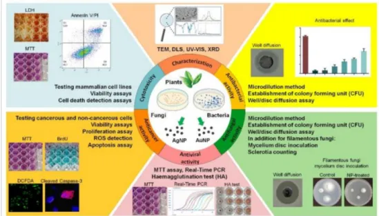
Figure 1.The general steps of green synthesis of inorganic nanoparticles using plant extracts. The figure was created withBioRender.com.
Table 2.Characteristics and biological activities of plant-mediated silver nanoparticles.
Organism Used for the
Synthesis Particle Size/Shape Antimicrobial Effect/Sensitive Species
Effect on Mammalian
Cell Lines Ref.
Ziziphus mauritianaleaf 3–20 nm, spherical
Escherichia coli,Staphylococcus aureus,Pseudomonas aeruginosa,
Bacillus subtilis
n.a. [62]
Sugarcane leaf (Saccharum
officinarum) 20–50 nm, spherical Phytophthora capsici,Colletotrichum
acutatum, Cladosporium fulvum n.a. [61]
Salvia spinosaplant 19–125 nm, rounded Bacillus subtilis,Bacillus vallismortis,
Escherichia coli n.a. [63]
Coffea arabicaseed 10–40 to 20–150 nm Escherichia coli, Staphylococcus
aureus n.a. [64]
Achillea biebersteiniiflower 10–40 nm, spherical and
pentagonal n.a. Cytotoxic (MCF-7) [65]
Lantana camaraleaf 14–27 nm, spherical Escherichia coli,Pseudomonasspp.,
Bacillusspp.,Staphylococcusspp. n.a. [66]
Black tea leaf 9–15 nm, spherical n.a. Cytotoxic (MCF-7) [67]
Green tea leaf (Richun Tea) 20–90 nm, spherical Escherichia coli n.a. [68]
Green tea leaf (R.
Twining), coffee seed (Tchibo Family)
3–12 nm, spherical
Bacillus cereusvar.mycoides, Micrococcus luteus,Escherichia coli,
Pseudomonas aeruginosa, Saccharomyces cerevisiae,Candida
parapsilosis,Candida albicans, Cryptococcus neoformans
Anti-proliferative, cytotoxic (HeLa and
NIH/3T3)
[69]
Green tea leaf (R. Twining) 5–15 nm, spherical, polyhedron
Escherichia coli, Bacillus megaterium, Cryptococcus neoformans
Cytotoxic (A549 and
NIH/3T3) [70]
Azadirachta indicaleaf ~34 nm, spherical Escherichia coli, Staphylococcus
aureus n.a. [71]
Mussaenda glabrataleaf 55 nm, spherical and triangular
Bacillus pumilus,Staphylococcus aureus,Pseudomonas aeruginosa, Escherichia coli,Aspergillus niger,
Penicillium chrysogenum
n.a. [72]
Plumbago zeylanicabark 10–25 nm, spherical
Escherichia coli,Pseudomonas aeruginosa, Bacillus subtilis, Staphylococcus aureus,Candida
tropicalis
Cytotoxic (Dalton
Lymphoma Ascites) [73]
Panax ginseng
fresh leaf 5–15 nm, spherical n.a.
Non-cytotoxic (HaCaT 20µg/mL) Cytotoxic (3T3-L1
20µg/mL)
[74]
Panax ginseng
berry 10–20 nm, spherical n.a. Cytotoxic (B16)
Less cytotoxic (HDF) [75]
Dimocarpus longandried
peel 8–22 nm, spherical n.a.
Cytotoxicin vitroand in vivo(H1299) Less cytotoxicin vitro
(VCaP, BxPC-3)
[76]
Dendropanax morbiferaleaf 100–200 nm; polygonal
and hexagonal n.a.
Cytotoxic (A549) Cytotoxic in high concentration (HaCaT)
[77]
Anemarrhena asphodeloides rhizome
20 nm, crystalline
face-centered cubic n.a.
Cytotoxic (A549, HT29, MCF-7) Slightly toxic (3T3-L1)
[78]
Artemisia turcomanicaleaf
extract 20–60 nm, spherical n.a. Cytotoxic (AGS, L929) [79]
Juglans regiawalnut green
husk 30–50 nm, spherical Escherichia coli,Pseudomonas aeruginosa,Staphylococcus aureus
Cytotoxic (MCF-7)
Non-cytotoxic (L929) [80]
Table 2.Cont.
Organism Used for the
Synthesis Particle Size/Shape Antimicrobial Effect/Sensitive Species
Effect on Mammalian
Cell Lines Ref.
Black tea (Tetley, England) 30–40 nm, spherical n.a.
Cytotoxic (A2780) Slightly cytotoxic (HCT116, primary human
fibroblast)
[81]
Fenugreek leaf 20–30 nm, spherical Escherichia coli,Staphylococcus
aureus Non-cytotoxic (HaCaT) [82]
Pimpinella anisumseed 3–16 nm (average 8.3 nm), spherical
Staphylococcus pyogenes, Acinetobacter baumannii,Klebsiella
pneumoniae,Salmonella typhi, Pseudomonas aeruginosa
Cytotoxic (HT115, hSSCs) [83]
Ficus religiosaleaf extract 3–28 nm (average 21 nm), spherical
Escherichia coli,Pseudomonas fluorescens,Bacillus subtilis,
Salmonella typhi
Cytotoxic (A549, Hep2, HeLa, COLO205,
SH-SY5Y)
[84]
Alternanthera dentataleaf 20–90 nm, spherical
Enterococcus faecalis,Escherichia coli, Klebsiella pneumoniae,Pseudomonas
aeruginosa
n.a. [85]
Boerhaavia diffusaplant 25 nm, spherical
Aeromonas hydrophila, Flavobacterium branchiophilum,
Pseudomonas fluorescens
n.a. [86]
Tribulus terrestrisdried
fruit 16–28 nm, spherical
Bacillus subtilis,Escherichia coli, Pseudomonas aeruginosa, Staphylococcus aureus,Streptococcus
pyogenes
n.a. [87]
Cocos nuciferainflorescence 22 nm, spherical
Bacillus subtilis,Klebsiella pneumoniae,Pseudomonas aeruginosa,Salmonella paratyphi
n.a. [88]
Abutilon indicumleaf 7–17 nm, spherical
Escherichia coli,Bacillus subtilis, Salmonella typhi,Staphylococcus
aureus
n.a. [89]
Cymbopogon citratusfresh
leaf 32 nm, spherical
Escherichia coli,Klebsiella pneumoniae,Proteus mirabilis, Salmonella typhi,Staphylococcus aureus, Aspergillus niger,Candida
albicans
n.a. [90]
Tinospora cordifoliastem 83 nm, spherical Staphylococcus aureus n.a. [91]
Eucalyptus citriodoraleaf 8–15 nm, spherical Acinetobacter baumannii n.a. [92]
Argemone mexicanaleaf 20 nm, spherical Escherichia coli, Pseudomonas
aeruginosa n.a. [93]
Solanum torvumleaf 14 nm, spherical
Pseudomonas aeruginosa, Staphylococcus aureus, Aspergillus
flavus,Aspergillus niger
n.a. [94]
Aloe veraplant 70–192 nm, spherical Pseudomonas aeruginosa,
Streptococcus epidermidis Non-cytotoxic (PBMC) [95]
Trianthema decandraroot 36–74 nm, spherical
Bacillus subtilis,Enterococcus faecalis, Escherichia coli,Proteus vulgaris,
Pseudomonas aeruginosa, Staphylococcus aureus,Streptococcus
faecalis,Yersinia enterocolitica, Candida albicans
n.a. [96]
Pongamia pinnatafresh
bark 5–55 nm, spherical
Bacillus subtilis,Klebsiella planticola, Klebsiella pneumoniae, Staphylococcus aureus
n.a. [97]
Ocimum sanctumleaf 18 nm, spherical Escherichia coli,Staphylococcus
aureus n.a. [98]
Catharanthus roseusleaf 48–67 nm, spherical
Bacillus cereus,Escherichia coli, Klebsiella pneumoniae,Pseudomonas
aeruginosa,Staphylococcus aureus
n.a. [99]
Cochlospermum gossypium 3 nm, spherical Escherichia coli,Pseudomonas
aeruginosa,Staphylococcus aureus n.a. [100]
Table 2.Cont.
Organism Used for the
Synthesis Particle Size/Shape Antimicrobial Effect/Sensitive Species
Effect on Mammalian
Cell Lines Ref.
Olive leaf 20–25 nm, spherical Escherichia coli,Pseudomonas
aeruginosa,Staphylococcus aureus n.a. [101]
Withania somniferaleaf 5–30 nm, spherical
Aspergillus niger,Staphylococcus aureus, Escherichia coli,Aspergillus
flavus, Candida albicans
n.a. [102]
Datura stramoniumleaf 15–20 nm, spherical Escherichia coli,Staphylococcus
aureus n.a. [103]
Emblica officinalisfruit 15 nm, spherical
Bacillus subtilis,Escherichia coli, Klebsiella pneumoniae, Staphylococcus aureus
n.a. [104]
Crataegus douglasiifruit 29 nm, spherical Escherichia coli,Staphylococcus
aureus n.a. [105]
Acalypha indicaleaf 20–30 nm, spherical Escherichia coli,Vibrio cholerae n.a. [106]
Solanum indicumplant 10–50 nm, spherical Klebsiellasp.,Staphylococcussp. Cytotoxic (rat splenocytes) [107]
Citrus sinensispeel 35 nm, 10 nm, spherical Escherichia coli,Pseudomonas
aeruginosa,Staphylococcus aureus n.a. [108]
Hibiscus rosa-sinensispetal 76 nm, spherical
Escherichia coli,Klebsiella pneumoniae,Staphylococcus aureus,
Vibrio cholerae
n.a. [109]
Daucus carotafresh extract 20 nm, spherical
Bacillus cereus,Klebsiella pneumoniae,Pseudomonas aeruginosa,Staphylococcus aureus
Non-cytotoxic (EAC cells) [110]
Melissa officinalisleaf 12 nm, spherical Escherichia coli,Staphylococcus
aureus n.a. [111]
Phoenix dactyliferaroot hair 15–40 nm, spherical Escherichia coli, Candida albicans Cytotoxic (MCF-7) [112]
Annona muricataroot bark 22 nm
Bacillus subtilis,Staphylococcus aureus, Klebsiella pneumoniae, Escherichia coli, Pseudomonas
aeruginosa
n.a. [113]
Terminalia mantalyfresh leaf, stem bark and root
11–80 nm anisotropic
Haemophilus influenzae,
Streptococcus pneumoniae n.a. [114]
Acacia rigidulastem and root
22.46 nm spherical
Escherichia coli,Pseudomonas aeruginosa,Bacillus subtilis
Non-toxic inin vivomouse
model [115]
Lampranthus coccineus aerial part
10.12–27.89 nm
spherical HAV-10, HSV-1, CoxB4
Non-toxic (HeLa) [116]
Malephora luteaaerial part 8.91–14.48 nm
spherical HAV-10, CoxB4
See notes on the cell lines at Table1.
Table 3.Characteristics and biological activities of plant-mediated gold nanoparticles.
Organism Used for the
Synthesis Particle Size/Shape Antimicrobial
Effect/Sensitive Species
Effect on Mammalian
Cell Lines Ref.
Zingiber officinalerhizome 5–15 nm, spherical n.a. No aggregation with
human blood cells [117]
Mussaenda glabrataleaf 10–12 nm, spherical
Bacillus pumilus, Staphylococcus aureus, Pseudomonas aeruginosa, Escherichia coli,Aspergillus
niger, Penicillium chrysogenum
n.a. [72]
Alpinia nigraleaf 20–55 nm, spherical and hexagonal Bacillus subtilis,Escherichia
coli,Candida albicans n.a. [118]
Table 3.Cont.
Organism Used for the
Synthesis Particle Size/Shape Antimicrobial
Effect/Sensitive Species
Effect on Mammalian
Cell Lines Ref.
Uncaria gambir Roxb.leaf 11–31 nm, hexagonal, triangular Escherichia coli,
Staphylococcus aureus n.a. [119]
Plumbago zeylanicabark 3–52 nm, spherical
Escherichia coli, Pseudomonas aeruginosa,
Bacillus subtilis, Staphylococcus aureus,
Candida tropicalis, Aspergillus flavus
Cytotoxic (Dalton
Lymphoma Ascites) [73]
Olax scandensleaf 5–100 nm, mostly spherical n.a. Antiproliferative (A549,
MCF-7 and COLO 205) [120]
Panax ginseng
fresh leaf 10–20 nm, spherical n.a. Non-cytotoxic (HaCaT,
3T3-L1) [74]
Panax ginseng
berry 5–10 nm, spherical n.a. Non-cytotoxic (HDF, B16
murine cell line) [75]
Trichosanthes kirilowiiplant ∼50 nm, spherical n.a. Cytotoxic (HCT-116) [121]
Marsdenia tenacissimaplant ∼50 nm, spherical (anisotropic
shapes) n.a. Cytotoxic (A549) [122]
Nerium oleander
stem bark 10–100 nm, mostly spherical n.a.
Cytotoxic (MCF-7) Non-cytotoxic (Human
lymphocytes)
[123]
Tribulus terrestrisdried
fruit 7 mm and 55 mm, mostly spherical
multi-drug resistant strains ofHelicobacter
pylori
GNP7 cytotoxic at 200 µg/mL (AGS) GNP55 less cytotoxic
(AGS) GNP7 and GNP55 non-cytotoxic at the MIC
ofH. pylori(AGS)
[124]
Peltophorum pterocarpum
leaf ∼55 nm, primarily spherical n.a. Non-cytotoxic (HUVEC,
ECV-304) [125]
Dendropanax morbiferaleaf 10–20 nm; polygonal and hexagonal n.a. Non-cytotoxic (A549,
HaCaT) [77]
Anemarrhena asphodeloides
rhizome 10 nm, crystalline face-centered cubic n.a.
Non-cytotoxic, toxic only in high concentration (100
ug/mL) (A549, HT29, MCF-7, 3T3-L1)
[78]
Potato starch powder 20–30 nm, quasi-spherical
MDRE. coli, MRSA
Non-toxic (human dermal fibroblasts, melanoma
cells) [126]
Ag-Au composite: 9–10 nm, quasi-spherical
Cytotoxic (human dermal fibroblasts) Enhanced cytotoxic
(melanoma cells) Trapa natanspeel extract Ag-Au composite: 26–90 nm,
hexagonal, triangular, spherical n.a.
Cytotoxic (HeLa, MDA-MB-231, HCT116
cancer cells)
[127]
Trianthema decandraroot 33–65 nm, spherical, triangular, hexagonal and cubical
Bacillus subtilis, Enterococcus faecalis, Escherichia coli,Proteus
vulgaris,Pseudomonas aeruginosa,Staphylococcus
aureus,Streptococcus faecalis,Yersinia enterocolitica, Candida
albicans
n.a. [96]
Ananas comosusfruit 16 nm, anisotropic Streptobacillussp.,
Escherichia coli n.a. [128]
Table 3.Cont.
Organism Used for the
Synthesis Particle Size/Shape Antimicrobial
Effect/Sensitive Species
Effect on Mammalian
Cell Lines Ref.
Annona muricataleaf 25.5 nm, spherical
Staphylococcus aureus, Enterococcus faecalis, Klebsiella pneumoniae, Clostridium sporogenes, Aspergillus flavus, Candida
albicans, Fusarium oxysporum, Penicillium
camemberti
n.a. [129]
Allium cepa 11 nm, spherical measles virus n.a. [130]
Azima tetracanthaleaf 80 nm, spherical
Aeromonas liquefaciens, Enterococcus faecalis,
Micrococcus luteus, Salmonella typhimurium,
Candida albicans, Cryptococcussp., Microsporum canis, Trichophyton rubrum
n.a. [131]
Caulerpa racemosagreen
seaweed 13.7–85.4 nm, spherical to oval Aeromonas veronii,
Streptococcus agalactiae Cytotoxic (HT-29) [132]
Nepenthes khasianaleaf 50–80 nm, spherical
Escherichia coli,Bacillussp., Aspergillus niger,Candida
albicans
n.a. [133]
Salix alba 63 nm, spherical
Staphylococcus aureus, Alternaria solani, Aspergillus niger, Aspergillus flavus
n.a. [134]
Curcumin 11.95 nm, lattice respiratory syncytial virus
(RSV) n.a. [135]
Abelmoschus esculentus
pulp 14 nm, spherical
Bacillus subtilis, Bacillus cereus, Pseudomonas aeruginosa, Micrococcus
luteus, Escherichia coli
Cytotoxic (Jurkat cells) [136]
Spirulina platensisgreen
alga 5 nm, spherical Staphylococcus aureus,
Bacillus subtilis [137]
Halymenia dilatatared alga 16 nm, spherical, triangular Aeromonas hydrophila Cytotoxic (HT-29) [138]
Acanthophora spicifera
seaweed >20 nm, spherical Vibrio harveyi,
Staphylococcus aureus Cytotoxic (HT-29) [139]
Gelidium pusillumseaweed ∼55 nm, spherical n.a. Cytotoxic (MDA-MB-231)
Non-cytotoxic (HEK-293) [140]
See notes on the cell lines at Table1.
The possible mechanisms of nanoparticle formation using plant extracts have been examined by several authors [63]. Two main theoretical directions have been suggested: 1.
Some studies proposed that the bioreduction of the metal ions was the result of trapping these on the protein surface due to electrostatic interactions between the metal ions and the proteins in the plant material extract. Proteins would reduce the metal ions, which ultimately leads to a change in the secondary structure of proteins and also to the formation of metal nanoparticle seeds or, as these are called, nuclei. The formed nuclei increase gradually in size upon accumulation and further reduction of metal ions on the nuclei, leading to the formation of nanoparticles [141]. 2. The second, generally more accepted approach is that the key mechanism behind the plant-mediated synthesis of nanoparti- cles is a plant-assisted reduction of the metal ions due to various phytochemicals [142].
Based on available literature data, it is probably not one biomolecule that is responsible for the reduction of metal ions, but several plant components and secondary metabolites to- gether are accountable [106,143]. Such active components include various proteins, among them numerous enzymes, amino acids, vitamins, polysaccharides, alkaloids, polyphenols,
flavonoids and organic acids, which are known to be non-toxic and biodegradable, and during nanoparticle synthesis, these can act both as reducing and capping agents, thus promoting the formation of nanoparticles and inhibiting their agglomeration [144].
On Au+-dihydromyricetin [145] and Ag+-hydrolysable tannin [89] pairs, the mech- anism of particle formation upon the reduction of metal ions with a plant extract has been analyzed in detail. Metal ions first form complexes with the phenolic hydroxyl groups in the biomolecule; then, the ions are reduced to zero oxidation state metals, while the biomolecule is oxidized, which is then capable of stabilizing the particles in parallel (“capping”). Therefore there is no requirement for the addition of further capping and stabilizing agents during the synthesis. These syntheses are generally very simple: the nanoparticles form spontaneously by mixing the metal salt solution with the plant extract.
The formation time of the particles varies between a few minutes and a day, depending on the metal–plant extract pair used. It is also worth noting that the conjugatedπ-electron system of polyphenols and flavonoids allows the donation of electrons, or hydrogen atoms, from the hydroxyl groups to various free radicals, so these molecules have an antioxidant capacity, which can also extend the life of nanoparticles [146].
The properties and biological performance of silver and gold nanoparticles, which have been generated using plant extracts, are summarized in Tables2and3[147]. In 2003, Shankar and colleagues were among the first to report the rapid green production of silver nanoparticles [148]. In their experiments, large amounts of silver nanoparticles were synthesized using plant extracts made from the leaves of Geranium (Pelargonium graveolens) and Indian lilac (Azadirachta indica) mixed with an aqueous solution of silver nitrate [149].
Among the indisputable advantages of the process, the authors highlighted first of all the speed of the synthesis. The nanoparticles formed much faster during the reduction with the plant extract than in their previous experiments using microorganisms. In the same year, as another step toward plant-mediated nanobiotechnology, Gardea-Torresdey and colleagues reported the production of AgNP particles using Alfalfa (Medicago sativa) [150].
Since then, the synthesis of metal NPs has been performed by different research groups utilizing a great variety of plants, plant extracts and their molecular components, where the bioactive materials were used as reducing and stabilizing agents in the production of silver nanoparticles, e.g., extracts of camphor tree (Cinnamomum camphora), lemon balm (Melissa officinalis), peppers (Capsicum annuum), Japanese red pine (Pinus densiflora), ginkgo (Ginkgo biloba), kobus magnolia (Magnolia kobus), oriental planetree (Platanus orientalis) and common grape vine (Vitis vinifera) [4,151]. These plants contain large amounts of active compounds (e.g., polyphenols, flavonoids) that are suitable for the reduction of metal ions [64,152]. Baharara et al. suggested that phenolic groups and proteins in plant extracts are responsible for the reduction of silver ions [65]. Ajitha et al. showed that the reduction of silver ions can be attributed to the hydroxyl and carbonyl groups in the active components (e.g., flavonoids, terpenoids, phenols, proteins) of plant extracts [66].
Furthermore, they revealed that proteins and peptides form a protective coating around the particles, thereby increasing the stability of the particles and preventing their aggregation.
Nadagouda and colleagues were the first to produce silver nanoparticles using coffee and tea extract. In their work, they demonstrated that in addition to the use of plant extracts, no other stabilizing agent was required, as the active ingredients of the extracts served as both reducing and stabilizing agents during the synthesis [153]. This simple one-step synthesis method has also been successfully extended to produce palladium, gold and platinum nanoparticles.
Silver nanoparticles have also been produced with aqueous-alcoholic solutions of roasted coffee (Coffea arabica), green tea and black tea [54,58,59]. The authors found that caffeine and theophylline in the extracts were responsible for stabilizing the produced nanoparticles. Moreover, Dhand et al. described that chlorogenic acid is the major phenolic component in the coffee extract that plays an essential role in the reduction of silver ions [64].
Importantly, it has been proposed that the quality as well as the quantity of the potential reducing or stabilizing components of the plant extract used for the synthesis determine
the properties of the resulting particles (e.g., size, morphology), including their reactivity in subsequent reactions. Ashokkumar and co-workers observed that particle size decreased with increasing plant extract concentration, while—as described in another study—the number of particles formed correlated with the amount of plant extract used [89]. Moreover, shape selectivity was observed by varying the dose of the bioreducing agent. Chandran et al.
also achieved shape and size selectivity of produced silver nanoparticles by modulating the concentration of the starting metal salt solution and aloe vera plant extract [154]. Loo et al. produced round-shaped silver nanoparticles with green tea extract [155]. They made the observation that when the concentration of the extract was increased, the size of the nanoparticles decreased while the number of the particles increased [156].
Recent works pointed out that besides the nature of the plant extracts and the types and concentrations of the active biomolecules within, several factors, including reaction time and temperature, pH and the electrochemical potential, can have an effect on the reduction process [157]. For instance, it was demonstrated that increasing the temperature can improve the nucleation rate, leading to the synthesis of smaller AgNPs and to increased synthesis rate of AgNPs. Furthermore, it was also proven that proteins in the plant extract significantly affect the shape, size and yield of nanoparticles during synthesis [158,159].
Green synthesis of silver nanoparticles was performed using the aqueous solution of Ziziphus mauritianaleaves extract as a bio-reducing agent. In this study, the effects of the leaf extract and silver nitrate concentrations, as well as of the temperature on the preparation of nanoparticles were investigated in detail [62].
These green synthesized silver nanoparticles were often produced for specific ap- plication purposes—not necessarily for medical utilizations—and sometimes, only the possibility of nanoparticle synthesis using a given plant extract was tested. However, by their application, these nanoparticles could come into contact with living systems; therefore, several research groups following the generation of nanoparticles, rightly, examined their impact on different biological systems [160]. Despite these attempts to assess the effects of the as-prepared nanomaterial on some living organism, only a few studies have examined thoroughly, or compared the complex (antibacterial, antifungal, antiviral and cytotoxic) biological activity of the produced nanoparticles [161]. To follow the most approved char- acterization procedure, we have investigated the chemical and biological characteristics of nanoparticles prepared by coffee and tea extracts [69]. Our results clearly showed that green materials used for stabilization and for reduction of metal ions have a defining role in the biological activity of the obtained nanomaterial against bacteria, fungi or human cells.
Based on our results, we have recommended to obtain a circumspect selection of the green extracts used for the synthesis of nanoparticles, and suggested that a comprehensive screen of the products should be carried out prior to their applications to delineate their behavior in the presence of living systems. Based on today’s nanotechnology results, combining the available methods of biology, chemistry and material sciences, more complex inves- tigations can be carried out [162,163]. Using such an approach, systematic examinations regarding AgNP aggregation behavior with simultaneous measurements of its effect on biological activity can be performed to offer new frontiers to preserve nanoparticle toxicity by enhancing colloidal stability [70].
The synthesis and utilization of gold nanoparticles is an emerging research area due to the unique and tunable surface plasmon resonance and the electrical conductivity, excellent catalytic activity and the biomedical potential of AuNPs, including drug delivery, molecular imaging and biosensing [164]. Therefore, there is also a growing need for environmentally benign synthesis processes of these particles without losing sight of the major aim, i.e., to provide a safe application and avoid adverse effects in medical applications. To date, several examples of gold nanoparticles produced by plant extracts have been reported in the literature (see Table3) [165,166]. We observed that the same types of plants and their respective components are generally exploited for the synthesis of AuNPs as for AgNPs. The first to report the fabrication of gold nanoparticles using living plants was in 2002 by Gardea-Torresdey and co-workers [150]. They described the formation and
growth of AuNPs inside live alfalfa plants. In their study, alfalfa plants were grown in a tetrachloroaurate ion-rich environment. The absorption of gold metal by the plants was proven by transmission electron microscopy and X-ray absorption measurements.
Sesbania drummondiiseedlings were also successfully applied in a similar system [167].
Beattie and Haverkamp revealed that the bioreduction of gold salts to metal occurs in chloroplasts [168].
Although the idea of utilizing living plants is revolutionary; nevertheless, the purifi- cation of the intracellularly formed nanoparticles proved to be a difficult task. Therefore, extracellular syntheses of nanoparticles, which utilize the extracts of plants, have gained immediate popularity [169]. As was mentioned before, one of the first studies on the biosynthesis of metallic silver and gold nanoparticles using plant extracts was performed by Shankar et al. who applied geranium leaf extract during the synthesis [148]. These reactions lasted for 2 days and the generated nanoparticles had various morphologies, such as spherical, triangular, icosahedral and spherical. This method was later optimized using other plant extracts (neem leaf), where a shorter reaction time (~2.5 h) was achieved [149].
The rapid green synthesis of monodispersed, spherical gold nanoparticles with dimensions of ~20 nm was observed usingMangifera indicaleaf extract [170]. The reduction of gold cations to gold nanoparticles by this extract was completed within 2 min and the obtained colloid was found to be stable for more than 5 months. Highly stable crystalline gold NPs were produced usingMomordica charantiaas well [171].
Dwivedi et al. also reported about rapid biosynthesis of metal nanoparticles using Chenopodium album, where the leaf extract was successfully applied to obtain silver and gold nanoparticles in the size range of 10–30 nm. They observed not only shape selectivity, but noted that the formation of spherical nanoparticles was more favorable at higher leaf extract concentrations [172]. The synthesis of gold nanoparticles of various shapes (spherical, hexagonal and triangle) via olive leaf extract as a reducing agent has also been demonstrated. The size and the shape of gold NPs were modulated by varying the ratio of plant extract and the initial metal salt in the reaction medium [173]. The authors emphasized the role of the high phenolic content of the hot water extract of olive leaves, which helped in the reduction. They observed that the generated spherical particles were capped by phytochemicals. It is well known that plant and plant-based phytochemicals are rich in various polyphenols, flavonoids, terpenoids, aldehydes, proteins, alkaloids, acids and alcoholic compounds [71,174]. These active components are assumed to participate in the reduction of chloroauric acid to form AuNPs and serve as stabilizing agents to prevent particle aggregation. Smitha et al. achieved shape diversity, whenCinnamomum zeylanicum leaf broth was used as a reducing agent: the plant extract at lower concentrations caused formation of prism-shaped particles, while at higher concentrations spherical particles dominated [175]. Tansy fruit extract was successfully employed for the development of silver and gold nanoparticles with spherical and triangular shapes with an average size of ~15 nm [176]. These nanoparticles were found to have a crystalline structure with face- centered cubic geometry verified by the XRD method. Plant extract ofPulicaria undulata (L.) was used as both reducing agent and stabilizing ligand for the rapid and green synthesis of gold, silver and gold-silver bimetallic alloy nanoparticles. These nanoparticles showed composition-dependent catalytic activity [177].
Regarding the collected data on metal nanoparticle synthesis, one thing is certain, the possibilities are endless—almost any plant, or part of it, can be put to use for producing nanoparticles; gold NPs were prepared by an environmentally friendly, one-step synthesis process using leaf extracts from coffee, mint, mango, tea, grapes, lemon, eucalyptus, neem, roses, aloe vera, tamarind, coriander and peppermint [178–181]. However, the careful analysis of the synthesis approach and the physico-chemical properties of the realized nanomaterials accentuate the attentive selection of plant material for nanoparticle synthesis, as these biocomponents are inherently responsible for the morphology, stability and biological properties of the formed NPs.