Review article
Paraneoplastic neuromyelitis optica spectrum disorder: A case report and review of the literature
Ádám Annus
a, Krisztina Bencsik
a, Izabella Obál
a, Zsigmond Tamás Kincses
a,b, László Tiszlavicz
c, Romana Höftberger
d, László Vécsei
a,e,⇑aDepartment of Neurology, Faculty of General Medicine, University of Szeged, Szeged, Hungary
bInternational Clinical Research Center, St. Anne’s University Hospital Brno, Brno, Czech Republic
cDepartment of Pathology, Faculty of General Medicine, University of Szeged, Szeged, Hungary
dInstitute of Neurology, Medical University of Vienna, Vienna, Austria
eMTA-SZTE Neuroscience Research Group, Szeged, Hungary
a r t i c l e i n f o
Article history:
Received 15 June 2017 Accepted 10 October 2017
Keywords:
Neuromyelitis optica Aquaporin-4 Paraneoplastic
a b s t r a c t
Neuromyelitis optica spectrum disorders (NMOSD) are demyelinating, autoimmune diseases affecting the central nervous system. Typically, recurrent optic neuritis and longitudinal extensive transverse myelitis dominates the clinical picture. In most cases NMOSD are associated with autoantibodies target- ing the water channel aquaporin-4 (AQP-4). NMOSD usually present in young adults. Clinical findings suggestive of NMOSD in elderly patients should raise the suspicion of a paraneoplastic etiology. To our knowledge, we report the first case of a 66 year-old female patient with paraneoplastic NMOSD that is associated with squamous cell lung carcinoma. Anti-AQP-4 was present in both the serum and cere- brospinal fluid of the patient. However, immunhistological staining of the malignant tissue did not show presence of AQP-4 on the surface of tumour cells.
Ó2017 Elsevier Ltd. All rights reserved.
1. Introduction
Neuromyelitis optica spectrum disorders (NMOSD) are demyelinating, autoimmune diseases affecting the central nervous system (CNS). Typically, recurrent optic neuritis and longitudinal extensive transverse myelitis dominates the clinical picture. In most cases NMOSD are associated with autoantibodies (IgG) tar- geting the water channel aquaporin-4 (AQP-4) [1]. AQP-4 is a transmembrane protein found on the surface of astrocyte projec- tions constituting to the blood-brain barrier (BBB) and is concerned with regulating water movement between the cerebrospinal fluid (CSF), blood and brain[2]. NMOSD usually present in young adults.
Clinical findings suggestive of NMOSD in elderly patients should raise the suspicion of paraneoplastic aetiology.
To our knowledge, we report the first case of paraneoplastic NMOSD secondary to squamous cell lung carcinoma. We also aimed to summarize previously reported cases of paraneoplastic NMOSD.
2. Case report
A 66 year-old female patient presented to the Emergency Department with complaints of sudden onset left lower extremity weakness, numbness and reduced sensation that started 3 days prior to presentation. She was unable to walk on the second day of her illness. She also complained of sudden onset urinary incontinence.
She had a history of myelodysplastic syndrome (MDS) with refractory anaemia for which she received regular blood transfu- sions every 3–4 weeks. She smoked a pack of cigarette a day. She denied regular alcohol consumption or illicit drug use.
On admittance she had severe monoparesis (2/5) of the left leg with diminished reflexes and sensory loss. She had urinary incon- tinence. Contrast enhanced MRI of the lower thoracic and lumbar spine showed a long T2 hyperintense lesion extending from 4th to 10th thoracic vertebrae (Fig. 1). As an additional finding, a neo- plasm (approximately 20 mm in diameter) in the 6th segment of the left lung was suspected. A CT guided biopsy of the lesion was not successful. After consultation with a haematologist, despite of a possible lung tumour, to treat a suspected funicular myelosis, parenteral vitamin B12 supplementation was initiated. Over the next few days, the patient’s condition did not improve. Paresis and sensory disturbance of the right leg also developed. In the
https://doi.org/10.1016/j.jocn.2017.10.030 0967-5868/Ó2017 Elsevier Ltd. All rights reserved.
⇑ Corresponding author at: Department of Neurology, Albert Szent-Györgyi Clinical Center, University of Szeged, Semmelweis u. 6, 6725 Szeged, Hungary.
E-mail address:vecsei.laszlo@med.u-szeged.hu(L. Vécsei).
Journal of Clinical Neuroscience 48 (2018) 7–10
Contents lists available atScienceDirect
Journal of Clinical Neuroscience
j o u r n a l h o m e p a g e : w w w . e l s e v i e r . c o m / l o c a t e / j o c n
CSF mildly increased protein level (0.57 g/l; normal range: 0.2–0.4 g/l) and white cell count (8 white cells, mostly lymphocytes) were found. Furthermore, matching oligoclonal bands (OCB) in both the serum and CSF of the patient were detected. Tests for mycobacte- ria, CMV, HSV and EBV were negative. The patient’s autoimmune panel (including ANA, anti-dsDNA, anti-SS-A and SS-B, anti-Scl- 70, anti-Jo-1, anti-RNP-70, anti-RNP/Sm, anti-centromere B) was normal. The tumour marker, carcinoembryonic antigen (CEA) was elevated (8.17 ng/ml, normal range <4.70 ng/ml). Bone marrow- biopsy did not show signs of transformation of MDS to acute mye- loid leukaemia.
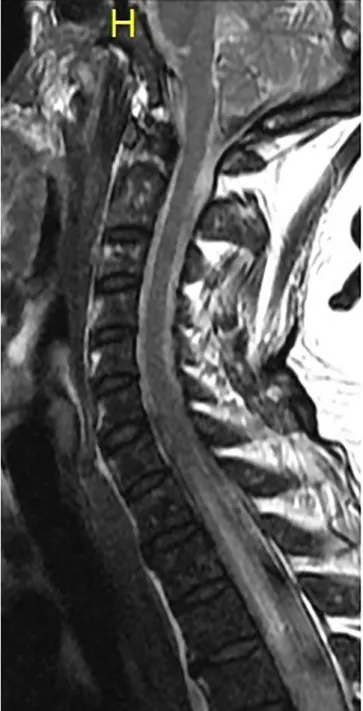
A further worsening of the patient’s condition was detected, paresis and sensory loss presented on the upper extremities and trunk. A contrast enhanced MRI of the cervical and upper thoracic spine found T2 hyperintense lesion in the spinal cord between the 7th cervical and the 10th thoracic segment (Fig. 2).
Later on, the patient complained of blurry vision on both eyes, more severely affecting the left eye. MRI of the brain showed T2
hyperintense lesions in both optic nerves, affecting the left nerve more profoundly. A repeated lumbar puncture found a protein level of 0.85 g/l and 15 white cells (dominantly lymphocytes) in the CSF. Furthermore, Lactobacillus crispatus cultured from the sample that was thought to be contamination, but antibiotic treat- ment, ceftriaxone was initiated.
Despite our efforts, the patient’s condition deteriorated further.
In the meantime we repeated the lung biopsy (only a small tissue sample was obtained), which confirmed a squamous cell carci- noma. AQP-4 autoantibodies were detected in the serum and in the CSF. No other autoantibodies (amphiphysin, CV2-CRMP5 [col- lapsin response mediator-protein-5], Ma2 [Ta], RI [ANNA2], Yo [PCA1], Hu [ANNA1]) were detected in the serum of the patient.
Based on the patient’s progressing neurological state, MRI findings (optic neuritis of both optic nerves and long extensive transverse Fig. 1.MRI of the lower thoracic and lumbar spine showing a long T2 hyperintense
lesion in the thoracic segment of the spinal cord.
Fig. 2.MRI of the cervical and upper thoracic spine showing T2 hyperintense lesion in the spinal cord below the 7th cervical segment.
8 Á. Annus et al. / Journal of Clinical Neuroscience 48 (2018) 7–10
myelitis) lung cancer, and the AQP-4 autoantibodies, we hypothe- sized that the patient had paraneoplastic NMOSD. The patient was in a severe condition (she had dyspnoea due to muscle weakness), therefore surgical removal of the tumour or plasmapheresis could not be performed due to the high mortality risk of these proce- dures. Intravenous immunoglobulin treatment was given, but no improvement was detected. Intravenous methylprednisolone had no beneficial effect either. Unfortunately, the patient died a few days later and the family did not consent to autopsy. Post- mortem immunhistological staining of the minute malignant tis- sue sample did not show presence of AQP-4 on the surface of tumour cells.
3. Discussion
NMOSD most commonly affect young adults (between 35 and 45 years of age) and there is a female predominance[3]. NMOSD can have a monophasic or relapsing course. The latter is more com- mon, presenting in approximately 80–90% of the cases. Anti-AQP-4 IgG is detected in the serum of most patients. Anti-AQP-4 has a specificity of more than 90% and a sensitivity of approximately 70% for NMOSD[4]. AQP-4 is found not only in astrocytic foot pro- cesses, but in the membrane of skeletal muscle, epithelia of breast and salivary glands, tracheal and bronchial epithelium, basolateral membranes of distal collecting tubules, parietal cells of the stom- ach, and epithelium of the colon[5]. Interestingly, these organs are rarely affected in anti-AQP-4 seropositive NMOSD cases. Chan et al. demonstrated that AQP-4 is also present on the surface of thymoma cells, but not in the membrane of normal thymus tissue [6]. Furthermore, they showed that anti-AQP-4 taken from serum of NMOSD patients bound to the receptors expressed by thymoma cells[6]. These findings could explain the frequent simultaneous presentation of myasthenia gravis and NMOSD[7,8]. Also, studies showed that AQP-4 is expressed in breast cancer, low-grade glio- mas and non-small cell lung cancer (NSCLC) cells even in patients not showing neurological symptoms[9–11]. AQP-4 might play a role in adhesion, migration and invasiveness of tumour cells.
NMOSD of paraneoplastic aetiology usually present in older ages compared to typical NMOSD and are subacute in onset[12].
Clinical signs of NMOSD in elderly patients, especially with a his- tory of smoking and autoimmune disease or cancer in the previous history or family, should raise the suspicion of paraneoplastic aeti- ology. Anti-AQP-4 detection in the serum and/or the CSF of patients can further direct the attention of clinicians towards para- neoplastic NMOSD. However, in approximately 10–20% of patients with clinical signs of NMOSD, anti-AQP-4 is absent in the serum [13]. There are a few proposed explanations to this finding[14].
(a) The detection methods might not be sensitive enough. (b) The patients might suffer from multiple sclerosis rather than NMOSD.
(c) Symptoms might not be related to autoimmune lesions in the CNS. (d) Other antibodies are responsible for CNS damage. It has been reported that paraneoplastic syndromes of optic neuropathy and myelitis associated with anti-CV2-CRMP5 (collapsin response mediator-protein-5), ANNA-1 (antineural nuclear antibody) or anti- amphyphisin can mimic NMOSD[15]. A few cases have been reported where anti-CV2/CRMP5 onconeural antibodies were pre- sent in anti-AQP-4 seronegative patients with symptoms of NMOSD[16,17]. Other antibodies in seropositive patients included ANNA-1, acetylcholine receptor-antibody, and N-type calcium channel (seeTable 1for references, which summarizes previously reported cases of paraneoplastic NMOSD). It is unknown whether these antibodies have an actual role in the development of CNS lesions in NMOSD or are just coincidental findings. Apart from anti-AQP-4, we could not detect any other autoantibodies in the serum of our patient.
Paraneoplastic neurological syndromes are the consequence of autoimmune response to neural antigens expressed by tumour cells[18]. The immune system recognizes antigens found on the surface of tumour cells which are also present in healthy tissues.
The immune system cannot differentiate between the two and attacks both. Immunhistological staining of our patient’s tissue sample taken from the lung tumour did not show AQP-4 on the surface of malignant cells. However, since the tissue sample was a very small one, we cannot rule out the possibility of the presence of AQP-4 in the membrane of tumour cells in other areas of the neoplasm. We base this hypothesis on the simultaneous presence of lung squamous cell carcinoma, characteristic symptoms of NMOSD and anti-AQP-4 antibodies in both the serum and CSF of the patient. We do not believe that these findings could be unrelated.
The development of paraneoplastic NMOSD is regulated by both the innate and adaptive immune system[3]. AQP-4 expressed on the surface of tumour cells, is possibly recognized as a foreign anti- gen by the immune system and therefore induces IgG production (anti-AQP-4) in the periphery. Anti-AQP-4 can access the CNS through the circumventricular organs[19]. This hypothesis is sup- ported by the fact that in NMOSD the area postrema and hypotha- lamus can be affected causing symptoms of nausea, hiccups, vomiting and diencephalic syndrome. Once in the CNS, anti-AQP- 4 binds to astrocytic foot processes containing AQP-4 to form an antigen-antibody complex. This leads to internalization and endolysosomic degradation of AQP-4 [19]. This finding is sup- ported by pathologic investigations which show reduced AQP-4 expression in CNS lesions of NMOSD patients[20]. The antigen- antibody complex causes complement activation and antibody dependent cellular cytotoxicity (ADCC) mediated by granulocytes and natural killer cells. The activation of the complement cascade leads to the formation of membrane attack complex and thus com- plement dependent cytotoxicity (CDC) and recruitment of more inflammatory cells. The degranulation of leukocytes augments inflammation in the CNS, disrupting the BBB and therefore initiat- ing a vicious circle by allowing more anti-AQP-4 to access the CNS.
ADCC and CDC directly cause astrocyte cell death, while it is believed that demyelination, oligodendroglial and neuronal cell death are secondary to inflammatory processes[21].
Kitazawa et al. reported a case of an 87 year-old patient who developed symptoms of NMOSD after the diagnosis of prostate adenocarcinoma and also after injection of 23-valent pneumococ- cal vaccination [22]. They hypothesized that the patient was an asymptomatic anti-AQP-4 carrier, until the prostate tumour induced CD4 T-lymphocyte activation and INF-
c
secretion. Accord- ing to the authors, anti-AQP-4 and T-cell mediated response alto- gether have resulted in NMOSD. Also, injection of the vaccine may have caused complement activation and together with the presence of anti-AQP-4 caused a relapse in the patient’s condition.This hypothesis is supported by two experiments, which found that injection of anti-AQP-4 from NMOSD patient into mice alone did not cause NMOSD-like lesions, only after induction of experi- mental autoimmune encephalomyelitis (EAE) or co-injection of human complement [23,24]. Armagan et al. reported a case of breast carcinoma and possibly paraneoplastic long extensive trans- verse myelitis (LETM) with anti-AQP-4 seropositivity[9]. Interest- ingly, they also reported 3 other cases of the same type of breast carcinoma with anti-AQP-4 in the patients’ serum; however, they did not develop neurological symptoms. These findings indicate that the presence of anti-AQP-4 in the serum alone is not enough for the development of NMOSD. Antibody induced T-cell activation and/or complement activation is also required. It is yet to be eluci- dated why anti-AQP-4 causes NMOSD symptoms in some individ- uals, and why it can be a coincidental finding in symptomless patients. It is possible that the presence of anti-AQP-4 needs a
Á. Annus et al. / Journal of Clinical Neuroscience 48 (2018) 7–10 9
longer period of time to provoke an autoimmune response leading to profound lesions in the CNS and causing the symptoms of NMOSD.
4. Conclusion
NMOSD in elderly patients are infrequent findings that should always raise the suspicion of a paraneoplastic aetiology and war- rants thorough investigations in search for an underlying cancer.
Risk factors include smoking, previous cancer or autoimmune dis- ease in the patient’s history and/or in the patient’s family history.
Treatment options comprise of oncologic therapy to eradicate the tumour and also immunosuppressive therapy (intravenous ster- oids, plasmapheresis, intravenous immunoglobulin, azathioprine or rituximab). The sooner the treatment is initiated, the better the prognosis for the patient, therefore early diagnosis of paraneo- plastic NMOSD is imperative for better outcome. It is also impor- tant to note, that the presence of anti-AQP-4 is not a necessity in the diagnosis of paraneoplastic NMOSD. Autoimmune reactions to other onconeural antigens, including CV2/CRMP5, ANNA-1 and amphyphisin can also cause symptoms of NMOSD.
Acknowledgements
The authors declare that there are no conflicts of interest. The study was supported by the ‘‘Neuroscience Research Group of the Hungarian Academy of Sciences and University of Szeged,” project FNUSA-ICRC (no. CZ.1.05/1.1.00/02.0123) from the European Regional Development Fund, the National Brain Research Program (Grant No. KTIA_13_NAP-A-II/20.) and an OTKA [PD 104715]c grant.
We are thankful for the Department of Immunology and Biotechnology, University of Pécs, Pécs, Hungary for the measure- ments of anti-AQP-4 in the serum and CSF of the patient using Western blot and immunofluorescence techniques.
Appendix A. Supplementary data
Supplementary data associated with this article can be found, in the online version, athttps://doi.org/10.1016/j.jocn.2017.10.030.
References
[1]Wingerchuk DM, Banwell B, Bennett JL, Cabre P, Carroll W, Chitnis T, et al.
International consensus diagnostic criteria for neuromyelitis optica spectrum disorders. Neurology 2015;85:177–89.
[2]Freitas E, Guimarães J. Neuromyelitis optica spectrum disorders associated with other autoimmune diseases. Rheumatol Int 2015;35:243–53.
[3]Pereira WL, Reiche EM, Kallaur AP, Kaimen-Maciel DR. Epidemiological, clinical, and immunological characteristics of neuromyelitis optica: a review.
J Neurol Sci 2015;355:7–17.
[4]Lennon VA, Wingerchuk DM, Kryzer TJ, Pittock SJ, Lucchinetti CF, Fujihara K, et al. A serum autoantibody marker of neuromyelitis optica: distinction from multiple sclerosis. Lancet 2004;364:2106–12.
[5]Pittock SJ, Lennon VA. Aquaporin-4 autoantibodies in a paraneoplastic context.
Arch Neurol 2008;65:629–32.
[6]Chan KH, Kwan JS, Ho PW, Ho SL, Chui WH, Chu AC, et al. Aquaporin-4 water channel expression by thymoma of patients with and without myasthenia gravis. J Neuroimmunol 2010;227:178–84.
[7]Kister I, Gulati S, Boz C, Bergamaschi R, Piccolo G, Oger J, et al. Neuromyelitis optica in patients with myasthenia gravis who underwent thymectomy. Arch Neurol 2006;63:851–6.
[8]McKeon A, Lennon VA, Jacob A, Matiello M, Lucchinetti CF, Kale N, et al.
Coexistence of myasthenia gravis and serological markers of neurological autoimmunity in neuromyelitis optica. Muscle Nerve 2009(39):87–90.
[9]Armag˘an H, Tüzün E, Içöz S, Birisßik O, Ulusoy C, Demir G, et al. Long extensive transverse myelitis associated with aquaporin-4 antibody and breast cancer:
favorable response to cancer treatment. J Spinal Cord Med 2012;35:267–9.
[10]Warth A, Muley T, Meister M, Herpel E, Pathil A, Hoffmann H, et al. Loss of aquaporin-4 expression and putative function in non-small cell lung cancer.
BMC Cancer 2011;11:161.
[11]Warth A, Simon P, Capper D, Goeppert B, Tabatabai G, Herzog H, et al.
Expression pattern of the water channel aquaporin-4 in human gliomas is associated with blood-brain barrier disturbance but not with patient survival. J Neurosci Res 2007;85:1336–46.
[12]McKeon A, Pittock SJ, Lennon VA. CSF complements serum for evaluating paraneoplastic antibodies and NMO-IgG. Neurology 2011;76:1108–10.
[13]Jarius S, Wandinger KP, Borowski K, Stoecker W, Wildemann B. Antibodies to CV2/CRMP5 in neuromyelitis optica-like disease: case report and review of the literature. Clin Neurol Neurosurg 2012;114:331–5.
[14]Papadopoulos MC, Verkman AS. Aquaporin 4 and neuromyelitis optica. Lancet Neurol 2012;11:535–44.
[15]Cai G, He D, Chu L, Dai Q, Xu Z, Zhang Y. Paraneoplastic neuromyelitis optica spectrum disorders: three new cases and a review of the literature. Int J Neurosci 2015;126:660–8.
[16]Cross SA, Salomao DR, Parisi JE, Kryzer TJ, Bradley EA, Mines JA, et al.
Paraneoplastic autoimmune optic neuritis with retinitis defined by CRMP-5- IgG. Ann Neurol 2003;54:38–50.
[17]Ducray F, Roos-Weil R, Garcia PY, Slesari J, Heinzlef O, Chatelain D, et al.
Devic’s syndrome-like phenotype associated with thymoma and anti-CV2/
CRMP5 antibodies. J Neurol Neurosurg Psychiatry 2007;78:325–7.
[18]Iorio R, Lennon VA. Neural antigen-specific autoimmune disorders. Immunol Rev 2012;248:104–21.
[19]Hinson SR, Pittock SJ, Lucchinetti CF, Roemer SF, Fryer JP, Kryzer TJ, et al.
Pathogenic potential of IgG binding to water channel extracellular domain in neuromyelitis optica. Neurology 2007;69:2221–31.
[20]Roemer SF, Parisi JE, Lennon VA, Benarroch EE, Lassmann H, Bruck W, et al.
Pattern-specific loss of aquaporin-4 immunoreactivity distinguishes neuromyelitis optica from multiple sclerosis. Brain 2007;130:1194–205.
[21]Ratelade J, Verkman AS. Neuromyelitis optica: aquaporin-4 based pathogenesis mechanisms and new therapies. Int J Biochem Cell Biol 2012;44:1519–30.
[22]Kitazawa Y, Warabi Y, Bandoh M, Takahashi T, Matsubara S. Elderly-onset neuromyelitis optica which developed after the diagnosis of prostate adenocarcinoma and relapsed after a 23-valent pneumococcal polysaccharide vaccination. Int Med 2012;51:103–7.
[23]Bradl M, Misu T, Takahashi T, Watanabe M, Mader S, Reindl M, et al.
Neuromyelitis optica: pathogenicity of patient immunoglobulin in vivo. Ann Neurol 2009(66):630–43.
[24]Saadoun S, Waters P, Bell BA, Vincent A, Verkman AS, Papadopoulos MC. Intra- cerebral injection of neuromyelitis optica immunoglobulin G and human complement produces neuromyelitis optica lesions in mice. Brain 2010;133:349–61.
10 Á. Annus et al. / Journal of Clinical Neuroscience 48 (2018) 7–10