Micron 140 (2021) 102959
Available online 10 October 2020
0968-4328/© 2020 The Authors. Published by Elsevier Ltd. This is an open access article under the CC BY-NC-ND license
(http://creativecommons.org/licenses/by-nc-nd/4.0/).
Simplified and rapid staining of leafhopper salivary sheaths in plant tissues for electrical penetration graph waveform correlations
Regina Gerstenbrand
a,b, D ´ avid Fül op ¨
b, Ferenc Samu
b,*, Gergely Tholt
baDoctoral School of Biology Sciences, Faculty of Agricultural and Environmental Sciences, Szent Istv´an University, 1 P´ater K´aroly Str., G¨od¨oll˝o, Pest, H-2100 Hungary
bPlant Protection Institute, Centre for Agricultural Research, 15 Herman Ott´o Str. Budapest, H-1022 Hungary
A R T I C L E I N F O Keywords:
Salivary sheath Hand sectioning Psammotettix alienus Acid fuchsin Electropenetrography
A B S T R A C T
Herbivorous insects in the order Hemiptera use piercing-sucking mouthparts to utilize plant sap. Among them salivary sheath feeders penetrate into the plant by their flexible stylets to reach vascular elements. Manoeuvering stylets in plant tissues is aided by the creation of salivary sheaths, which solidify from proteinous gelling saliva and remain as lasting artefacts in the plant tissues. Studying their structure reveals hidden details of the feeding behaviour and the transmission of pathogens in case of vector insects. One important aspect of studying salivary sheaths is that it can be used to confirm the biological function of electropenetrography (EPG) waveform pat- terns. Previously, complex and vaguely documented histological methods have been used to observe salivary sheath structure. Building on existing methodologies, we report a simplified histological procedure where each step was optimized to offer a rapid process that does not require special equipment, can be applied to many samples, has good success rate and a low cost of errors in terms of time and materials. We describe the procedure, using a Psammotettix alienus – barley model system, in three steps. (i) Clarification of entire plant parts and pre- staining salivary sheaths with aqueous fuchsin. This step allows to identify salivary sheath starting points on the surface. (ii) Knowing salivary sheath location, using hand sectioning, produce a single c. 60 μm section that contains the entire salivary sheath. (iii) Counterstain the section with methylene green and, after further clari- fication, study under light microscope in a glycerol - ethanol embedding solution, without fixed mounting.
1. Introduction
Herbivorous hemipteran insects penetrate their piercing-sucking mouthparts into plant tissues, to feed on plant sap (Backus, 1988).
Among them, many species ingest sap from vascular tissues, most commonly the xylem and the phloem. In order to aid the extra- and intracellular movements of their highly flexible stylets which try to localize specific target tissues, different groups within Hemiptera (all Sternorrhyncha and several groups of Auchenorrhycnha) create a pro- teinous sheath encapsulating the stylets, called a salivary sheath (Mor- gan et al., 2013; Sharma et al., 2014). Salivary sheaths remain in the plant tissues much longer than the duration of the feeding process which creates them. They can be detected for at least seven days, but in some cases even up to 50 days (Ammar et al., 2018) after the feeding.
Detailed experimental examination of the hidden sap feeding process is only possible by using electropenetrography (EPG) method, which provides real time representation of hemipterans’ feeding behaviour (Tjallingii, 1985). The EPG method is probably the most important and
most frequently used technique in Hemiptera-plant interaction studies.
In an EPG measurement an electrical circuit is established between the plant and the insect when the insect contacts the plant. The continuously fluctuating voltage flow generated can be divided into typical sections, called EPG waveforms, which represent various penetration phases and identify corresponding stylet tip positions within the plant tissues (Tjallingii and Hogen Esch, 1993). The interpretation of waveforms varies among taxa, and for such studies taxon specific interpreted waveform libraries are needed to be established by waveform verifica- tion. In the verification process, correlating waveforms with the physical development of the feeding process can be achieved by interrupting feeding at the first occurrence of a waveform and, in the case of salivary sheath feeders, study the structure of the salivary sheath produced upto that point (Carpane et al., 2011; Walker and Backus, 2000; Civolani et al., 2011; Seo et al., 2009).
A number of studies report more or less documented histological methodologies for the visualisation of salivary sheaths in plant tissues (SI Table 1). When attempting to study salivary sheath structure for EPG
* Corresponding author at: Plant Protection Institute, Centre for Agricultural Research, Zoology 15thHerman Ott´o str. Budapest, Pest, H-1022 Hungary.
E-mail address: samu.ferenc@agrar.mta.hu (F. Samu).
Contents lists available at ScienceDirect
Micron
journal homepage: www.elsevier.com/locate/micron
https://doi.org/10.1016/j.micron.2020.102959
Received 4 March 2020; Received in revised form 4 September 2020; Accepted 4 September 2020
verification, we face three major tasks. First, we need to localize the concrete sheath which was produced during EPG recording. Second, we need effective and preferably easy and quick method to section the plant material at the place of the penetration. Third, we have to make visible the salivary sheath (parts) contained in these sections by staining them.
Most approaches follow few stages: (i) creating a series of sections at the approximate penetration site and reconstruct salivary sheath topology from these; (ii) on the sections various staining, clearing and fixing techniques - often including dehydrating series - are applied; (iii) embedding sections for microsopy studies, mostly by fixed mounting (SI Table 1). As complexity, the number of sections and steps needed for the procedure grow, the failure rate increases multiplicatively. Therefore, the creation of simple procedures involving the fewest number of steps on the least number of sections is vital. Low error rate is especially important for EPG verification. This is, because the order of different waveforms is only semi-deterministic, therefore if stylet position during a certain waveform is needed to be ascertained, then it might require a considerable number of EPG trials. When after many trials we end with the first occurrence of the right waveform, then in such cases performing the sectioning and staining as much error free as possible is an important time saver.
Here we aim to provide a low error rate methodology by describing step-by-step a procedure using a model system of Psammotettix alienus (Dahlbom, 1850) (Hemiptera: Auchenorrhyncha: Cicadellidae) feeding on barley (Hordeum vulgare L.). Psammotettix alienus is one of the most abundant oligophagous leafhopper pest in cereals in the Holarctic re- gion. It is a salivary sheath feeder leafhopper which feeds on phloem sap of its host plants (Tholt et al., 2015). Its direct damage due to feeding is negligible, however, it can cause significant losses in cereals by spreading the Wheat Dwarf Virus (WDV). Initial EPG waveform char- acterisation of P. alienus has already been performed (Tholt et al., 2015).
The procedure to be described provides a quick and easy way to visualise entire intact salivary sheaths within the plant tissues.
2. Materials and methods 2.1. Plants and insect materials
A stock population of Psammotettix alienus was maintained on barley (variety ‘Conchita’), in climate chamber, at 21 ◦C temperature and a photoperiod of L16:D8. Sample animals were regularly tested with PCR to be free from carrying WDV. Leafhoppers were kept on potted plants covered with a cage of fine nylon mesh. All plants were grown in a general glasshouse soil mixture (‘J´ofold¨ ’ type ‘erk´elyl´ad´aba’). For more details of keeping the stock population see Tholt et al. (2018).
2.2. Solutions used
Concentrated aqueous acid fuchsin solution (5% w/v) was made from Acid Fuchsin CAS [3244− 88-0] (Reanal labor) pigment, prepared with deionized water. As we used water as solvent and not glacial acid, the solution needed gentle shaking till the powder dissolved completely.
The fuchsin solution was then leached on filter paper in order to filter out any undissolved particles. Aqueous methylene green (1% w/v) was made from Methyl Green CAS [7114− 03-6] (Sigma-Aldrich) pigment.
We followed the same steps as with the fuchsin. To make Phosphate- buffered saline (PBS) solution, one PBS tablet (Sigma-Aldrich) was solved in 200 mL of deionized water; complete dissolving was helped by magnetic stirrer. Unused PBS solution was stored in well-closed dish at 5
◦C. Clarification of cut-off leaf pieces was performed in potassium hydroxid (KOH) solution (20 % w/v) (CAS [1310− 58-3] Reanal labor).
Clarification of sectioned slices was carried out in 1,5 cm diameter evaporating dish, filled with DL-Lactic acid puriss. (CAS [598− 82-3]
Reanal labor). The embedding solution was made of Ethanol 96 % puriss. (CAS [64− 17-5] Reanal labor) and Glycerol 87 % a.r. (CAS [56− 81-5] Reaal labor) (v:v 1:1). To create the solution, 5 mL ethanol
and 5 mL of glycerol was shaken thoroughly.
2.3. Exposing leaves to leafhopper feeding
To limit the area where we were looking for salivary sheaths, leaf- hoppers were placed in micro isolators on individual leaves of two leaves stage barley plants. We applied one isolator per plant using only the first leaf. The isolators were 5 cm long, 3 cm wide, made from sections of comb binding spines (item number: 555.4995, Office Depot), where the open cylindrical surface of the spine was covered with nylon mesh (thread diameter: 0.07 mm, opening size: 0.23 ×0.33 mm, air pene- tration: 62 %; rayher.hu) glued on with hot melt adhesive. To seal off leafhoppers, in the two open ends we used two polyurethane foam plugs which were cut in half, and the leaf was led through these cuts. Two leafhoppers were put into this closed system for 24 h, during which time they produced several salivary sheaths (and sometimes eggs, too) in the plant tissue.
2.4. Localizing salivary sheaths by initial staining
To determine the exact location of the salivary sheaths, the 3 cm previously enclosed piece was cut off from the leaf. To clarify the cut-off piece, it was placed in KOH solution for 4 min at 40 ◦C, in an Eppendorf tube. Next, the piece was washed first by sinking it in 200 mL of deionized water for 1 min, until any KOH residue soaked out, then in PBS solution. In case we did not process the cut-off piece immediately, until clarification it had to be stored in water to prevent rapid drying out. After the above treatment, we soaked the leaf in fuchsin solution for 60 min at 23 ◦C. A smoother result could be achieved if during the staining we were shaking the solution gently in a one-axis horizontal shaker (60 shake/min), which prevents the coagulation of pigments and results in a smoother staining. Further, shaking resulted in a deeper penetration of fuchsin into the salivary sheath. After staining we washed the pieces completely again with 200 mL of water for 1 min.
2.5. Sectioning of single salivary sheaths by hand microtomy
Localization of salivary sheaths starting points, marked by magenta spots on the surface after the previous treatments, was done by exam- ining the leaf samples under a stereo microscope (Olympus SZX-TR30).
Sectioning was performed so that the starting point was included in the section, in a direction perpendicular to the leaf vein. We made hand sectioning with a sharp razor blade (Wilkinson Sword Classic blade).
The aim was to produce slices as thin as possible, but still containing intact salivary sheaths, a process that needed few iterative trials. To control the direction of the blade we used the tip of the index finger and used a gentle but firm movement to cut the slice (SI Fig. 1). It was important to make sectioning on fresh samples, because leaf pieces dried up fast. The sectioned slice was clarified in lactic acid heated up in dryblock thermostat to 90 ◦C for 30–40 min (depending on the amount of chlorophyll). Clarifying the slice without heating was possible, but a much slower process.
2.6. Microscopic investigation of counterstained sections
When the section was perfectly transparent, except for the salivary sheath, we counter stained it with methylene green. Two drops of dye was put on a microscope slide with Pasteur pipette. The slice was sunk totally in the pigment drops for c. 0.5− 1 min. After removing the stained section with a sharp needle, it was thoroughly washed in water till the excess dye was cleaned out. The counterstained slice was then placed on a microscope slide (Thermo scientific microscope slides, 76 ×26 mm) into the ethanol-glycerol embedding solution. The slice was laid on one of the sectioned surfaces in a way that the stained salivary sheath was closer to the upper surface. To manipulate the sample under a stereo- microscope, we used a tungsten needle, or Dumont #3 fine forceps
[11231− 30]. To analyse the samples, we used an Olympus BX40F4 light microscope.
2.7. Storage and re-staining
It is possible to store stained slices in embedding solution in sealed containers for a few weeks. For long-term storage the samples can be preserved in glycerol at +5 ◦C, although the stained areas fade away after a few months. It is possible to re-stain such samples. After removal from the storage medium, first they need a complete washing in water. If cells collapse during the washing, they can be restored with lactic acid, but then it has to be washed out again in water. Re-staining is done on washed samples in fuchsin for 0.5− 1 min, while trying to avoid over- staining. Washing the slices in water takes out the surplus stain. The vascular bundles are also re-stainable with methylene green. After washing in water, slices can be put into the embedding solution and examined further the same way as with samples dyed for the first time.
3. Results
We have conducted methodological trials to optimise the procedure of localisation and visualisation of P. alienus salivary sheaths within barley leaf tissues. With the presented method we successfully stained 122 pieces of salivary sheaths (created by 113 leafhopper individuals) out of 147 salivary sheaths that we targeted during our trials, which amounts to a 83 % success rate. The distribution of the gender of the insects, chosen randomly from the stock population, was 30 males and 83 females. To refine the procedure we also initially tried 35 different combinations of stains (lignin pink, fuchsin), calrifying materials (lactic acid, KOH, glycerol, water) and contrasting stains (Safranin, Crystal Violet, Toluidine Blue, Coomassie Brilliant Blue, Methyl Green, Methyl Blue). In order that others can avoid sub-optimal procedures, we give a summary of these trials in Supplementary Information (SI) textually and summarised in SI Table 2. In SI we also give examplar photo docu- mentation (SI Fig. 2) of samples resulting from different staining method, clarification and contrasting combinations, out of which some are more-or-less acceptable, others are clearly inferior compared to re- sults arising from the final recommended procedure. We also present a
“cheat sheet” flowchart with the main steps of the final procedure (SI Fig. 3).
The duration of the complete final procedure was about 1–1.5 hours.
The development included optimisation of all three stages of the pro- cedure: (i) localization by initial staining; (ii) hand sectioning of the selected salivary sheath; (iii) microscopic investigation of sections. The sectioned samples could be stored long-term and re-stained if necessary.
In the first stage of the procedure, we developed a method to spe- cifically stain all salivary sheaths in the leaf piece (initial staining) and localize their starting points on the leaf surface. This was achieved by a clearing step applying KOH and a consecutive staining step in which the gelling saliva that build up the sheaths was stained with aqueous fuchsin. Starting points of sheaths, appearing as magenta dots, were simple to spot under stereo microscope on the cleared cut-off piece of the leaf. This made it possible to strategically cut only one section that contained the whole, often branching, salivary sheath that we intended to study.
The second stage was the sectioning. Here, apart from the optimi- zation that producing a single section was enough, the simplest method of hand sectioning was applied without the need of using freezing microtome or embedding the sample in fixing material. The exact technique of hand sectioning is depicted in SI Fig. 1. Our sectioning method produced leaf slices of 60 μm thickness. If the sectioning was executed properly (salivary sheath starting point in the middle of the section, straight cutting surfaces, perpendicular to the vein), then the branching structure of the salivary sheath was always entirely contained within the 60 μm thickness of the slice.
The third stage was the visualisation of salivary sheaths in the
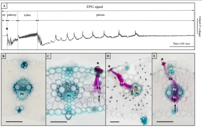
sectioned samples. The sheaths themselves had been stained already during the first stage with fuchsin. In this stage, visualisation was assisted by methylene green counterstaining, which stained the thick- ened cell walls, including those of the vascular bundles, to create a fine contrast between the fuchsin coloured sheaths and the glaucous vascular bundles. Simplicity in the third stage was also advanced by the method of microscopic observation. Here stained samples were simply immersed in ethanol-glycerol embedding solution (Fig. 1). By this microscopic technique, we could visualise the entire salivary sheath structure and determine the exact point where a salivary sheath ended, including making distinction between xylem and phloem endings.
4. Discussion
During the experimental trials, we have developed a complete pro- cedure that made it possible to quickly and easily visualise the position and topology of salivary sheaths (Fig. 1). The simplicity and rapidity of the procedure was reached by the optimisation of its steps that involved the localization of salivary sheath starting points on a leaf surface, hand sectioning of a selected salivary sheath and a simplified microscopic investigation method. For a flowchart style summary of the proposed procedure see SI Fig. 3. The conducted literature survey indicated that, even though there were similar attempts concerning the individual steps, to date there has not been any comprehensive methodology published that could achieve the same results with the present simplicity and rapidity.
In the center of our procedure is an initial staining step that serves double purpose of localizing and deep staining salivary sheaths. The reported aqueous fuchsin method stained the sheath branches properly even in the innermost of the plant tissues. Many of the previously described procedures use staining methods with McBride’s solution (Almeida and Backus, 2004). The solvent of this solution is diethyl ether, which is highly volatile, flammable and toxic, resulting in the need to work under chemical hood. It is also hard to work with, as it has low surface tension. Replacing it with a water-based solvent solves both of these problems. However, a pre-filtration of this water based dye was a necessary step, because when the filtration step was left out, several unsolved pigment particles remained on the leaf surface, resembling the starting points of salivary sheaths. Fuchsin could also be substituted with lignin pink, but that provided less contrast, as it stained vascular bun- dles, as well (see SI “Salivary sheath staining” section). Concerning the clarification steps, at first, we wanted to use only acidic components in the whole procedure, but using KOH (which is a basic component) resulted in better overall results during the first clarification step. Even though, lactic acid was useable to make the epidermis more transparent, during KOH treatment the epidermis got more transparent, the leaf stayed more rigid and resulted in a better consistency for sectioning, further, KOH proved to be a better degreaser, as well.
In the second stage, the main difficulty was how to avoid the need to produce a series sectioning. Leafhoppers almost always position them- selves parallel with the leaf veins during feeding. Due to its anatomy the stylets can be moved most easily sideways (Zhao et al., 2010), which means that the branching salivary sheath is largely in one plane, which is perpendicular to the leaf vein. Consequently, if we know where to cut and in which direction, then that eliminates the need for creating a series sectioning, which was an important step in the optimization of the complete procedure. Sectioning success was also dependent on the thickness of the samples. Even though the aim was to cut as thin slices as possible, too thin samples were unanalysable as we cut out some parts of salivary sheath. Reaching optimal sectioning thickness was not difficult, but it needed few iterative trials. During sectioning, we preferred to use a background light on the microscope, which helped us to find the salivary sheath, but the type of illumination used depends on personal preferences. The hand sectioning method in other studies was mostly applied in plants with more sclerotized leaves, such as in Yuccasp. and Pinus sp. (Brennan et al., 2001). The present study showed that the
method was successfully applicable to barley, a herbaceous mono- cotyledon plant, which has relatively soft leaves. Salivary sheath orientation might be less obvious in dicotyledons with branching and less straight veins.
After sectioning, before counterstaining, another clarification step was necessary. Unlike in the first clarification step, here the application of KOH resulted in collapsed leaf tissues, therefore in the final protocol we used lactic acid in this step, so that cell walls stayed intact and flexible. Lactic acid also has the advantage, that it restores the shape of the cells that might have been distorted during sectioning.
The third stage served the visualisation of salivary sheaths in the sectioned samples. The sheaths themselves had been stained already during the first stage with fuchsin. The reported method stained salivary sheath branches properly even in the innermost of the plant tissues. In this stage visualisation was assisted by methylene green counterstaining, which stained the thickened cell walls, including those of the vascular bundles, as well. This created a fine contrast between the colour of fuchsin stained sheaths and the glaucous vascular bundles (Fig. 1). The method worked well in unravelling the branching pattern in other salivary sheath feeders, such as in the aphid Rhopalosiphum padi (L.) (Hemiptera: Aphididae) (G. Tholt and R. Gerstenbrand, unpublished results).
The greatest advantage of the third step was that by using an embedding solution of ethanol and glycerol, we could avoid fixed mounting for the microscopic investigation. Fixed mounting would have greatly impeded the quick examination of the sectioned material. The use of solidifying embedding media with cover slips (like Canada bal- sam, Permount mounting medium, glycerinated gelatin, Euparal or others) result in a slower process which often requires additional steps of dehydrating through ethanol series or the use of other solvents (Brennan et al., 2001; Lucini and Panizzi, 2016; Saxena and Chada, 1971; Wirth and Marston, 1968). Once a mounting medium solidifies, the cover slip and the sample can only be removed by a lengthy procedure and further
solvents. The application of the presented embedding solution had the advantage that embedding is not permanent, the slices remain intact and re-stainable for later observations. This might be needed due to the commonly occurring fading of microscopic stains after longer storage.
The ethanol component reduced the dissolution of methylene green and the fading of fuchsin, while the glycerol component aided observation, because it has the same refractive index as glass. Many of the published techniques use epifluorescent, TEM or SEM to analyse the samples (Ahmad et al., 2012; Ammar et al., 2018; Backus et al., 2005). Epi- fluorescent microscopy gives good results and it is relatively easy to use but it doesn’t allow us to examine samples many times because the slices lose their excitability after several usage. However, if we get this far in the proposed methodology, the prepared samples can also be used with fluorescent microscopy, as fuchsin and methylene green have different excitation and emission wavelengths.
The described staining and sectioning procedure makes it possible to rapidly and routinely visualise the entire structure of salivary sheaths within plant tissues. This artefact of leafhopper feeding behaviour al- lows us to deduce whether feeding occurred from xylem or from phloem vascular elements, and the branching structure also gives an estimate how many attempts were needed to find the actual location of feeding.
One of the greatest advantages of studying salivary sheaths is that, coupled with the interruption of the feeding, it makes possible to pre- cisely calibrate EPG waveforms and thereby ascertain the correlation between waveform types and the physical location of the feeding pro- cess. Hemipterans are major pests in various plant cultivars, not only as a result of their direct damage, rather because they are the vectors of 90
% of insect-borne plant diseases (Fereres and Moreno, 2009; Weintraub and Beanland, 2006). Their feeding process is intimately coupled with their vector efficiency, therefore studying details of their feeding can provide opportunities to disrupt pathogen transmission.
Fig. 1. An examplar EPG waveform of Psammotettix alienus with stained salivary sheaths. The salivary seath developments (where they end) can be connected to certain waveforms representing different stages of the penetration process. (SS) Salivary sheath going through epidermis (*) Starting point of the salivary sheath. (↑) Tip of the salivary sheath.(Xy) Xylem. (Ph) Phloem. Sub figures: A) EPG waveform, B) Non penetration, C) pathway, D) Xylem, E) Phloem. Scalebars are 100 μm.
Declaration of Competing Interest
The authors report no declarations of interest.
Acknowledgments
We are grateful for Endre Toth for providing the microscopic dyes. ´ The study was financed by NKFI OTKA grants K116062 and K134811.
Appendix A. Supplementary data
Supplementary material related to this article can be found, in the online version, at doi:https://doi.org/10.1016/j.micron.2020.102959.
References
Ahmad, A., Kaushik, S., Ramamurthy, V.V., Lakhanpaul, S., Ramani, R., Sharma, K.K., Vidyarthi, A.S., 2012. Mouthparts and stylet penetration of the lac insect Kerria lacca (Kerr) (Hemiptera:tachardiidae). Arthropod Struct. Dev. 41, 435–441. https://doi.
org/10.1016/j.asd.2012.04.001.
Almeida, R.P.P., Backus, E.A., 2004. Stylet penetration behaviors of Graphocephala atropunctata (Signoret) (Hemiptera, Cicadellidae): EPG waveform characterization and quantification. Ann. Entomol. Soc. Am. 97, 838–851. https://doi.org/10.1603/
0013-8746(2004)097[0838:Spboga]2.0.Co;2.
Ammar, E.D., Hall, D.G., Shatters Jr., R.G., 2018. Salivary sheaths of the asian Citrus psyllid show signs of degradation 3-4 weeks following their deposition into Citrus Leaves by the feeding psyllids. J. Microsc. Ultrastruct. 6, 129–133. https://doi.org/
10.4103/JMAU.JMAU_13_18.
Backus, E.A., 1988. Sensory systems and behaviours which mediate hemipteran plant- feeding: a taxonomic overview. J. Insect Physiol. 34, 151–165. https://doi.org/
10.1016/0022-1910(88)90045-5.
Backus, E.A., Habibi, J., Yan, F.M., Ellersieck, M., 2005. Stylet penetration by adult Homalodisca coagulata on grape: electrical penetration graph waveform characterization, tissue correlation, and possible implications for transmission of Xylella fastidiosa. Ann. Entomol. Soc. Am. 98, 787–813. https://doi.org/10.1603/
0013-8746(2005)098[0787:Spbahc]2.0.Co;2.
Brennan, E.B., Weinbaum, S.A., Pinney, K., 2001. A new technique for studying the stylet tracks of homopteran insects in hand-sectioned plant tissue using light or epifluorescence microscopy. Biotech. Histochem. 76, 59–66. https://doi.org/
10.1080/bih.76.2.59.66.
Carpane, P., Wayadande, A., Backus, E., Dolezal, W., Fletcher, J., 2011. Characterization and correlation of new electrical penetration graph waveforms for the corn leafhopper (Hemiptera: cicadellidae). Ann. Entomol. Soc. Am. 104, 515–525.
https://doi.org/10.1603/an10052.
Civolani, S., Leis, M., Grandi, G., Garzo, E., Pasqualini, E., Musacchi, S., Chicca, M., Castaldelli, G., Rossi, R., Tjallingii, W.F., 2011. Stylet penetration of Cacopsylla pyri;
an electrical penetration graph (EPG) study. J. Insect Physiol. 57, 1407–1419.
https://doi.org/10.1016/j.jinsphys.2011.07.008.
Fereres, A., Moreno, A., 2009. Behavioural aspects influencing plant virus transmission by homopteran insects. Virus Res. 141, 158–168. https://doi.org/10.1016/j.
virusres.2008.10.020.
Lucini, T., Panizzi, A.R., 2016. Waveform characterization of the soybean stem feeder Edessa meditabunda: overcoming the challenge of wiring pentatomids for EPG.
Entomol. Exp. Appl. 158, 118–132. https://doi.org/10.1111/eea.12389.
Morgan, J.K., Luzio, G.A., Ammar el, D., Hunter, W.B., Hall, D.G., Shatters Jr, R.G., 2013.
Formation of Stylet Sheaths in aere (in air) from eight species of phytophagous hemipterans from six families (Suborders: auchenorrhyncha and Sternorrhyncha).
PLoS One 8, e62444. https://doi.org/10.1371/journal.pone.0062444.
Saxena, P., Chada, H.L., 1971. The greenbug, Schizaphis graminum. 1. Mouth parts and feeding habits. Ann. Entomol. Soc. Am. 64, 897–904. https://doi.org/10.1093/aesa/
64.4.897.
Seo, B.Y., Kwon, Y.-H., Jung, J.K., Kim, G.-H., 2009. Electrical penetration graphic waveforms in relation to the actual positions of the stylet tips of Nilaparvata lugens in rice tissue. J. Asia Pac. Entomol. 12, 89–95. https://doi.org/10.1016/j.
aspen.2009.02.002.
Sharma, A., Khan, A.N., Subrahmanyam, S., Raman, A., Taylor, G.S., Fletcher, M.J., 2014. Salivary proteins of plant-feeding hemipteroids – implication in phytophagy.
Bull. Entomol. Res. 104, 117–136. https://doi.org/10.1017/S0007485313000618.
Tholt, G., Samu, F., Kiss, B., 2015. Feeding behaviour of a virus-vector leafhopper on host and non-host plants characterised by electrical penetration graphs. Entomol. Exp.
Appl. 155, 123–136. https://doi.org/10.1111/eea.12290.
Tholt, G., Kis, A., Medzihradszky, A., Szita, ´E., T´oth, Z., Havelda, Z., Samu, F., 2018.
Could vectors’ fear of predators reduce the spread of plant diseases? Sci. Rep. 8, 8705. https://doi.org/10.1038/s41598-018-27103-y.
Tjallingii, W.F., 1985. Electrical nature of recorded signals during stylet penetration by aphids. Entomol. Exp. Appl. 38, 177–186. https://doi.org/10.1111/j.1570- 7458.1985.tb03516.x.
Tjallingii, W.F., Hogen Esch, T., 1993. Fine structure of aphid stylet routes in plant tissues in correlation with EPG signals. Physiol. Entomol. 18, 317–328. https://doi.
org/10.1111/j.1365-3032.1993.tb00604.x.
Walker, G.P., Backus, E.A., 2000. Principles and Applications of Electronic Monitoring and Other Techniques in the Study of Homopteran Feeding Behavior. Entomological Society of America, Annapolis, USA, pp. 1–260.
Weintraub, P.G., Beanland, L., 2006. Insect vectors of phytoplasmas. Annu. Rev.
Entomol. 51, 91–111. https://doi.org/10.1146/annurev.ento.51.110104.151039.
Wirth, W.W., Marston, N., 1968. A method for mounting small insects on microscope slides in Canada Balsam. Ann. Entomol. Soc. Am. 61, 783–784. https://doi.org/
10.1093/aesa/61.3.783.
Zhao, L., Dai, W., Zhang, C., Zhang, Y., 2010. Morphological characterization of the mouthparts of the vector leafhopper Psammotettix striatus (L.) (Hemiptera:
cicadellidae). Micron 41, 754–759. https://doi.org/10.1016/j.micron.2010.06.001.