— 5 —
Genetic Diseases and Aberrations
GEORG BENZ1
Entomological Laboratory, Swiss Federal Institute of Technology, Zurich, Switzerland
I. Introduction 161 II. Genetic and Cy to logical Terms and Symbols 163
III. Genetic Pathology 165 A. Terminology 165 B. T h e Significance of Selected Examples 166
C. T h e Spectrum of Viability 168 D. Harmless Aberrations 169 E. Malformations 170 F. Sterility Factors 171 G. Subvital Factors 173 H. Semilethal and Lethal Factors 174
IV. T h e Occurrence of Genetic Diseases in Populations of
Wild Insects 181 V. Genetic Diseases in Laboratory Breeding 184
VI. Conclusion 184 References 185
I. INTRODUCTION
Each character of an organism, i.e., every single feature and each chemical event taking place in an organism, is evoked and directed by one or several units of the genetic material, called the genes. In insects, as in all higher organisms, the genes are located in the chromosomes.
They are transmitted in the well-known cytological manner from one generation to the next, where the genes manifest themselves again with
ι T h e author is greatly indebted to his former teacher, Professor Dr. E . Hadorn from the Zoological Institute of the University of Zurich, to whom he owes most of the ideas expressed in this chapter. Warmest thanks are also extended to him for reading the manuscript and for constructive criticism.
161
their specific characters, called phenes (cf. Kühn, 1942; Hadorn, 1955). In order to guarantee the proper functioning of a living system and its off
spring systems, the genetic material has to be rather stable. How
ever, changes do occur; they are known as mutations. In the fruit fly Drosophila melanogaster Meigen, the total rate of mutation per genera
tion is at least 5 percent under natural conditions, a finding that applies to other organisms as well (Muller, 1950). T h e spontaneous mutation rate may be considerably increased by irradiation of the germ cells with X rays (Muller, 1927) or other ionizing rays (Mickey, 1954), and by the treatment of the germ cells with mutagenic chemicals, such as mustard gas (Auerbach and Robson, 1947). An individual in which the mu
tated genetic material becomes manifest in altered phenes, is called a mutant. Each new mutant sets up a highly specific experiment, which sheds light on the functional relationship between individual mutational states of the genetic material and the processes leading to the formation of characters or phenes.
Since the maintenance of a living system depends on the proper co
operation of so many genes, the chances are small or minimal that the changed genetic material in a mutant will fulfill its task as well or better than the proved and well-balanced normal genetic material. W e therefore expect that most mutations entail a risk for the mutant.
Investigations on the viability of mutants show that most mutations somehow intervene with the proper functioning of the organism and lead to more or less severe pathological conditions. T h e contribution of gene transformations without harmful effects might be estimated at only a few percent of the total number of mutations (Hadorn, 1955, 1961).
It was found early in work on mutations that, considering any given morphological character, mutations with smaller effects occur more fre
quently than those with larger effects (Altenburg and Muller, 1920;
Muller, 1923). T h i s is equally true for "invisible" mutations affecting the viability of the mutant. Quantitative tests of the frequency of lethal and of invisible detrimental mutations of varying grades, induced by X rays, showed the ''detrimentals'' to be induced with two to four times the frequency of the complete lethals (Muller, 1934; Kerkis, 1938;
Timofeeff-Ressovsky, 1935; Käfer, 1952).
We have said above that the spontaneous occurrence of mutations in insects is not at all a rare event. However, the severe and reckless elim
inating power of selection usually does not allow the mass occurrence of pathological mutants in natural populations. Thus, our knowledge of hereditary diseases is practically limited to the two species domesticated by sericulturists and apiculturists [Bombyx mori (Linnaeus) and Apis mellifera Linnaeus], and to a few species kept \n laboratories for genetic
5. GENETIC DISEASES AND ABERRATIONS 163 and physiological studies, i.e., to several species of the genus Drosophila, Culex pipiens Linnaeus, Ephestia (Anagasta) kühniella Zeller, and the parasitic wasp Bracon hebetor Say [ ( = Microbracon hebetor (Say) and Habrobracon juglandis (Ashmead) ].
I I . G E N E T I C AND CYTOLOGICAL T E R M S AND SYMBOLS
In this section the necessary genetic and cytological terms are intro
duced. T h e reader who is familiar with genetics and genetic nomen
clature may, therefore, pass over this part of the chapter.
In most bisexually reproducing insects, the germ cells or gametes (egg and sperm) contain one set of chromosomes; they are haploid.
After the fertilization of the egg cell by the sperm, a zygote is produced, containing two sets of chromosomes, a maternal and a paternal one.
T h e zygote is diploid, and develops into a diploid insect. In some groups of insects, the males develop parthenogenetically from unfertil
ized eggs; they are haploid organisms. Yet in the majority of insects, the sex determination is achieved by the distribution of sex chromosomes
(XY and X O types). X and Y chromosomes are called heterosomes; the other chromosomes, autosomes. T h e sex which possesses two identical sets of chromosomes produces but one type of gametes and is therefore called the homogametic sex (in most insects the female, in Lepidoptera the male). T h e sex with two different sets of chromosomes produces two types of gametes ( X gametes, and Y gametes or Ο gametes) and is called the heterogametic sex.
If a zygote has a gene A from both father and mother, its genetic constitution for this chromosome locus is A/A; it is homozygous for A.
If in one of the parents the gene A has mutated to a, and is carried over to the zygote by the germ cell, the zygote has the genetic constitution A/a; it is heterozygous for both A and a. A and a represent two differ
ent mutational stages of the same chromosome locus; they are called alleles. We know of cases where for the same locus of a chromosome a number of different mutational stages have been found; i.e., several different alleles of one gene locus do exist. Such series of alleles are called multiple alleles, of which a diploid individual can carry just two.
T h e normal allele of any chromosome locus is usually symbolized by the sign Thus a wild-type insect should have the genetic constitu
tion + / + · Many mutated genes produce characteristic phenes only in homozygous condition, i.e., m/m (m stands for mutated gene). If they are in heterozygous condition, (i.e. -\-/m), the carrier looks like a + / + wild type. In this case the action of the -\- allele is dominant over m , or: - j -m is a dominant allele, m a recessive allele. Since differing genetic
constitutions may produce similar phenes, we must distinguish between the genotype and the phenotype of an individual.
In most instances the normal allele is dominant over the mutated alleles. However, dominant mutations do exist; we designate them with capital letters. Recessive alleles are symbolized by small letters. Thus A is dominant over -\-A, and dominant, over a (or aA > a+ > a). In a case where the genotype A/-\-A produces an intermediate phenotype between A/A and - \ -Α/ - \ -Α, w e sPe ak of semidominant alleles.
According to the definition given above, a recessive mutation would manifest its phenes only in homozygous condition. T h i s rule, however, does not apply to recessive genes in hemizygous and azygous condition, i.e., when they are not counterbalanced by another allele in the hetero- somes of the heterogametic sex, and in haploid organisms.
A mutation may involve one single gene, a group of genes, whole chromosomes, or whole sets of chromosomes. A single gene mutation may result in (1) the change of a gene from one comparatively stable state to another (point mutation); (2) the loss of a gene (one gene deficiency); or (3) the doubling of a gene (repeat). Mutations of gene complexes may result in (1) the loss of a more or less large piece of a chromosome (deficiency); (2) the doubling of a piece of a chromosome (duplication); (3) the inversion of a piece of a chromosome, causing the reversion of the original gene sequence (inversion); or (4) the translocation of a piece of a chromosome to another place in the same chromosome or to another chromosome (translocation). Mutations affecting the number of the chromosomes may result in (1) the loss of one or several chromosomes (monosomies in diploid organisms); (2) the doubling, tripling, etc., of one or several chromosomes (trisomies, ietrasomics, polysomics), or the doubling, tripling, etc., of all chromo
somes, leading to polyploidy. Since it is not always easy to determine whether the change of a single phene, or of a complex of phenes is caused by the mutation of one gene, or of a larger unit of the genetic material, we will often replace the word gene by the term hereditary factor.
T h e term mutation includes all possible changes mentioned above.
Yet as a rule, it will be used with the understanding that changes of whole chromosomes, or whole sets of chromosomes, are excluded unless the contrary is specified.
5. GENETIC DISEASES AND ABERRATIONS 165
I I I . G E N E T I C PATHOLOGY
A. Terminology
1. Aberrations and Diseases
Any variation of the phenotype of the species may be called an aberration. It may be produced either by environmental factors, or by the action of the mutated genetic material. In the latter case, we speak of a genetic aberration. I f an aberration proves to be harmful for the individual bearer, we speak of a disease. Thus, all hereditary transmitted biochemical, physiological, and morphological characters xvhich are harmful for the organism, are genetic diseases. However, we have to keep in mind that every species shows a more or less broad spec
trum of variations, and that in an organism, no clear-cut borderline exists between pathological and still normal physiologic conditions.
Therefore, it is sometimes difficult to distinguish between harmless genetic aberrations and genetic diseases. Besides, one and the same he
reditary factor may be harmless in a certain environment, but harmful under other conditions {conditional genetic diseases). Certain types of genetic diseases, discernible by harmful variations of the morphologic structures, but not perilous under ideal conditions (such as laboratory conditions may b e ) , are called genetic malformations.
2. Classification of Genetic Diseases
If an unfavorable mutation affects an essential vital factor, the mutant will die. A hereditary factor which causes the death of the mutant prior to its reproductive stage is called a lethal factor (Hadorn, 1949). Lethal factors lead to a "developmental death'' before repro
duction has taken place, so that with the death of the bearer the mutant chromosomes are lost.
Lethal factors, sensu stricto, are characterized by a penetrance of 100 percent. This means that all carriers of the lethal factor die during their development. However, numerous instances are known where an occasional individual overcomes the developmental crisis and continues to progress, in spite of its lethal constitution. Such exceptional indi
viduals are called "escapers" (Hadorn, 1945).
If the penetrance of a lethal factor is so much reduced that "es
capers" appear regularly and in definite proportions, the term semi- lethal factor should be applied. Semilethals are all those factors which cause death to at least 50 percent of the mutant genotypes. Hereditary factors whose lethal action is less than 50 percent are called subvital factors (Hadorn, 1948). Some hereditary factors affecting viability may produce 100 percent lethals under certain conditions, but allow the
development of "escapers" under other conditions. These factors are called conditional lethal factors; they are susceptible both to the geno- typic milieu which is determined by the entire gene complement, and to variations in the external environment, which is conditioned by fac
tors such as nutrition, population density, and temperature.
It should be pointed out that the differentiation between lethal fac
tors in the striot sense, semilethal factors, and subvital factors is, up to a point, dependent on the internal and external environment. There
fore the terms should be regarded as a classification of penetrance rather than as distinct groups of mutations.
T h e majority of the more thoroughly investigated lethal factors are characterized by a specific phase of action at a certain stage of development. In insects, we distinguish embryonic, larval, pupal, and early imaginal lethal factors. T h e lethal crisis often coincides with a critical stage of development, such as hatching from the egg, or any kind of molting. Hence many lethal factors are classified as boundary lethals, e.g., embryonic-larval lethals, larval-pupal lethals (Hadorn, 1951;
Hadorn and Chen, 1952). Other lethal factors produce a crisis at more than one stage of development; some mutants, for instance, may die at the embryonic stage whereas the survivors perish in the pupal stage. In such cases we speak of biphasic and multiphasic lethal factors. Multi
phasic factors may in extreme cases become aphasic lethal factors if they cause death at any time and stage of development. Since insects have to pass every once in a while through a critical developmental stage, the moltings and metamorphosis, these cases are very rare.
3. Sterility Factors
A mutant gene that causes sterility affects the population genetically in exactly the same way as a lethal factor. Its physiological effect, how
ever, differs from that of a lethal factor, since it does not endanger the life of its bearer, but merely interferes with reproduction.
B. The Significance of Selected Examples
It is impossible to discuss the wide field of hereditary diseases within the scope of a small chapter. T h e "law of homologous variation/' formulated by Vavilov (1922), however, allows us to treat our subject in a schematic way and to refer to a few selected examples only. Vavilov could show that mutations with similar effects may occur in different species of varying relationship. His law implies that if a number of mutations are known in one species, "parallel mutations" may be ex
pected in related species. T h e appearance of such corresponding pheno- types in mutants of different organisms, may be due to three different causes:
5. GENETIC DISEASES AND ABERRATIONS 167 (1) Mutation of homologous genes: Many processes in different species are of a general nature; they are controlled by homologous genes with identical function. Thus the vermillion+ genes (v+) of Drosophila melanogaster, D. simulans Sturtevant and D. pseudoobscura Frolova are homologous with the a + gene of Ephestia kiihniella, and the ivory + (i+) of Apis mellifera. All these genes control the conversion of trypto
phan into kynurenine, a precursor of brown eye-pigments (Butenandt et al., 1940; Kühn, 1936, 1942). T h e mutation of these genes prevents the formation of the brown eye-pigment in the above-mentioned species;
as a result, the eyes of the mutants are, respectively, bright red in Droso
phila and Ephestia, and white in Apis. Again the cinnabar+ (cn+) gene of D. melanogaster, the orangen (o+) genes of D. pseudoobscura and of the wasp Bracon hebetor, the snow+ ($+) gene of Apis mellifera, and the white^ (&>i+) gene of Bombyx mori (Kikkawa, 1941) are homo
logous genes, controlling the conversion of kynurenine into 3-oxykynure- nine, i.e., another precursor of the brown eye-pigment (Butenandt et al., 1949).
(2) Mutation of "chain genes": As a rule, a chain of chemical steps leads to the formation of a certain phene. In the case of the brown eye-pigments of many insects, we find the following sequence of chemical steps which are controlled by the following genes:
tryptophan > » kynurenine > 3-oxykynurenine > brown pigment
first group of second group of third, etc., groups of homologous genes homologous genes homologous genes
It is clear from this scheme that a mutation of the i + gene of Apis (first group) may have the same phenotypic effect as a mutation of the wx+ gene of Bombyx (second group).
(3) Mutation of genes with combinant relation: In Apis and Bombyx, the dark color of the eyes is the result of the combination of a brown eye-pigment (controlled by the genes i + and s+ in Apis, in Bombyx) with a pigment-bearing substance of protein character. T h e production of the brown eye-pigment has no apparent effect, if the pigment bearer is missing. A mutation which blocks the production of the pigment bearer will therefore produce the same phenotype as the mutation of any one of the pigment-controlling genes just mentioned.
Such interspecific relations show that the few selected examples which we are able to describe in this chapter, may have a more general meaning. They are taken from a few more thoroughly studied species.
But similar examples should be found in many other species all through the Class Insecta.
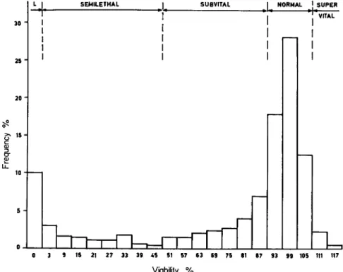
C. The Spectrum of Viability
Any large group of mutations, whether spontaneous or experimentally induced, will contain mutations with approximately normal viability, as well as numerous mutations which reduce viability in varying degrees.
In order to demonstrate the relative frequency of different mutations, in a wild population of D. pseudoobscura, Dobzhansky et al. (1942) investigated the survival of animals which had been made homozygous
ι SUPER
15 21 27 33 39 45 51 57 63 69 75 61 87 93 99 105 111 117
Viability %
FIG. 1. Distribution of relative viabilities of homozygotes for 3 2 6 second chro
mosomes from wild populations of Drosophila pseudoobscura. L = lethal. (After data by Dobzhansky et al., 1 9 4 2 . )
for the second chromosome and compared their viability with hete
rozygous controls which were kept under the same conditions and whose viability was regarded as "normal," or 100 percent. T h e results for the second chromosome are illustrated in Fig. 1.
T h e diagram shows that viability factors of all classes are present in a wild population. Of all the second chromosomes tested, about 10 percent carry lethal factors, 10 percent semilethal factors, 19 percent subvital factors, and only 58 percent are normal (90 to 105 percent viability). There are also a few supervitals whose viability exceeds the
5. GENETIC DISEASES AND ABERRATIONS 169 means of normal controls (110 to 115 percent viability). In the class of semilethals, half the cases show a strong penetrance (3 to 9 percent viability), whereas the weakly semilethals with a penetrance of 10 to 50 percent, are rare. T h e proportion of the sub vitals amounts to the sum of lethals and semilethals, a preponderance of cases having weak penetrance.
T h e diagram shows very clearly that no strict borderline exists between subvitals and normals. Similar results were obtained for the fourth chromosome of the same population.
It is interesting to note that the viability of mutants induced by X rays in the second chromosome and in the X chromosome of D.
melanogaster, show a surprisingly similar spectrum of viability (Käfer, 1952).
D. Harmless Aberrations
We have stated above that the percentage of harmless or even advantageous mutations are estimated at only a few percent. Therefore we may conclude that most genetic aberrations are more or less pathological. However, the occurrence of so many variations in nature, described as aberrations, forms, or subspecies, demonstrates that a con
siderable number of mutants are fully viable under natural conditions.
T h e actual preponderance of such forms in certain regions proves that, in a given environment, certain mutations may have a positive selection value (e.g., industrial melanism of Lepidoptera). Certain aberrations, classified as fully viable in laboratory experiments, may be fit for survival only when viewed from the standpoint of developmental physiology.
Under natural conditions, the same mutations are often subject to rigorous selection. Thus, the mutant blue-green larva of Colias philo- dice Latreille is fully viable when reared in the laboratory, but the frequency of the gene is kept low in natural populations because birds prey much more upon the mutant larvae than upon the grass-green wild- type larvae (Gerould, 1921).
How differently homologous mutations may be judged, according to their occurrence in either wild or domesticated insects, will be illustrated by two mutations of B. mori. T h e sex-limited, dominant mutation No glue (Ng) is responsible for a poor development of the mucous glands in the genital tract of the Bombyx female. Whereas most "normal"
females attach their eggs firmly to the substrate, the females of the Baghdad race, which are homozygous for Ng, attach their eggs only lightly to the substrate (Tanaka, 1953) .2 In the domesticated silkworm, this
2 Tanaka, in his excellent review on the genetics of the silkworm, mentions numerous mutations, reported in papers in the lapanese language. All mutations of Bombyx mentioned in this chapter without reference are cited by Tanaka (1953).
mutant is harmless; the character of loose eggs may even be desirable, while in many other species of free-living insects, the same character would be strictly negative. On the other hand, the relatively harmless mutation Naked b (Ndb), which affects the silkgland of Bombyx, is highly undesirable, because it reduces the silk production. This mutation has a penetrance of 50 percent; it causes 50 percent of the silkworms to pupate without spinning a cocoon.
An undesirable dominant mutation with imperfect penetrance may become a problem in mass rearings. While mutations with full pene
trance can be easily eliminated from the breeding stock, imperfect pene
trant mutations are not wholly detectable. T h e only answer to this problem is single-pair breeding, which enables us to eliminate the entire progeny of pairs which, among others, produce mutant individuals.
£. Malformations
Numerous mutations in insects interfere with a number of develop
mental processes without causing developmental death. They result in variations of the morphological structures, called malformations. In Drosophila, Bombyx, etc., malformations are known for practically each organ (e.g., no bristles or no scales, deformed or too small eyes, no eyes, vestigial wings, crippled legs, supernumerary legs, or legs in the place of antennae, deformed body).
Some of these mutants are fully viable in the laboratory, but would be subject to selection in nature. It is true that, under certain conditions, malformations may become a positive selection value. Thus, the loss of wings, which normally greatly impairs an insect, may be advantageous, and therefore favored, on a windy island where winged insects are easily carried into the sea.
Most dominant factors for malformations, are recessive lethal factors;
they cause in homozygous condition the death of the mutant. Thus, the dominant mutation Vestigial (Vg) of Bombyx, causes in heterozygous condition club-shaped or completely reduced wings in the moth, whereas in homozygous condition, it acts as an embryonic lethal factor (Tanaka, 1953). Or else, the gene Droopy (D) in A. mellifera, causes, in the heterozygous condition, the wings of worker bees and queens to be slightly extended from the body, and to droop, and therefore, D bees cannot fly. Since no D drones are known, it is probable that D acts as a lethal factor in haploid males, a hypothesis, which is supported by viability counts of unfertilized eggs (Rothenbuhler et al, 1953). T h e r e are many other similar cases; some of them will be discussed in later sections.
Some dominant mutations for malformations may have a recessive
5. GENETIC DISEASES AND ABERRATIONS 1 7 1
lethal action in a certain genetic milieu only. T h e factor Deformed gonad (Gd) of Bombyx in heterozygous condition, is responsible for
supernumerary ovarioles in the female, supernumerary testicular follicles in the male, short wings of the pupa, and split hind wings of the moth, in homozygous condition, the factor provokes death shortly before, or after, hatching from the egg. Crossings with a Chinese poly vol tine race gave a strain that showed only malformations, but no lethality, when homozygous. Many recessive factors for malformations have to be classi
fied as subvital factors. Thus, the recessive mutation fl (wingless) of Bombyx which affects the second and third thoracic segment of the pupa, accounts not only for the primitive state of the second and third thoracic legs and the lack of wings, but many fl mutants will die from loss of blood in the area of the wings during pupation or when the moth emerges. This mutation shows that differences in the expressivity of a gene bring very different results. T h e factor fl affects the imaginal discs of the legs and wings of the second and third thoracic segment. As a rule, only those parts of the imaginal discs that produce the appendages, are severely affected, while the parts which form the thoracic hypodermis are normal. In cases with high expressivity, the thoracic parts of the imaginal discs are affected too, and consequently the thorax becomes weak.
Another reduction of viability is found in the recessive mutation apodal (ap) of Bombyx. T h e mutant larvae have rudimentary legs which make feeding difficult. A considerable number of mutants die during ecdysis and pupation. T h e apodal moths are unable to walk, and males are sterile, being able to copulate only when assisted. More direct sterility may be effected by genes whose highest expressivity leads to malformations of the genital organs. T h e mutation crossveinless2 (cv2) in Drosophila subobscura Collin, which in most cases prevents the development of the anterior and posterior crossveins of the wings only, produces in about 23 percent of the males abnormalities of the repro
ductive organs. In light cases, certain deformities of the external and internal genitalia occur. In more severe cases, the sexual organs are absent. Obviously, all males with such malformed genitalia are sterile
(Bird, 1948).
F. Sterility Factors
Although sterility factors do not endanger the life of the individual, they have a profound influence on population genetics. Not only are chromosomes of sterile individuals lost for the species, but in species which copulate only once during life, the chromosomes of the mates are lost too. This last effect is utilized in practical insect control where
large numbers of artificially sterilized (irradiated) males are introduced in natural populations (Knipling, 1959, 1960).
Since the sterility of an individual can be traced back to many different causes, sterility factors constitute a very heterogeneous group of mutations. Only a few examples suffice to illustrate this.
(1) Sterility as a result of a defect in locomotion or guiding ap
paratus: T h e factor ap of B. mori, which belongs into this category, has been mentioned in the previous section. Although the males with the genetic constitution ap/ap are fully fertile when assisted for the copula, the apodal males never produce offspring without help and therefore must be considered sterile.
Another type of indirect sterility in drones of A. mellifera is caused by mutations of the eye color. White or other light-colored eyes so much impair the drones' vision that they seldom, if ever, succeed in mating (Kerr and Laidlaw, 1956).
(2) Sterility as a result of a defective sex apparatus: In males of Bombyx with the gen tic constitution sip/sip, one or two of the three pairs of copulatory muscles are degenerated (Umeya, 1936). As a result of this defect, the males are usually unable to copulate. T h e penetrance of this factor is conditioned by the rearing temperature.
T h e females of the lozenge-claxüless (lzlc) mutant of D. melanogaster are devoid of spermathecae and parovariae; hence they are completely sterile, although they lay eggs. Transplanted ovaries of lzcl/lzcl female larvae will produce fertile eggs, when grown in a female-sterile host
(Anders, 1955).
(3) Sterility as a result of defective gametes: Some genes of the Y chromosome of D. melanogaster ensure the motility of the sperm (Stern, 1929). Although X O individuals will develop into apparently normal males which produce morphologically normal sperm cells, these gametes lack motility and are unable to fertilize eggs.
Females of D. melanogaster with the genetic constitution bos/bos (bordo-sterile) (an eye-color mutant, isolated twice from wild popu
lations in Germany), produce sterile eggs of irregular shape and size.
Only the few normal-looking eggs may be fertilized, but they do not develop beyond the time of hatching (Fabian, 1948).
(4) Sterility for want of gamete production: In D. melanogaster, the gene female-sterile (fes) has no effect on males, but interferes with the production of eggs in females (Clancy and Beadle, 1937; Gloor and Hadorn, 1942).
(5) Sterility as a consequence of undeveloped gonads: Females of D.
subobscura with the genetic constitution gs/gs (grandchildless) lay eggs from which sterile offspring results. T h e sons and daughters of gs/gs
5. GENETIC DISEASES AND ABERRATIONS 173 females have no, or extremely reduced gonads, whether they are homozy
gous or heterozygous for the gene gs. This means that the sterility of the ¥1 of gs females is a maternal effect (Spurway, 1948; Suley, 1953).
(6) Sterility of intersexes: Crossings of certain species and races of the lepidopterous genera Lymantria and Solenobia result in intersex individuals which, as a rule, are sterile (Goldschmidt, 1931; Seiler, 1929, 1949).
In C. pipiens, a simple recessive autosomal factor changes males into sterile intersexes (Laven, 1955). Just the reverse sex-limited action is found in the intersex gene ix of D. subobscura, where homozygous ix/ix- males are fertile, while X X genotypes carrying ix/ix are sterile intersexes
(Spurway and Haldane, 1954).
G. Subvital Factors
Subvital factors with a viability of 50 to 95 percent provide a large proportion of mutants. Since many of them are "invisibles," i.e., with
out typical phenes, they are not very interesting for the student of developmental physiology and therefore are less thoroughly studied than other types of viability factors. Some of the subvital factors, however, provide excellent examples of genetic diseases which are moderately severe. Thus, the factor vomitor (vo) of Bombyx is responsible for the poor development of the mandibular muscles in fifth-ins tar larvae of the mutant. T h e mulberry leaves are swallowed in larger pieces by the mutant larvae than by normal larvae. T h e large pieces often interrupt the closure of the cardiac valves, which causes vomiting. This disease retards development, and weakly disposed individuals will not complete development.
Numerous cases of subvital factors with characteristic phenotypic appearance could be cited. W e restrict ourselves to a few examples.
Α semidominant factor of D. melanogaster (Kg — Kugel, = sphere) is a typical subvital factor. Kg larvae are shorter and thicker than normal larvae, and so are the pupae. Heterozygotes are viable to about 90 percent, whereas homozygotes reach a viability of approximately 67 percent (Benz, 1956). In Bombyx, the recessive gene compressed (cp) causes a similar phene in the full-grown larva. In this case, feeding is slow, and growth is retarded.
An interesting conditional subvital factor in D. melanogaster is represented by the mutation Jammed (J). T h e viability of both homo
zygotes and heterozygotes is about 70 percent (Bridges and Brehme, 1944). T h e wings of the mutants are jammed into a narrow strip at the posterior edge. This phenotype is fully realized only at temperatures
above 25°C; if reared at 19°C, neither homozygous nor heterozygous / flies can be distinguished from the wild type.
H. Semilethal and Lethal Factors
Most semilethal factors show a strong penetrance, i.e., a lethal action of 90 to 99 percent, and are therefore discussed together with the lethal factors. Besides, the penetrance and expressivity of many viability factors varies considerably under different conditions.
Lethal factors are of great interest from the standpoint of develop
mental physiology. Each lethal factor sets up a highly specific experi
ment which often could not be achieved by surgical or biochemical methods. If, for instance, a hereditary factor is responsible for the development or the proper functioning of certain cells, and mutants for this factor die, we know that those cells are essential for life. Thus the study of lethal factors provides knowledge on the general and special pathology of development. It also leads to a better understanding of the gene physiologic basis of normal development (Hadorn, 1951).
1. Complementary Lethal Systems
Some hereditary factors which have little or no influence on the viability of the mutant may act as lethal factors when combined with other harmless factors. T h e gene purploid (pd) in D. melanogaster for instance, has no action in single dose, but clears up the color of the eyes in the homozygous condition. Another gene Purploider (Pdr) enhances in single dose the action of pd, but has no visible influence when present in single dose without pd. Pdr/Pdr individuals without pd have somewhat rosier eyes than wild flies and are fully viable. Yet homozygous individuals for both pd and Pdr will die (Bridges and Brehme, 1944).
A similar, even more drastic, case was described by Sturtevant (1956).
T h e gene Prune-killer (K-pn) brings on the death of all individuals carrying the gene prune (pn) in homozygous or hemizygous condition, at the end of the second larval instar. T h i s action of the gene K-pn is dominant. Neither pn nor K-pn lower the viability of the mutants, when present in homozygous condition only. (K-pn/K-pn individuals do not even show visible or otherwise detectable mutant phenes.)
2. Conditional Lethal Factors
W e have already mentioned that some mutations cause death only under certain developmental conditions.
a. Penetrance as a function of temperature. It is a common occur
rence for penetrance to be affected by temperature. T h e eye-color mutant rosy (ry) of D. melanogaster is fully viable at relatively low temperatures,
5. GENETIC DISEASES AND ABERRATIONS 175 but semilethal at the standard temperature of 25°C (Hadorn and Schwinck, 1956). According to Dobzhansky (1946), a genotype of D.
pseudoobscura isolated from a wild population, is normal at 16.5°C, semilethal at 21°C, and lethal at 25.5°C. In Bracon hebetor the gene kidney (k) is lethal at 30°C, while the same genotype is fully viable at lower temperatures (Whiting, 1934).
A reversed action of temperature is reported by Tanaka (1953).
T h e white egg mutant ol/ol of Bombyx mori dies at the time of the fourth molt when reared below 23°C whereas it remains viable at higher temperatures.
T h e temperature-conditioned action of viability factors may be interpreted according to the gene physiologic findings in organisms like fungi, bacteria, and mammals (Wagner and Mitchell, 1955). Thus Horowitz and Sheng (1952) found a mutant of the mold Neurospora which produces no tyrosinase at 35°C, whereas at 25°C active enzyme is present. It could be demonstrated that the tyrosinase produced by the mutant at 25°C is thermolabile, while tyrosinase of normal strains is inactivated only at temperatures far above 35°C (Horowitz and Fling, 1953).
b. Penetrance as a function of other environmental factors. Schneider and Brügger (1946) described an apparently dominant mutation of A. mellifera which shows a penetrance of about 43 percent in working bees and a higher penetrance in azygous males, but no penetrance in queens. T w o interpretations of these facts are possible. Either the mutation manifests its phenes only in individuals reared without suffi
cient amounts of "royal jelly/' or the mutation is conditioned by the shape of the honeycomb cells (see also Section I I I , H, 4, d ) .
c. Penetrance as a function of the genotypic milieu. Differences in penetrance are often the result of modifying genes. In such cases, it is possible to isolate breeding lines in which the genotypic milieu becomes progressively more influential, i.e., in which modifying factors become accumulated and homozygous. Such experiments were carried out by Gloor (1945) with the lethal factor cryptocephal (crc) and by Benz
(1957) with the lethal factor letal-polymorph (Ipm) in D. melanogaster.
In the first example, starting with an initial stock with a penetrance of 50 percent, it was possible to reduce the lethality by selection to a value of only 1.5 percent. In the second case, starting from an initial stock with a lethal penetrance of 98 to 99 percent, the lethal action of the factor Ipm could be reduced to zero. Although these Ipm insects were fully viable, the Ipm factor was still present, but its lethal action was suppressed by genie modifiers. When flies of this "escaper" stock (Ipm/lpm and modifiers) were crossed with heterozygous Ipm flies from
a full y penetran t stoc k (-\-/lpm withou t modifiers) , letha l offsprin g resulted. Afte r thi s outcrossing , 5 0 percen t o f th e offsprin g individual s were homozygou s fo r Ipm, bu t n o longe r homozygou s fo r th e modifyin g genes, an d therefor e died .
d. Penetrance as a function of ploidy. Mackense n (1951 , 1955 ) ha s found a serie s o f a t leas t 1 1 haploviabl e homozygou s letha l allele s i n A. mellifera. Al l thes e allele s hav e n o harmfu l effec t i n azygou s
(haploid) males , bu t caus e mortalit y i n homozygou s femal e embryo s a t about 4 day s afte r th e egg s hav e bee n laid . Thus , th e diploi d workin g bees an d queen s ar e heterozygou s fo r thes e alleles . I t i s possibl e tha t the factor s hav e a sex-determinin g actio n simila r t o th e serie s o f multiple , allelic se x factor s foun d i n Bracon hebetor b y Whitin g (1940 , 1943) . According t o Whiting' s theor y o f se x determinatio n b y multipl e alleles , any heterozygot e fo r tw o member s o f th e serie s xa, xb, xc, etc. , i s female , and an y homozygot e o r azygot e i s male . Althoug h th e azygou s male s are full y viable , abou t 3 0 percen t o f th e diploi d male s wil l die . Thus , the sex-determinin g factor s o f Bracon coul d b e calle d a serie s o f haplo - viable homozygou s subvita l factors .
e. Penetrance as a function of sex. Sex-limite d lethalit y occur s whe n a recessiv e o r dominan t letha l facto r manifest s marke d difference s o f penetrance i n th e tw o sexes . Möh r an d Sturtevan t (1919 ) describe d a n autosomal recessiv e gen e i n Drosophila funebris (Fabricius ) whic h causes pupa l lethalit y almos t exclusivel y i n females . T h e abdome n o f the femal e mutant s i s generall y severel y malforme d and , accordingly , development i s stopped . Suc h factor s probabl y ac t o n a syste m presen t in on e o f th e sexe s only .
In Bombyx th e dominan t facto r Ml enhance s th e effec t o f severa l dark pigmen t mutations . I t act s a s a recessiv e semiletha l facto r i n females onl y (Beliajeff , 1937) .
3. Dominant Mutations with Recessive Lethal Effect
We hav e alread y discusse d a fe w example s o f dominan t factor s tha t produce malformation s whic h ac t a s letha l factor s i n homozygou s con - dition. I n £) . melanogaster i t wa s foun d tha t a t leas t two-third s o f dominant mutation s ar e letha l i n homozygou s o r hemizygou s condition . A fe w example s o f dominan t factor s whic h produc e n o malformation s may b e cite d here . T h e gen e Brown ursa (UBr = allel e t o U = Ursa) of Bombyx mori produce s i n heterozygou s conditio n numerou s dark - brown dot s o n a reddish-brow n groun d colo r o f th e ski n o f th e mutan t larvae. UBr/UBr individual s wil l di e durin g th e fourt h molt , becaus e these larva e ar e no t abl e t o cas t of f th e ol d ski n (Aruga , 1939 , 1940) . Two othe r dominan t mutation s o f th e colo r o f B. mori, Dark striped 4
5. GENETIC DISEASES AND ABERRATIONS 177 (Ds4) and Brown Body lethal (l-Bd), cause the larval skin to be even more intensely pigmented. These factors act as subvital factors in single dose, whereas the homozygotes die already during the first molt.
4. Lethal Factors with Monophasic Action
Most lethal factors discussed in the previous paragraph belong to the group of lethal factors with a definite phase of action. T h e study of the pathological phenomena in the developmental physiology of these mutations should explain why the mutant organism breaks down and dies at a certain stage of development. In most cases, however, only the immediately obvious cause of death is found, and the answer is super
ficial because the death of an individual may be prepared and determined long before the lethal crisis occurs. It is left to the gene physiologists to determine the primary cause of the lethal action. As a rule we may expect that hereditary factors which produce lethality in an early phase of development, act on the more fundamental systems of development, while late-acting lethal factors may interfere with less fundamental processes. As we will see later on, this rule has many exceptions, especially with postembryonic lethal factors.
a. Embryonic lethal factors. Ede (1956a) describes a recessive, sex- linked mutation of D. melanogaster which causes the death of the mutant at a rather early stage of development. In the mutant eggs, the cleavage nuclei migrate to the surface of the egg as normal, but instead of forming the monocellular layer of the blastoderm, the cells proliferate irregularly without further differentiation, so that after some time, the yolk is reduced to a central sphere. In some cells, the chromosomes divide several times without subsequent cell division, and this leads to the formation of large polyploid cells. T h i s mutation then prevents the formation of the blastoderm and, consequently, the differentiation into the embryo.
A very interesting mutation of D. melanogaster has been described by Gloor (1950). T h e autosomal factor Krüppel (Kr) causes, in single dose, numerous malformations in the thorax of the imagines, whereas homozygotes die as "full-grown" embryos. It could be shown that the lethal action leading to the death of the embryo, starts at the stage of blastokinesis, when the germ band does not correctly contract because the median primary body segments are imperfectly laid down. T h e chain of the ventral ganglia in the future thoracic region disintegrates, and the tracheal system in this region is highly defective. T h e analysis shows that the chromosome locus Kr+ is responsible for the proper anläge of the ectoderm in the future thoracic region.
T h e mutation Burnt (Bu) of B. mori is reported to stop the develop-
ment of homozygous embryos at the stage of blastokinesis, when the embryo is maximally extended. Owing to the poor development of the amnion, the navel of the embryo remains open (Aruga, 1939).
A less strictly phase-specific action is found in the mutant X20 of I). melanogaster (Ede, 1956b). This mutation produces four types of lethal embryos: (1) about 17 percent of the mutants show no blastoderm formation and no cellular differentiation; (2) about 13 percent of the mutants produce a malformed blastoderm and consequently a very irregular germ band; gastrulation movements are mostly abortive, even though in some embryos, rudiments of nervous tissue are produced;
(3) some 20 percent of the mutants develop a complete hypodermis, but the nervous system is completely or almost completely absent; (4) the remaining 40 percent of the mutants develop no hypodermis in the ventral region, and so the gut protrudes through the body wall. T h e analysis of this factor reveals that some fundamental process of differenti
ation is affected. T h e chromosome locus in question is responsible for the cleavage nuclei to form a proper blastoderm. In mutants with high expressivity (types I and I I ) , the blastoderm will be either absent or malformed. In mutants with lesser expressivity (types I I I and I V ) , the blastoderm seems to form normally, but the switch mechanism which controls the invagination of the nervous tissue, is out of balance. All ventral ectoderm cells become either hypodermis (type I I I ) or nervous tissue (type I V ) , whereas in the normal embryo the ventral ectoderm cells partly differentiate into hypoderm cells, partly into neuroblasts.
Before completion of this section, we shall discuss a mutation of B.
mori which acts in female imagines, but causes death of the embryos they produce. Crossings of individuals which are heterozygous for the gene kidney-shaped (ki) result in 25 percent homozygous ki/ki individuals.
Females of this genotype lay kidney-, or bean-shaped eggs. All embryos of such eggs, whether homozygous or heterozygous for ki, develop only ectodermal organs, such as hypodermis, mouthparts, but no internal organs (Tanaka, 1953). Since the recessive gene ki has no influence on the viability of the embryos of heterozygous females (ki/ki embryos may develop into ki/ki moths), it must act on the ovary of the adult female. T h e lethal effect in the embryo, namely the prevention of the gastrulation and segregation of the germ layers, is independent of the genotype of the embryo, and thus it must be caused somehow by faulty egg plasm.
b. Larval lethal factors. Numerous lethal factors are known to act as embryonic-larval lethals. Thus the factor lemon-lethal (lern1) in B. mori affects the hardening of the chitinous parts of the embryo and the larva. In cases of high expressivity, the mouthparts of the fully
5. GENETIC DISEASES AND ABERRATIONS 179 developed embryo are so soft that they cannot open the egg shell. Less expressive mutants manage to hatch, but the mouthparts are too soft for the ingestion of mulberry leaves, and the young larvae die of starvation (Tsujita, 1953).
T h e mutation lethal jawless (Ijl) in D. melanogaster has a similar effect. T h e first instar larvae have poorly formed or no mouthparts and starve to death at the beginning of the first larval instar (Oster, 1952).
A similar lethal factor (yellow lethal) which acts later on, is reported by Umeya and Tsujita (1951) for B. mori. T h e mutants complete their first larval molt, but then change their color to yellowish, and die from starvation, since they cannot eat mulberry leaves.
Melanie forms, as found in industrial melanism, are as a rule hardier than normal genotypes, as is exemplified by the geometrid moth Cleora repandata (Linnaeus) (Ford, 1940). It is therefore interesting to note that dark pigmentation often results in lethality. This is the case in a larval-pupal boundary lethal of C. pipiens described by Laven and Chen (1956). T h e dark-pigmented L4 die shortly before pupation, or as pupae ( = 2 percent). It seems that the factor affects the tyrosine metabolism. Since in the mutants large amounts of tyrosine are trans
formed into melanin, practically no free tyrosine is left in the hemo
lymph. T h e mutations l-Bb and Ds4 of B. mori (see Section I I I , H, 3) possibly act in a similar way.
Many lethal factors obviously interfere with the production of an enzyme. A well-studied larval lethal factor of D. melanogaster interfering with the proteolytic activity of the larval intestinal cells, was described by Schmid (1949) and Chen and Hadorn (1955). T h e factor letal- meander (Ime) causes the death of the mutant during the first half of the third larval instar. T h e Zme-larvae becomes only about half as long as the normal third-instar larvae, and the size of the fat body and of several organs is characteristically, though not uniformly, reduced. Fairly good morphological phenocopies are obtained from normal larvae which have been changed from standard food to a protein-free diet earlier than 70 hours after the deposition of the eggs.
An interesting semilethal factor of B. mori seems to affect the oenocytes of the first-instar larvae, and therefore to inhibit the first larval molt. Individuals homozygous for the factor nonmolting (nm) will perish after they have lived about 2 weeks without molting
(Yokoyama, 1936).
Some mutations are known to affect the hormone-producing glands of insects. Thus the investigation of the lethal factor lethal giant larva
(Igl) in D. melanogaster led to the discovery of the ring gland of higher Diptera, which contain the corpora allata, the corpora cardiaca, and
(presumably) the prothoracic gland (Hadorn, 1937). T h e Igl locus is responsible for an insufficient production of pupation hormone by the ring gland and thus prevents pupation, or retards it considerably
(Hadorn and Neel, 1938). A similar, even more striking case has been reported by Oster (1952). T h e factor lethal ring gland rudimentary
(Irr) causes a reduction of the ring gland with associated failure to undergo the third molt. T h e Irr larvae are normal otherwise.
c. Pupal lethal factors. Most lethal factors of the pupal stage act at early stages. They frequently prevent the completion of the pupation or metamorphosis. In Diptera these mutants often die as pseudopupae
(Hadorn, 1937), i.e., they form a puparium, but do not develop into pupae. An interesting case of D. melanogaster has been reported by Faber et al. (1954). These authors assume that in the mutant lethal non evaginated (hie) the secretion of the ring gland sets in too early, which results in the premature disintegration of the thoracic hypodermis.
Another pupal lethal factor cryptocephal (crc) in D. melanogaster prevents the evagination of the imaginal head. T h e eyes, which are fully differentiated but small, are visible through the thoracic wall. In addition wings and legs of the pupa are shortened and malformed (Hadorn and Gloor, 1943; Gloor, 1944). Since the muscles in the mutant are reduced, it is probable that the whole pleiotropic pattern of damage (Hadorn, 1945) results from this failure in muscle development
(compare Ipm below).
A late pupal factor of D. melanogaster has been studied by Benz (1954, 1957). Fully differentiated flies of the mutant letal-bluter (Ibl) are unable to open the lid of the puparium. After prolonged unsuccessful hatching movements, the flies lose hemolymph and die. If the lids of lb /-puparia are removed, normally viable and fertile flies hatch. T h e microscopical examination of sections of the puparia showed that the /b/-puparia are much thicker than normal ones. Since the puparia are composed of the skin of the third larval instar, we conclude that the harmful action of the factor Ibl affects the larvae, but causes death only at the very end of the pupal stage.
d. Early imaginal lethal factors. Lethal factors of this type are extremely rare in insects. As a rule, they cause malformations of the mouthparts and bring on death from starvation. A good example is the conditional viability factor of A. mellifera, mentioned in Section I I I , H, 2, b. This factor seems to interfere with the "cocoon" spinning of worker larvae and drones. Mutant bees and drones are characterized by deformations of the antennae, legs, and mouthparts. Such crippled bees can neither walk nor fly normally and are unable to feed; they are often unable to leave the honeycomb (Schneider and Brügger, 1946).
5. GENETIC DISEASES AND ABERRATIONS 181 T h e "Crippled" malformations are perfectly phenocopied if normal larvae are taken out of the cell after having spun their "cocoon" and are then reared on a polished surface in the thermos täte. Fyg (1957) therefore concludes that the larvae of the honey bee need their rudi
mentary "cocoon" for proper pupal molting. It is probable that the factor Crippled of A. mellifera is homologous with the factor Naked b of B. mori (see Section I I I , D ) ; both have a penetrance of about 50 percent.
5. Diphasic and Polyphasic Lethal Factors
We may assume that certain sensitive processes of development may be repeated in the course of ontogenesis. A hereditary factor that inter
feres with such a process, may show a multiphasic action. T h e more expressive mutants die when the process runs for the first time, but less expressive types may have the crisis later on.
A typical example of a diphasic lethal factor is the factor Igl already mentioned (Section I I I , H, 4, b). This factor not only prevents meta
morphosis, but kills a proportion of mutants already in the embryonic stage (Hadorn, 1940). Its interference with the hormone production of the ring gland is therefore second to its more fundamental action on the cellular differentiation, which becomes fatal either in embryogenesis or metamorphosis.
An interesting case of polyphasic lethality in D. melanogaster has been described by Ede (1956c). T h e factor S9 causes in some individuals embryonic, in other larval, and in the remainder pupal, mortality.
6. Aphasie Lethal Factors
Completely aphasic lethality with death occurring at all stages of development with equal probability, is practically unknown. However, the homozygotes of the factor lethal polymorph (Ipm) in D. melano
gaster, manifest a distribution of effective lethal phases, which, in some lines, extend from the first larval stage to the almost differentiated pupa (Benz, 1957). T h e mutants suffer from a muscular dystrophy. T h e larvae crawl slower, and if they pupate, they fail to contract and accordingly appear longer and narrower. T h e wide range of the mutant character results in a great variety of malformations: the less expressive pupae differentiate almost completely, whereas the more expressive mutants show more or less severely crippled legs and their head may be evaginated half, one-sided, or not at all (see crcf Section I I I , H, 4, c ) .
IV. T H E OCCURRENCE OF G E N E T I C DISEASES IN POPULATIONS OF
" W I L D " INSECTS
Mutations in the genetic material of "wild" populations arise con
tinually. Yet unfavorable dominant mutations as well as unfavorable
recessive mutations in the sex chromosome of the heterogametic sex will soon be eliminated by selection. (It will be shown later on that this is not true if a selective mechanism favors the heterozygotes compared with either of its corresponding homozygotes.) In haploid individuals, such as the males of many hymenopterous species, all mutations will be exposed to selection. On the other hand, recessive autosomal mutations will be ineffective in heterozygous condition. T h e probability for the occurrence of homozygous lethals, or homozygous mutants with mal
formations, is equal to the square of the specific gene frequency in the populations. Since the frequency of a specific lethal factor, or of a specific malformation, will usually be small in large populations, the mating of two individuals which both are heterozygous for the same unfavorable factor, will be rather unlikely. T h e rate of elimination is therefore insignificant for these factors.
Numerous recessive genes for hereditary diseases may accumulate in the chromosomes of wild populations. T h e very comprehensive surveys on American populations of D. melanogaster carried out by Ives (1945, 1954) revealed that in wild populations from Florida, lethal and semilethal factors were found in up to 60 percent of the second chromosomes and that in northern New England populations, 30 percent of the second chromosomes carried such factors. In Russian populations of D. melanogaster, no lethal factors could be found in 4740 X chromo
somes from wild females, but 10 percent of the second chromosomes carried at least one lethal factor (Dubinin et ah, 1936) (compare also Dobzhansky et al, 1942, Fig. 1 ) .
If we take into account that D. melanogaster possesses four chromo
somes, we may assume that practically every wild fly carries one or several lethal or semilethal factors in heterozygous condition. This may be equally true for other species of insects. In cross breeding experiments with the coccinellid beetle Adalia bipunctata (Linnaeus), an extraordi
nary high mortality during development was found by Lus (1947).
According to this author, the wild populations of A. bipunctata carry even more lethal factors than those of D. melanogaster.
In a natural population, the normal alleles are replaced by mutated alleles in a relatively short time only if the mutants have more offspring than the wild type. Such a situation is exemplified by industrial melanism. I f the mutation and the wild type have an equal survival value, the number of mutant individuals cannot greatly exceed the number of generations that have arisen since the occurrence of the mutation (Fisher, 1930). T h e replacement of one gene by another of equal survival value is such a rare and slow process that it will seldom be accomplished. Even if the less common of two alleles may occupy no
5. GENETIC DISEASES AND ABERRATIONS 183 more than 1 or 2 percent of loci available, we can be almost certain that it has some selective advantage. I f a specific lethal or semilethal factor is relatively frequent in a natural population, it must have a selective advantage either by itself, as in those cases where the heterozygotes are so much superior to the normal genotypes that the elimination of the homozygotes is more than counterbalanced, or the lethal factor may gain some protection by a favorable dominant mutation near the same chromosome locus. An example of the former type has been studied by Nabours and Kingsley (1934) in the grouse locust, Apotettix curycephalus Hancock, where the individuals heterozygous for a lethal factor, are much more vital than the normal genotypes. Examples of the second type are exemplified by some polymorphic species which are controlled by dominant factors (reviewed by Ford, 1953). Since there, too, the dominant factors tend to become frequent in heterozygous condition, while the homozygotes are rare, specific recessive lethal factors may accumulate around a favorable dominant locus where they are protected from selection. Such a case has been investigated in the pierid butterfly Colias chrysotheme (Esper). In some local populations of this species, a sex-limited dominant color factor (white) is combined with lethal or semilethal factors. However, the white females may attain up to 60 percent of the female population in some localities. Such a high frequency of specific lethal factors can be understood only by the assumption that the heterozygotes are extremely favored by selection.
It has been shown by Hovanitz (1944a, c) that the white females are more common in northern than in southern populations and at higher elevations (1945). Hovanitz (1944b) also found that the pale females regularly appeared before the orange. This seems to indicate that they develop faster, a property which may be favorable in cooler localities.
T h a t the better survival of the heterozygotes may greatly outweigh a lethal factor, is best demonstrated by some polymorphic species which mimic inedible insects. In the African nymphalid butterfly, Hypolimnas misippus (Linnaeus), the bimorphic females mimic the distasteful Danaus chrysippus (Linnaeus). Carpenter and Ford (1933) showed that the form misippus is heterozygous for a dominant factor and mimics D. chrysippus while the other form inaria (Cramer) is homozygous for the recessive allele and mimics D. chrysippus dorippus (Kluge). T h e homozygotes for the dominant allele are lethal. It seems that the elimination of the homozygous misippus, is counterbalanced by the positive selection value gained by the heterozygotes because they mimic D. chrysippus. Similar findings are exemplified by the oriental Papilio polytes Linnaeus whose females are trimorphic (Fryer, 1913). T h e form cyrus Fabricius is not mimic; it resembles the male (genotype p/p;
r/r or p/p;R/r); the form polytes mimics the distasteful Polydorus aristolochia (Fabricius) (genotype P/p; r/r or P/P; r/r), and the third form romulus Cramer mimics P. hector (Linnaeus) (genotype P/p;
R/r etc.). It was shown by Fisher (1927) that the homozygous dominants are probably much reduced in fertility. Again the mimicry of a dis
tasteful model gives so much advantage to the heterozygous dominants that the sterility of the homozygotes is of no significance for the species.
V. GENETIC DISEASES IN LABORATORY BREEDING
It will be clear from the foregoing section that insects collected in nature, after a few generations may yield poor breeding results in the laboratory. Hereditary diseases, which in the parent insects were present in heterozygous condition, may become homozygous and mani
fest. In mass rearings, where one starts from large numbers of wild parents, and where for each new generation a great number of parents are used, the difficulties are less grave; however, in laboratory rearings started from a few collected individuals, only proper selection over a few generations (single-pair breeding and selection of the offspring of the best breeding pairs) will grant good breeding stocks. T h e same may be said for the breeding of domesticated insects. Generally, homozygosity should be avoided in insect breeding, since heterozygous individuals are as a rule more vigorous (heterosis effect). T h e practical value of heterosis has been known to European silkworm raisers since the latter half of the nineteenth century. Statistical investigations by Japanese scientists on the value of heterosis in silkworm breeding show that the rearing period is shorter, mortality lower, cocoon fiber longer, and thicker, reeled fiber weight heavier, and double cocoon percentage higher in ¥1 generations compared with the averages of pure parental races (Osawa and Harada, 1944). Since genotypes, heterozygous for lethal factors, may be more vigorous than the normal type (see above), care must be taken not to select such factors. It has already been mentioned that not fully penetrant mutations with undesirable characters must be removed by adequate selection.
V I . CONCLUSION
T h e scope of this chapter is far too small for genetic pathology to be treated comprehensively. But since our knowledge of the fundamental processes of gene physiology is still incomplete, it would, apart from this, not be possible to give a full concept of the principles involved in genetic diseases. Our task has been to collect fragments to the general study of gene physiology and gene pathology. T h e reader who is interested in more detailed information, is referred to the books on lethal factors by
5. GENETIC DISEASES AND ABERRATIONS 185 Hadorn (1955, in German; 1961, in English). Although genetic diseases do not play an essential part in population dynamics or in insect control, the study of genetic pathology contributes to the general picture of insect pathology and at the same time provides a definite stimulus for the study of physiopathology and histopathology. It has been pointed out by Steinhaus (1962) that only a handful of the known genetic abnormalities in insects have received detailed pathological study, and that to hasten progress there needs to be an increased interest on the part of pathologists in the genetic diseases of insects, and a greater collaboration between geneticists and insect pathologists to attack the problems involved.
REFERENCES
Altenburg, Ε . , and Muller, Η. J . 1920. T h e genetic basis of truncate wing, an inconstant and modifiable character in Drosophila. Genetics, 5, 1-59.
Anders, G. 1955. Untersuchungen über das pleiotrope Manifestationsmuster der Mutante lozenge-clawless (Izd) von Drosophila melanogaster. Z. Induktive Abstam- mungs-u. Vererbungslehre, 87, 113-186.
Aruga, H. 1939. Genetical studies on mutants obtained from silkworms treated with X-rays. Bull. Sericult. Expt. Sta. (Tokyo), 9, 295-352.
Aruga, H. 1940. Genetical studies on mutants obtained from silkworms treated with X-rays: V. Bull. Sericult. Expt. Sta. (Tokyo), 9, 495-520.
Auerbach, C , and Robson, J . M. 1947. T h e production of mutations by chemical substances. Proc. Roy. Soc. Edinburgh, B 6 2 , 284-291.
Beliajeif, Ν. K. 1937. A genetic analysis of the colour patterns in the silkworm moth (Bombyx mori L.). Biol. Zhur., 6, 51-68.
Benz, G. 1954. Der Faktor letal-bluter (Ibl) bei Drosophila melanogaster. Arch.
Julius Klaus-Stift. Verer bungs forsch. Sozialanthropol. u. Rassenhyg., 29, 346-352.
Benz, G. 1956. Der Erbfaktor Kugel (Kg) bei Drosophila melanogaster. Rev. Suisse Zool., 63, 208-216.
Benz, G. 1957. Untersuchungen über die Wirkung der Letalfaktoren letal-bluter (Ibl) und letal-polymorph (Ipm) von Drosophila melanogaster. Z. Induktive Abstammungs-u. Vererbungslehre, 88, 78-114.
Bird, Μ. J . 1948. Genetics and cytology of Drosophila subobscura. V. T h e genital abnormalities associated with the sex-linked recessive crossveinless*. J. Genet., 49,
141-150.
Bridges, C B., and Brehme, Κ. S. 1944. T h e mutants of Drosophila melanogaster.
Carnegie Inst. Wash. Publ., 552, pp. 105 and 148.
Butenandt, Α., Weidel, W., and Becker, E . 1940. α-Oxytryptophan als "Prokynu- renin" in der zur Augenpigmentbildung führenden Reaktionskette bei Insekten.
Naturwissenschaften, 28, 447-448.
Butenandt, Α., Weidel, W., and Schlossberger, H. 1949. 3-Oxy-kynurenin als cn+ genabhängiges Glied im intermediären Tryptophan-Stoffwechsel. Z. Natur
forsch., 4b, 242-245.
Carpenter, G. D. H., and Ford, Ε . B . 1933. "Mimicry," 134 pp. Methuen, London.
Chen, P. S., and Hadorn, E . 1955. Zur Stoffwechselphysiologie der Mutante letal- meander (Ime) von Drosophila melanogaster. Rev. suisse zool., 62, 338-347.