https://doi.org/10.1177/2324709620930930 Journal of Investigative Medicine High Impact Case Reports
Volume 8: 1–6
© 2020 American Federation for Medical Research
DOI: 10.1177/2324709620930930 journals.sagepub.com/home/hic
Creative Commons Non Commercial CC BY-NC: This article is distributed under the terms of the Creative Commons Attribution- NonCommercial 4.0 License (https://creativecommons.org/licenses/by-nc/4.0/) which permits non-commercial use, reproduction and distribution of the work without further permission provided the original work is attributed as specified on the SAGE and Open Access pages (https://us.sagepub.com/en-us/nam/open-access-at-sage).
Case Report
Background and Importance
Septic cerebral emboli are very hard to diagnosis as they can mimic features of metastatic brain lesions, and due to their rarity as well as high occurrence rate of metastatic tumors, they are often mistaken and misdiagnosed for met- astatic lesions. Corynebacterium mucifaciens is known as normal flora of the skin and it can also be isolated from sterile body fluids, yet pathogenic strains of this bacteria have been identified in recent years and some cases of severe deadly infections caused by C mucifaciens have been reported.1,2 This strain of Corynebacterium can be considered a human pathogen in immunocompromised patients; therefore, we find it necessary to investigate this bacteria in patients who have a defective immune system.
In this case, a septic cerebral embolus was almost com- pletely ruled out, due to the fact that the brain computed tomography (CT) and magnetic resonance imaging (MRI)
scans showed a metastatic cystic tumor-like lesion, and the authors considered a septic embolus caused by C mucifa- ciens highly unlikely as no case of septic cerebral embolus caused by this strain of bacteria has ever been reported;
therefore, a neuronavigation-guided needle biopsy was per- formed to give a solid diagnosis based on the histopathologic findings.
To our best knowledge, this is the first case of septic cere- bral embolus caused by C mucifaciens reported.
1University of Szeged, Szeged, Hungary
Received February 12, 2020. Revised March 10, 2020. Accepted April 25, 2020.
Corresponding Author:
Seyed Arad Senaobar Tahaei, Neurosurgery, University of Szeged Faculty of Medicine, Semmelweis utca 6, Szeged 6725, Hungary.
Email: red.stradivarius@gmail.com
An Unusual Case (Metastatic-Like Lesion) of Septic Cerebral Embolus Caused
by Corynebacterium mucifaciens in a Diabetic Patient
Seyed Arad Senaobar Tahaei, MD
1, Zoltan Mencser, MD
1, Zita Reisz, MD
1, and Pal Barzo, MD, PhD, DSc
1Abstract
Septic cerebral emboli can be a challenging diagnosis to give, especially if atypical bacterial infections are the cause of it.
Correct diagnosis of this condition can change the management route of the patient and result in a nonsurgical treatment.
To our best knowledge, this is the first case of septic cerebral embolus caused by Corynebacterium mucifaciens reported.
In this study, a 65-year-old diabetic patient who have developed ketoacidosis and went into coma was investigated for a case of septic cerebral embolization. The patient developed a sudden right-sided hemiparesis, and the radiological findings showed a tumor-like lesion on the left hemisphere at the level of the internal capsule. At first glance, presence of a metastatic tumor could not be excluded; therefore, further laboratory tests and examinations were done to rule out metastatic lesions. The blood culture of the patient revealed a case of bacteremia caused by Corynebacterium mucifaciens and then a septic cerebral embolus was suspected, but due to the rarity of this pathogen causing such complications as well as the similarity of the lesion to a metastatic brain tumor, a biopsy was performed and the histopathological findings confirmed the diagnosis of a septic cerebral embolus. Corynebacterium mucifaciens should be considered a human pathogen in immunocompromised patients and it can cause cerebral septic embolization. Metastatic brain tumors and tuberculomas should be excluded; if the uncertainty of a metastatic tumor remains, biopsy can be performed and histological findings can amplify the diagnosis of septic cerebral embolus.
Keywords
Corynebacterium mucifaciens, diabetic patient, immunosuppressed patient, metastatic-like lesion, septic cerebral embolism, tumor-like lesion
Clinical Presentation History and Examination
A 65-year-old male patient with a known history of type 1 diabetes mellitus that was treated with metformin tablets for years was admitted to the emergency room due to a sudden loss of consciousness. Primary examinations and laboratory tests revealed a pH of 7.083 (normal range = 7.350-7.450), blood glucose level of 39.7 mmol/L (normal range = 4.5-5.5 mmol/L), a base excess (BE act) value of −25.4 mmol/L (BE ecf = −27.40 mmol/L), a normal lactate level, minimal increase in C-reactive protein, a negative procalcitonin value, and a significant leukocytosis (25.19 Giga/L). The dipstick urinalysis revealed a significant ketonuria, and further inves- tigation had revealed that the patient had abandoned his ther- apy for 3 weeks prior to the diabetic insult; therefore, a diagnosis of diabetic ketoacidosis was established, and as a consequence of diabetic ketoacidosis, the patient went into an acidotic coma within hours after his admission. After sta- bilizing the patient and normalizing the blood glucose levels, the patient gained a normal level of consciousness with a Glasgow Coma Scale of 15. Further laboratory tests includ- ing a chest X-ray, urine analysis, and blood culturing were performed to find the source of leukocytosis; all tests showed a negative result for a possible source of leukocytosis and an empirical antibiotic therapy with meropenem was started (the susceptibility of the organism for meropenem on the later antibiogram was not tested). Days after the therapy con- trol, laboratory tests showed a reduction of white blood cell count. After 5 days from stabilizing, the patient had a sudden right-sided 3+/5 hemiparesis occurring with no other neuro- logical deficits. An emergency head CT scan was performed to rule out an acute stroke.
Radiological Findings
The brain CT scan revealed a left-sided lesion in the level of the basal ganglia invading the internal capsule with edema (Figure 1). Based on the initial brain CT scans, we could not exclude metastatic lesions as well as presence of a tubercu- loma; therefore, abdominal and pelvis ultrasonography examinations were performed to rule out any source of pri- mary tumor. Both the abdominal-pelvic ultrasonography and the chest X-ray performed a couple of days prior to the new neurologic insult did not show any alteration.
To visualize the lesion and its extent of infiltration as well as to exclude multiplex brain metastasis, a contrast-enhanced brain MRI was done.
The contrast-enhanced MRI showed a well-circumcised cystic-like lesion with contrast enhancement of the wall of the lesion in the basal ganglia of the left hemisphere with involvement of the internal capsule. On T1WI, the peripheral rim of the lesion showed hyperintensity with hypointense content (Figure 2). On T2WI, the peripheral hypointense rim with hyperintense content were seen; the FLAIR sequence showed a hypointense peripheral rim with hypointense sig- nal content and diffusion restriction was observed on the diffusion-weighted imaging sequences.3 It is important to mention that surrounding white matter edema was observed in the above-mentioned sequences (Figure 3).
Surgical Preparation and Treatment of the Patient
The brain MRI scans showed a single well-circumcised con- trast-enhancing lesion that resembled a metastatic tumor lesion as well as an abscess. While preparing the patient for surgery, control laboratory tests showed a significant increase Figure 1. The contrast-enhanced computed tomography scans showing a cystic-like lesion at the level of the internal capsule on the left side.
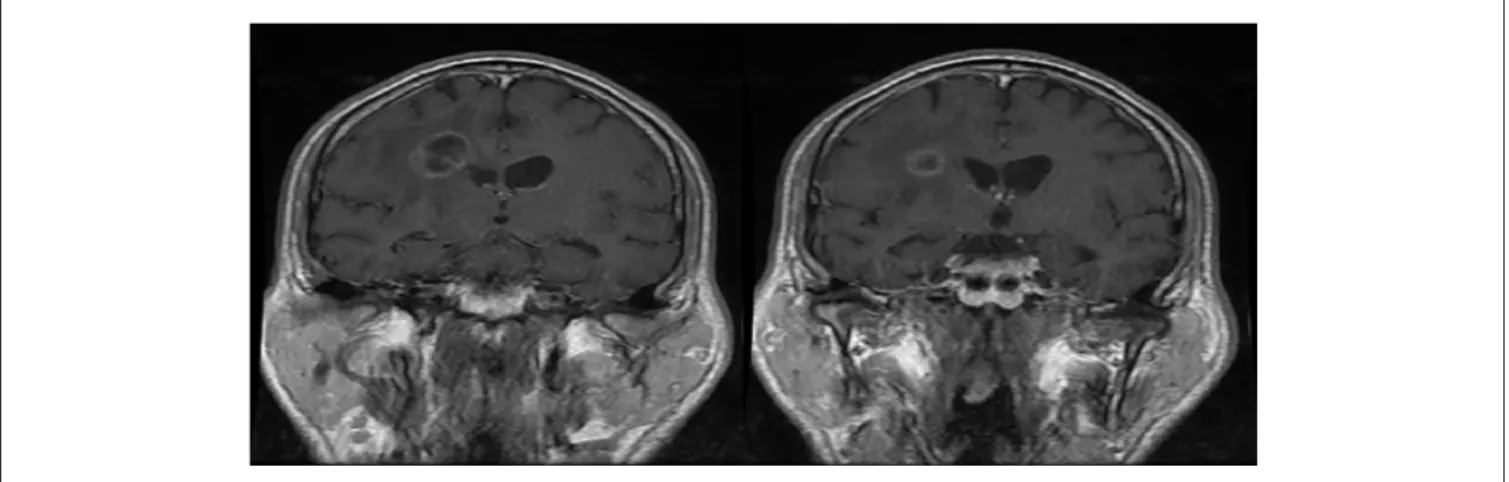
Figure 2. The coronal view on the T1WI shows a peripheral hyperintense rim with hypointense signals within the lesion.
Figure 3. The post-Gd, T2 FLAIR and DWI sequences of the lesion, showing peripheral rim enhancement with surrounding white matter edema.
tion, we tested the patient for bacteremia for a second time by sending 3 pairs of aerobic and anaerobic blood cultures.
After 3 days, the aerobic blood culture gave a positive result for C mucifaciens that explained the patient’s leukocytosis.
The resistance scale of the bacteria showed bacterial vulner- ability to several antibiotics (Table 1), and the patient was treated with intravenous clindamycin for 7 days. Once the patient’s status was stabilized, awake neuronavigation- guided needle biopsy was performed to obtain samples from the lesion, no bleeding was observed during the operation, and newer neurological deficits or insults were not observed during the postoperative care of the patient. The patient’s right-sided hemiparesis did not worsen or improve.
Histopathological Findings
Histological examination showed mainly the central part of a cerebral abscess, characterized by confluent necrosis com- posed of amorphous necrotic material and numerous neutro- phils. There were small areas of edematous brain tissue also present with swollen reactive astrocytes, but with no obvious granulation tissue formation (Figure 4). Special stains for microorganisms revealed a few Gram-positive aerobic bacilli, while the Grocott and periodic acid-Schiff stains were nega- tive for fungus infection. The overall appearances of these samples are in keeping with acute confluent suppurative encephalitis with confluent central necrosis (stage: days 2-7).
The Follow-up
Once the concrete diagnosis of the septic cerebral embolism was established, we wanted to rule out any suspicion of infective endocarditis even though the patient did not have any cardiologic signs or symptoms; therefore, a transesopha- geal cardiac echography as well as a blood culture to rule out the HACEK group bacteremia was done and neither of examinations resulted in any abnormal findings.
After the diagnosis was given, the patient was treated with intravenous clindamycin for an additional 7 days and then was discharged. His oral antibiotic (clindamycin) therapy was continued at home and a minimal dose of steroids (only
for 2 days) were prescribed for him to reduce the edema. The blood sugar levels were controlled on a daily basis. The patient had a control brain MRI in 3 months and the MRI scans showed a significant decrease in the mass of the lesion as well as a significant decrease in the peripheral edema (Figure 5). The patient had gained back his muscle strength almost completely with no other neurological signs and symptoms other than a right-sided 4/5 monoparesis of the forearm. The patient was controlled after 1 year, the physical examination did not reveal any neurological deficits, and the contrast-enhanced brain MRI showed a complete regression of the lesion (Figure 6).
Discussion
Diagnosing a septic cerebral embolus can be very challeng- ing and hard, due to the fact that several diseases such as metastatic tumors, tuberculomas, or even brain abscesses can mimic the cerebral septic emboli. Therefore, it is of great importance to have a solid differential diagnosis based on the patient’s clinical findings and the laboratory results. It is vital to mention that correlated morbidities such as diabetes, chronic immunosuppression, obesity, renal failure, and exist- ing malignancies play an essential role in formation of a sep- tic emboli.4
Figure 4. The microscopic findings and features of the biopsied tissue, showing confluent central necrosis containing necrotic debris and neutrophils as well as poor demarcation from the adjacent brain parenchyma.
It is a known fact that diabetic patients have a higher rate of infections (especially opportunistic infections) and a more complicated course of healing due to impairment of the adaptive cellular immunity. Among diabetic patients, those who develop ketoacidosis, in 75% of the cases the course of the disease is complicated or followed by infec- tions that has a mortality rate of 43%. Also in a different prospective study, it was shown that among 1000 studied patients with bacteremia, two thirds of the patients were diabetic patients.5
Septic cerebral emboli in most of the cases are originat- ing from cardiac vegetations, in fact 40% of patients with infective endocarditis are suspected to have neurological
symptoms due to cerebral embolization and in up to 47% of the patients neurological complications could be the first sign of infective endocarditis.6
Bacteremia is another common cause of septic cerebral emboli; therefore, it is strongly suggested that, in patients with bacteremia who develop sudden neurological symp- toms, septic cerebral emboli should be taken into consider- ation. These septic emboli can also cause mycotic aneurysms as well as secondary cerebral abscess formation. The forma- tion of mycotic aneurysms in patients with infective endo- carditis varies between 1% and 10%.6
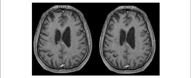
Most of the septic cerebral emboli are caused by Staphylococcus aureus,6 therefore there is very less Figure 5. The control postoperative magnetic resonance imaging scans after 3 months show a significant regression of the lesion.
Figure 6. The contrast-enhanced magnetic resonance imaging scans after 1 year show a complete regression of the lesion on the left side. Some artifacts of the biopsy can be seen in the surroundings of the surgical site.
done very cautiously to exclude other types of lesions.
Precise radiological examinations are needed to rule out multiple brain metastasis as well as tuberculomas of the brain. Yet as septic cerebral embolism is not seen often, and metastatic tumor lesions happen to have a high occurrence rate on CT and MRI scans, it is often mistaken for meta- static tumors, and in this case, despite the fact that the patient’s clinical findings were supporting a septic cerebral embolus, the neurosurgical team could not be convinced that the C mucifaciens bacteremia could be the cause of such a lesion; to establish a solid diagnosis and exclude metastatic alterations, we decided to perform a neuronavi- gation-guided biopsy and confirm the diagnosis by the microscopic features of the lesion.
Conclusion
Immunocompromised patients should be investigated further for opportunistic infections and diabetic patients are no exclusion of this fact. C mucifaciens as a normal skin flora can be a human pathogen in certain conditions; therefore, its recognition as a human pathogen in certain conditions is important and can play an important role in the diagnosis of a septic cerebral embolism. Also as septic cerebral emboli can mimic features of metastatic brain tumors as well as tuberculomas on radiological findings, it is important to consider a cerebral septic embolization in such patients as the course of treatment can be drastically changed toward
vidual cases or case series.
Informed Consent
Verbal informed consent was obtained from the patient(s) for their anonymized information to be published in this article.
ORCID iD
Seyed Arad Senaobar Tahaei https://orcid.org/0000-0002-3078 -8658
References
1. Cantarelli VV, Brodt TC, Secchi CA, Inamine E, Pereira Fde S, Pilger DA. Fatal case of bacteremia caused by an atypi- cal strain of Corynebacterium mucifaciens. Braz J Infect Dis.
2006;10:416-418.
2. Djossou FE, Bézian MC, Moynet DA, Le Flèche-Matéos A, Malvy D. Corynebacterium mucifaciens in an immunocom- petent patient with cavitary pneumonia. BMC Infect Dis.
2010;10:355.
3. Akshay PE, Nandish KU, Ketan RA, Bahri NU. Septic embolic encephalitis. Eur Radiol. 2017;15183.
4. Stawicki SP, Firstenberg MS, Lyaker MR, et al. Septic embo- lism in the intensive care unit. Int J Crit Illn Inj Sci. 2013;3:
58-63.
5. Geerlings SE, Hoepelman AI. Immune dysfunction in patients with diabetes mellitus (DM). FEMS Immunol Med Microbiol.
1999;26:259-265.
6. Zakher NA, Castillo MA, Torres CA. Unusual cerebral emboli.
Neuroimage Clin N Am. 2016;26:147-163.