FREE AND ZEOLITE-IMMOBILIZED PROBIOTIC MIXTURE VERSUS SODIUM VALPROATE IN PREVENTION OF OXIDATIVE STRESS AND MODULATION OF THE L- ARGININE INTRACELLULAR METABOLIC PATHWAYS IN
THE RAT BRAIN AND BLOOD FOLLOWING DEXAMPHETAMINE- INDUCED BIPOLAR DISORDER
N. Kh. Alchujyan
[a]*, M. R. Hovhannisyan
[a], N. H. Movsesyan
[a], R. A. Madoyan
[2], H. H.
Sargsyan
[a], A. A. Aghababova
[a], G. H. Minasyan
[c], H. L. Hairapetyan
[a], R. G. Kevorkian
[a], S. G.
Chailyan
[a]and G. A. Kevorkian
[a]Keywords: Arginase, bipolar disorder, d-amphetaminelipid peroxidation, nitric oxide synthase, probiotics, sodium valproate.
Experimental bipolar disorder (BD) was induced by repeated daily injection of the increasing doses of d-amphetamine sulfate (AMPH) (2-4 mg kg-1, 18 injections) in male young adult Wistar rats characterized by temporal arousal mimicked mania, and reduced exploratory and locomotor activities associated with behavioural depression under the condition of withdrawal of AMPH. At the end of the injection course, a stimulation of the lipid peroxidation processes and alterations in the mitochondrial and cytoplasmic activities of both arginase and nitric oxide synthase (NOS) were observed in the regions of brain corticolimbic system (prefrontal cortex, striatum, hippocampus and hypothalamus) and blood leukocytes. We have shown for the first time that a reversal treatment with the mixture of the specific probiotics with psycho- and antifungal activities in free (PMF) and zeolite-immobilized (PMZ) forms, and/or with a mood stabilizer, sodium valproate (VPA) inhibited oxidative stress and modulated differentially the L-arginine metabolic pathways in the brain and blood following AMPH- induced BD. Both PMF and PMZ efficiently normalized the activities of arginase isoforms and upregulated the suppressed intracellular NOS along with the gut microbiota restoration and prevention of the histopathological changes in the brain regions accompanied by normalization of rat behaviour.
* Corresponding Authors Fax:
E-Mail: alchujyan@mail.ru
[a] H. Buniatian Institute of Biochemistry NAS RA, 5/1 P.
Sevak St., 0014, Yerevan, Republic of Armenia [b] VITAMAX-E, LTD Co, Yerevan, Republic of Armenia [c] A.L. Mnjoyan Institute of fine organic chemistry (FOC) of
scientific technological center of organic and pharmaceutical chemistry NAS, Republic of Armenia
INTRODUCTION
Bipolar disorder (BD), a complex, psychiatric disorder is one of the leading causes of disability, low quality of life both among men and women, affecting about 60 million people worldwide. 1,2 In recent years the incidence of BD has increased, the mean age of patients decreased up to 42 years, and without treatment approximately 15 % of patients with BD commit suicide.3 Nowadays, the multiple character of the BD etiology is accepted involving oxidative stress, mitochondrial dysfunction, inflammation, cell signalling, apoptosis, impaired neurogenesis, etc., that are controlled by current mood stabilizers such as valproate, lithium, lamotrigine.4 It has also become obvious, that the microbiome alterations are implicated in stress response, memory functions, social behaviour, and mood contributing to the pathophysiology of BD and other neuropsychiatric disorders.5,6 Moreover, gut microbiota can affect cognition and behaviour through a number of immune-related mechanisms.7 Since, normalizing of microbiota with probiotics (live useful bacteria) is showing antipsychotic and
antidepresssant effects, their possible adjunctive therapeutic role in mood-related psychiatric symptoms has been suggested.8,9
Previously, we have shown that administration of zeolite- immobilized probiotics may protect from a development of depression/anxiety and cognitive deficit in stressed rats.10 We have also demonstrated the changes in the behaviour, gut microbiota, brain morphology and redox homeostasis are accompanied by perturbations in the L-arginine intracellular metabolic pathways in the regions of corticolimbic system and blood leukocyte following d- amphetamine-induced BD.11-13
In this study we show for the first time the effect of reversal treatment with the mixture of specific probiotics in free (PMF) and zeolite-immobilized (PMZ) forms on the mitochondrial and cytoplasmic activity of arginine- metabolizing enzymes, arginase and nitric oxide synthase (NOS) compared to reversal treatment with sodium valproate (VPA), a multiple action anticonvulsant and mood stabilizer that is also known as an effective drug in the treatment of BD.14
EXPERIMENTAL
Materials
Depakine, containing sodium valproate (VPA) (Sanofi- Aventis U.S. LLC) as an active substance, d-amphetamine
Eur. Chem. Bull. 2018, 7(1) 42-51 DOI: 10.17628/ecb.2018.7.42-51
43
sulfate (AMPH) (Sigma, St. Louis, Mo.), NG-monomethyl- L-arginine hydrochloride (Calbiochem La Jolla, CA), Dextran (Mr ̴ 70 000) (Serva, Heidelberg Germany), bovine serum albumin was from Carl Roth (GmbH, Karlsruhe) were used. All other reagents were purchased from Sigma- Aldrich Chemical Co. (St. Louis, MO, USA).
Commercially available probiotics, a concentrated source of naturally occurring microorganisms were used.
Lactobacillus rhamnosus strain ВКПМ В-6778, L.
salivarius strain B-7701(VITAMAX-E (LTD Co, Yerevan RA), L. plantarum strain ЦМПМ В-2353, L. acidophilus strain ИНМИА 9602 (РЦДМ), Bifidobacterium bifidum strain ВКПМ AC-1666, and E. coli strain М17 were rehydrated in sterile 0.85% NaCl and routinely propagated at 37° C in MRS medium (Hi Media, India) and/or MRS medium supplemented with 5 % MNM. Limiting dilution assay (by McCrady) was used for the separation, characterization, and quantitation of bacteria.21 The probiotic mixture contained (6 x 10⁹ CFU mL-1) with equal quantities of the mentioned microorganisms.
Animals and treatments
All procedures involving animals were in accordance with the International Laboratory Animal Care and the European Communities Council Directive (86/809/EEC) and approved by the respective local committee on biomedical ethics (H.
Bunyatyan institute of biochemistry, Yerevan, NAS RA).
Two-to 3-month-old maleWistar rats from our breeding colony were used. All animals were maintained on a 12 h light/dark cycle at normal room temperature and housed in groups of 6 per cage with free access to food and tap water.
Experimental design
The animals were divided into control group - native rats and experimental groups, in which BD was reproduced by repeated intramuscular (i.m.) injection of non-neurotoxic escalating doses of AMPH (2-4 mg kg-1).15,16 Rats received AMPH once a day on each weekday (but not on weekends), in total 18 injections. After the ninth injection the experimental animals were divided into four groups, an AMPH-group, in which rats continued receive AMPH only, VPA group, in which in parallel with AMPH injection animals were orally gavaged with VPA at a dose of 200 mg kg-1, and PMF and PMZ groups in which in parallel with AMPH injection animals were fed with 1 mL (6 x 10⁹ CFU mL-1) of the PMF and/or PMZ. Toward the end of the treatment, all of the animals underwent a behavioral testing in open field (OP) and elevated plus-maze (EPM).
Stereotypy ratings were also scored. Thereafter, rats were decapitated.
Open field (OF) test
The rats were placed singly into an OF (diameter 1m, divided by 2 concentric circles into 16 equal sections on the floor of the arena) and observed in 3 min to measure locomotor activity (the number of sectors crossed with all paws (crossing), exploratory behavior i.e., the number of rears (posture sustained with hind-paws on the floor) and grooming (including washing or mouthing of forelimbs,
hind-paws, body and genitals), and boluses (anxiety) counted manually/visually.17
Elevated plus-maze test
Immediately after the OF test the rats were placed singly into a common central platform (10 cm × 10 cm) of elevated plus-maze comprised two open and two closed arms (45 cm x 10 cm x 10 cm) and elevated to a height of 80 cm above the floor. During a 3-min observation period, the following parameters were measured: number of open arms entries and number of closed arm entries. Exploration (grooming and rearing) and risk assessment (number of hanging over the open arms).18 At the end of each trial, the open field and elevated plus-maze were wiped clean with ethanol.
Stereotypy ratings were scored as previously described.19
Microbiota
After decapitation trunk blood was collected, each animal was opened aseptically, samples of faeces from the lower part of the gut and washouts of brain were immediately placed into an anaerobic chamber for bacteriological analysis. Samples were incubated in sucrose broth at 37 °C for 24 h (blood was diluted by 1:5 v/v), then examined by microscopy, inoculated to the solid culture media, agar plates (Endo, sucrose, and blood agar), and incubated for 24 h. Blood samples were incubated for 5 days to facilitate a growth of microbes. The characteristics such as morphology and color of the colonies, as well as hemolysis, plasma- coagulation, aerobic fermentation of mannitol were examined for identification of microorganisms.20
Histopathological analysis
Formalin fixed brain region tissues were stained with hematoxylin and eosin (H&E) and examined for any histopathological changes. Pathological diagnosis of each brain specimen was assessed and analyzed by specialized histopathologist in a blinded manner.
Composition of chemically modified natural minerals
The multielemental composition, chemical modification, grinding (about 50 μm powder) of zeolite, bentonite and diatomite were previously dtermined, similarly dose- dependent effect on growth promotion in cultures of specific strains of Lactobacilli and Bifidobacteria and efficiency of their immobilization have been studied.10 Selected probiotics were cultured and immobilized using composition of micronized modified natural minerals (MNM) (zeolite (80 %), diatomite (10 %), and bentonite (10 %).
Brain cytoplasmic and mitochondria
Preparation of brain cytoplasmic and mitochondrial fractions was performed by differential centrifugation.22 Brains were rapidly removed from the skulls, placed on a cold plate, and prefrontal cortex (PFC), striatum, hippocampus and hypothalamus were dissected and homogenized in ice-cold 20 mM HEPES buffer pH 7.4, containing 0.25 M sucrose, (1:10, w/v) using Potter
homogenizer (1500 rpm for 3 min). Homogenates were centrifuged at 3000 rpm for 10 min to remove nuclear fraction. Supernatants were collected, centrifuged at 15000 rpm for 20 min, and cytoplasm in the supernatants and mitochondria in the pellets were obtained. Mitochondria were washed twice using the above mentioned buffer.
Isolation of blood leukocyte
Freshly obtained blood was drawn into 3.8 % sodium citrate anticoagulant, then mixed with 6 % dextran (prepared in 0.9 % NaCl) and incubated at 37 °C for 60 min to remove erythrocytes from blood by gravity sedimentation, and decanted layer was centrifuged at 1000 rpm for 5 min and the pellet containing leukocytes was washed twice and used, whereas plasma was obtained from supernatant by centrifugation at 6000 rpm for 20 min at 4 °C.23
Preparation of leukocyte cytoplasmic and mitochondrial fractions was performed by differential centrifugation of the leukocyte homogenates.22 Leukocytes were resuspended and homogenized in ice-cold 20 mM HEPES buffer pH 7.4, containing 0.25 M sucrose, (1:10, w/v) using Potter homogenizer (1500 rpm for 3 min), then centrifuged at 1200 rpm for 10 min at 4 °C to remove nuclei and cell debris.
Pellet was discarded and the supernatant further was centrifuged at 11000 rpm for 20 min at 4 оС to yield the crude mitochondrial preparation which was washed twice, resuspended and homogenized in the buffer used. The cytoplasm was in the supernatant fraction.
Arginase assay
The samples were added to the reaction mixture containing 20 mM HEPES buffer (pH 7.4), 3.9 mM MnCl2·4H2O, 15.4 mM L-arginine·HCl and incubated at 37 °C for 60 min, followed by the addition of 10 % TCA to stop the reaction.24 Parallel control experiments were conducted in the presence of 20 mM L-valine, a non- selective inhibitor of the arginase isoforms. Following a centrifugation (15000 rpm, 3 min) the protein-free supernatants were sampled and analyzed for L-ornithine content. The arginase activity expressed as produced in an hour L-ornithine per mg of total protein.
Measurement of L-ornithine
Samples were mixed with 4.5 % ninhydrin (1:1, v/v), heated (90 °C, 20 min), cooled to the room temperature and the absorbance was measured at 505 nm wavelength against reagent blank containing all the reagents minus the sample.24
Nitric oxide synthase assay
A total NOS activity was assessed by measuring stable intermediate of NO, nitrite (NO2-) accumulated during a long-term incubation of samples (37 °C for 22 h) in 20 mM HEPES buffer pH 7.4 in the presence of NOS substrate, 15.4 mM L-arginine·HCl, and cofactors included 0.2 mM NADPH, 6 µM FAD, 5.5 µM FMN, 20 µM ((6R)-5,6,7,8- tetrahydro-L-biopterin dihydrochloride) (BH4) and 1.7 mM CaCl2.25 Parallel control experiments were conducted in the presence of 15 mM NG–monomethyl-L-arginine·HCl, non-
selective inhibitor of all the NOS isoforms. Reaction was initiated by addition of samples to the incubation medium and terminated by subsequent addition of 0.5 N NaOH and 10 % ZnSO4·7H2O. Following a centrifugation (15000 rpm, 3 min) the protein-free supernatants were sampled and analyzed for nitrite content. The NOS activity is expressed as produced in 22 h nitrite per mg of total protein.
Measurement of nitrite
Samples were deproteinized with 0.5 N NaOH and 10 % ZnSO4·7H2O. Following a centrifugation (15000 rpm, 3 min), the protein-free supernatants were sampled and analyzed for nitrite using colorimetric technique based on diazotization reaction. Samples were mixed in equal parts with Griess-Ilosvay reagent (1:1 mixture of 0.17 % sulfanilic acid and 0.05 % α-naphthylamine in 12.5 % acetic acid) and measured at 546 nm wavelength against reagent blank containing all the reagents minus the sample.26
Indices of oxidative stress referring to lipid peroxidation processes were established by measuring malondialdehyde (MDA) using thiobarbituric acid (TBA).27 Samples were deproteinized with 10 % TCA and the precipitates were removed by centrifugation at 15000 rpm for 3 min, supernatants were mixed with 0.6 N HCl and 0.72 % TBA, heated for 15 min in boiling water bath that resulted in the formation of pink-colored secondary product of MDA and the absorbance was measured at 535 nm wavelength against reagent blank containing all the reagents minus the sample.
Protein was determined using crystalline bovine serum albumin as standard.28
Statistical analysis
All data were analyzed using a one-way analysis of variance (ANOVA) followed by post hoc Holm-Sidak test (SigmaStat 3.5 for Windows). Data are expressed as the mean ± S.E.M. Differences are considered statistically significant at a probability level of P < 0.05.
Results and discussion
Amphetamine-induced behaviors and underlying brain changes are considered as an endophenotype of BD.29 Previously, we have established a rat model of dexamphetamine (AMPH)-induced BD and revealed an overgrowth of Candida albicans, manifestation of Staphylococcus aureus contributed to a reduction in the number of beneficial bacteria, as well as changes in histopathological and biochemical patterns in brain and blood.11-13 Elevation of C. albicans and its association with worse positive psychiatric symptoms in patients with BD and schizophrenia have been demonstrated.30 We showed that preventive treatment with a mixture of the specific probiotic have benefits in the AMPH-induced BD.31 This probiotic mixture was composed of psychobiotics, L.
rhamnosus and Bifidobacterium bifidum, and Lactobacilli with fungicidal activity, as well as E. coli М17, which plays a pivotal role in the modulation of microbiota and maintaining homeostasis.32,33 However, a major concern for
Eur. Chem. Bull. 2018, 7(1) 42-51 DOI: 10.17628/ecb.2018.7.42-51
45
the use of probiotics in vivo is that they must survive and sustain transit through the detrimental factors of the gut in large quantities to facilitate their colonization in the host and confer in vivo health benefits, and immobilization of probiotics may protect them from the harmful gut factors and enable their transport and normal functioning in gut.34,35 Natural minerals such as zeolites, diatomite and bentonite with absorbent and ion-exchange properties containing macro- and microelements have been effectively used as carriers and promoters of bacterial growth.36 Natural minerals can also replenish the need of organism for minerals and used as enterosorbents improving metabolism via absorption of toxins from the intestine, and even from blood due to a diffusion through the intestine.37
In addition, natural minerals do not exert mutagenic effects, they are non-toxic, effective, versatile and economical, therefore bentonite and diatomite are E558, E551 food additives approved in EU as anti-caking agents.
Notably, potential benefit of a micronutrient treatment (consisting mainly of vitamins and minerals) is shown for various psychiatric symptoms, including bipolar II disorder with co-occurring attention- deficit/hyperactivity disorder.38 Based on this, we immobilized the above probiotics using the micronized chemically modified natural minerals composition (MNM) with domination of zeolite (see Experimental) and conducted a comparative study of the specific probiotic mixture in free (PMF) and MNM- immobilized (PMZ) forms versus VPA in reversal treatment of AMPH-induced BD.
Effect of treatment with probiotics vs. sodium valproate on histopathological changes in the regions of corticolimbic system
Our results show that reversal treatment with both probiotics and/or VPA prevented bacterial translocation and mainly normalized microbiota and rat behavior. However, in the gut of VPA-treated animals single colonies of S. aureus were found. It is in line with finding that sodium valproate is selectively potent in vitro against C. albicans, while it exerted low activity against S. aureus.39
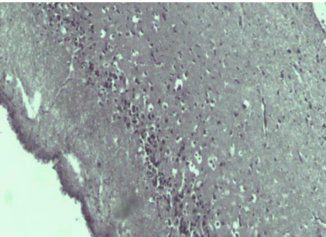
Restoration of balanced microflora via treatment with probiotics and VPA apparently contributed to amelioration and prevention of histopathological changes in the brain regions of corticolimbic system which were examined using H&E staining. Reversal treatment with both PMF and PMZ showed the similar effect on the brain regions morphology like in prevention treatment with the same mixture of probiotics, i.e., in the most of regions were detected only unremarkable changes from control.31
However, after reversal treatment with PMF in the hypothalamus were observed proliferation and edema, and in the PFC were seen multiple blood microvessels, following PMZ-treatment, presumably, related to protective capillary creation (Figure 1, A, B). As shown in Figure 1C-F, after reversal treatment with VPA, edema and compensatory full-blooded vessels were detected in the PFC, and interfibrillar edema, proliferation, cellular polymorphism were observed in the rest of brain regions.
Biochemical pattern associated with the effects of the probiotics and VPA was also studied.
Effect of treatment with free and immobilized probiotics vs.
sodium valproate on the lipid peroxidation processes in brain and blood
Overproduction of reactive oxygen species accompanied by protein oxidation, lipid peroxidation and oxidative damage to DNA/RNA plays crucial role in the pathophysiology of BD.40,41 Thiobarbituric acid reactive substances (TBARS) are formed as a byproduct of lipid peroxidation, and TBARS levels reflect the oxidative stress state which increases both in the acute phase of BD (mania/hypomania and depression) and with BD progression stage.42 We used assay of TBARS to measure MDA level formed via the decomposition of certain primary and secondary lipid peroxidation products and is a marker of oxidative stress. AMPH-induced BD was associated with an elevation of MDA level of a 2.7, 2.2, 2.0 and 4.5-fold in the PFC, striatum, hippocampus and hypothalamus and by about 6 and 3 times in the leukocyte and plasma compared respectively to ontrol. In reversal treatment administration all of preparations, PMF, PMZ and VPA decreased the MDA content in brain and blood (Figure 2).
PMF normalized the MDA content in the PFC and striatum. Both PMF and PMZ didnot influence MDA level in hippocampus, but reduced it almost halved in thehypothalamus, in which it remained 2.4 times above the control. At the same time, PMF diminished the MDA content in theleukocytes by 1.6 times, compared to control, whereas it was normalized by PMZ and VPA. Notably, VPA reduced the MDA content by 1.7, 1.8 and 1.5 times below the control in thePFC, striatum and hippocampus respectively, and normalized in hypothalamus.
Antidepressant mechanism of VPA is shown perhaps linked to an inhibition of oxidative damage via improvement serum MDA level, and serum catalase and superoxide dismutase activities, and upregulation of tyrosine hydroxylase and tryptophan hydroxylase in the PFC of rats exposed to chronic unpredicted stress.43 Both PMF and PMZ reduced drastically the MDA level in plasma by approximately 7 and 4.6 times compared to control, while after VPA treatment it was twice the norm.
Thus, probiotics and VPA suppress differently AMPH- induced lipid peroxidation, i.e., system-wide oxidative stress response there through preventing oxidative damage in the brain regions responsible for cognitive function, emotion and mood, as well as in blood leukocyte and plasma.
However, it should be considered that a drop of the MDA level below the norm could decrease the physiological level of oxidant challenge essential for governing life processes through redox signaling.44
Effect of treatment on the arginase activity in brain and blood Recent research has identified inflammatory agents and reactive oxygen species as drivers of the pathologic elevation of arginase activity and expression.45 Arginase hydrolyzes L-arginine to urea and L-ornithine, and exists in 2 isoforms, cytoplasmic (A1) and mitochondrial (A2).46 Immunolocalization studies have shown the presence of both A1 and A2 in brain, especially in hippocampal neurons.47 Differential expression of the arginase isoforms could provide a means to preferentially direct ornithine either to proline or excitatory amino acid glutamate
synthesis via ornithine aminotransferase in cytoplasm or to polyamine synthesis via ornithine decarboxylase in mitochondria.48 A significant increase in the concentration of polyamines in some structures of the limbic system and reticular formation in autopsy specimens of the brain of patients with schizophrenia has been found.49 Previously, we have shown that activation of lipid peroxidation processes was accompanied by a region-specific stimulation of the arginase isoforms in the cytoplasm and mitochondria in the brain corticolimbic system regions and blood leucocyte following AMPH-induced BD.12,13 Here we observed that
Figure 1A. Effect of treatment with PMF on proliferation and edema in hypothalamus.
Figure 1B. Effect of treatment with PMZ on multiple blood microvessels of PFC.
Figure 1C. Effect of treatment with VPA on edema and full- blooded vessels of PFC.
reversal treatment with VPA, and free and immobilized probiotic mixture exerted a modulatory effect on the intracellular arginase activity in brain and blood following AMPH-induced BD. 24 hours after discontinuation of treatment with probiotics and VPA and injection of dexamphetamine, A1 and A2 activities were mainly reduced in the brain regions studied, with exception for the A1
Figure 1D. Effect of treatment with VPA on Striatum showing intensive interfibrillar edema.
Figure 1E. Effect of treatment with VPA on Hippocampus showing proliferation, cellular polymorphism and the presence of large cells.
Figure 1 F. Effect of treatment with VPA on Hypothalamus showing interfibrillar edema and cellular polymorphism. (H&E stain, 100X).
Eur. Chem. Bull. 2018, 7(1) 42-51 DOI: 10.17628/ecb.2018.7.42-51
47
Figure 2. Effect of treatment with PMF, PMZ and VPA on the lipid peroxidation processes in the brain corticolimbic system regions and blood leukocyte.
activity in hippocampus resistant to any treatment used (Figure 3). Oxidized lipoproteins can upregulate A1 in mouse macrophages.50 Superoxide anion (O2•−) and hydrogen peroxide (H2O2) can also enhance mRNA content and A1 activity in the rat alveolar macrophages.51 A decrease of arginase isoforms activity partially is due to suppression of lipid peroxidation processes by the preparations. Of interest, both PMF and PMZ did not also decrease lipid peroxidation in the hippocampus following AMPH-induced BD. However, despite the fact that VPA decreased the level of MDA, the A1 activity was not reduced in the hippocampus of VPA-treated rats indicating the existence of other factors that may affect the expression
and activity of the enzyme. Figure. 3. Effect of treatment with PMF, PMZ and VPA on the arginase activity in the cytoplasm and mitochondria of the brain corticolimbic system regions.
It should be noted that on one hand A2 may contribute to oxidative stress via stimulation of mitochondrial reactive oxygen species (O2•− and H2O2) production and there through promote macrophage inflammatory responses.52 On the other hand, A2 preferentially direct ornithine to putrescine which suppresses lipid peroxidation, and support brain functions in adaptation to extreme environmental conditions.53 This complicates the picture studied. So, both PMZ and VPA equally reduce the MDA content in the leukocyte, but they differentially decrease the arginase isoforms activity (Figure 3).
It should be noted that during BD enhanced arginase activity and a subsequent decrease in the L-arginine levels can activate a stress kinase pathway that impairs function of T lymphocytes and also can inhibit the mitogen-activated protein kinase signaling pathway required for macrophage production of cytokines in response to bacterial endotoxin/lipopolysaccharide.54
VPA decreased the A1 and A2 activities by 1.9 and 4 times below control values in the PFC. PMZ caused a decrease in the activity of A1 and A2 of a 2.4 and 3.2-fold below the norm in the leukocytes respectively. Such suppression can affect the functions of arginase, which plays a role in protection against NH3 toxicity and cell growth and repair. So, hyperammonemia is caused by valproate therapy or overdose, and L-arginine could be potentially used therapeutically to correct this phenomenon.55
Figure 4. Effect of treatment with PMF, PMZ and VPA on the in the arginase activity in the cytoplasm and mitochondria of leukocyte.
Effect of treatment with probiotics and sodium valproate on the nitric oxide synthase activity in brain and blood
Arginase and nitric oxide synthase (NOS) share common substrate L-arginine, and another likely mechanism which may also contribute to the arginase effects is influence on the nitric oxide (NO) production via quenching of L- arginine and limiting its supply or via synthesis of urea, which inhibits a dimerization of the inducible NOS (iNOS) monomers to active form.56,57 Moreover, arginase-derived L- ornithine is converted to putrescine and then to the polyamines, spermidine and spermine, which inhibits iNOS translation and NO overproduction.58 NO is a versatile messenger molecule, with the characteristics of neurotransmitters, that may influence the levels of dopamine, noradrenaline, serotonine, acetylcholine, GABA.59 Moreover, NOS/NO system appeared to be involved in the pathophysiology of BD.41
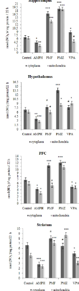
We have previously demonstrated that a total NOS activity was decreased in the cytoplasm and mitochondria in the regions of corticolimbic system and blood leucocyte following AMPH-induced BD.12 Reversal treatment with VPA and free and immobilized probiotic mixture not only prevented an inhibition of NO production, but also stimulated the latter (Figure 5).
A total NOS activity has increased approximately equally over shooting the control values in the cytoplasm and mitochondria of striatum and hippocampus following treatment with PMF and PMZ. Pronounced differences in the influence of PMF and PMZ on total NOS are observed in the mitochondria of PFC, and in the cytoplasm of hypothalamus leveled after a week post-treatment (data not shown). The tendency to normalization of the NOS in the cellular compartments of hippocampus and hypothalamus predominated following VPA treatment, whereas in the PFC and striatum a total NOS activity was not significantly affected by VPA. This indicates that an increase in the activity of NOS is not necessarily related to the inhibition of the activity of arginase isoforms by preparations studied.
Moreover, a stimulation of NOS could contribute to inhibition of arginase reaction, as the first intermediate of NO synthesis, NG-hydroxy-L-arginine is a well-known arginase inhibitor.60
Figure 5. Effect of treatment with PMF, PMZ and VPA on the total nitric oxide synthase activity in the cytoplasm and mitochondria of the brain corticolimbic system.
Eur. Chem. Bull. 2018, 7(1) 42-51 DOI: 10.17628/ecb.2018.7.42-51
49
The most pronounced drop about thrice in the NOS activity observed in the cytoplasm and mitochondria of blood leukocyte was prevented following reversal treatment with probiotics and VPA at AMPH-induced BD (Figre 6). VPA modulate a NOS activity in the cytoplasm and increased it in the mitochondria above the norm. PMF also caused a significant increase in the intracellular NO production, whereas PMZ had almost no effect. Nevertheless, a total NOS activity normalized in the cell compartments of leukocytes of both PMF- and PMZ-treated rats, a week after treatment in contrast to self-recovery group (data not shown).
Figure 6. Effect of treatment with PMF, PMZ and VPA on the total nitric oxide synthase activity in the cytoplasm and mitochondria of leukocyte.
It should be noted that increased arginase activity following AMPH-induced BD could restrict the supply of L- arginine required for NO production, and NOS will become uncoupled and use molecular oxygen to form superoxide, which reacts rapidly with any available NO to form peroxynitrite, further decreasing NO and further uncoupling NOS by oxidizing the co-factor BH4.61,62 Of interest, a negative correlations between NOS activity and free radical generation were revealed in the active rat cerebral cortex (animals selected using the emotional resonance test).63 The antioxidant effects of NO is a consequence of direct reaction with alkoxyl and peroxyl radical intermediates during lipid peroxidation, thus terminating lipid radical chain propagation reactions.64
CONCLUSION
Taken together the data presented in this report provide further support to the claim that psychoactive and antifungal probiotics mixture both in free and immobilized forms may normalize gut microbiota and histopathological changes in the brain corticolimbic system, as well as may efficiently suppress oxidative stress and modulate the L-arginine metabolic pathways in region-specific manner in the brain, and in blood leukocyte following dexamphetamine induced BD. Further study is needed to confirm whether L-arginine intracellular alternative metabolic pathways are represent new targets for developing methods to diagnose and treat BD, and whether PMF and PMZ are effective for BD, both as mono-therapy and in combination with mood stabilizers.
ACKNOWLEDGMENTS
The authors greatly thank Prof. Manukyan E.V. for hystopathological analysis and Ms. Ani Hakobyan (Master of English language and literature) for editing the manuscript.
References
1WHO/Mental disorders. 2016.
http://www.who.int/mediacentre/factsheets/fs396/en/
2Kim, Y., Santos, R., Gage, F. H., Marchetto, M. C., Molecular Mechanisms of Bipolar Disorder: Progress Made and Future Challenges, Front. Cell. Neurosci., 2017, 11, 30.
http://doi.org/10.3389/fncel.2017.00030
3Medici, C. R., Videbech, P., Gustafsson, L. N., Munk-Jørgensen, P., Mortality and secular trend in the incidence of bipolar disorder J. Affect. Disord., 2015, 183, 39. doi:
10.1016/j.jad.2015.04.032.
4Data-Franco, J., Singh, A., Popovic, D., Ashton, M., Berk, M., Vieta, E., Figueira, M. L., Dean, O. M., Beyond the therapeutic shackles of the monoamines: New mechanisms in bipolar disorder biology, Prog. Neuropsychopharmacol. Biol.
Psychiatry, 2017, 72, 73. doi: 10.1016/j.pnpbp.2016.09.004.
5Dickerson, F., Severance, E., Yolken R., The microbiome, immunity, and schizophrenia and bipolar disorder, Brain Behav. Immun., 2017, 62, 46. doi: 10.1016/j.bbi.2016.12.010.
6Latalova, K., Hajda, M., Prasko, J., Can gut microbes play a role in mental disorders and their treatment, Psychiatr.
Danub., 2017, 29 (1), 28.
hdbp.org/psychiatria_danubina/pdf/dnb_vol29_no1/dnb_vol2 9_no1_28.pdf
7Rescigno, M., Intestinal microbiota and its effects on the immune system, Cell Microbiol., 2014, 16, 1004. doi:
10.1111/cmi.12301.
8Bravo, J. A., Julio-Pieper, M., Forsythe, P., Kunze, W., Dinan, T.
G., Bienenstock, J., Cryan, J. F., Communication between gastrointestinal bacteria and the nervous system, Curr. Opin.
Pharmacol., 2012, 12(6), 667. doi:
10.1016/j.coph.2012.09.010.
9Gros, D. F., Antony, M. M., McCabe, R. E., Swinson, R. P., Frequency and severity of the symptoms of irritable bowel syndrome across the anxiety disorders and depression, J.
Anxiety Disord., 2009, 23(2), 290.
DOI:10.1016/j.janxdis.2008.08.004
10Barsegyan, K. A., Sargsyan, H. H., Madoyan, R. A., Alchujyan, N. Kh., Guevorkian, A. G., Movsesyan, N. H., Hayrapetyan, H. L., Hovhannisyan, M. R., Khachatryan, H. F., Barseghyan, V. A., Kevorkian, G. A., Combined effect of lactic acid bacteria and modified natural mineral composite substances on the chronic stress-induced depression and cognitive deficit. J. Applied Biochem. Photon, 2013, 106, 157.
ISJN: 3742-1863
11Kevorkian G. A., Hakobyan A. M., Movsesyan H. A., Agababova A. A., Amphetamine treatment affects the rat gut microbiota. Reports NAS RA, 2016, 116(3), 304. ISSN 0321- 1339
12Movsesyan, H. A., Alchujyan, N. Kh., Movsesyan, N. H., Aghababova, A. A., Hovhannisyan, M. R., Hakobyan, A. M., Minasyan, G. G., Khachatryan, H. F., Kevorkian, G. A., Metabolic changes in the corticolimbic system and blood following d-amphetamine-induced bipolar disorder.
Alternative pathways of L-arginine conversion. Med. Sci.
Arm., 2017, 57(2), 12. ISSN 0514-7484
13Movsesyan, H. A., Alchujyan, N. Kh., Movsesyan, N. H., Hakobyan, A. M., Aghababova, A. A., Hovhannisyan, M. R., Minasyan, G. G., Kevorkian, G. A., Metabolic changes in the corticolimbic system and blood following d-amphetamine- induced bipolar disorder. Oxidative stress and creatine kinase system. Med. Sci. Arm., 2017, 57(3), 37. ISSN 0514-7484
14Chateauvieux, S., Morceau, F., Dicato, M., Diederich, M., Molecular and Therapeutic Potential and Toxicity of Valproic Acid, J. Biomed. Biotech., 2010, 479364.
http://doi.org/10.1155/2010/479364
15Frey, B. N., Valvassori, S. S., Réus, G. Z., Martins, M. R., Petronilho, F. C., Bardini, K., Dal-Pizzol, F., Kapczinski, F., Quevedo, J., Effects of lithium and valproate on amphetamine-induced oxidative stress generation in an animal model of mania, J. Psychiatry Neurosci., 2006, 31(5), 326. PMID:16951735 PMCID:PMC1557682
16Robinson, T. E., Camp, D. M., Long-lasting effects of escalating doses of d-amphetamine on brain monoamines, amphetamine-induced stereotyped behavior and spontaneous nocturnal locomotion, Pharmacol. Biochem. Behav., 1987, 26(4), 821. PMID:2440058
17Buresh, Ya., Bureshova, O., Hyuston, P., Methods and basic Experiments on Brain and Behavior Study, Moscow, 1991, 399.
18Augustsson, H., Meyerson, B., Exploration and risk assessment: a comparative study of male house mice (Mus musculus musculus) and two laboratory strains, Physiol. Behav., 2004, 81(4), 685. DOI:10.1016/j.physbeh.2004.03.014
19Costall, B., Nylor, R., Olley J., Altitude Does Not Reduce Concussion Incidence, Eur. J. Pharmacol., 1972, 18, 95.
PMID:4555563
20Pokrowsky, M. N., Methodical instructions on microbiological diagnostics of the diseases caused by Enterobacteriacea, Moscow. 1986, 152.
21Aristovskaya T.V., Vladimirskaya M.E., Hollerbakh,Katanskaya G.A., Kashkin P.N., Practical Guide on Microbiology, (Ed., Seliber, G. L.), High School, Moscow, 1962, 491.
22Dizhe, G. P., Eshchenko, N. D., Dizhe, A. A., and Krasouskaya, I.
E., Introduction to the techniques of biochemical experiments, SPb. 2003, 86.
23Frik, G., Preisner, Z. S., Iensen, G. L., Burmeister, Yu., Immunological Methods (Ed. Friemel, H.), Mir, Moscow, 1979, 372.
24Iyamu, E. W., Asakura, T., Woods, G. W., A colorimetric microplate assay method for high-throughput analysis of arginase activity in vitro, Anal. Biochem., 2008, 383(2), 332.
DOI:10.1016/j.ab.2008.08.016
25Gagnon, C., Leblond, F. A., Filep, J. G., Peroxynitrite production by human neutrophils, monocytes and lymphocytes challenged with lipopolysaccharide, FEBS Lett., 1998, 431(1), 107. PMID:9684875
26Schmidt, H. H. H. W., Kelm, M., Determination of nitrite and nitrate by the Griess reaction. in Methods in Nitric Oxide Research, (Ed. Feelisch M., Stamler J. S.), Wiley, Chichester, 1996, 491.
27Buege, J. A., Aust, S. D., Methods Enzymol., Microsomal lipid peroxidation, 1978, 52, 302. PMID:672633
28Lowry, O. H., Rosebrough, N. J., Farr, A. L., Randall, R. J., Protein measurement with the Folin phenol reagent., J. Biol.
Chem., 1951, 193, 265. PMID:14907713
29Einat, H., Shaldubina, A., Bersudsky, Y., Belmaker, R. H., Prospects for the Development of Animal Models for the Study of Bipolar Disorder in Bipolar disorders: basic mechanisms and therapeutic implications (Ed. J.C. Soares, J.
C., A.H. Young, A. H.), 2nd ed. Informa Healthcare USA, Inc.
New York, London, 2007, 19-31.
30Severance, E. G., Gressitt, K. L., Stallings, C. R., Katsafanas, E., Schweinfurth, L. A., Savage, C. L., Adamos, M. B., Sweeney, K. M. , Origoni, A. E. , Khushalani, S., Leweke, F. M., Dickerson, F. B., Yolken, R. H., Candida albicans exposures, sex specificity and cognitive deficits in schizophrenia and bipolar disorder, NPJ Schizophrenia, 2016, 2, 16018. http://doi.org/10.1038/npjschz.2016.18
31Alchujyan, N. Kh., Aghababova, A. A., Movsesyan, N. H., Hovhannisyan, M. R., Movsesyan, H. A., Hakobyan, A. M., Kevorkian, G. A., Modulatory effect of the selected probiotics cocktail on the intracellular metabolic changes in the dynamics of dexamphetamine-induced bipolar disorder. Biol. J. Arm., 2017, 69 (3), 25. ISSN 0366-5119
32Lodinová-Zádníková, R., Cukrowska, B., Tlaskalova-Hogenova, H., Oral Administration of Probiotic Escherichia coli after Birth Reduces Frequency of Allergies and Repeated Infections Later in Life (after 10 and 20 Years), Int. Arch.
Allerg. Immunol., 2003, 131 (3), 209.
DOI:10.1159/000071488
33Hudault, S., Guignot, J., Servin, A. L., Escherichia coli strains colonising the gastrointestinal tract protect germfree mice against Salmonella typhimurium infection, Gut, 2001, 49(1), 47. PMID: 11413110 PMCID: PMC1728375
34Ding, W. K., Shah, N. P., Acid, Bile, and Heat Tolerance of Free and Microencapsulated Probiotic Bacteria, J. Food Sci., 2007, 72(9), M446. DOI:10.1111/j.1750-3841.2007.00565.x
35Singh, P. K., Kaur, I. P., Synbiotic (probiotic and ginger extract) loaded floating beads: a novel therapeutic option in an experimental paradigm of gastric ulcer, J. Pharm.
Pharmacol., 2012, 64 (2), 207. DOI:10.1111/j.2042- 7158.2011.01397.x
36Mery, C., Guerrero, L., Alonso-Gutierrez, J., Figueroa, M., Lema, J. M., Montalvo, S., Mena, C.. Borja, R., Evaluation of natural zeolite as microorganism support medium in nitrifying batch reactors: Influence of zeolite particle size, J.
Environ. Sci. Health A Tox. Hazard. Subst. Environ. Eng., 2012, 47(3), 420. DOI:10.1080/10934529.2012.646129
37Weiß, S., Lebuhn, M., Andrade, D., Zankel, A., Cardinale, M., Birner-Gruenberger, R., Somitsch, W., Ueberbacher, B. J., Guebitz, G. M., Activated zeolite--suitable carriers for microorganisms in anaerobic digestion processes, Appl.
Microbiol. Biotechnol., 2013, 97 (7), 3225.
DOI:10.1007/s00253-013-4691-6
38Rucklidge, J. J., Kaplan, B. J., Broad-spectrum micronutrient formulas for the treatment of psychiatric symptoms: a systematic review, Exp. Rev. Neurother., 2013, 13 (1), 49.
DOI:10.1586/ern.12.143
39Esiobu, N., Hoosein, N., An assessment of the in vitro antimicrobial effects of two antiepileptic drugs--sodium valproate and phenytoin, Antonie Van Leeuwenhoek, 2003, 83 (1), 63. PMID:12755481
40Steckert, A. V., Valvassori, S. S., Moretti, M., Dal-Pizzol, F., Quevedo, J., Role of oxidative stress in the pathophysiology of bipolar disorder, Neurochem. Res., 2010, 35 (9), 1295.
DOI:10.1007/s11064-010-0195-2.
41Brown, N. C., Andreazza, A. C., Young, L. T., An updated meta- analysis of oxidative stress markers in bipolar disorder, Psychiatry Res., 2014, 218 (1-2), 61.
DOI:10.1016/j.psychres.2014.04.005
42Siwek, M., Sowa-Kucma, M., Styczen, K., Misztak, P., Szewczyk, B., Topor-Madry, R., Nowak, G., Dudek, D., Rybakowski, J .K., Thiobarbituric Acid-Reactive Substances:
Markers of an Acute Episode and a Late Stage of Bipolar Disorder, Neuropsychobiol., 2016, 73 (2),116-22. doi:
10.1159/000444491.
43Qiu, H. M., Yang, J. X., Jiang, X. H., Hu, X. Y., Liu, D., Zhou, Q.
X., Enhancing tyrosine hydroxylase and tryptophan hydroxylase expression and improving oxidative stress involved in the antidepressant effect of sodium valproate on rats undergoing chronic unpredicted stress, Neuroreport, 2015, 26 (18), 1145. doi: 10.1097/WNR.0000000000000482.
Eur. Chem. Bull. 2018, 7(1) 42-51 DOI: 10.17628/ecb.2018.7.42-51
51
44Sies, H., Hydrogen peroxide as a central redox signaling molecule in physiological oxidative stress: Oxidative eustress
Redox Biol., 2017, 11, 613.
http://doi.org/10.1016/j.redox.2016.12.035
45Caldwell, R. B., Toque, H. A., Narayanan, S. P., Caldwell, R. W., Arginase: an old enzyme with new tricks, Trends Pharmacol.
Sci., 2015, 36(6), 395.
http://doi.org/10.1016/j.tips.2015.03.006
46Morris, S. M. Jr., Enzymes of arginine metabolism, J. Nutr., 2004, 134(10), 2743S-2747S. PMID:15465778
47Peters, D., Berger, J., Langnaese, K., Derst, C., Madai, V. I., Krauss, M., Fischer, K. D. Veh, R. W., Laube, G., Arginase and Arginine Decarboxylase – Where Do the Putative Gate Keepers of Polyamine Synthesis Reside in Rat Brain ? PLoS ONE, 2013, 8(6), e66735.
http://doi.org/10.1371/journal.pone.0066735
48Cederbaum, S. D., Yu, H., Grody, W. W., Kern, R. M., Yoo, P., Arginases I and II: do their functions overlap? Mol. Genet.
Metab., 2004, 81(Suppl 1), S38.
DOI:10.1016/j.ymgme.2003.10.012
49Svinarev, V. I., Syatkin, S. P., Frolov, V. A., Zaletok, S., Golomazova, K. A., Shevchenko, A. A, Fedoronchuk, T. V., Neborak, K.. Natroshvili, N., The Role of Polyamines in Etiopathogenesis of Schizophrenia, Amino Acids, 2007, 33, 43.
50Gallardo-Soler, A., Gomez-Nieto, C., Campo, M. L., Marathe, C., Tontonoz, P., Castrillo, A., Corraliza, I., Arginase I Induction by Modified Lipoproteins in Macrophages: A Peroxisome Proliferator-Activated Receptor-γ/δ-Mediated Effect that Links Lipid Metabolism and Immunity, Mol. Endocrinol., 2008, 22(6), 1394. DOI:10.1210/me.2007-0525
51Matthiesen, S., Lindemann, D., Warnken, M., Juergens, U. R., Racke, K., Inhibition of NADPH oxidase by apocynin inhibits lipopolysaccharide (LPS) induced up-regulation of arginase in rat alveolar macrophages, Eur. J. Pharmacol., 2008, 579 (1-3), 403. DOI:10.1016/j.ejphar.2007.10.043
52Ming, X-F., Rajapakse, A. G., Yepuri, G., Xiong, Y., Carvas, J.
M., Ruffieux, J., Scerri, I., Wu, Z., Popp, K., Arginase II Promotes Macrophage Inflammatory Responses Through Mitochondrial Reactive Oxygen Species, Contributing to Insulin Resistance and Atherogenesis, J. Am. Heart Assoc.
2012, 1, e000992. doi: 10.1161/JAHA.112.000992.
53Zomkowski, A. D, Santos, A. R., Rodrigues, A. L., Putrescine produces antidepressant-like effects in the forced swimming test and in the tail suspension test in mice, Prog. Neuropsych..
Biol. Psych., 2006, 30, 1419.
DOI:10.1016/j.pnpbp.2006.05.016
54Morris S. M. Jr., Arginine metabolism: boundaries of our knowledge, J. Nutr., 2007, 137, 1602S. PMID:17513435
55Schrettl, V., Felgenhauer, N., Rabe, C., Fernando, M., Eyer, F., L-Arginine in the treatment of valproate overdose – five clinical cases, Clin. Toxicol (Phila),2017, 55(4), 260. doi:
10.1080/15563650.2017.1284333.
56Mori, M., Regulation of nitric oxide synthesis and apoptosis by arginase and arginine recycling, J., Nutr., 2007, 137, 1616S.
PMID:17513437
57Moeslinger, T., Friedl, R., Volf, I., Brunner, M., Baran, H., Koller, E., Spieckermann P. G., Urea induces macrophage proliferation by inhibition of inducible nitric oxide synthesis, Kidney Int., 1999, 56(2), 581. DOI:10.1046/j.1523- 1755.1999.00570.x
58Bussiere, F. I., Chaturvedi, R., Cheng, Y., Gobert, A. P., Asim, M., Blumberg, D. R.., Xu, H., Kim, P. Y., Hacker, A., Casero, R. A., Wilson, K. T., Spermine Causes Loss of Innate Immune Response to Helicobacter pylori by Inhibition of Inducible Nitric-oxide Synthase Translation, J. Biol. Chem., 2005, 280(4), 2409. DOI:10.1074/jbc.C400498200
59Philippu, A., Nitric Oxide: A Universal Modulator of Brain Function, Curr. Med. Chem., 2016, 23 (24), 2643.
PMID:27356532
60Hecker M., Nematollahi H., Hey C., Busse R., Racke K.
Inhibition of arginase by NG-hydroxy-L-arginine in alveolar macrophages: implications for the utilization of L-arginine for nitric oxide synthesis, FEBS Lett., 1995, 359(2-3), 251.PMID: 7532597
61Romero, M. J., Platt, D. H., Tawfik, H. E., Labazi, M., El- Remessy, A. B., Bartoli, M., Caldwell, R. B., Caldwell, R.
W., Diabetes-induced Coronary Vascular Dysfunction Involves Increased Arginase Activity, Circ. Res., 2008, 102(1), 95. DOI:10.1161/CIRCRESAHA.107.155028
62Katusic, Z. S., d’Uscio, L., Nath, K. A., Vascular protection by tetrahydrobiopterin: progress and therapeutic prospects, Trends Pharmacol. Sci., 2009, 30 (1), 48.
http://doi.org/10.1016/j.tips.2008.10.003
63Gulyaeva, N. V., Onufriev, M.V., Stepanichev, M. Yu., NO synthase and free radical generation in brain regions of old rats: correlations with individual behaviour., Neuro Report, 1994, 6 (1), 94. PMID:7535579
64Rubbo, H., Radi, R., Trujillo, M., Telleri, R., Kalyaaraman, B., Barnes, S., Kirk, M., Freeman, B.A., Nitric oxide regulation of superoxide and peroxynitrite-dependent lipid peroxidation.
Formation of novel nitrogen-containing oxidized lipid derivatives, J. Biol. Chem., 1994, 269 (42), 26066.
PMID:7929318
Received: 06.03.2018.
Accepted: 10.01.2018.