Cancers 2021, 13, 2333. https://doi.org/10.3390/cancers13102333 www.mdpi.com/journal/cancers
Article
Pharmacogenetics of the Central Nervous System—Toxicity and Relapse Affecting the CNS in Pediatric Acute
Lymphoblastic Leukemia
Judit C. Sági 1, András Gézsi 2,3, Bálint Egyed 4, Zsuzsanna Jakab 4, Noémi Benedek 5, Andishe Attarbaschi 6, Stefan Köhrer 7, Jakub Sipek 8, Lucie Winkowska 8,9, Marketa Zaliova 8,9, Stavroula Anastasopoulou 10,11, Benjamin Ole Wolthers 12, Susanna Ranta 10,11, Csaba Szalai 1,13, Gábor T. Kovács 4, Ágnes F. Semsei 1,*,† and Dániel J. Erdélyi 4,*,†
1 Department of Genetics, Cell and Immunobiology, Semmelweis University, H-1089 Budapest, Hungary;
sagi.judit@med.semmelweis-univ.hu (J.C.S.); szalai.csaba@med.semmelweis-univ.hu (C.S.)
2 Department of Measurement and Information Systems, Budapest University of Technology and Economics, H-1111 Budapest, Hungary; gezsi.andras@gmail.com
3 MTA-SE Immune-Proteogenomics Extracellular Vesicle Research Group, Semmelweis University, H-1089 Budapest, Hungary
4 2nd Department of Pediatrics, Semmelweis University, H-1094 Budapest, Hungary;
egyed.balint@med.semmelweis-univ.hu (B.E.); jakab.zsuzsanna@med.semmelweis-univ.hu (Z.J.);
kovacs.gabor1@med.semmelweis-univ.hu (G.T.K.)
5 Department of Pediatrics, Pécs University, H-7623 Pécs, Hungary; noemi.benedek@gmail.com
6 Department of Pediatric Hematology and Oncology, St. Anna Children’s Hospital, Medical University of Vienna, A-1090 Vienna, Austria; andishe.attarbaschi@stanna.at
7 Renate Panzer-Grümayer: Leukemia Biology Group, Children’s Cancer Research Institute, A-1090 Vienna, Austria; stefan.koehrer@ccri.at
8 Department of Paediatric Haematology and Oncology, 2nd Faculty of Medicine, Charles University and University Hospital Motol, CZ-150 06 Prague, Czech Republic; jakub.sipek@fnmotol.cz (J.S.);
lucie.winkowska@lfmotol.cuni.cz (L.W.); marketa.zaliova@lfmotol.cuni.cz (M.Z.)
9 CLIP—Childhood Leukemia Investigation Prague, Department of Paediatric Haematology and Oncology, 2nd Faculty of Medicine, Charles University and University Hospital Motol, CZ-150 06 Prague, Czech Republic
10 Childhood Cancer Research Unit, Department of Women’s and Children’s Health, Karolinska Institutet, S-171 77 Stockholm, Sweden; stavroula.anastasopoulou@ki.se (S.A.); susanna.ranta@ki.se (S.R.)
11 Astrid Lindgren Children’s Hospital, Karolinska University Hospital, S-14186 Stockholm, Sweden
12 Department of Paediatrics and Adolescent Medicine, Rigshospitalet, DK-2100 Copenhagen, Denmark;
benjamin.ole.wolthers@regionh.dk
13 Central Laboratory, Heim Pál Children’s Hospital, H-1089 Budapest, Hungary
* Correspondence: semsei.agnes@med.semmelweis-univ.hu (A.F.S.);
erdelyi.daniel@med.semmelweis-univ.hu (D.J.E.).
† Ágnes F. Semsei and Dániel J. Erdélyi contributed equally to this work as co-last authors.
Simple Summary: Despite recent improvements in cure rates, pediatric acute lymphoblastic leuke- mia (ALL) patients remain at risk to develop relapse disease or suffer from therapy-associated side effects. Over 5% of adverse events appear in the central nervous system (CNS) and can impact sur- vival or quality of life of the patients. Inherited genetic variations are possible predictive factors for these adverse events. This retrospective study aimed to investigate if inherited genetic variations in genes encoding drug-metabolizing enzymes and drug transporters localized in the blood-brain bar- rier are predictive for CNS events. Our results suggest that certain ABCB1, ABCG2 and GSTP1 gene polymorphisms influence CNS toxicity and CNS relapse. A more effective drug-clearance could lead to less toxicity but contribute to a higher chance of relapse and vice versa. Genetic variants in ABCB1, ABCG2 or GSTP1 genes are promising candidates for personalized medicine.
Citation: Sági, J.C.; Gézsi, A.;
Egyed, B.; Jakab, Z.; Benedek, N.;
Attarbaschi, A.; Köhrer, S.; Sipek, J.;
Winkowska, L.; Zaliova, M.; et al.
Pharmacogenetics of the Central Nervous System—Toxicity and Relapse Affecting the CNS in Pediatric Acute Lymphoblastic Leukemia. Cancers 2021, 13, 2333.
https://doi.org/10.3390/
cancers13102333
Received: 27 March 2021 Accepted: 7 May 2021 Published: 12 May 2021
Publisher’s Note: MDPI stays neu- tral with regard to jurisdictional claims in published maps and institu- tional affiliations.
Copyright: © 2021 by the authors. Li- censee MDPI, Basel, Switzerland.
This article is an open access article distributed under the terms and con- ditions of the Creative Commons At- tribution (CC BY) license (http://crea- tivecommons.org/licenses/by/4.0/).
Abstract: Despite improving cure rates in childhood acute lymphoblastic leukemia (ALL), thera- peutic side effects and relapse are ongoing challenges. These can also affect the central nervous sys- tem (CNS). Our aim was to identify germline gene polymorphisms that influence the risk of CNS events. Sixty single nucleotide polymorphisms (SNPs) in 20 genes were genotyped in a Hungarian non-matched ALL cohort of 36 cases with chemotherapy related acute toxic encephalopathy (ATE) and 544 controls. Five significant SNPs were further analyzed in an extended Austrian-Czech- NOPHO cohort (n = 107 cases, n = 211 controls) but none of the associations could be validated.
Overall populations including all nations’ matched cohorts for ATE (n = 426) with seizure subgroup (n = 133) and posterior reversible encephalopathy syndrome (PRES, n = 251) were analyzed, as well.
We found that patients with ABCB1 rs1045642, rs1128503 or rs2032582 TT genotypes were more prone to have seizures but those with rs1045642 TT developed PRES less frequently. The same SNPs were also examined in relation to ALL relapse on a case-control matched cohort of 320 patients from all groups. Those with rs1128503 CC or rs2032582 GG genotypes showed higher incidence of CNS relapse. Our results suggest that blood-brain-barrier drug transporter gene-polymorphisms might have an inverse association with seizures and CNS relapse.
Keywords: encephalopathy; CNS toxicity; CNS relapse; PRES; genetic polymorphisms; childhood leukemia
1. Introduction
Cancer is the leading cause of illness-linked deaths in childhood in the developed countries [1]. The most common pediatric malignancy is acute lymphoblastic leukemia (ALL). Its effective chemotherapy regimens yield more than 90% long term survival rates in developed countries [2]. Although, adverse drug reactions are still a challenge [3]. Phar- macokinetics and pharmacodynamics of the drugs are also influenced by germline gene variants, some already indicated in drug labels [4,5]. The most important genetic risk fac- tors are already described in pediatric ALL as well, however, they failed to reliably predict prognosis [6]. Relapse, and toxicity-related deaths still limit outcome, therefore under- standing their genetic background and finding predictive biomarkers are still important [7,8]. Approximately 5–15% of children with ALL were reported to suffer from acute cen- tral nervous system (CNS) toxicity during the treatment, while isolated and/or combined CNS relapse occurred in 3–8% [9–11]. Neurotoxicity is the second most common adverse event to trigger dose-reduction of chemotherapeutic agents [12]. However, there are lim- ited number of publications investigating prognostic factors of acute CNS toxicity or phar- macogenetics of CNS relapse in ALL [13–15].
There are a few suggested pharmacogenetic risk factors for relapse or neurotoxicity in ALL. SNPs in a MTX (methotrexate) pathway enzyme, methylene tetrahydrofolate re- ductase (MTHFR) were reported to associate with neurotoxicity [16], and so were
CYP3A5*1/*3 alleles (cytochrome P450 family 3 subfamily A member 5) that play a role invincristine metabolism [17]. Central nervous system toxicity during cancer therapy was also associated with genetic variants of ABCB1, ABCG2 and GSTP1 (glutathione S-trans- ferase 1) [18–20]. Variants in GSTP1 and ABCB1 genes were also associated with CNS re- lapse or overall relapse [15,21]. Vincristine-pathway genes were not associated with re- lapse of ALL, but with peripheral neurotoxicity [22].
Symptoms of CNS toxicities vary based on etiology and the affected areas. Adverse complications within the brain may cause acute, subacute, or delayed encephalopathy [6].
Toxic encephalopathy may be reversible or permanent and can lead to neurocognitive
impairment [23]. The diagnosis of acute toxic encephalopathy is based on clinical features
and specific MRI findings [24]. Differential diagnosis requires the exclusion of peripheral
neuropathy, CNS infection, intracranial vascular events, CNS malignancy, effect of seda-
tive medications, or metabolic disturbances e.g., liver failure [25]. A nomenclature estab- lished in 2016 defined some typical CNS toxic events of childhood ALL therapy: metho- trexate-related stroke-like syndrome (SLS), seizures, posterior reversible encephalopathy syndrome (PRES), and depressed level of consciousness [26]. PRES and SLS have specific clinical and/or radiological patterns. Suspicion for PRES or SLS might be triggered by any CNS symptom with unique MRI lesions [10,26]. Chemotherapy related neurotoxicity in children with ALL appeared most often among females and at younger age [27]. Moreo- ver, it was also described that risk for PRES and seizures is higher in older children (>10 years) [28,29]. Toxicity of intrathecal chemotherapy was associated with age above 3 years in a different study [30]. However CNS involvement did not associate with MTX neuro- toxicity [31].
Patients with relapsed ALL face unfavorable outcome, their 5-year overall or event- free survival (OS, EFS) varies around 30–50% [32,33]. Approximately 30% of patients with relapsed ALL have CNS leukemia (combined or isolated) [15,34]. Repeated doses of in- trathecal chemotherapy (CNS treatment of CNS negative ALL patients) [27,34] in combi- nation with CNS directed systemic chemotherapy has reduced the CNS relapse rate to 5%
for the nineties [35]. Intrathecal dose intensification by CNS status at diagnosis could im- prove the prevention of CNS relapses [36–41].
Systemic and CNS directed treatment of ALL are known to be neurotoxic both in the short and in the long term [27,34,42]. Vincristine, methotrexate, cytarabine, l-asparaginase, iphosphamide, and glucocorticoids (prednisone and dexamethasone) are thought to exert the most acute adverse effects in the CNS [13,27]. It is usually hard to find single cause- effect relationships as multi-agent chemotherapy cycles are used, and other factors like drug-drug interactions, cranial irradiation, CNS-infiltration must also be considered [13].
Therefore, biomarkers for predicting CNS complications are much needed [34].
In 2007, we published a study on BBB pharmacogenetics of CNS toxicity in childhood ALL [20]. Acute toxic encephalopathy (ATE, any ≥ grade 3 CNS toxicity directly evoked by chemotherapy) was found to be more frequent among patients homozygous for the
ABCB1 rs1045642 T allele; and the association was stronger with a combination of ABCB1rs1045642 TT and ABCG2 rs2231142 CA/AA genotypes. In this study, our aims were to (1) reexamine this question on a larger patient cohort, with an extended set of SNPs relevant in pharmacogenetics; and (2) to examine the association of the same SNPs with leukemia CNS relapse. We hypothesized that a functional SNP leading to a higher concentration of chemotherapeutics in the brain would increase the risk of CNS toxicity but reduce the chance of CNS relapse, or vice versa.
2. Materials and Methods 2.1. Patients
We enrolled to all study cohorts children treated for frontline ALL, at ages 0–18 years
(1–18 years for toxicity analyses to avoid infant patients on different chemotherapy regi-
mens; 0–18 years for analyzing relapses) at diagnosis in Hungary, Austria, Czech Republic
and in the NOPHO group (Denmark, Norway, Sweden, Finland, Iceland, Lithuania, Es-
tonia) [43]. We excluded children with any previous chemotherapy, any major deviations
from ALL protocol to focus on pharmacogenetic effects. Clinical data were collected from
the medical records of the patients retrospectively. Data collection sheets of the PdL ‘Ret-
rospective Investigation of Children with ALL/LBL with Central Neurotoxicity Related to
Therapy’ study were used (with complements to Christina Halsey and the Ponte di Legno
Toxicity Working Group) as all four contributing groups are participating in that ongoing
study. See Tables 1–3, and Table S7 for characteristics of cohorts.
Table 1. Basic characteristics of the studied populations with acute encephalopathy (AE) and acute toxic encephalopathy (ATE).
Study Cohort Hungarian Austrian Czech NOPHO 4
Combined Joined Validation Cohort
Non-matched Matched Matched Matched
Phenotype AE ATE ATE ATE ATE
Number of
patients n 626 580 108 62 119 137 426
ATE Cases/controls n (%)
82/544 36/544 36/72 21/41 39/80 47/90 143/283
(13/87) (6/94) (33/67) (34/66) (49/51) (34/66) (34/66)
Seizure only n 21 20 20 8 10 6 44
SLS 1 n 6 6 6 1 6 7 20
Toxic PRES 2 n 3 3 3 12 18 33 66
Gender n (%) 339 317 52 26 53 74 205
Male (54) (55) (48) (42) (45) (54) (48)
Period of ALL
diagnosis y 1990–2015 1990–2015 1992–2015 2010–2018 2003–2017 2008–2015 1992–2018 Age at diagnosis n
(%) 104 88 35 30 42 29 136
>10 yr n (17) (15) (32) (48) (35) (21) (32)
Median (range) yr 5.0 (1–18) 5.0 (1–18) 7.7 (1–18) 9.9 (1.8–17.7) 7.1 (1.3–18) 7.0 (1–16) 7.6 (1–18)
Risk group (HR 3) n 75 69 17 29 15 41 102
(%) (12) (12) (16) (47) (13) (30) (24)
Abbreviations: AE: acute encephalopathy; ATE: acute toxic encephalopathy; 1 SLS: Stroke-like syndrome; 2 PRES: Posterior re-versible encephalopathy syndrome; 3 HR: high risk, as per patient’s treatment protocol; 4 NOPHO: Nordic Society for Pediatric Hematology and Oncology.
Table 2. Basic characteristics of the studied population of posterior reversible encephalopathy syndrome (PRES).
Study Cohort Austrian Czech Hungarian NOPHO 2 Combined
Matched cohorts
Number of patients n 39 62 18 132 251
Cases/controls n (%) 13/26 19/43 6/12 44/88 82/169
(33/67) (31/69) (33/66) (33/66) (33/67)
Gender n (%) 18 43 9 76 146
Male (46) (69) (50) (58) (58)
Period of ALL diagnosis y 2010–2017 2003–2017 1998–2013 2008–2015 1998–2017
Age at diagnosis n (%) 14 16 9 23 62
>10 yr n (36) (26) (50) (17) (25)
Median (range) yr 9.0(1.8–16.9) 5.68(1.3–14.5) 10.5(4–15) 8.0(1–15) 8.0(1–16.9)
Risk group (HR 1) n 21 7 3 48 79
(%) (54) (11) (17) (36) (32)
Abbreviations: 1 HR: high risk; 2 NOPHO: Nordic Society for Pediatric Hematology and Oncology.
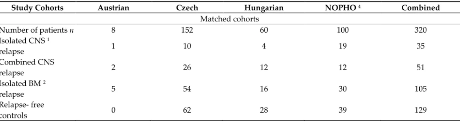
Table 3. Basic characteristics of the studied population of central nervous system first relapse (CNS relapse).
Study Cohorts Austrian Czech Hungarian NOPHO 4 Combined
Matched cohorts
Number of patients n 8 152 60 100 320
Isolated CNS 1
relapse 1 10 4 19 35
Combined CNS
relapse 2 26 12 12 51
Isolated BM 2
relapse 5 54 16 30 105
Relapse- free
controls 0 62 28 39 129
Gender n (%) 4 102 42 62 210
Male (50) (67) (70) (62) (66)
Period of ALL
diagnosis y 2010–2014 1996–2017 1992–2013 2008–2015 1992–2017
Age at diagnosis n
(%) 3 29 22 24 78
>10 yr n (40) (19) (37) (24) (24)
Median (range) yr 9.5 (5.8–15.9) 4.2 (0.1–17.8) 7.4 (1–17) 5.0 (1–16) 4.9 (0.1–17.8)
Risk group (HR 3) n 5 38 17 27 87
(%) (63) (25) (28) (27) (27)
Abbreviations: 1 CNS: central nervous system; 2 BM: bone marrow; 3 HR: high risk 4 NOPHO: Nordic Society for Pediatric Hematology and Oncology.
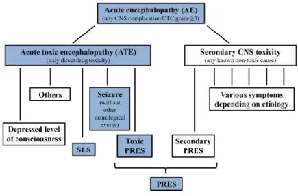
The two main studied phenotypes were adverse CNS symptoms called acute enceph- alopathy (AE) and CNS relapse. The definition of AE was any evolving adverse CNS symptom at least grade 3 as per Common Terminology Criteria for Adverse Events (CTCAE) v.4.0 occurring after the first dose of anti-leukemic treatment but within 3 weeks after the last dose of i.v. chemotherapy [44]. Patients with preceding CNS diseases; with uncertain, or mild neurologic symptoms were excluded from all analyses targeting neu- rotoxicity.
CNS adverse events with no known secondary etiology are defined as acute toxic encephalopathy (ATE.), as subgroup of AE. AE cases with identified underlying systemic causes including cerebrovascular events, CNS infections, actual CNS leukemia not in re- mission, metabolic alterations (e.g., severe electrolyte disturbance, hepatic encephalopa- thy, hypoglycemia or diabetic ketoacidosis) or insufficient CNS circulation (e.g., hyper- tensive encephalopathy, increased intracranial pressure, severe anemia or sepsis with hy- potension or hypoxia) possibly causing CNS symptoms were excluded. See more details in Supplementary Materials Patient Criteria. Hence, only events with suspected direct chemotherapy-related CNS adverse toxic effects were stratified as drug-induced ATE.
These patients could be classified into the overlapping Delphi consensus definitions of stroke-like syndrome (SLS), seizures without other neurological events, depressed level of consciousness, posterior reversible encephalopathy syndrome (PRES), however, these symptoms could also be observed with known secondary cases in the AE cohort [26] (Fig- ure 1.). Two controls per case were enrolled. Controls were pediatric patients with ALL who experienced none of these events, had no comorbidities, medical history, or co-med- ication that may have influenced the occurrence of CNS complications or drug pharma- cokinetics.
We categorized each event of AE according to four different types of chemotherapy cycles taking into account during or after what type of chemotherapy the CNS complica- tion evolved (see more details in Table S1b).
Boxes of studied phenotypes are highlighted with blue background. Note: symptoms of ATE subgroups may overlap, see definitions at Reference [26]. Further rare manifesta- tions of ATE are not demonstrated in the Figure 1., e.g., ataxia, extrapyramidal move- ments, steroid evoked psychosis, etc. Secondary CNS toxicities may present with differ- ent, similar or same symptoms as ATE. E.g. PRES can be caused by hyponatremia or by severe hypertension, but may also present without these.
For the CNS relapse case-control analysis, 1st ALL relapse cases were selected, both
isolated CNS and combined medullary plus CNS, and other extramedullary plus CNS
relapses. Three controls per one case were matched: two non-relapsed patients with ALL
and one isolated BM first relapse case. See Supplementary Materials Patient Criteria for
details.
2.2. Study Design, Overview
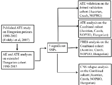
Following the 2007 publication, further Hungarian ALL patients were enrolled be- tween 2005 and 2015. Sixty SNPs in 20 genes encoding drug-metabolizing enzymes and transporters were studied on the whole 1990–2015 Hungarian non-matched patient cohort (n = 580). To validate prior results, we organized a European case-control matched cohort with Austrian, Czech, and Nordic Society of Pediatric Hematology and Oncology (NOPHO) groups for validation of the ATE—genotype associations found in the Hungar- ian population (validation cohort: 107 ATE cases and 211 controls). SLS, seizure without other neurological events, toxic PRES, altered consciousness, and their overlap cases were requested, and two matched controls for each case. The same enrolment criteria were used for all of the study groups when selecting patients for the Joined validation cohort. In the same study, we also examined another AE phenotype, PRES, which included cases with toxic or secondary causes (82 PRES cases, 169 controls). Together, the four groups had enough cases to test for the effect of the same SNPs on CNS relapse, as well (86 CNS re- lapse cases (isolated or combined), 105 isolated bone-marrow (BM) relapse cases, 129 con- trols). The number of patients to be involved was designed based on the results of the discovery population with the statistical power of 0.8. For the demonstration of the study elements of CNS toxicity in a flow chart see Figure 1.
Figure 1. Classification of CNS toxicities during childhood ALL therapy.
Throughout the paper, by ‘Joined cohort’, we mean the Austrian-Czech-NOPHO
case-control ATE validation population. By ‘Combined cohort’ of ATE or PRES or CNS
relapse, we mean the matched study populations of all the four study groups (Hungarian,
Austrian, Czech and NOPHO), respectively. See Figure 2.
Figure 2. Study design.
2.3. Ethical Considerations
The study was conducted according to the principles expressed in the Declaration of Helsinki for all nations. Written informed consent was requested from all patients or the parents or guardians of the minors involved in the study. The study was approved by the ethical committees in the participating countries. These are: Ethics Committee of the Med- ical University of Vienna on 3 August 2010 (No. 641/2010) (Austrian patients); Ethics Com- mittee of University Hospital Motol (approval file number NV15-30626A, approved in August 2014) (Czech patients); Ethics Committee of the Hungarian Medical Research Council (approval file number 12988-52-1018/-EKU, Date: 29 September 2003, 23310–
1/2011/EKU, Date: 19 January 2012, ad. 60106-1/2015/EKU, Date: 21 December 2015) (Hun- garian patients). The database containing phenotype data was approved by the Swedish Ethical Review Authority (731-10 (date 17 January 2011), the regional ethical review board of The Capital Region of Denmark (H-2-2010-022), the Danish Data Protection Authorities (j.nr.: 2012-58-0004), and by relevant regulatory authorities in all participating countries.
Genotype data were stored at the Technical University of Denmark’s server Comput- erome (NOPHO patients).
2.4. Laboratory Methods
DNA was isolated from peripheral blood taken in remission and 60 SNPs (Table S1a) of drug-metabolizing or transporting genes were selected and genotyped in the Hungar- ian population. The main features of the studied SNPs are summarized in Table S1a. Gen- otype data of 5 SNPs were requested from collaboration partners. In the Austrian cohort genotyping was performed via Sanger sequencing of remission bone marrow samples (see Table S1c for primer sequences). Major proportion of Czech patients was genotyped using KASPar (KBioscience Competitive Allele-Specific Polymerase chain reaction)-on-Demand prevalidated assays (LGC Biosearch Technologies , Hoddesdon, United Kingdom). Minor proportion was genotyped using the single nucleotide polymorphism arrays (Hu- manOmni Express BeadChip from Illumina, San Diego, CA, United states and CytoScan HD arrays from Affymetrix, Santa Clara, CA, United States) as described previously [45].
Genotyping of Hungarian patients was conducted using TaqMan
®OpenArray™ Geno-
typing System (Thermo Fisher Scientific, Waltham, MA, United States) or using KASPar-
on-Demand prevalidated assays (LGC Biosearch Technologies, Hoddesdon, United King-
dom) following the manufacturer’s instructions as described earlier [46]. The genotyping
of NOPHO patients were performed using Omni2.5exome-8-BeadChip arrays (Illumina,
San Diego, CA, United States) and described in detail in the article of Wolthers et al. [47]
and Hojfeldt et al. [48].
2.5. Statistical Analysis
In the Hungarian cohort, multivariate logistic regression models (for case-control analysis) and Cox proportional hazards regression models (for survival analysis) were used to investigate the influence of genetic polymorphisms on the neurological symptoms affecting the brain and CNS relapse. In the Joined cohort and during the analysis of the Combined cohort, conditional logistic regression models (for case-control analysis on co- horts with matched controls) and Cox proportional hazard regression models for nested case-control data (for survival analysis on cohorts with matched controls) were used. We calculated the OS, EFS in every disease cohort (AE sub-phenotypes, CNS relapse). Fisher exact test was used to evaluate the association between NOPHO or BFM-protocols and occurrence of the studied phenotypes. Detailed study constructions of CNS events are shown in Figure 2. The number of cases with depressed level of consciousness was below 10, so we did not analyze this sub-phenotype separately. Confounders used in analyses are shown in Table S1b. Allele frequencies were estimated by allele counting and tested for deviation from Hardy-Weinberg equilibrium (HWE) by the on-line software (Tests for deviation from Hardy-Weinberg equilibrium
p. https://ihg.gsf.de/cgi-bin/hw/hwa1.pl.).Significant violation of HWE was considered if p ≤ 1.13E-02. Confidence intervals (CI) or hazard ratios (HR) were calculated at the 95% level. The analyses were performed study- ing the genotypes separately (11 vs. 12; 11 vs. 22), using dominant (11 vs. 12/22) or reces- sive (11/12 vs. 22) models, with the common homozygotes signed as 11 and the rare (2) allele supposed to be dominant. If the number of rare homozygote patients was n ≤ 10, we merged them with heterozygote patients for the analyses. Genotype combinations were determined based on the results of these merged groups. Multiple testing corrections were performed using the Benjamini-Hochberg false discovery rate (FDR) method with a type I error rate of 13% [49,50]. Alpha levels of p ≤ 1.13E-02 were considered significant after FDR correction in multiple testing for the studied SNPs (with 465 analyses performed for 60 or 5 SNPs and each phenotype). Results reported without mentioning the used model were studied in additive model. Analyses were performed using IBM SPSS Statistics 25.0 (IBM Corporation, Armonk, NY, United States) and R statistical software (version 3.6.3, R Foundation for Statistical Computing, Vienna, Austria). Conditional logistic regression analyses were performed by the clogit function of the survival package of R [51]. Cox proportional hazards regression analyses for nested case-control data were performed by the multipleNCC package [52]. Power analysis was conducted by PS: Power and Sample Size Calculation 3.1.2.
3. Results
3.1. Chemotherapy Related Adverse Neurological Symptoms
3.1.1. Case-Control Analyses
Acute encephalopathy and its sub-phenotype, ATE, were first studied in the Hun-
garian discovery cohort. The following genotypes below were found to associate with
both AE and ATE: ABCB1 rs1045642 TT, rs1128503 TT and the combination of
ABCB1rs1045642 TT genotype with ABCG2 rs2231142 CA or AA genotypes. GSTP1 rs1695 AG +
GG genotype associated with decreased risk for AE and ATE in this cohort.
ABCB1rs2032582 TT associated with ATE only. The other examined 55 SNPs showed no signifi-
cant association with ATE. The summary of the results is shown in Table S2a and Table
S4a. When analyzing the 5 selected SNPs and ATE in the Joined validation cohort (Aus-
trian, Czech and NOPHO case-control cohort), none of the associations could be con-
firmed. The relation of GSTP1 rs1695 and ATE was actually the opposite of that found in
the Hungarian cohort, while tests with the ABC SNPs were largely non-significant (see
Table S2a, Table S4b).
The Combined cohort of patients including both the matched Hungarian ATE cohort and the Joined validation cohort was large enough for more detailed analyses of neuro- toxicity phenotypes: seizure without other neurological events, SLS, and toxic PRES. T alleles of ABCB1 rs1045642, rs1128503 and rs2032582 polymorphisms appear to be associ- ated with seizures, and particularly with seizures during Induction-like chemotherapy cycles (see Table S2a and Table S4c). On the other hand, the ABCB1 rs1045642 CT genotype might be protective against PRES and toxic PRES. In addition to the genetic variations, CNS 2 status was also predictive for PRES (OR = 5.08, CI 95% (2.10–12.29)) (see Table S2a, Table S4c and Table S4d). PRES and toxic PRES were more frequent in the NOPHO cohort compared to those of the countries using BFM-protocols (OR = 2.14, CR95% (1.25–3.67), OR = 2.98, CI95% (1.33–6.65)) (see Table S4e). SLS did not associate with the studied SNPs.
3.1.2. Survival Analyses on the Neurotoxicity Case-Control Cohorts
OS and EFS were studied on cohorts with adverse neurological symptoms and in association with SNPs. A higher risk for death was associated with AE in the studied un- matched Hungarian cohort (HR = 2.51, CI 95% (1.32–4.76)). Among the 82 AE cases, in our database two cases died related to neurotoxicity (9.5% of all exits). Examining SNPs with survival on the unmatched Hungarian cohorts of AE or ATE, patients with
CYP3A5rs4646450 T allele had worse outcome (both OS and EFS). This risk was even higher in patients with TT genotype. CYP3A4 rs3735451 GG genotype associated with poorer OS and EFS (see Table S2b, Table S5a). Analyzing the Combined matched cohort of ATE in which only 5 SNPs were genotyped, GSTP1 rs1695 GG + AG genotype was associated with better outcome (OS), and this association remained significant in the seizure sub- phenotype cohort, and in the ATE cohort during Induction-like cycles (see Table S2b, Ta- ble S5b). Analyzing EFS of the Combined cohort in PRES, the worse outcome was associ- ated with ABCB1 rs2032582 TT genotype and with the combination of ABCB1 rs1045642 TT genotype with ABCG2 rs2231142 CA or AA genotypes (see Table S2b, Table S5c).
3.2. Central Nervous System Relapse
We analyzed the impact of SNPs in metabolizing enzymes and transporters on the prevalence of CNS relapse, using the Combined relapse case-control cohort. When com- paring patients with isolated or combined CNS relapse to non-relapsed controls, the
ABCB1 rs2032582 GT and the rs1128503 TT + CT genotype seemed to be protectors againstCNS relapse. The results are shown in Table S3a and Table S6a. Analyzing the survival of the Combined relapse case-control cohort, we have not found any significant SNPs in as- sociation with CNS relapse. The summary of the results is shown in Table S3b. The full set of results can be found in Table S6b.
3.3. Inverse Association of SNPs with Chemotherapy Related Adverse Neurological Events and CNS Relapse
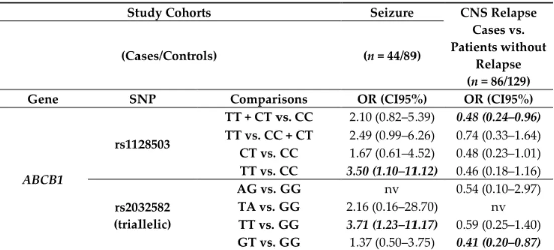
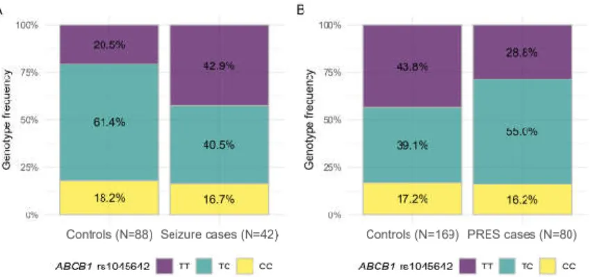
Examining Combined cohorts of ATE and CNS relapse including case-control matched cohorts from all groups, we have found that patients with ABCB1 rs1128503 TT or rs2032582 TT genotypes were more prone to have toxicity related seizures but lower incidence of CNS relapse. For more details see Figure 3 and Table 4.
ABCB1
rs1045642 TT was also in inverse association with seizure and PRES in the Combined cohort (p = 0.011, OR = 0.34, CI95% (0.15–0.78), p = 0.017, OR = 2.10, CI95% (1.14–
3.87), respectively) (Figure 4).
Table 4. Summary of the results of toxic seizure and CNS relapse analyses in Combined cohort.
Study Cohorts Seizure CNS Relapse
Cases vs.
Patients without Relapse (n = 86/129)
(Cases/Controls) (n = 44/89)
Gene SNP Comparisons OR (CI95%) OR (CI95%)
ABCB1
rs1128503
TT + CT vs. CC 2.10 (0.82–5.39) 0.48 (0.24–0.96) TT vs. CC + CT 2.49 (0.99–6.26) 0.74 (0.33–1.64) CT vs. CC 1.67 (0.61–4.52) 0.48 (0.23–1.01) TT vs. CC 3.50 (1.10–11.12) 0.46 (0.18–1.16)
rs2032582 (triallelic)
AG vs. GG nv 0.54 (0.10–2.97) TA vs. GG 2.16 (0.16–28.70) nv TT vs. GG 3.71 (1.23–11.17) 0.59 (0.25–1.40) GT vs. GG 1.37 (0.50–3.75) 0.41 (0.20–0.87) Abbreviations: nv: not valid; CNS: central nervous system; REL: relapse. Results with p ≤ 0.05 are shown with bold italics characters, significant results with p ≤ 1.13E-02 are shown with bold char- acters.
Figure 3. Inverse associations of blood-brain-barrier SNPs with toxic seizure or CNS relapse in case-control analyses. The studied populations were the Combined case-control cohorts of ATE and CNS relapse, respectively. (A) Genotype fre- quencies between cases and controls regarding association of ABCB1 rs1128503 and seizure, (B) Genotype frequencies between cases and controls regarding association of ABCB1 rs1128503 and CNS relapse, (C) Genotype frequencies between cases and controls regarding association of ABCB1 rs2032582 (triallelic) and seizure, (D) Genotype frequencies between cases and controls regarding association of ABCB1 rs2032582 (triallelic) and CNS relapse. Colors refer to genotypes.
Figure 4. Genotype distributions of ABCB1 rs1045642 in seizure or PRES Combined cohorts. (A) Genotype frequencies between cases and controls regarding association of ABCB1 rs1045642 and seizure, (B) Genotype frequencies between cases and controls regarding association of ABCB1 rs1045642 and PRES. Colors refer to genotypes.
4. Discussion
In this study, we evaluated the association of SNPs in drug-metabolizing and trans- porting genes with acute CNS toxicity and CNS relapse episodes in patients with child- hood acute lymphoblastic leukemia. In the Hungarian cohort, we found that
ABCB1rs1045642, rs1128503, and rs2032582 TT genotypes, the combination of ABCB1 rs1045642 TT genotype with ABCG2 rs2231142 CA or AA genotypes, and GSTP1 rs1695 AA geno- type may increase the risk of chemotherapy-related adverse neurological symptoms.
These associations were not confirmed in the Austrian-Czech-NOPHO Joined validation cohort, however, still appeared as significant in the seizure subgroup of the Combined cohort. Interestingly, there appears to be an inverse association of the SNP rs1045642 with PRES and seizure in Combined cohorts. Our results with ABCB1 rs1128503 and rs2032582 in relation with seizure and CNS relapse suggest that blood-brain-barrier drug transporter gene-polymorphisms might have an inverse association with CNS toxicity and CNS re- lapse. The Hungarian AE cases had lower OS, CYP3A5 rs4646450 and CYP3A4 rs3735451 associated with worse OS and EFS in the Hungarian AE and ATE cohorts.
Patients with CNS toxicity had worse survival than control patients in our analysis.
The direct contribution of neurotoxic events to the deaths were negligible. This is in par- allel with findings of other studies and may be related to treatment delays, dose-reduc- tions or omissions of intrathecal or systemic chemotherapy after the neurotoxic event, or enzyme inducing antiepileptic therapies increasing the metabolism of chemotherapy [53–
56]. Delays in intrathecal MTX treatment caused by MTX neurotoxicity associated with increased risk of CNS relapse [31]. Similar strategies were applied indeed unfortunately in some hospitals at the time when our study cohort was treated [56].
ABC transporters are important in the resistance to methotrexate, cytarabine, vincris-
tine, anthracyclines, and dexamethasone, influence response to treatment and survival
[57–59]. Genetic variants of ABCB1 were studied in hematological malignancies, a broad
variety of conclusions regarding their function was observed but their true clinical impact
is still under debate [60]. ABCB1 rs1045642 TT + CT vs CC alleles were associated with
higher 24 h plasma MTX concentration [61]. In contrast, rs1045642 CC genotype associated
with higher MTX plasma level and with relapse investigating high risk ALL patients in
another study [62]. ABCB1 genetic variants can influence cerebrospinal fluid (CSF) drug
levels. The CSF concentration of MTX was different between the rs1045642 C allele (CC +
CT) carriers and TT homozygous patients [63]. ABCB1 SNPs were found to associate with
vincristine-related neurotoxicity in a study, however, they have found no association with
SNPs included in our analysis [64]. Another ABCB1 SNP, rs4728709 T allele was also pro-
tective against neurotoxicity in the study of Ceppi et. al. [22]. ABCB1 rs1045642 (C3435T)
and rs2032582 (G2677T) TT genotype associated with worse EFS and the same trend was
observed if rs1128503 T allele was also included in the analysis [22].
GSTP1 protectsagainst oxidative stress, GSTP1 rs1695 is a missense variant, decreases the enzyme activity [65]. GSTP1 rs1695 GG genotype associated with CNS toxicity and also with attention def- icit in ALL survivors [19,66]. GSTP1 rs1695 G allele in two different studies increased and reduced the risk for CNS relapse in ALL [15,67–70].
This study has multiple limitations. The retrospective data collection may have re- sulted in skewed populations. Another difficulty lies in the categorization of neurotoxic events into phenotype subgroups (like SLS, seizures, PRES), or into etiology groups (toxic or secondary). Various subsets of tests were missing (not performed or not available in retrospect) for some of the neurotoxicity cases, e.g., blood pressure, miscellaneous labor- atory results and imaging. The differentiation of toxic PRES (direct drug toxicity) and sec- ondary PRES (e.g., PRES evoked by hyponatremia or hypertension which had been caused by chemotherapy) is especially difficult and may just be theoretical. We aimed to be consistent and applied the logic described in Figure 1. and Supplementary Materials Patient Criteria and also took both the original opinion of the treating physician and the Delphi definitions [26] into consideration. The categorization of events was unambiguous in the large majority of the cases, so we think these factors don’t undermine the results of the study. Discrepancies in CNS2 status classification among study groups are well known, this confounding factor could not be avoided. Treatment heterogeneity may also have influenced our results. NOPHO protocols use higher and more frequent dosing of vincristine than BFM-based protocols applied in the other groups, the high rate of PRES among NOPHO patients may also relate to this.
Despite some identified associations that are concordant with our original hypothe- sis, we can’t formulate practical clinical guidance based on our results. As neurotoxic ep- isodes proved to be reversible with only very few exceptions, surely CNS relapse should be the main focus in any future attempt on therapy individualization. There is great need for even larger international studies for studying these very rare events in more homoge- nous cohorts.
5. Conclusions
In the present study SNPs were investigated in a European international collabora- tion to assess their role in CNS complications and related survival. Key BBB genes ABCB1 and GSTP1 and their SNPs rs1045642, rs1128503, rs2032582 and rs1695 came to focus. Our findings suggest that genetic variations which are associated with a lower or higher risk of CNS complications can also associate with better or worse outcomes in respect of sur- vival, respectively.
Supplementary Materials: The following are available online at www.mdpi.com/2072- 6694/13/10/2333/s1, Supplementary Patient Criteria, Table S1a: Information about the studied SNPs, Table S1b: Confounders and stratification listed for every analyses, Table S1c: Primer sequences used in Sanger-sequencing of the Austrian population, Table S2a: Results of the case-control analy- sis of chemotherapy related adverse neurological events, Table S2b: Survival analysis on the CNS adverse event case-control cohort, Table S3a: SNPs analyzed in the CNS relapse Combined case- control cohort, Table S3b: Results of the survival analysis of CNS relapse Combined cohort, Table S4a: Results of acute encephalopathy and acute toxic encephalopathy case-control study in the Hun- garian cohort, Table S4b: Results of acute toxic encephalopathy case-control study in the joined val- idation cohort, Table S4c: Results of acute toxic encephalopathy case-control study in the combined cohort, Table S4d: Results of PRES case-control study in the combined cohort, Table S4e. Results of treatment protocol-comparisons among acute toxic encephalopathy subphenotypes in the com- bined cohort, Table S5a: Results of the survival study in the Hungarian cohort with acute encepha- lopathy and acute toxic encephalopathy, Table S5b: Results of the survival study in the combined cohort with acute toxic encephalopathy, Table S5c: Results of the survival study in the combined cohort with PRES, Table S6a: Results of case-control study in combined cohort with CNS relapse, Table S6b: Results of survival study in combined cohort with CNS relapse, Table S7: Applied pro- tocols in the studied populations.
Author Contributions: Conceptualization, J.C.S., Á.F.S. and D.J.E.; Data curation, J.C.S., A.G. and Á.F.S.; Formal analysis, A.G.; Funding acquisition, C.S. and D.J.E.; Investigation, J.C.S., Z.J. and N.B.;
Methodology, J.C.S., Á.F.S. and D.J.E.; Resources, J.S. and S.A.; Software, A.G. and B.E.; Supervision, G.T.K.; Validation, S.K., L.W. and B.O.W.; Visualization, A.G.; Writing—original draft, J.C.S., Á.F.S.
and D.J.E.; Writing—review & editing, A.A., M.Z., S.R. and C.S. All authors have read and agreed to the published version of the manuscript.
Funding: The project was supported by the St. Anna Kinderkrebsforschung in case of the Austrian group. Czech research infrastructure was supported by the NCMG-LM2018132. The Hungarian study part was supported by the New National Excellence Program of the Ministry for Innovation and Technology Grant No. UNKP-19-3-IV (JC Sagi); by the National Research, Development and Innovation Office (NKFIH) Grants No. PD109200 (Á.F.S.), PD134449 (A.G.) and K115861 (D.J.E.), by the Hungarian Paediatric Oncology Network (07/MGYH-MGYGYT/2018), and by the János Bolyai Research Scholarship of the Hungarian Academy of Sciences (A.G.). The NOPHO study was sup- ported by the Swedish Childhood Cancer Fund (KP2017-0010, TJ2020-0082, TJ2019-0031), by the Danish Childhood Cancer Fund and by the Danish Cancer Society (R150-A10181). The funders had no role in study design, data collection and analysis, decision to publish, or preparation of the man- uscript.
Institutional Review Board Statement: The study was conducted according to the principles ex- pressed in the Declaration of Helsinki for all nations. Written informed consent was requested from all patients or the parents or guardians of the minors involved in the study. The study was approved by the ethical committees in the participating countries. The study was approved by the ethical committees in the participating countries. These are: Ethics Committee of the Medical University of Vienna on 3 August 2010 (No. 641/2010) (Austrian patients); Ethics Committee of University Hos- pital Motol (approval file number NV15-30626A, approved in August 2014) (Czech patients); Ethics Committee of the Hungarian Medical Research Council (approval file number 12988-52-1018/-EKU, Date: 29 September 2003, 23310–1/2011/EKU, Date: 19 January 2012, ad. 60106-1/2015/EKU, Date: 21 December 2015) (Hungarian patients). The database containing phenotype data was approved by the regional ethical review board of The Capital Region of Denmark (H-2-2010-022), the Danish Data Protection Authorities (j.nr.: 2012-58-0004), and by relevant regulatory authorities in all participat- ing countries. Genotype data were stored at the Technical University of Denmark’s server Comput- erome (NOPHO patients).
Informed Consent Statement: All patients or legal guardians of the patients provided written in- formed consent in accordance with the Helsinki Declaration. The written informed consent was ob- tained from the participants or the legal guardians of participants under the age of 16 before they entered the study.
Data Availability Statement: The datasets analyzed during the current study are available from the corresponding author on reasonable request.
Acknowledgments: We are thankful to all the patients and control subjects, nurses, physicians who took part in this study. We thank to Mónika Sándorné Vángor the sample collection and prepara- tion, for Anna Artner genotyping relapse samples, Shira Ben Neriah searching the scientific back- ground of CNS toxicity.
Conflicts of Interest: Andishe Attarbaschi: Honoraria: Jazz Pharmaceuticals, Amgen, Novartis, MSD; Consulting or Advisory Role: Jazz Pharmaceuticals, Amgen, Novartis, MSD, Gilead; Travel, Accommodations, Expenses: Jazz Pharmaceuticals. The other authors declare no conflict of interest.
The funders had no role in the design of the study; in the collection, analyses, or interpretation of data; in the writing of the manuscript, or in the decision to publish the results.
References
1. Filbin, M.; Monje, M. Developmental origins and emerging therapeutic opportunities for childhood cancer. Nat. Med. 2019, 25, 367–376, doi:10.1038/s41591-019-0383-9.
2. Pui, C.; Pei, D.; Campana, D.; Cheng, C.; Sandlund, J.; Bowman, W.; Hudson, M.; Ribeiro, R.; Raimondi, S.; Jeha, S.; et al. A Revised Definition for Cure of Childhood Acute Lymphoblastic Leukemia. Leukemia 2014, 28, 2336–2343, doi:10.1038/leu.2014.142.A.
3. Coleman, J.J.; Pontefract, S.K. Adverse drug reactions. Clin. Med. 2016, 16, 481–485, doi:10.1016/B978-0-323-48110-6.00007-7.
4. Kearns, G.L.; Abdel-Rahman, S.M.; Alander, S.W.; Blowey, D.L.; Leeder, J.S.; Kauffman, R.E. Developmental pharmacology - Drug disposition, action, and therapy in infants and children. N. Engl. J. Med. 2003, 349, 1157–1167, doi:10.1056/NEJMra035092.
5. Kim, J.A.; Ceccarelli, R.; Lu, C.Y. Pharmacogenomic Biomarkers in US FDA-Approved Drug Labels ( 2000 – 2020 ). J. Pers. Med.
2021, 11, 179, doi:10.3390/ jpm11030179.
6. Cunningham, L.; Aplenc, R. Pharmacogenetics of acute lymphoblastic leukemia treatment response. Expert Opin. Pharmacother.
2007, 8, 2519–2531, doi:10.1517/14656566.8.15.2519.
7. Stary, J.; Zimmermann, M.; Campbell, M.; Castillo, L.; Dibar, E.; Donska, S.; Gonzalez, A.; Izraeli, S.; Janic, D.; Jazbec, J.; et al.
Intensive chemotherapy for childhood acute lymphoblastic leukemia: Results of the randomized intercontinental trial ALL IC- BFM 2002. J. Clin. Oncol. 2014, 32, 174–184, doi:10.1200/JCO.2013.48.6522.
8. Marchiano, R.D.M.; Sante, G. Di; Piro, G.; Carbone, C.; Tortora, G.; Boldrini, L.; Pietragalla, A.; Daniele, G.; Tredicine, M.; Cesario, A.; et al. Translational Research in the Era of Precision Medicine : Where We Are and Where We Will Go. J. Pers. Med. 2021, 11, 216, doi:10.3390/jpm11030216.
9. Pui, C.H.; Howard, S.C. Current management and challenges of malignant disease in the CNS in paediatric leukaemia. Lancet Oncol. 2008, 9, 257–268, doi:10.1016/S1470-2045(08)70070-6.
10. Parasole, R.; Petruzziello, F.; Menna, G.; Mangione, A.; Cianciulli, E.; Buffardi, S.; Marchese, L.; Nastro, A.; Misuraca, A.; Poggi, V. Central nervous system complications during treatment of acute lymphoblastic leukemia in a single pediatric institution.
Leuk Lymphoma. 2010, 51, 1063–1071, doi:10.3109/10428191003754608.
11. Kembhavi, S.A.; Somvanshi, S.; Banavali, S.; Kurkure, P.; Arora, B. Pictorial essay: Acute neurological complications in children with acute lymphoblastic leukemia. Indian J. Radiol. Imaging 2012, 22, 98–105, doi:10.4103/0971-3026.101080.
12. Magge, R.S.; DeAngelis, L.M. The double-edged sword: Neurotoxicity of chemotherapy. Blood Rev. 2015, 29, 93–100, doi:10.1016/j.blre.2014.09.012.
13. Vagace, J.M.; de la Maya, M.D.; Caceres-Marzal, C.; Gonzalez de Murillo, S.; Gervasini, G. Central nervous system chemotoxicity during treatment of pediatric acute lymphoblastic leukemia/lymphoma. Crit. Rev. Oncol. Hematol. 2012, 84, 274–286, doi:10.1016/j.critrevonc.2012.04.003.
14. de Carvalho, D.C.; Wanderley, A.V.; dos Santos, A.M.R.; Fernandes, M.R.; Cohen Lima de Castro, A. de N.; Leitão, L.P.C.; de Carvalho, J.A.N.; de Souza, T.P.; Khayat, A.S.; dos Santos, S.E.B.; et al. Pharmacogenomics and variations in the risk of toxicity during the consolidation/maintenance phases of the treatment of pediatric B-cell leukemia patients from an admixed population in the Brazilian Amazon. Leuk. Res. 2018, 74, 10–13, doi:10.1016/j.leukres.2018.09.003.
15. Stanulla, M.; Schäffeler, E.; Arens, S.; Rathmann, A.; Schrauder, A.; Welte, K.; Eichelbaum, M.; Zanger, U.M.; Schrappe, M.;
Schwabb, M. GSTP1 and MDR1 genotypes and central nervous system relapse in childhood acute lymphoblastic leukemia. Int.
J. Hematol. 2005, 81, 39–44, doi:10.1532/IJH97.E0418.
16. Mahadeo, K.M.; Dhall, G.; Panigrahy, A.; Lastra, C.; Ettinger, L.J. Subacute methotrexate neurotoxicity and cerebral venous sinus thrombosis in a 12-year old with acute lymphoblastic leukemia and methylenetetrahydrofolate reductase (MTHFR) C677T polymorphism: Homocysteine-mediated methotrexate neurotoxicity via direct end. Pediatr. Hematol. Oncol. 2010, 27, 46–52, doi:10.3109/08880010903341904.
17. Egbelakin, A.; Ferguson, M.; MacGill, E.; Lehmann, A.; Topletz, A.; Quinney, S.; Li, L.; McCammack, K.; Hall, S.; Renbarger, J.
Increased risk of vincristine neurotoxicity associated with low CYP3A5 expression genotype in children with acute lymphoblastic leukemia. Pediatr Blood Cancer 2011, 56, 7, doi:10.1002/pbc.22845.
18. Froklage, F.E.; Reijneveld, J.C.; Heimans, J.J. Central neurotoxicity in cancer chemotherapy: Pharmacogenetic insights.
Pharmacogenomics 2011, 12, 379–395, doi:10.2217/pgs.10.197.
19. Kishi, S.; Cheng, C.; French, D.; Pei, D.; Das, S.; Cook, E.H.; Hijiya, N.; Rizzari, C.; Rosner, G.L.; Frudakis, T.; et al. Ancestry and pharm acogenetics of antileukemic drug toxicity. Blood 2007, 109, 4151–4157, doi:10.1182/blood-2006-10-054528.
20. Erdilyi, D.J.; Kámory, E.; Csókay, B.; Andrikovics, H.; Tordai, A.; Kiss, C.; Filni-Semsei, A.; Janszky, I.; Zalka, A.; Fekete, G.; et al. Synergistic interaction of ABCB1 and ABCG2 polymorphisms predicts the prevalence of toxic encephalopathy during anticancer chemotherapy. Pharmacogenomics J. 2008, 8, 321–327, doi:10.1038/sj.tpj.6500480.
21. Talaat, R.M.; El-Kelliny, M.Y.K.; El-Akhras, B.A.; Bakry, R.M.; Riad, K.F.; Guirgis, A.A. Association of C3435T, C1236T and C4125A polymorphisms of the MDR-1 Gene in Egyptian children with acute lymphoblastic leukaemia. Asian Pacific J. Cancer Prev. 2018, 19, 2535–2543, doi:10.22034/APJCP.2018.19.9.2535.
22. Ceppi, F.; Langlois-pelletier, C.; Gagné, V.; Rousseau, J.; Lorenzo, S. De; Kevin, K.M.; Cijov, D.; Sallan, S.E.; Lewis, B.
Polymorphisms of the vincristine pathway and response to treatment in children with childhood acute lymphoblastic leukemia.
Pharmacogenomics 2014, 15, 1105–1116, doi:10.2217/pgs.14.68.
23. Cole, P.D.; Beckwith, K.A.; Vijayanathan, V.; Roychowdhury, S.; Smith, A.K.; Kamen, B.A. Folate Homeostasis in Cerebrospinal Fluid During Therapy for Acute Lymphoblastic Leukemia. Pediatr. Neurol. 2009, 40, 34–41, doi:10.1016/j.pediatrneurol.2008.09.005.
24. Özütemiz, C.; Roshan, S.K.; Kroll, N.J.; Benson, J.C.; Rykken, J.B.; Oswood, M.C.; Zhang, L.; McKinney, A.M. Acute toxic leukoencephalopathy: Etiologies, imaging findings, and outcomes in 101 patients. Am. J. Neuroradiol. 2019, 40, 267–275, doi:10.3174/ajnr.A5947.
25. Beitinjaneh, A.; McKinney, A.M.; Cao, Q.; Weisdorf, D.J. Toxic Leukoencephalopathy following Fludarabine-Associated Hematopoietic Cell Transplantation. Biol. Blood Marrow Transplant. 2011, 17, 300–308, doi:10.1016/j.bbmt.2010.04.003.
26. Schmiegelow, K.; Attarbaschi, A.; Barzilai, S.; Escherich, G.; Frandsen, T.L.; Halsey, C.; Hough, R.; Jeha, S.; Kato, M.; Liang, D.C.;
et al. Consensus definitions of 14 severe acute toxic effects for childhood lymphoblastic leukaemia treatment: a Delphi consensus.
Lancet Oncol. 2016, 17, e231–e239, doi:10.1016/S1470-2045(16)30035-3.
27. Fulbright, J.M.; Raman, S.; McClellan, W.S.; August, K.J. Late effects of childhood leukemia therapy. Curr. Hematol. Malig. Rep.
2011, 6, 195–205, doi:10.1007/s11899-011-0094-x.
28. Anastasopoulou, S.; Eriksson, M.A.; Heyman, M.; Wang, C.; Niinimäki, R.; Mikkel, S.; Vaitkevičienė, G.E.; Johannsdottir, I.M.;
Myrberg, I.H.; Jonsson, O.G.; et al. Posterior reversible encephalopathy syndrome in children with acute lymphoblastic leukemia: Clinical characteristics, risk factors, course, and outcome of disease. Pediatr. Blood Cancer 2019, 66, 1–10, doi:10.1002/pbc.27594.
29. Anastasopoulou, S.; Heyman, M.; Eriksson, M.A.; Niinimäki, R.; Taskinen, M.; Mikkel, S.; Vaitkeviciene, G.E.; Johannsdottir, I.M.; Myrberg, I.H.; Jonsson, O.G.; et al. Seizures during treatment of childhood acute lymphoblastic leukemia: A population- based cohort study. Eur. J. Paediatr. Neurol. 2020, 27, 72–77, doi:10.1016/j.ejpn.2020.04.004.
30. Olmos-Jiménez, R.; Díaz-Carrasco, M.S.; Galera-Miñarro, A.; Pascual-Gazquez, J.F.; Espuny-Miró, A. Evaluation of standardized triple intrathecal therapy toxicity in oncohematological pediatric patients. Int. J. Clin. Pharm. 2017, 39, 126–132, doi:10.1007/s11096-016-0408-0.
31. Mateos, M.K.; Marshall, G.M.; Barbaro, P.M.; Quinn, M.C.; George, C.; Mayoh, C.; Sutton, R.; Revesz, T.; Giles, J.E.; Barbaric, D.;
et al. Methotrexate-related central neurotoxicity: clinical characteristics, risk factors and genome-wide association study in children treated for acute lymphoblastic leukemia. Haematologica 2021, doi:10.3324/haematol.2020.268565.
32. Ritchey, A.K.; Pollock, B.H.; Lauer, S.J.; Andejeski, Y.; Buchanan, G.R. Improved survival of children with isolated CNS relapse of acute lymphoblastic leukemia: A pediatric oncology group study. J. Clin. Oncol. 1999, 17, 3745–3752, doi:10.1200/JCO.1999.17.12.3745.
33. Tsurusawa, M.; Yumura-Yagi, K.; Ohara, A.; Hara, J.; Katano, N.; Tsuchida, M. Survival outcome after the first central nervous system relapse in children with acute lymphoblastic leukemia: A retrospective analysis of 79 patients in a joint program involving the experience of three Japanese study groups. Int. J. Hematol. 2007, 85, 36–40, doi:10.1532/IJH97.06085.
34. Frishman-Levy, L.; Izraeli, S. Advances in understanding the pathogenesis of CNS acutelymphoblastic leukaemia and potential for therapy. Br J Haematol. 2016, 176, 157–167, doi:10.1111/bjh.14411.
35. Krishnan, S.; Wade, R.; Moorman, A. V.; Mitchell, C.; Kinsey, S.E.; Eden, T.O.B.; Parker, C.; Vora, A.; Richards, S.; Saha, V.
Temporal changes in the incidence and pattern of central nervous system relapses in children with acute lymphoblastic leukaemia treated on four consecutive Medical Research Council trials, 1985-2001. Leukemia 2010, 24, 450–459, doi:10.1038/leu.2009.264.
36. Gao, R.W.; Dusenbery, K.E.; Cao, Q.; Smith, A.R.; Yuan, J. Augmenting Total Body Irradiation with a Cranial Boost before Stem Cell Transplantation Protects Against Post-Transplant Central Nervous System Relapse in Acute Lymphoblastic Leukemia. Biol.
Blood Marrow Transplant. 2018, 24, 501–506, doi:10.1016/j.bbmt.2017.11.013.
37. Fukano, R.; Nishimura, M.; Ito, N.; Nakashima, K.; Kodama, Y.; Okamura, J.; Inagaki, J. Efficacy of prophylactic additional cranial irradiation and intrathecal chemotherapy for the prevention of CNS relapse after allogeneic hematopoietic SCT for childhood ALL. Pediatr. Transplant. 2014, 18, 518–523, doi:10.1111/petr.12276.
38. Laver, J.H.; Barredo, J.C.; Amylon, M.; Schwenn, M.; Kurtzberg, J.; Camitta, B.M.; Pullen, J.; Link, M.P.; Borowitz, M.;
Ravindranath, Y.; et al. Effects of cranial radiation in children with high risk T cell acute lymphoblastic leukemia: A Pediatric Oncology Group report. Leukemia 2000, 14, 369–373, doi:10.1038/sj.leu.2401693.
39. Piette, C.; Suciu, S.; Bertrand, Y.; Uyttebroeck, A.; Vandecruys, E.; Plat, G.; Paillard, C.; Pluchart, C.; Sirvent, N.; Maurus, R.; et al. Long-term outcome evaluation of medium/high risk acute lymphoblastic leukaemia children treated with or without cranial radiotherapy in the EORTC 58832 randomized study. Br. J. Haematol. 2020, 189, 351–362, doi:10.1111/bjh.16337.
40. Jastaniah, W.; Elimam, N.; Abdalla, K.; AlAzmi, A.A.; Algamal, A.; Felimban, S. Intrathecal dose intensification by CNS status at diagnosis in the treatment of children with acute lymphoblastic leukemia. Hematology 2019, 24, 369–377, doi:10.1080/16078454.2019.1590962.
41. Pui, C.H.; Mahmoud, H.H.; Rivera, G.K.; Hancock, M.L.; Sandlund, J.T.; Behm, F.G.; Head, D.R.; Relling, M. V.; Ribeiro, R.C.;
Rubnitz, J.E.; et al. Early intensification of intrathecal chemotherapy virtually eliminates central nervous system relapse in children with acute lymphoblastic leukemia. Blood 1998, 92, 411–415, doi:10.1182/blood.v92.2.411.
42. Kobayashi, K.; Ito, M.; Miyajima, T.; Fujii, T.; Okuno, T. Successful management of intractable epilepsy with intravenous lidocaine and lidocaine tapes. Pediatr. Neurol. 1999, 21, 476–480, doi:10.1016/S0887-8994(99)00026-0.
43. NOPHO Participating institutions Available online: https://www.nopho.org/organization/treating_clinics/Participating Clinics 2019.pdf (accessed on Oct 1, 2020).
44. NIH. Common terminology criteria for adverse events v4.0. NIH Publ. 2009, 2009, 1–71.
45. Zaliova, M.; Stuchly, J.; Winkowska, L.; Musilova, A.; Fiser, K.; Slamova, M.; Starkova, J.; Vaskova, M.; Hrusak, O.; Sramkova, L.; et al. Genomic landscape of pediatric B-other acute lymphoblastic leukemia in a consecutive European cohort. 2019, 104, doi:10.3324/haematol.2018.204974.
46. Sági, J.C.; Egyed, B.; Kelemen, A.; Kutszegi, N.; Hegyi, M.; Gézsi, A.; Herlitschke, M.A.; Rzepiel, A.; Fodor, L.E.; Ottóffy, G.; et al. Possible roles of genetic variations in chemotherapy related cardiotoxicity in pediatric acute lymphoblastic leukemia and osteosarcoma. BMC Cancer 2018, 18, 1–14, doi:10.1186/s12885-018-4629-6.
47. Wolthers, B.O.; Frandsen, T.L.; Abrahamsson, J.; Albertsen, B.K.; Helt, L.R.; Heyman, M.; Jónsson, G.; Kõrgvee, L.T.; Lund, B.;
Raja, R.A.; et al. Asparaginase-associated pancreatitis: A study on phenotype and genotype in the NOPHO ALL2008 protocol.
Leukemia 2017, 31, 325–332, doi:10.1038/leu.2016.203.
48. Højfeldt, S.G.; Wolthers, B.O.; Tulstrup, M.; Abrahamsson, J.; Gupta, R.; Harila-Saari, A.; Heyman, M.; Henriksen, L.T.; Jónsson, Ò.G.; Lähteenmäki, P.M.; et al. Genetic predisposition to PEG-asparaginase hypersensitivity in children treated according to NOPHO ALL2008. Br. J. Haematol. 2019, 184, 405–417, doi:10.1111/bjh.15660.
49. Storey J.D. A direct approach to false discovery rates. J. R. Stat. Soc. 2002, 64, 479–498.
50. Benjamini, Y.; Hochberg, Y. Controlling the false discovery rate: a practical and powerful approach to multiple testing. J. R. Stat.
Soc. 1995, 57, 289–300.
51. Therneau, T. A Package for Survival Analysis in R. R package version 3.2-7; R Core Team: Vienna, Austria, 2020.
52. Stoer, N.C.; Samuelsen, S.O. Weighted Cox-Regression for Nested Case-Control Data. R package version 1.2-2; R Core Team:
Vienna, Austria, 2020.
53. Watanabe, K.; Arakawa, Y.; Oguma, E.; Uehara, T.; Yanagi, M.; Oyama, C.; Ikeda, Y.; Sasaki, K.; Isobe, K.; Mori, M.; et al.
Characteristics of methotrexate-induced stroke-like neurotoxicity. Int. J. Hematol. 2018, 108, 630–636, doi:10.1007/s12185-018- 2525-0.
54. Baker, D.K.; Relling, M. V.; Pui, C.H.; Christensen, M.L.; Evans, W.E.; Rodman, J.H. Increased teniposide clearance with concomitant anticonvulsant therapy. J. Clin. Oncol. 1992, 10, 311–315, doi:10.1200/JCO.1992.10.2.311.
55. Schrøder, H.; østergaard, J.R. Interference of high-dose methotrexate in the metabolism of valproate? Pediatr. Hematol. Oncol.
1994, 11, 445–449, doi:10.3109/08880019409140546.
56. Hough, R.E.; Kirkwood, A.A.; Samarasinghe, S.; Rowntree, C.J.; Goulden, N.J.; Vora, A. Major Deviations in the Delivery of Induction Chemotherapy Are Associated with an Increased Risk of Relapse in Acute Lymphoblastic Leukaemia: Results from UKALL2003. In Proceedings of the Blood; 2019; Vol. 134, pp. 2579–2579.
57. Aberuyi, N.; Rahgozar, S.; Pourabutaleb, E.; Ghaedi, K. Selective dysregulation of ABC transporters in methotrexate-resistant leukemia T-cells can confer cross-resistance to cytarabine, vincristine and dexamethasone, but not doxorubicin. Curr. Res. Transl.
Med. 2020, 69, 103269, doi:10.1016/j.retram.2020.09.003.
58. Ramos-Peñafiel, C.; Olarte-Carrillo, I.; Maldonado, R.C.; de la Cruz Rosas, A.; Collazo-Jaloma, J.; Martínez-Tovar, A. Association of three factors (ABCB1 gene expression, steroid response, early response at day + 8) on the response to induction in patients with acute lymphoblastic leukemia. Ann. Hematol. 2020, 99, 2629–2637, doi:10.1007/s00277-020-04277-y.
59. Olarte Carrillo, I.; Ramos Peñafiel, C.; Miranda Peralta, E.; Rozen Fuller, E.; Kassack Ipiña, J.J.; Centeno Cruz, F.; Garrido Guerrero, E.; Collazo Jaloma, J.; Nacho Vargas, K.; Martínez Tovar, A. Clinical significance of the ABCB1 and ABCG2 gene expression levels in acute lymphoblastic leukemia. Hematology 2017, 22, 286–291, doi:10.1080/10245332.2016.1265780.
60. Ankathil, R. ABCB1 genetic variants in leukemias: Current insights into treatment outcomes. Pharmgenomics. Pers. Med. 2017, 10, 169–181, doi:10.2147/PGPM.S105208.
61. Ma, C.X.; Sun, Y.H.; Wang, H.Y. ABCB1 polymorphisms correlate with susceptibility to adult acute leukemia and response to high-dose methotrexate. Tumor Biol. 2015, 36, 7599–7606, doi:10.1007/s13277-015-3403-5.
62. Ramírez-Pacheco, A.; Moreno-Guerrero, S.; Alamillo, I.; Medina-Sanson, A.; Lopez, B.; Moreno-Galván, M. Mexican Childhood Acute Lymphoblastic Leukemia: A Pilot Study of the MDR1 and MTHFR Gene Polymorphisms and Their Associations with Clinical Outcomes. Genet. Test. Mol. Biomarkers 2016, 20, 597–602, doi:10.1089/gtmb.2015.0287.
63. Zhang, Y.X.; Ma, Y.; Zhang, H.; Zhang, W.P.; Yang, X.Y. Genetic polymorphism in MDR1 C3435T is a determinant of methotrexate cerebrospinal fluid concentrations in Chinese children with acute lymphoblastic leukemia. Int. J. Clin. Pharmacol.
Ther. 2020, 58, 254–260, doi:10.5414/CP203462.
64. Lopez-Lopez, E.; Gutierrez-Camino, A.; Astigarraga, I.; Navajas, A.; Echebarria-Barona, A.; Garcia-Miguel, P.; Garcia De Andoin, N.; Lobo, C.; Guerra-Merino, I.; Martin-Guerrero, I.; et al. Vincristine pharmacokinetics pathway and neurotoxicity during early phases of treatment in pediatric acute lymphoblastic leukemia. Pharmacogenomics 2016, 17, 731–741, doi:10.2217/pgs-2016-0001.
65. Watson, M.A.; Stewart, R.K.; Smith, G.B.J.; Massey, T.E.; Bell, D.A. Human glutathione S-transferase P1 polymorphisms:
Relationship to lung tissue enzyme activity and population frequency distribution. Carcinogenesis 1998, 19, 275–280, doi:10.1093/carcin/19.2.275.
66. Krull, K.R.; Bhojwani, D.; Conklin, H.M.; Pei, D.; Cheng, C.; Reddick, W.E.; Sandlund, J.T.; Pui, C.H. Genetic mediators of neurocognitive outcomes in survivors of childhood acute lymphoblastic leukemia. J. Clin. Oncol. 2013, 31, 2182–2188, doi:10.1200/JCO.2012.46.7944.
67. B. Leonardi, D.; Abbate, M.; Riccheri, M.C.; Nuñez, M.; Alfonso, G.; Gueron, G.; De Siervi, A.; Vazquez, E.; Cotignola, J.
Improving risk stratification of patients with childhood acute lymphoblastic leukemia: Glutathione-S-Transferases polymorphisms are associated with increased risk of relapse. Oncotarget 2017, 8, 110–117, doi:10.18632/oncotarget.8606.
68. Krajinovic, M.; Labuda, D.; Sinnett, D. Childhood acute lymphoblastic leukemia: genetic determinants of susceptibility and disease outcome. Rev Env. Heal. 2001, 16, 263–279, doi:10.1515/reveh.2001.16.4.263.
69. Anderer, G.; Schrappe, M.; Brechlin, A.M.; Lauten, M.; Muti, P.; Welte, K.; Stanulla, M. Polymorphisms within glutathione S- transferase genes and initial response to glucocorticoids in childhood acute lymphoblastic leukaemia. Pharmacogenetics 2000, 10, 715–726, doi:10.1097/00008571-200011000-00006.
70. Krajinovic, M.; Labuda, D.; Sinnett, D. Glutathione S-transferase P1 genetic polymorphisms and susceptibility to childhood acute lymphoblastic leukaemia. Pharmacogenetics 2002, 12, 655–658, doi:10.1097/00008571-200211000-00010.