Other uses, including reproduction and distribution, or selling or licensing copies, or posting to personal, institutional or third party
websites are prohibited.
In most cases authors are permitted to post their version of the article (e.g. in Word or Tex form) to their personal website or institutional repository. Authors requiring further information
regarding Elsevier’s archiving and manuscript policies are encouraged to visit:
http://www.elsevier.com/authorsrights
Regular Article
Improved circulating microparticle analysis in acid-citrate dextrose (ACD) anticoagulant tube
Bence György
a,⁎ , Krisztina Pálóczi
a, Alexandra Kovács
a, Eszter Barabás
b, Gabriella Bek ő
b, Katalin Várnai
b, Éva Pállinger
a, Katalin Szabó-Taylor
a, Tamás G. Szabó
a, Attila A. Kiss
c, András Falus
a, Edit I. Buzás
a,⁎⁎
aSemmelweis University, Department of Genetics, Cell- and Immunobiology, Budapest, Hungary
bSemmelweis University, Department of Laboratory Medicine, Budapest, Hungary
cMilitary Hospital, National Health Institute, Department of Obstetrics and Gynecology, Budapest, Hungary
a b s t r a c t a r t i c l e i n f o
Article history:
Received 15 July 2013
Received in revised form 13 September 2013 Accepted 18 November 2013
Available online 25 November 2013
Keywords:
ACD
Extracellular vesicle Flow cytometry Microparticles Microvesicles
Introduction:Recently extracellular vesicles (exosomes, microparticles also referred to as microvesicles and apo- ptotic bodies) have attracted substantial interest as potential biomarkers and therapeutic vehicles. However, analysis of microparticles in biologicalfluids is confounded by many factors such as the activation of cells in the blood collection tube that leads toin vitrovesiculation. In this study we aimed at identifying an anticoagulant that preventsin vitrovesiculation in blood plasma samples.
Materials and Methods:We compared the levels of platelet microparticles and non-platelet-derived microparti- cles in platelet-free plasma samples of healthy donors. Platelet-free plasma samples were isolated using different anticoagulant tubes, and were analyzed byflow cytometry and Zymuphen assay. The extent ofin vitrovesicula- tion was compared in citrate and acid-citrate-dextrose (ACD) tubes.
Results:Agitation and storage of blood samples at 37 °C for 1 hour induced a strong release of both platelet mi- croparticles and non-platelet-derived microparticles. Strikingly,in vitrovesiculation related to blood sample han- dling and storage was prevented in samples in ACD tubes. Importantly, microparticle levels elevatedin vivo remained detectable in ACD tubes.
Conclusions:We propose the general use of the ACD tube instead of other conventional anticoagulant tubes for the assessment of plasma microparticles since it gives a more realistic picture of thein vivolevels of circulating microparticles and does not interfere with downstream protein or RNA analyses.
© 2013 Elsevier Ltd. All rights reserved.
Introduction
Extracellular vesicles (EVs) are membrane surrounded structures of various sizes (30-5000 nm) that have received significant attention re- cently[1]. EVs may be classified on the basis of their biogenesis, diame- ter and membrane markers. The two best characterized types of EVs include exosomes of endosomal origin and plasma membrane-derived microparticles (MPs) (recently often referred to also as microvesicles or ectosomes in the literature)[1]. EVs are present in all biologicalfluids
including blood plasma, synovialfluid, cerebrospinalfluid, urine, tears and breast milk[1]. MPs are in between 100 and 1000 nm in diameter, and they are also detectable byflow cytometry[2,3]. Thus, MP profiles are easily analyzed in the routine clinical laboratory practice, and repre- sent novel biomarkers of various diseases. In circulation, most MPs are derived from platelets, red blood cells, endothelial cells and leukocytes.
Because of their abundance, platelet-derived MPs (PMPs) received the highest attention during the past few years. Elevated PMP counts are characteristic for nearly all autoimmune disorders[4,5], and also for several cardiovascular and metabolic diseases[1]. EVs not only contain proteins but also RNA molecules[1]. Extracellular RNA (exRNA) in blood plasma is encapsulated in EVs, or bound to either proteins or HDL molecules[6]. exRNAs, particularly miRNAs, are specific and sensi- tive biomarkers of various diseases (for a review see[7]).
Although MPs represent promising novel biomarkers, their precise analysis is confounded by several pre-analytical factors (e.g. blood sampling, transportation and centrifugation of blood[8,9]) and analytical issues[10]. Well-known confounding factors include i) artificial,in vitroactivation of platelets in the blood collection tube induced by agitation or transportation[3], ii) residual platelets present in„platelet-free plasma”[11], iii) detection threshold of the used Abbreviations:ACD, acid‐citrate‐dextrose; AX, annexin V; CTAD, citrate -
theophylline - adenosine - dipyridamole; CPDA, citrate- phosphate -dextrose - adenin; EDTA, ethylenediaminetetraacetate; EV, extracellular vesicles; ISTH, International Society on Thrombosis and Haemostasis; FITC,fluorescein isothiocya- nate; MP, microparticles; PerCP, peridinin chlorophyll protein; PCR, polymerase chain reaction; PFP, platelet free plasm; PMP, platelet microparticle; PS, phosphatidylserine; RMS, root mean square; RT, room temperature.
⁎ Correspondence to: B. György, Nagyvárad tér 4, H-1089 Budapest, Hungary. Tel.: +36 1 4591500/56432; fax: +36 1 3036968.
⁎⁎ Correspondence to: E.I. Buzás, Nagyvárad tér 4, H-1089 Budapest, Hungary. Tel.: +36 1 2102929; fax: +36 1 3036968.
E-mail addresses:gyorgyben@gmail.com(B. György),edit.buzas@gmail.com (E.I. Buzás).
0049-3848/$–see front matter © 2013 Elsevier Ltd. All rights reserved.
http://dx.doi.org/10.1016/j.thromres.2013.11.010
Contents lists available atScienceDirect
Thrombosis Research
j o u r n a l h o m e p a g e : w w w . e l s e v i e r . c o m / l o c a t e / t h r o m r e s
flow cytometer[2]and iv) presence of protein aggregates[12]or calci- um phosphate microprecipitates[13]that mimic MPs duringflow cy- tometry. Recently, in joint efforts supervised by the International Society on Thrombosis and Haemostasis (ISTH), we and others sug- gested standard pre-analytical and analytical procedures for the detec- tion of MPs[2,3,10]. This will facilitate the improvedflow cytometry measurement of these structures. However, there are remaining debat- ed questions including the use of anticoagulants for MP analysis. Al- though most studies used sodium-citrate tubes, some groups applied ACD (acid-citrate-dextrose) tubes in their studies[14–16]. A recent re- port by Jayachandran et al. suggested that neither citrate nor ACD tubes were suitable for MP measurement as these anticoagulants may elimi- nate MPs from plasma[17]. In contrast to the ISTH protocol, the above authors suggested the use of heparin as an anticoagulant. Furthermore, they proposed that published data on blood plasma MP levels using so- dium citrate or ACD as an anticoagulant may need re-evaluation[17].
This debate prompted us to carry out a systemic analysis on the effect of anticoagulants on blood plasma MP levels. In this study we found that ACD stabilizes MP count in the blood collection tube by inhibiting in vitrovesiculation. Therefore we suggest the use of ACD tubes in order to obtain a realistic picture ofin vivoMP levels without interfering with RNA analysis.
Materials and Methods Blood Donors
We collected venous blood samples from healthy volunteers into blood collection tubes containing different anticoagulants. The donors did not take any medications in the last 3 months prior to blood sampling, nor did they suffer from any chronic or acute disease at the time of venipuncture. Smoking habit was documented. For the comparison of different anticoagulants, we tested 30 healthy indi- viduals (14 females, 16 males, mean age ± s.d.: 30.2 ± 11.0 years, range: 22-59 years, 4 smokers). For these individuals, a routine blood test was performed, and hemoglobin, platelet count, white blood cell count, blood sugar, C-reactive protein, creatinine, gamma-glutamyl transpeptidase, alanine aminotransferase and aspartate aminotransfer- ase values were documented. These parameters were in the normal range of all included subjects. To test the effect of citrate and ACD tubes on artificially induced,in vitrovesiculation, we collected blood plasma from further 6 healthy individuals (3 females, 3 males, mean age ± s.d.: 24.8 ± 5.3 years, age range: 18-34 years, non- smokers). To test the individual components of ACD (citric acid and dextrose) on the inhibition of artificially induced vesiculation, we also recruited 10 healthy individuals (7 women, 3 men, mean age ± s.d.:
38.8 years ± 10.5 years, age range: 23-53 years, all non-smokers).
Resistive pulse sensing experiments were carried out testing the blood plasma of 3 individuals (2 women, 1 man, mean age ± s.d.:
33.0 ± 15.9 years). Furthermore, we collected blood plasma samples from non-smoker pregnant women (6 women, mean age ± s.d.:
30.8 ± 1.3 years, range: 29-33 years, all in the third trimester), and 6 age matched healthy, non-pregnant, non-smoker women as controls (mean age ± s.d.: 27.8 ± 5.2 years, range: 23-37 years). During the entire investigation period, we followed the guidelines and regulations of the Helsinki Declaration in 1975, and the experiments were approved by the Hungarian Scientific and Research Ethics Committee; all tested individuals signed an informed consent form.
Blood Collection
During the entire investigation, we rigorously followed the guidelines ISTH on blood sampling and handling for MP analysis [3,9], except for the use of various blood collection tubes containing different anticoagulants. In the present study we used tubes containing citrate (sodium-citrate 3.2%, 3.5 ml volume, art. no.: 454332, Greiner
Bio-One, Frickenhausen, Germany), heparin (lithium-heparin, 3 ml volume, art no.: 454244, Greiner Bio-One), EDTA (K3EDTA, 3 ml vol- ume, art. no.: 454217), acid-citrate dextrose tube (ACD-A tube, 9 ml volume, art. no.: 455055, Greiner Bio-One), citrate-theophylline- adenosine-dipyridamole tube (CTAD, 3.5 ml volume, art. no.:
454462) and citrate phosphate dextrose adenine tube (CPDA, 6 ml volume, art. no.: 456057). The venipuncture was performed in the morning (8-11 hours am), while the donors were fasting. We used a 21 Gauge needle for the venipuncture of an antecubital vein after applying a light tourniquet. Thefirst few milliliters of blood were discarded, and were not used subsequently for MP analysis. The blood collection tubes were turned up and downfive times gently in order to mix anticoagulants with blood. The venipuncture was performed within the same laboratory as blood sampling, thus, sam- ples were not transported. The blood collection tubes were held in a rack in an upright position until centrifugation at room temperature (RT). During blood sampling and centrifugation the sequence of order of tubes with different anticoagulants was randomized. The centrifugation was performed within one hour after venipuncture.
The different anticoagulant tubes were handled exactly the same manner by the same investigator.
Preparation of Platelet-Free Plasma (PFP)
We applied the ISTH protocol for preparation of PFP[3,9]. The blood was centrifuged at 2,500 g at room temperature using a Hermle Z206A table-top centrifuge (Hermle Labortechnik GmbH, Wehingen, Germany).
After 15 minutes of centrifugation, the platelet-poor plasma (PPP) was aspirated. At least 500μL of PPP was left in the tube in order to minimize contamination with cells. The PPP samples were then centrifuged once again at 2,500 g for 15 minutes. The PFP was then collected (again, at least 100μL was left in the polypropylene tubes), aliquoted, snap frozen in liquid nitrogen, and stored at -80 °C de- grees until analysis. Snap freezing of the samples did not result in any sig- nificant alteration in the MP count measured byflow cytometry in the case of citrate and ACD tubes (n = 9, Supplementary Fig. 1).
Flow Cytometric Analysis of PFP Samples
PFP samples were analyzed using a FACSCaliburflow cytometer (BD Biosciences, Franklin Lakes, NJ, USA). Theflow cytometry instru- ment settings and MP gating were adopted from previous works [2,12,16]. We used Megamix beads (BioCytex, Marseille, France) to determine the gate for MPs, as described previously[2,18]. Briefly, 0.5μm, 0.9μm and 3μm beads were detected on an SSC/FL2 plot (Supplementary Fig. 2A). Next, we selected bead regions, and back- gated them on to the FSC/SSC plot (Supplementary Fig. 2B). MP gate was set on the FSC/SSC plot as described previously[2,12,18]
(Supplementary Fig. 2B and C). Counting beads (3μm in diameter, Partec GmbH, Münster, Germany) were detected on the SSC/FL2 plot (Supplementary Fig. 2D). We applied our earlier protocol for staining of MPs in biologicalfluids[12]. Briefly, 1μg of anti-CD42a- PerCP antibody (BD Biosciences) was added to 20μL PFP, and incu- bated for 30 minutes at RT in dark. The samples were then diluted up to 400μL in 0.9% sodium-chloride. Next, we added 2μL of annexin V-FITC (AX-FITC, BD Biosciences) to the samples. The amounts of AX- FITC and anti-CD42a antibodies were determined in preliminary tests. Before measurement, Ca2 +was added to the samples up to a concentration of 2.5 mM, and samples were incubated for 5 minutes.
This calcium concentration resulted in maximal AX binding (as de- termined in preliminary measurements), in accordance with previ- ous works[19], except for EDTA tubes. EDTA is a strong chelator of Ca2 +, whose presence interferes with AX binding. Therefore we ex- cluded EDTA samples from subsequent analysis in order to analyze all samples at the same Ca2 +concentration. Event numbers of equal sample volumes were counted for 60 seconds at mediumflow rate.
Background fluorescence was compared with that of the isotype- control antibody. When detecting AX binding, 5 mM EDTA-containing annexin-binding buffer solution was used to determine the background fluorescence. PMP (CD42+AX+) and non-PMP (CD42–AX+) counts were enumerated (Supplementary Fig. 2E). They were referred to known amounts offluorescent counting beads detected on the FL2 plot (Supplementary Fig. 2D). MP concentration was calculated using the following formula: (MP concentration) = (MP count/bead count) x bead concentration x plasma dilution. To verify if we detected MPs (vesicular structures), and to exclude the presence of immune com- plexes or protein aggregates, we added 0.1% Triton X-100 to the sam- ples, as we described previously[5,12]. This step resulted in prompt disappearance offluorescent event counts (Supplementary Fig. 2F) sug- gesting the presence of membranous structures within the MP gate. All measurements were carried out in NaCl solution instead of PBS in order to avoid calcium microprecipitation[13].
Resistive Pulse Sensing
To test the size distributions and concentrations of MPs in blood plasma anticoagulated with citrate or ACD, we performed resistive pulse sensing analysis using a qNano instrument (Izon Science Ltd., Christchurch, New Zealand)[20]. The use of the 200 nm nanopore membrane enabled us to cover the typical MP size range in the blood plasma[12,21]. PFP samples were diluted 1:1 in 0.1μmfiltered PBS.
This dilution was selected by previous serial dilution experiments test- ing samples from undiluted ones to 1:1000 dilution. MPs were counted for 5 minutes using 7.15 mbar pressure. Voltage was set in between 0.1-0.25 Volts in order to achieve a stable 100 nA current. Particle histo- grams were recorded when RMS noise was below 12 pA, and particle rate in time was linear.
Prothrombinase Assay
PFP samples were also analyzed by Zymuphen MP-Activity Kit (Hyphen, BioMed, Neuville-sur-Oise, France), a functional assay of the procoagulant activity of MPs. Briefly, 1:20 diluted PFP samples were applied onto plates coated with streptavidine and biotinylated AX-V. After incubation at 37 °C for one hour, plates were washed 5 times with wash buffer provided by the manufacturer. Next, bovine factor Xa–Va mixture and human prothrombin were added, and plates were incubated for 10 minutes at 37 °C. After the addition of the thrombin-specific chromogenic substrate, the reaction was stopped with 2% citric acid, and absorbance was measured at 405 nm. Calibra- tors were used, and values were expressed in phosphatidylserine (PS) equivalents (nM PS).
Artificial Induction of In Vitro Vesiculation
We tested artificially induced release of MPs from platelets and other cells in whole blood. We applied gentle, high frequency (50 Hz), low amplitude shaking (similar to motor-vehicle transpor- tation) using a plate shaker (Denley WeWarm1, Cat No.: WI-031, DJB Labcare Ltd., Newport Pagnell, UK) for 1 hour at RT. We also tested the effects of 1 hour incubation both at 37 °C and RT. As a control, we measured PMP and non-PMP levels in PFP samples prepared immedi- ately after blood sampling from the same donors.
Calibrated Automated Thrombogram
To analyze platelet function in citrate and ACD tubes, we used a calibrated automated thrombogram (CAT) assay. Platelet rich plas- ma (PRP) was prepared from blood plasma of 4 healthy individuals (3 females, 1 male, mean age ± s.d.: 45.5 ± 11.7 years) using 180 g centrifugation for 6 minutes. Platelet count was determined and plate- let concentration was set to 150,000/μl by diluting the samples with
PFP. Reaction was started by mixing 80μl PRP with 20μl activator (con- taining 1 pM tissue factor, Diagnostica Stago SAS, Nanterre, France) and 20μlfluorescent substrate (Diagnostica Stago), according to the manu- facturer. Fluorescence was detected for 60 minutes using a Fluoroskan Ascent CAT machine (Thermo Scientific, Waltham, MA, USA).
RNA Isolation and miRNA Analysis
To analyze miRNA content of plasma and blood plasma derived EVs, we collected blood plasma samples into citrate and ACD tubes from 5 healthy donors (3 females, 2 males, mean age ± s.d.: 28 ± 3.7 years).
Total RNA was isolated from 150μl of PFP (filtered through a 0.8μmfil- ter) using Qiagen miRNeasy Mini Kit (Qiagen, Germantown, MD, USA).
We also isolated RNA from EVs isolated from the same PFP samples (8 mL volume, these vesicle samples contained both MPs and exosomes in the 100,000 g for 60 minutes pellets) using Qiagen RNeasy Plus Micro Kit (Qiagen). RNA quality was tested using RNA 6000 Pico Kits (Agilent, Santa Clara, CA, USA) and small RNA fractions were deter- mined using a small RNA kit (Agilent) on an Agilent 2100 Bioanalyzer.
In order to detect miRNAs both in PFP samples and EVs, we performed real-time PCR assays for miR16, miR24, miR451 and let7a. Reverse tran- scription of RNA was carried out using miRNA-specific stem-loop primers (hsa-miR-16 [RT 391]; hsa-miR-24 [RT 402]; hsa-miR-451 [RT 1105]; hsa-let-7a [RT 377], Life Technologies, Carlsbad, CA, USA) and TaqMan MicroRNA Reverse Transcription Kit (Life Technologies) on an Applied Biosystems GeneAmp® PCR System 9700 machine. qPCR was performed using SensiFAST™Probe Hi-ROX One-Step Kit (Bioline, Taunton, MA, USA) and miRNA-specific primers and probes (Life Technologes) on an Applied Biosystems 7900HT Fast Real-Time PCR machine. cDNA product was visualized by gel electrophoresis in 10%
polyacrylamide gels. We used a GeneRuler TM Ultra Low Range DNA Ladder (0,5μg/lane)(Thermo Scientific). For analysis, we used a FluorChemTM 8000 Advanced Fluorescence machine (Alpha Innotech Corporation, San Jose, CA, USA).
Statistical Analysis
We compared multiple groups using Kruskal-Wallis one way analysis of variance on Ranks. Pairwise comparisons were carried out using Signed Rank test. To compare two non-related sample groups, we used Mann-Whitney Rank Sum test. For correlations, we used Pearson product-moment correlation. For statistical testing, we used the SigmaPlot for Windows, version 11.0 (Systat Software, Inc., San Jose CA, USA).
Results
Comparison of MP Counts in Different Anticoagulant Blood Collection Tubes
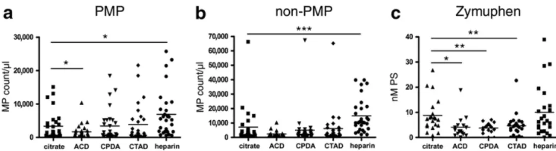
First we enumerated PMPs (CD42+AX+MPs) and non-PMPs (CD42–AX+MPs) byflow cytometry in PFP samples collected into tubes with different anticoagulants (Fig. 1A and B). The PMP concen- tration in the citrate plasma samples was around 102-104/μl in our study (median (1st quartile to 3rd quartile): 1,152/μl (251/μl-4,553/μl)), con- sistent with previousfindings[10]. The type of anticoagulant signifi- cantly influenced both the PMP and non-PMP levels (p = 0.002 for PMPs and pb0.001 for non-PMPs, Kruskal-Wallis one way analysis of variance on Ranks). The highest PMP and non-PMP counts were found in heparin tubes, and differed significantly from PMP and non-PMP counts in citrate tubes (p = 0.012 and pb0.001, respective- ly, Signed Rank test). The citrate, CTAD and CPDA tubes contained near- ly equal amounts of MPs. The lowest MP count was detected in ACD tubes. The PMP count in this tube differed significantly from that of cit- rate tube (p = 0.04, Signed Rank test). Furthermore, inter-individual differences (reflected by standard deviation) of healthy samples were also the lowest in the case of the ACD tube samples. Using the
Zymuphen assay, we obtained similar results (Fig. 1C). The median (1st quartile to 3rd quartile) PS concentration was 7.4 nM (4.7- 10.1 nM) in the citrate samples, which was consistent with the healthy range determined by the manufacturer. Similarly toflow cytometry results, the plasma samples in heparin tubes showed the highest PS concentration (however, the difference was not signifi- cant compared to citrate tubes). On the contrary, ACD, CTAD, CPDA tubes contained significantly less PS as compared to citrate tubes (p = 0.017, p = 0.003 and p = 0.006, respectively, Signed Rank test).
Next we assessed if there was a correlation betweenflow cytometry and Zymuphen assay results (Supplementary Fig. 3). Zymuphen assay detects all PS positive structures including small MPs and exosomes in- visible for manyflow cytometers. We analyzed the correlation between all AX+MPs detected byflow cytometry (the sum of PMPs and non- PMPs) and Zymuphen assay results. There was only a tendency between the two methods in the case of citrate tubes (R = 0.385, Pearson corre- lation), and there was no correlation in the case of CPDA, CTAD and hep- arin tubes. Of note, there was a correlation in the case of ACD tubes (R = 0.794, p = 0.00005, Pearson correlation).
The lowest MP numbers and lowest standard deviation detected in the ACD tubes might indicate that artificial,in vitrovesiculation is limited in ACD tubes, compared to other tubes. Based on these findings we selected ACD tube as a candidate optimal anticoagulant, and in the rest of this study, we compared artificialin vitrovesicula- tion in ACD tube and citrate tubes (the latter being suggested by the latest ISTH recommendation[3]).
Artificially Induced Vesiculation in Citrate and ACD Tubes
We inducedin vitrorelease of MPs in whole blood by gentle agitation and incubation of the blood samples at various temperatures. In ac- cordance with previous reports [9], agitation of whole blood in citrated tubes resulted in a dramatic, however, highly variable increase (3-29 fold) of both PMP and non-PMP counts compared to untreated controls (Fig. 2A and B) (p = 0.03 and p = 0.03, respectively, Signed Rank test), measured byflow cytometry.
Similarly, incubation for one hour at 37 °C resulted in 2-3 fold elevation of PMP and non-PMP counts in citrate tubes (p = 0.03
and p = 0.03, respectively, Signed Rank test). Strikingly, in ACD tubes, handled exactly the same manner as citrate tubes, there was only mod- erate, non significant elevation of PMPs and non-PMPs both after gentle agitation and 37 °C incubation (Fig. 2A and B). The incubation of whole blood at RT for one hour did not result in significant changes in MP levels either in the case of citrate or ACD tubes. Using the Zymuphen assay, we observed similar results: in plasma samples from citrate tubes, PS concentration was elevated after agitation and 37 °C incuba- tion (n = 3) (Fig. 2C). Similarly, one hour incubation at RT also resulted in an average of 5-fold elevation in PFP samples in citrate tubes. Most importantly, in ACD tubes, PS concentration remained unchanged after both agitation and incubation either at 37 °C or RT (Fig. 2C).
These data strongly support that in ACD tubes, artificially induced (in vitro) vesiculation of blood cells is successfully prevented.
The Effect of Citric Acid and Dextrose on Artificially Induced Vesiculation
To investigate which component of the ACD tubes was responsible for the inhibition ofin vitrovesiculation, we analyzed the effect of citric acid and dextrose. These substances were added separately to the blood, drawn into conventional citrate tubes up to a concentration characteris- tic for the commercial ACD tube (8 g/L citric acid and 24.5 g/L dextrose).
Then, the tubes were subjected to gentle agitation or 37 °C 1 hour incu- bation, as described above. The addition of citrate successfully inhibited the release of PMPs during both agitation and 37 °C incubation (Signed Rank test, p = 0.01 and p = 0.016, respectively) (Fig. 3A). The forma- tion of non-PMP counts was also inhibited by citric acid during 37 °C in- cubation (p = 0.016), but the release of PMPs was not inhibited during shaking (Fig. 3B). Dextrose alone could not inhibit the formation of ei- ther PMPs or non-PMPs counts during any of the tested conditions (Fig. 3A and B).
These data suggest that the active component of ACD tubes that inhibits artificialin vitrovesiculation, is citric acid which decreases the pH of the blood sample to approximately 6.0[22].
To confirm that platelets are inhibited in ACD tubes (that do not con- tain direct platelet inhibitors), we performed CAT measurement of PRP samples in citrate or ACD tubes. Clearly, there was a significant delay in the activation of platelets, the velocity of activation was smaller and the Fig. 1. Comparison of the effect of different anticoagulants on the MP counts measured byflow cytometry and Zymuphen assay.PMP (CD42a+AX+) (a) and non-PMP (CD42a-AX+) (b) counts enumerated byflow cytometry, (c) results of Zymuphen assay. Horizontal lines represent mean values. *pb0.05, **pb0.01, ***pb0.001, n = 30.
Fig. 2. The effect of citrate or ACD on thein vitrorelease of MPs.Counts of PMPs (a) and non-PMPs (b) measured byflow cytometry, and Zymuphen assay (c). Theyaxis represents fold increase compared to PFP samples isolated immediately after blood sampling. RT denotes room temperature. Mean ± s.d. values are shown. *pb0.05 compared to untreated control sam- ples (Signed Rank test), n = 6.
amount of total thrombin activity was also impaired in tubes with ACD (Supplementary Fig. 4). These results indicate that platelet inhibition explains the fewer numbers of PMPs in the PFP samples from ACD tubes.
Detection of Pregnancy Related Elevation of PMP and non-PMP Counts is not Confounded in ACD Tubes
Next, we assessed if the MP count elevatedin vivowas also detect- able in ACD tubes in pregnancy (a condition where elevated levels of PMPs and non-PMPs were reported previously in citrate tubes[23]). In- deed, there was a significant elevation in the PMP and non-PMP counts in pregnant women compared to non-pregnant healthy controls in cit- rate tubes (p = 0.002 and p = 0.004, respectively, Mann-Whitney Rank Sum test) (Fig. 4). Importantly, the pregnancy-related elevation was also detectable in ACD tubes (p = 0.015 for PMPs and p = 0.002 for non-PMPs, Mann-Whitney Rank Sum test) (Fig. 4).
Size Distribution of MPs in Citrate and ACD Tubes
Using the qNano instrument, we determined the size distribu- tions of blood plasma MPs in both citrate and ACD tubes (Fig. 5).
We did not detect any significant difference between the vesicle sizes in the two types of anticoagulant tubes. The mean (± s.d.) di- ameter of particles was 242.3 nm (± 5.8) nm in the case of citrate tubes and 230.8 (±9.2) nm in the case of ACD tubes. Concentration of particles was lower in ACD tubes (Fig. 5) as compared to those in citrate tubes (in accordance with data obtained by flow cytometry and Zymuphen assay).
RNA Analysis of Blood Plasma and EVs from Citrate and ACD Tubes
Based on the above data, we propose the use of ACD tubes for the assessment of circulating MPs. Recently, several studies analyzed exRNA content of vesicles or blood plasma (or serum)[24,25]. We tested ACD tubes for downstream RNA analysis. Blood plasma and total EV (including both exosomes and MPs in the pellets) RNA pro- files were similar between conventional citrate and ACD tubes (Sup- plementary Fig. 5A). We detected almost exclusively small RNA molecules in all samples, 18S and 28S RNAs were absent. We next applied small RNA analysis to determine the percentage of miRNAs in EV samples. We found that 46.8 ± 0.8% (175.1 ± 51.9 pg/μL) and 45.2 ± 2.3% (164.3 ± 77.1 pg/μL) of small RNAs were miRNAs Fig. 3. The effect of ACD components on thein vitrorelease of PMPs (a) and non-PMPs (b) determined byflow cytometry.Citric acid or dextrose was added separately to the tubes, and blood samples were subsequently subjected to agitation or 1 hour incubation at 37 °C. Mean ± s.d. values are shown. *pb0.05, n = 10.
Fig. 4. MP counts in pregnancy measured in citrate and ACD tubes determined byflow cytometry.Mean ± s.d. values are shown. *pb0.05, ** pb0.01, n = 6 in each group.
in citrate and ACD tubes, respectively. To further confirm the presence of miRNAs in the samples, we performed qRT-PCR analysis specific for miR16, miR24, miR451 and let7a. The presence of these miRNAs in blood or in vesicles has been confirmed earlier[24,25]. We found that all analyzed miRNAs were detectable in EVs and in blood plasma, both in the case of citrate and ACD tubes (Supplementary Fig. 5B).
There was no significant difference in CT values for the above ana- lyzed miRNAs between ACD and citrate tubes (equal amount of plas- ma was used for vesicle isolation or for RNA isolation).
Discussion
In this systemic study, we assessed whether the use of various anticoagulants affected the measured MP counts andin vitrovesicu- lation of blood cells. Indeed, we found that MP levels, determined by flow cytometry and Zymuphen assay, were significantly different in tubes with various anticoagulants. In accordance with previous results, the highest MP count was detected in tubes with heparin, and the low- est was found in ACD tubes[17]. This may be due to the fact that unlike other anticoagulants used in this study, heparin is not a calcium che- lator, and calcium is known to play a crucial role in vesiculation [26,27]. We demonstrated that the use of citrate, a weak chelator of calcium, resulted in a significantly reduced number of MPs compared to heparin in the blood collection tube. Interestingly, we found com- parable levels of PMP and non-PMP counts. This is in contrast with previousfindings[1], as most previous works found PMPs the main MP population in blood plasma. However, many of these earlier works did not control for the presence of residual platelets, and did not use pre-analytical parameters suggested by the ISTH[3]. Thus, many of these vesicles might have corresponded toin vitroformed MPs. Recent standard protocols[3]use very efficient centrifugation and rigorous pre-analytical settings in order to remove most of platelets, resulting in lower number of PMPs.
In Zymuphen assay, tubes containing platelet inhibitors (CTAD, CPDA and ACD) showed decreased MP counts. In this assay, citrate tubes gave comparable results to heparin in contrast to data obtained byflow cytometry. This controversy may be explained by the fact that Zymuphen assay does not only detect all types of AX+EVs (even exosomes), but also any PS positive cells that may possibly re- main in the PFP samples[11]. In contrast, duringflow cytometry we gated on the MV size range only, and smaller or larger particles were
thus, excluded from detection. By using the resistive pulse sensing approach, we detected much higher number of events than byflow cytometry. This is in accordance with previous reports[20], and is explained by the fact thatflow cytometry is able to detect events above 200-300 nm, while the threshold for qNano detection is lower. Furthermore, van der Pol et al. reported the presence of mul- tiple vesicles in the laser beam (swarm effect) that was responsible for an up to 1000-fold underestimation of MPs byflow cytometry [20].
Next, we focused on ACD and citrate tubes. Apparently, every single step from blood sampling to analysis may affect MP counts[8]. It has been shown previously that intense agitation of the tubes results in a strong elevation in MP count, and a significant decrease in the clotting time[9]. Furthermore, unsupported transportation and a delay between blood collection and thefirst centrifugation of samples also resulted in a significant increase in MP counts[9]. In the present study, we applied a high frequency and low amplitude gentle agitation, which induced an increase of PMP counts in citrate but not in ACD tubes. We also assessed the effect of blood sample storage at 37 °C which increased both PMP and non-PMP counts in citrate tubes but not in ACD ones. Our results in- dicate thatin vitroplatelet vesiculation is prevented in ACD tubes com- pared to the commonly used citrate tubes. Most interestingly, tubes containing direct platelet inhibitors, like theophylline, adenosine and dipyridamole (CTAD tubes) could not inhibitin vitrovesiculation. ACD tubes contain dextrose and citric acid, which components were sep- arately analyzed in our work. Dextrose had no effect on the counts of vesicles. Both in the case of PMPs and non-PMPs, citric acid was found responsible for the inhibition of vesiculation. The ACD tube was reported to result in pH 6.0 value of the blood sample[22]. The authors reported that platelets prepared in ACD tubes, showed significantly fewer signs of activation compared to platelets in sodium-citrate tubes.
Platelets retained their resting discoid morphology in ACD tubes, and the expression of the activation marker GPIIb-IIIa was also inhibited [22].
The inhibitory activity of local acidosis on platelets has been described long time ago[28]. Low pH was shown to inhibit ADP- induced change in platelet shape, adhesion, spreading and interaction withfibrinogen,fibronectin or collagen[29]. The mechanism is likely due to inhibition of the store-operated calcium influx into platelets in extracellular acidosis[30]. Lower intracellular calcium concentration results in the inhibition of haemostatic functions of platelets. As calcium Fig. 5. Particle size distributions measured by resistive pulse sensing (qNano).Histograms show a representative PFP sample (diluted 1:1 in PBS), n = 3.
is known to be required for vesiculation[26,27], it is not surprising that in vitrovesiculation was successfully dampened in the acidic envi- ronment. In this study, using CAT we also confirmed that platelets are markedly inhibited in ACD tubes. This inhibition might explain why PMP levels were generally lower in these tubes and why fur- ther effects related to blood handling failed to inducein vitrovesi- cle formation from platelets. However, a direct link between extracellular acidosis and vesiculation is yet to be established, and the inhibition of blood cells other than platelets in ACD tubes is yet to be confirmed.
Taken together, ACD tubes appear more suitable for MP analysis than any other types of anticoagulant tubes as they inhibitin vitro vesiculation. Importantly, detection ofin vivoelevated MP counts was not prevented in ACD tubes.
Based on our data, if sufficient care is taken to handle blood tubes, if samples are centrifuged shortly after blood sample collection and benchtop storage temperature is at around RT, both citrate and ACD tubes give comparable results.
In the past couple of years, hundreds of articles assessed MPs from blood plasma usingflow cytometry. The data, particularly on PMP counts, are often controversial; some studies show elevated levels of PMPs in a given condition, while some others fail to show an association (e.g. venous thromboembolism,[31]) One possible explanation for these results is thein vitrovesiculation of blood cells induced by many physical factors.
RNA profiles were found similar in citrate and ACD tubes. Mostly small RNAs were detected, and detailed Bioanalyzer analysis revealed that small RNA fractions contained miRNA in high amounts. The presence of miR16, miR24, miR451 and let7a in plasma has already been described[24,25], and we found no difference in the quantity of these RNAs in citrate and ACD tubes. Thus, as there was no differ- ence in citrate and ACD tubes in terms of exRNA profiles and quanti- ties of the studied specific miRNAs, ACD tubes are not only suitable for protein analysis of MVs, but also for exRNA extraction and subse- quent analysis.
Taken together, we suggest the use of ACD tubes for clinical lab- oratory assessments of MP counts because in practice a strict con- trol of transportation, temperature and delay between sample collection and analysis, is not always feasible. Conclusively, sample collection in ACD tubes may represent an important step towards standardization of both fresh and biobanked plasma samples for MP studies.
Supplementary data to this article can be found online athttp://dx.
doi.org/10.1016/j.thromres.2013.11.010.
Conflict of Interest Statement
The authors declare no conflicting interests.
Acknowledgements
This work was supported by OTKA K 73247, NK 84043 and K77537, Kerpel-Fronius Ödön Fellowship, Baross Gábor (REG-KM-09-1-2009- 0010) and FP7-PEOPLE-2011-ITN - PITN-GA-2011-289033“DYNANO” and BM1202 European Network on Microvesicles and Exosomes in Health and Disease (ME-HAD). Bence György and Tamás G Szabó are Kerpel-Fronius Ödön Fellows. This research was realized in the frames of TÁMOP 4.2.4. A/1-11-1-2012-0001„National Excellence Program– Elaborating and operating an inland student and researcher personal support system”. The project was subsidized by the European Union and co-financed by the European Social Fund.
Disclosure None declared.
References
[1]György B, Szabó TG, Pásztói M, Pál Z, Misják P, Aradi B, et al. Membrane vesicles, cur- rent state-of-the-art: emerging role of extracellular vesicles. Cell Mol Life Sci 2011;68:2667–88.
[2]Lacroix R, Robert S, Poncelet P, Kasthuri RS, Key NS. Dignat-George F; ISTH SSC Workshop. Standardization of platelet-derived microparticle enumeration byflow cytometry using calibrated beads: results of ISTH SSC collaborative workshop. J Thromb Haemost 2010;8:2571–4.
[3] Lacroix R, Judicone C, Mooberry M, Boucekine M, Key NS, Dignat-George F. The ISTH SSC Workshop. Standardization of pre-analytical variables in plasma microparticle determination: results of the International Society on Thrombosis and Haemostasis SSC Collaborative workshop. J Thromb Haemost Apr 2013.http://dx.doi.org/
10.1111/jth.12207[print copy in press. Available from: URL:http://onlinelibrary.
wiley.com/doi/10.1111/jth.12207/abstract;jsessionid=A9B21C784EABD90B7B1556 CAFCBE8027.d02t02] [Epub ahead of print]PMID: 23551930 [PubMed - as supplied by publisher].
[4]Beyer C, Pisetsky DS. The role of microparticles in the pathogenesis of rheumatic dis- eases. Nat Rev Rheumatol 2010;6:21–9.
[5]György B, Szabó TG, Turiák L, Wright M, Herczeg P, Lédeczi Z, et al. Improvedflow cytometric assessment reveals distinct microvesicle (cell-derived microparticle) sig- natures in joint diseases. PLoS One 2012;7:e49726.
[6]Vickers KC, Palmisano BT, Shoucri BM, Shamburek RD, Remaley AT. MicroRNAs are transported in plasma and delivered to recipient cells by high-density lipoproteins.
Nat Cell Biol 2011;3:423–33.
[7]Weiland M, Gao XH, Zhou L, Mi QS. Small RNAs have a large impact: circulating microRNAs as biomarkers for human diseases. RNA Biol 2012;9:850–9.
[8]Yuana Y, Bertina RM, Osanto S. Pre-analytical and analytical issues in the analysis of blood microparticles. Thromb Haemost 2011;105:396–408.
[9]Lacroix R, Judicone C, Poncelet P, Robert S, Arnaud L, Sampol J, et al. Im- pact of pre-analytical parameters on the measurement of circulating microparticles: towards standardization of protocol. J Thromb Haemost 2012;10:437–46.
[10]Robert S, Poncelet P, Lacroix R, Arnaud L, Giraudo L, Hauchard A, et al. Standard- ization of platelet-derived microparticle counting using calibrated beads and a Cytomics FC500 routineflow cytometer: afirst step towards multicenter studies?
J Thromb Haemost 2009;7:190–7.
[11]Artoni A, Merati G, Padovan L, Scalambrino E, Chantarangkul V, Tripodi A. Residual platelets are the main determinants of microparticles count in frozen-thawed plas- ma. Thromb Res 2012;130:561–2.
[12]György B, Módos K, Pállinger E, Pálóczi K, Pásztói M, Misják P, et al. Detection and isolation of cell-derived microparticles are compromised by protein complexes due to shared biophysical parameters. Blood 2011;117:e39–48.
[13]Larson MC, Luthi MR, Hogg N, Hillery CA. Calcium-phosphate microprecipitates mimic microparticles when examined withflow cytometry. Cytometry A 2013;83:242–50.
[14]van Ierssel SH, Van Craenenbroeck EM, Conraads VM, Van Tendeloo VF, Vrints CJ, Jorens PG, et al. Flow cytometric detection of endothelial microparticles (EMP): ef- fects of centrifugation and storage alter with the phenotype studied. Thromb Res 2010;125:332–9.
[15]Smalley DM, Root KE, Cho H, Ross MM, Ley K. Proteomic discovery of 21 proteins expressed in human plasma-derived but not platelet-derived microparticles.
Thromb Haemost 2007;97:67–80.
[16]Boilard E, Nigrovic PA, Larabee K, Watts GF, Coblyn JS, Weinblatt ME, et al. Platelets amplify inflammation in arthritis via collagen-dependent microparticle production.
Science 2010;327:580–3.
[17]Jayachandran M, Miller VM, Heit JA, Owen WG. Methodology for isolation, identifi- cation and characterization of microvesicles in peripheral blood. J Immunol Methods 2012;375:207–14.
[18]Emmerechts J, Jacobs L, Van Kerckhoven S, Loyen S, Mathieu C, Fierens F, et al. Air pollution-associated procoagulant changes: the role of circulating microvesicles. J Thromb Haemost 2012;10:96–106.
[19]Connor DE, Exner T, Ma DD, Joseph JE. The majority of circulating platelet-derived microparticles fail to bind annexin V, lack phospholipid-dependent procoagulant ac- tivity and demonstrate greater expression of glycoprotein Ib. Thromb Haemost 2010;103:1044–52.
[20]van der Pol E, van Gemert MJ, Sturk A, Nieuwland R, van Leeuwen TG. Single vs.
swarm detection of microparticles and exosomes byflow cytometry. J Thromb Haemost 2012;10:919–30.
[21]van der Pol E, Böing AN, Harrison P, Sturk A, Nieuwland R. Classification, func- tions, and clinical relevance of extracellular vesicles. Pharmacol Rev 2012;64:676–705.
[22]Maurer-Spurej E, Pfeiler G, Maurer N, Lindner H, Glatter O, Devine DV.
Room temperature activates human blood platelets. Lab Invest 2001;81:
581–92.
[23]Bretelle F, Sabatier F, Desprez D, Camoin L, Grunebaum L, Combes V, et al. Circulating microparticles: a marker of procoagulant state in normal pregnancy and pregnancy complicated by preeclampsia or intrauterine growth restriction. Thromb Haemost 2003;89:486–92.
[24]Arroyo JD, Chevillet JR, Kroh EM, Ruf IK, Pritchard CC, Gibson DF, et al.
Argonaute2 complexes carry a population of circulating microRNAs inde- pendent of vesicles in human plasma. Proc Natl Acad Sci U S A 2011;
108:5003–8.
[25]Redova M, Poprach A, Nekvindova J, Iliev R, Radova L, Lakomy R, et al. Circulating miR-378 and miR-451 in serum are potential biomarkers for renal cell carcinoma.
J Transl Med 2012;10:55.
[26]Henriksson CE, Klingenberg O, Hellum M, Landsverk KS, Joø GB, Westvik AB, et al.
Calcium ionophore-induced de-encryption of tissue factor in monocytes is associat- ed with extensive cell death. Thromb Res 2007;119:621–30.
[27]Crespin M, Vidal C, Picard F, Lacombe C, Fontenay M. Activation of PAK1/2 dur- ing the shedding of platelet microvesicles. Blood Coagul Fibrinolysis 2009;
20:63–70.
[28]Rogers AB, Des Prez RM. The effect of pH on human platelet aggregation induced by epinephrine and ADP. Proc Soc Exp Biol Med 1972;139:1100–3.
[29]Nachmias VT, Yoshida K, Glennon MC. Lowering pH in blood platelets dissociates myosin phosphorylation from shape change and myosin association with the cyto- skeleton. J Cell Biol 1987;105:1761–9.
[30]Marumo M, Suehiro A, Kakishita E, Groschner K, Wakabayashi I. Extracellular pH affects platelet aggregation associated with modulation of store-operated Ca(2 +) entry. Thromb Res 2001;104:353–60.
[31]Owens III AP, Mackman N. MP's and VTE's: Fact orfiction. Thromb Res 2011;
128:505–6.