Acta Microbiologica et Immunologica Hungarica
(20 ) , 1– 5 DOI:10.1556/030.66.2019.041
©2019The Author(s)
ORIGINAL ARTICLE
* Corresponding author:
Erika Orosz
Department of Parasitology, National Public Health Center, Albert Fl´orián út 2-6, H-1097 Budapest, Hungary Phone: +36 1 4761100/2231;
E-mail:orosz.erika@nnk.gov.hu
Genotyping of Acantamoeba spp. from rhisophere in Hungary
ERIKA OROSZ
1* and KATALIN POSTA
21Department of Parasitology, National Public Health Center, Budapest, Hungary
2Institute of Genetics, Microbiology and Biotechnology, Szent István University, Gödöllo, Hungary
Received: June 17, 2019•Accepted: October 14, 2019
ABSTRACT
The protistaAcanthamoebais a free-living amoeba existing in various environments. A number of species among protista are recognized as human pathogens, potentially causing Acanthamoeba keratitis (AK), granulomatous amoebic encephalitis (GAE), and chronic granulomatous lesions. In this study, 10 rhizosphere samples were collected from maize and alfalfa plants in experimental station at Institute of Genetics, Microbiology and Biotechnology, Szent István University. We detected Acanthamoebabased on the quantitative real-time PCR assay and sequence analysis of the 18S rRNA gene. All studied molecular biological methods are suitable for the detection of Acanthamoeba infection in humans. The quantitative real-time PCR-based methods are more sensitive, simple, and easy to perform; moreover, these are opening avenue to detect the effect of number of parasites on human disease.Acanthamoebaspecies were detected infive (5/10; 50%) samples. AllAcanthamoeba strains belonged to T4 genotype, the main AK-related genotype worldwide. Our result confirmed Acanthamoebastrains in rhizosphere that should be considered as a potential health risk associated with human activities in the environment.
KEYWORDS
Acanthamoeba, rhizosphere isolates, genotypes
INTRODUCTION
Acanthamoeba is a genus of free-living amoebae widely distributed in various ecological environments [1–3]. The life cycle of Acanthamoeba species (sp.) consists of the active trophozoites and dormant cysts stages.Acanthamoebatrophozoites have a size between 20 and 40 μm, although this range can vary significantly among isolates of different species genotypes. Cysts are double-walled and range in size from 10 and 20μm. This difference in size between the cyst and trophozoite involves a significant loss of cell volume mail due to cellular dehydrations.Acanthamoebaspp. are thermotolerant, which are resistant to extreme temperature, pH conditions, UV, as well as to chlorine and other disinfectant media.
Most of the environmental studies are focusing on pathogenic Acanthamoeba sp.
taxonomic and pathogenic markers, geographic distribution, ecology, and transmission dynamic [4–6]. Unlike obligate parasites, pathogenicAcanthamoebaspp. can complete their life cycle, environmental performance without having to enter the human or animal host [7,8]. The genusAcanthamoeba has been currently classified into 21 different genotypes, T1–T21, based on 18S rRNA nucleotide sequence [9,10]. Some genera of Acanthamoeba cause different infections, which produceAcanthamoebakeratitis (AK), subacute or chronic granulomatous amoebic encephalitis, and skin infections. Human infections with these amoebae have been reported from all over the world [11]. The first cases that clearly established Acanthamoeba as causative agents of disease in humans have been reported in the early 1970s [12]. In many cases, AK infections occur after water exposure or a history of swimming in lakes, following contact with soil or plants, or while wearing contact lenses [13,14].
67 20 3 17 17
In general,Acanthamoebaare metabolically active and use a wide variety of bacteria, fungi, and organic matter as a food source [15].
Therefore, in this study, high microbial activities showing rhizosphere soil used for isolation protozoan organisms to test their occurrences are not in human host. Moreover, the isolated strains morphologically characterized by electron microscopy molecularly characterized based on the 18S rRNA gene sequence and the robust phylogenetic analysis was also measured.
MATERIALS AND METHODS Samples collection
Rhizosphere samples were collected from experimental station at Institute of Genetics, Microbiology and Biotech- nology, Szent István University (longitude: 19°21′39.85″, latitude: 47°35′37′63″) in June 27, 2018. Rhizosphere samples were taken from the depth of 0–20 cm. During the sampling period, altogether 10 samples from rhizosphere of maize and alfalfa plants samples were taken.
The sampling was performed, in which 10 samples were taken from rhizosphere of maize and alfalfa plants (notation:
K1_1, K1_2, K1_3; K2_1, K2_2, K2_3; and L1_1, L1_2, L1_3, L2_1).
Culture-con fi rmed detection method
To concentrate Acanthamoeba spp., the samples were filtered, eluted, and centrifuged. Soil samples (1 g) collected from rhizosphere of maize and alfalfa plants were dissolved in 10 ml of sterile physiological saline solution (0.85%) buffer and 500μl of each sample was inoculated onto PAGE agar 9-cm plates seeded with heat-killed Escherichia coli and incubated at 36 °C [16].
Microscopic detection
Samples were examined under a microscope for 72–96 h at 400×with an inverted ZEISS microscope (Figure 1).
Molecular analysis
TheAcanthamoebaspecies were isolated by dilution method.
For this purpose, the samples of soil (1 g) were suspended in 10 ml of sterile physiological saline solution (0.85%). After preparation, the DNA extraction was treated with High Pure PCR Template Preparation Kit (Germany), according to the instructions of the manufacturer. If further processing was delayed, the isolates were stored at 4 °C for 24 h or at–20 °C for a longer period. The DNA amplification was performed using genus-specific primers and genus-specificfluorescence resonance energy transfer (FRET) hybridization probes, previously described by Orosz et al. [17]. Each experiment included one reaction mixture without DNA as a negative control; positive control and each specimen were run in duplicate for real-time PCR assay in parallel. We have used serial dilutions ofAcanthamoeba(GenBank accession num- ber: KC434439) strain to determine the calibration curve that the liquid chromatography device could determine the addi- tional samples parasite number in copy numbers.
PCR products were purified with PCR Clean up-M Kit (Viogene, Sunville, CA). The sequence of each amplicon was determined by cycle sequencing with primers for the 5′-NTR region and with primers with BigDye Terminator v3.1 Cycle Sequencing Kit (Applied Biosystems, Germany), according to the manufacturer’s instruction. The electrophoresis was carried out on Applied Biosystems 3500 Genetic Analyzer (Applied Biosystems, Budapest, Hungary).
The 5′-NTR and VP1 gene sequences were subject to nucleotide–nucleotide BLAST analysis [18] using the online server at the National Center for Biotechnology Information (http://blast.ncbi.nlm.nih.gov/Blast).
The unknown sequences were aligned with known published sequences of the major genotypes using the align- ment program MULTALIN (http://multalin.toulouse.inra.fr/
multalin) [19]. The genotypes of samples were determined based on this comparison.
The phylogenetic tree was constructed by the neighbor- joining method of genetic distance calculated by the MEGA 6 (http://www.megasoftware.net) [20].
Genotype identification was carried out with a real-time FRET PCR assay based on sequence analysis of the 18S rRNA gene, and sensitivity and specificity were evaluated in com- parison with traditional parasitological techniques.
Figure 1.Photomicrograph of Acanthamoeba trophozoites (A) and cysts (B) with 400×magnification. Photographer: Erika Orosz Acta Microbiologica et Immunologica Hungarica6767676 6767 –
172 67 (2020) 3, 171 175
RESULTS
Microscopic detection
All investigated samples revealedAcanthamoeba were able to grow at 36 °C, the approximate temperature of the human organism. Microscopically 5 out of the 10 samples were declared asAcanthamoebapositive (Medicago sativa– L1_2, L1_3 and Zea mays – K1_2, K2_1, K2_3). Five rhizosphere samples (Medicago sativa – L1_1, L1_3 and Zea mays – K1_1, K1_3, K2_2) were microscopically negative. Further examination of the obtained results was conducted by FRET PCR.
Molecular analysis
This study reports successful PCR amplification for 5 (5/10;
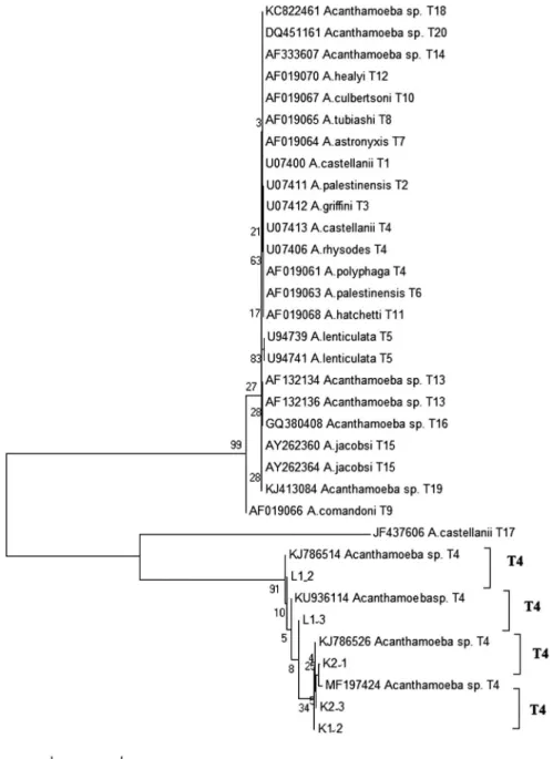
50.0%) positive cases. The samples offive Acanthamoeba – positive samples, detected by PCR method, were sequenced to identify the species. Sequence analysis using a BLAST search indicated an identity of>98% withAcanthamoeba18r rRNA gene reference sequences. It was found that all obtained sequences of amoebae isolates from the cases belong to the different T4 genotypesAcanthamoebaspp. Neighbor-joining analysis inferred relationships between the PCR products isolated from rhizosphere samples reference strains obtained from NCBI GenBank, shown in Figure2, respectively.
Figure 2.Phylogenetic relations ofAcanthamoebaspecies PCR product sample L1_2, sample L1_3, sample K2_1, sample K2_1, sample K2_3, and reference strains from NCBI GenBank inferred by neighbor-joining analysis from pairwise comparisons (180-bp fragments)
Acta Microbiologica et Immunologica Hungarica 67 (2020) 3, 171 175– 173
DISCUSSION AND CONCLUSIONS
Studies ofAcanthamoebahave grown exponentially. To the best of our knowledge, this is the second study of occurrence of Acanthamoeba similar to T4 genotypes in rhizosphere samples from Hungary. These organisms have gained attention from the broad scientific community studying environmental biology, molecular biology, and biochemistry.
Literature describes T4 genotypeAcanthamoeba, as the most common in the environment. These results are consistent with previous findings indicating that T4 is worldwide predominant [21–24].
However, the correct understanding of the factors influ- encing the occurrence of the different species appears of great concern, as these amoebae are free-living organisms, and their potential capabilities to cause severe infections of the central nervous system, ocular keratitis, and other disorders are now ascertained worldwide.
All the isolates in this study exhibited morphological features of the genusAcanthamoebaconfirmed by means of quantitative real-time PCR. Quantitative real-time PCR with FRET hybrid- ization probes method is the most sensitive with a short turnaround time. It is possible even to estimate the parasite number in the samples with method. Therefore, only molecular methods allow reliable differentiation of the Acanthamoeba species. Based on rRNA gene sequences, the genusAcantha- moebais divided into 21 different genotypes to date (T1–T21).
Each genotype exhibits 5% or more sequence divergence between different genotypes. Five isolates were characterized as similar to genotype T4 due to their strict correspondence to the reference sequences of this genotype (GenBank accession number: KJ786514, KU936114, KJ786526, and MF197424).
Sequence date indicate that the vast majority of them causes human infections. Contrary to data on Acanthamoeba infections in humans, little is known about infections in rhizosphere. It has been concluded that the rhizosphere isolates are most closely related to strains commonly isolated from human infections, especially AK [25–29].
In conclusion, our results confirm and support previous report on Acanthamoeba genotype free-living amoeba in rhizosphere soil. A homologous analysis of the 18S rRNA of fiveAcanthamoebaspecies isolated from rhizosphere of maize and alfalfa was identified into one genotype, namely T4. These genotypes were associated with AK or encephalitis; therefore, the presence of Acanthamoeba should be considered as potential health threat associated with human activity in soil.
Acknowledgements: This work was supported by Higher Education Institutional Excellence Program (NKFIH-1159- 6/2019) awarded by the Ministry of Human Capacities within the framework of water-related researches of Szent István University.
Conflict of Interest: There is no conflict of interest. The corresponding author assures that there are no links with a company whose product is mentioned in the article or a company that distributes a competing product. The presen- tation of the topic is independent and the presentation of the content is product-neutral.
REFERENCES
1. Shatilovich, A., Shmakova, L., Gubin, S., Goodkov, A., Gilichinsky, D.: Viable protozoa in late Pleistocene and Holo- cene permafrost sediments. Dokl Biol Sci401, 136–138 (2005).
2. Siddiqui, R., Khan, N. A.: Biology and pathogenesis of Acanthamoeba. Parasit Vectors10, 6 (2012).
3. Reyes-Batlle, M., Zamora-Herrera, J., Vargas-Mesa, A., Valer´on- Tejera, M. A., Wagner, C., Martín-Navarro, C. M., L´opez- Arencibia, A., Sifaoui, I., Martínez-Carretero, E., Valladares, B., Pinero, J. E., Lorenzo-Morales, J.:˜ Acanthamoebagenotypes T2, T4, and T11 in soil sources from El Hierro island, Canary Islands, Spain. Parasitol Res115, 2953–2956 (2016).
4. Podlipaeva, J., Shmakova, L., Gilichinski, D., Goodkov, A.: Heat shock protein of hsp70 family revealed in some contemporary freshwater amoebae and inAcanthamoebasp. from cysts iso- lated from permafrost samples. Tsitologiia48, 691–694 (2006).
5. Nuprasert, W., Putaporntip, C., Pariyakanok, L., Jongwutiwes, S.: Identification of a novel t17 genotype of Acanthamoeba from environmental isolates and t10 genotype causing keratitis in Thailand. J Clin Microbial48, 4636–4640 (2010).
6. Visvesvara, G. S., Hercules, M., Schuster, F. L.: Pathogenic and opportunistic free-living amoebae:Acanthamoebaspp.,Bala- muthia mandrillaris,Naegleria fowleriandSappinia diploidea.
FEMS Immunol Med Microbiol50, 1–26 (2007).
7. Alves, D. S., Moraes, A. S., Nitz, N., de Oliveira, M. G., Hecht, M. M., Gurgel-Gonçalves, R., Cuba, C. A.: Occurrence and characterization ofAcanthamoebasimilar to genotypes T4, T5, and T2/T6 isolated from environmental sources in Brasília.
Exp Parasitol131, 239–244 (2012).
8. Karakavuk, M., Aykur, M., ¸Sahar, E. A., Karaku¸s, M., Aldemir, D., Döndüren, Ö., Özdemir, H. G., Can, H., Gürüz, A. Y., Da˘gcı, H., Dö¸skaya, M.: First time identification ofAcantha- moeba genotypes in the cornea samples of wild birds; Is Acanthamoebakeratitis making the predatory birds a target?
Exp Parasitol183, 137–142 (2017).
9. Montoya, A., Mir´o, G., Saugar, J. M., Fernández, B., Checa, R., Gálvez, R., Bailo, B., Marino, V., Pinero, J. E., Lorenzo-Morales,˜ J., Fuentes, I.: Detection and molecular characterization of Acanthamoeba spp. in stray cats from Madrid, Spain. Exp Parasitol188, 8–12 (2018).
10. Corsaro, D., Köhsler, M., Di Filippo, MM., Venditti, D., Monno, R., Di Cave, D., Berrilli, F., Walochnik, J.: Update on Acanthamoeba jacobsigenotype T15, including full-length 18S rDNA molecular phylogeny. Parasitol Res 116, 1273–1284 (2017).
11. Khan, N. A., Jarroll, E. L., Paget, T. A.: Molecular and physio- logical differentiation between pathogenic and nonpathogenic Acanthamoeba. Curr Microbiol45, 197–202 (2002).
12. Jones, D., Visvesvara, G., Robinson, N.: Acanthamoeba polyphaga keratitis andAcanthamoeba uveitisassociated with fatal menin- goencephalitis. Trans Ophthalmol Soc UK95, 221–231 (1975).
13. Schroeder, J. M., Booton, G. C., Hay, J., Niszl, I. A., Seal, D. V., Markus, M. B., Fuerst, P. A., Byers, T. J.: Use of subgenic 18S ribosomal DNA PCR and sequencing for genus and genotype identification of Acanthamoebae from humans with keratitis and from sewage sludge. J Clin Microbial39, 1903–1911 (2001).
Acta Microbiologica et Immunologica Hungarica6767 67 –
174 67 (2020) 3, 171 175
14. Walochnik, J., Scheikl, U., Haller-Schober, E. M.: Twenty years ofAcanthamoebadiagnostics in Austria. J Eukaryot Microbiol 62, 3–11 (2015).
15. Neelam, S., Niederkorn, J. Y.: Pathobiology and immunobiol- ogy ofAcanthamoebakeratitis: Insights from animal models.
Yale J Biol Med90, 261–268 (2017).
16. Page, F. C.: A New Key to Freshwater and Soil Gymnamoebae.
Freshwater Biological Association, Ambleside, Cumbria, 1988, 122 p.
17. Orosz, E., Farkas, Á., Ködöböcz, L., Becsák, P., Danka, J., Kucsera, I., Füleky, G.: Isolation of Acanthamoebafrom the rhizosphere of maize and lucerne plants. Acta Microbiol Immunol Hung60, 29–39 (2013).
18. Altschul, S. F., Gish, W., Miller, W., Myers, E. W., Lipman, D. J.:
Basic local alignment search tool. J Mol Biol215, 403–410 (1990).
19. Corpet, F.: Multiple sequence alignment with hierarchical clustering. Nucleic Acids Res16, 10881–10890 (1988).
20. Tamura, K., Stecher, G., Peterson, D., Filipski, A., Kumar, S.:
MEGA6: Molecular evolutionary genetics analysis version 6.0.
Mol Biol Evol30, 2725–2729 (2013).
21. Montalbano Di Filippo, M., Santoro, M., Lovreglio, P., Monno, R., Capolongo, C., Calia, C., Fumarola, L., D’Alfonso, R., Berrilli, F., Di Cave, D.: Isolation and molecular characteriza- tion of free-living amoebae from different water sources in Italy. Int J Environ Res Public Health12, 3417–3427 (2015).
22. Üstüntürk-Onan, M., Walochnik, J.: Identification of free- living amoebae isolated from tap water in Istanbul, Turkey.
Exp Parasitol195, 34–37 (2018).
23. Reyes-Batlle, M., Hernández-Pinero, I., Rizo-Liendo, A.,˜ L´opez-Arencibia, A., Sifaoui, I., Bethencourt-Estrella, C. J., Chiboub, O., Valladares, B., Pinero, J. E., Lorenzo-Morales,˜ J.: Isolation and molecular identification of free-living amoebae from dishcloths in Tenerife, Canary Islands, Spain. Parasitol Res118, 927–933 (2019).
24. Orosz, E., Farkas, Á., Kucsera, I.: Laboratory diagnosis of Acanthamoebakeratitis in Hungary. Acta Microbiol Immunol Hung63, 293–299 (2016).
25. Khan, N. A.:Acanthamoeba: Biology and increasing impor- tance in human health. FEMS Microbiol Rev 30, 564–595 (2006).
26. Corsaro, D., Walochnik, J., Köhsler, M., Rott, M. B.:Acantha- moeba misidentification and multiple labels: Redefining genotypes T16, T19, and T20 and proposal forAcanthamoeba micheli sp. nov. (genotype T19). Parasitol Res 114, 2481–2490 (2015).
27. Orosz, E., Szentmáry, N., Kiss, H. J., Farkas, Á., Kucsera, I., Nagy, Z. Z.: First report ofAcanthamoebagenotype T8 human keratitis. Acta Microbiol Immunol Hung65, 73–79 (2018).
28. Gyenes, A., Orosz, E., Sándor, G. L., Fries, F. N., Seitz, B., Nagy, Z. Z., Szentmáry, N.: Early diagnosis and successful medical treatment ofAcanthamoebakeratitis. Klin Monbl Augenheilkd 235, 1407–1410 (2018).
29. Orosz, E., Krisk´o, D., Shi, L., Sándor, G., Kiss, H. J., Seitz, B., Nagy, Z. Z., Szentmáry, N.: Clinical course ofAcanthamoeba keratitis isolates T4 and T8 in Hungary. Acta Microbiol Immunol Hung66, 289–300 (2019).
Acta Microbiologica et Immunologica Hungarica 67 (2020 3) ,171–175 175
Open Access statement. This is an open-access article distributed under the terms of the Creative Commons Attribution 4.0 International License (https://creativecommons.org/licenses/by/4.0/), which permits unrestricted use, distribution, and reproduction in any medium, provided the original author and source are credited, a link to the CC License is provided, and changes – if any – are indicated. (SID_1)