Studies of Laboulbeniales on Myrmica ants (IV): host-related diversity and thallus distribution patterns of Rickia wasmannii
Danny Haelewaters1,2,3,*, Peter Boer4, Ferenc Báthori5, Zoltán Rádai5, Ana Sofia P.S. Reboleira6, András Tartally5, Walter P. Pfliegler7, André De Kesel8, and Oldřich Nedvěd2
1 Farlow Reference Library and Herbarium of Cryptogamic Botany, Harvard University, 22 Divinity Avenue, Cambridge, MA 02138, USA
2 Faculty of Science, University of South Bohemia, Branišovská 31, 37005České Budějovice, Czech Republic
3 Department of Botany and Plant Pathology, Purdue University, 915 W. State Street, West Lafayette, IN 47907, USA
4 Gemene Bos 12, 1861 HG Bergen, The Netherlands
5 Department of Evolutionary Zoology and Human Biology, University of Debrecen, Egyetem tér 1, 4032 Debrecen, Hungary
6 Natural History Museum of Denmark, University of Copenhagen, Universitetsparken 15, 2100 København Ø, Denmark
7 Department of Molecular Biotechnology and Microbiology, University of Debrecen, Egyetem tér 1, 4032 Debrecen, Hungary
8 Meise Botanic Garden, Nieuwelaan 38, 1860 Meise, Belgium
Received 6 February 2019, Accepted 20 April 2019, Published online 20 May 2019
Abstract –Fungal species identities are often based on morphological features, but current molecular phylogenetic and other approaches almost always lead to the discovery of multiple species in single morpho-species. According to the morphological species concept, the ant-parasitic fungusRickia wasmannii(Ascomycota, Laboulbeniales) is a single species with pan-European distribution and a wide host range. Since its description, it has been reported from ten species ofMyrmica(Hymenoptera, Formicidae), of which two belong to therubra-group and the other eight to the phylogenetically distinctscabrinodis-group. We found evidence forR. wasmanniibeing a single phylogenetic species using sequence data from two loci. Apparently, the original morphological description (dating back to 1899) represents a single phylogenetic species. Furthermore, the biology and host-parasite interactions ofR. wasmanniiare not likely to be affected by genetic divergence among different populations of the fungus, implying comparability among studies conducted on members of different ant populations. We found no differences in total thallus number on workers be- tweenMyrmicaspecies, but we did observe differences in the pattern of thallus distribution over the body. The locus of infection is the frontal side of the head inMyrmica rubraandM. sabuletiwhereas inM. scabrinodisthe locus of infec- tion differs between worker ants from Hungary (gaster tergites) and the Netherlands (frontal head). Possible explana- tions for these observations are differences among host species and among populations of the same species in (i) how ant workers come into contact with the fungus, (ii) grooming efficacy, and (iii) cuticle surface characteristics.
Key words:Ant-associated fungi, Laboulbeniomycetes, Molecular evolution, Ribosomal DNA, Thallus density.
Re´sume´ –Études des Laboulbeniales sur les fourmisMyrmica(IV) : Diversité liée à l’hôte et schémas de dis- tribution des thalles de Rickia wasmannii. L’identification des espèces fongiques est souvent basée sur des caractéristiques morphologiques, mais les approches phylogénétiques moléculaires et autres conduisent presque toujours à la découverte d’espèces multiples dans une seule morpho-espèce. Selon le concept d’espèce morphologique, le champignon parasite de fourmisRickia wasmannii(Ascomycota, Laboulbeniales) est une espèce unique ayant une répartition paneuropéenne et une large gamme d’hôtes. Depuis sa description, il a été signalé chez dix espèces de Myrmica (Hymenoptera, Formicidae), dont deux appartiennent au groupe rubra et les huit autres au groupescabrinodis, phylogénétiquement distinct. Nous avons trouvé que R. wasmannii était une seule espèce phylogénétique en utilisant les données des séquences de deux loci. Apparemment, la description morphologique originale (datant de 1899) représente une seule espèce phylogénétique. De plus, la biologie et les interactions hôte-parasite de R. wasmannii ne devraient pas être affectées par une divergence génétique entre différentes populations du champignon, ce qui implique une comparabilité entre les études conduites sur des membres de différentes populations de fourmis. Nous n’avons trouvé aucune différence dans le nombre total de thalles chez les ouvrières entre les espèces deMyrmica, mais nous avons observé des différences dans le schéma de distribution des thalles sur le corps. Le locus d’infection est le front de la tête chez Myrmica rubra et
*Corresponding author:danny.haelewaters@gmail.com Parasite26, 29 (2019)
ÓD. Haelewaters et al., published byEDP Sciences, 2019 https://doi.org/10.1051/parasite/2019028
Available online at:
www.parasite-journal.org
This is an Open Access article distributed under the terms of the Creative Commons Attribution License (http://creativecommons.org/licenses/by/4.0), which permits unrestricted use, distribution, and reproduction in any medium, provided the original work is properly cited.
OPEN ACCESS
RESEARCHARTICLE
M. sabuleti, alors que chez M. scabrinodis, le lieu d’infection diffère entre les ouvrières de Hongrie (tergites abdominaux) et des Pays-Bas (front de la tête). Les explications possibles de ces observations sont les différences entre les espèces hôtes et entre les populations de la même espèce en ce qui concerne (i) le contact des ouvrières avec le champignon, (ii) l’efficacité du toilettage et (iii) les caractéristiques de la surface de la cuticule.
Introduction
Ants harbour a vast diversity of microbial parasites and pathogens. Fungal species of ants are usually pathogenic, but some species, notably members of Laboulbeniales (Ascomy- cota), are ectoparasitic and do not cause the death of the hosts.
Laboulbeniales are developmentally unique in that they do not produce mycelia; instead, they produce multicellular units, thalli, which attach externally to the integument of the host.
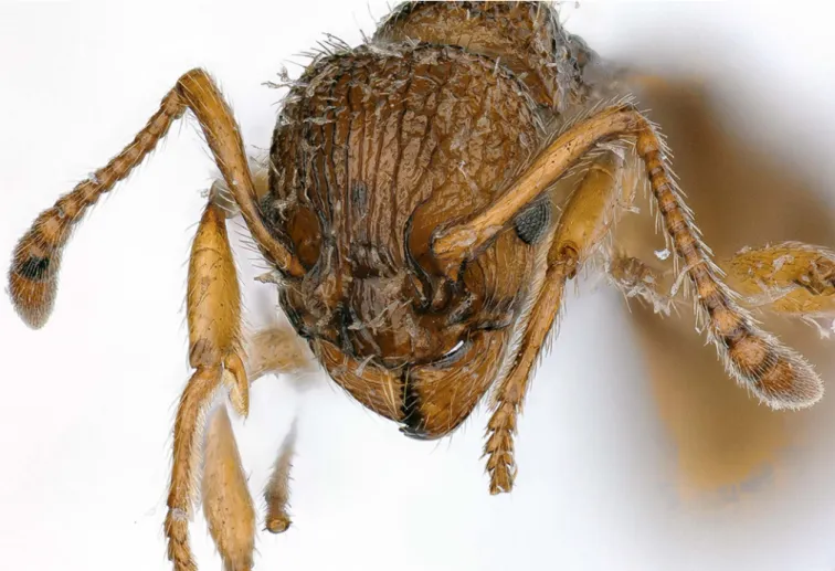
Rickia wasmannii Cavara, 1899 [9] (Fig. 1) is a species of Laboulbeniales that infects diverse ants in the genusMyrmica Latreille, 1804 (Hymenoptera, Formicidae) in Europe. Knowl- edge on the biology ofR. wasmanniiis accumulating and this species has quickly become one of the most thoroughly researched species of Laboulbeniales [2–5,11–13,15,29,32, 52,53,70].
Non-random positional patterns on the integument [48], variation in host usage across geographical regions [29], and habitat specificity [53] have recently been explored for R. wasmannii. On the other hand, the phylogenetic diversity ofR. wasmanniifrom different host species remains unknown.
This question deserves to be explored, as it was shown recently for a few Laboulbeniales examples that there is phylogenetic structuring within presumed species. For species in the genera Gloeandromyces Thaxt. and Hesperomyces Thaxt., phyloge- netic segregation by host species has been observed. For exam- ple, in bothG. pageanusHaelew. andG. streblaeThaxt., two phylogenetic clades can be found: one clade for isolates removed fromTrichobius dugesioidesWenzel, 1966 (Diptera, Streblidae) and another clade for isolates from T. joblingi Wenzel, 1966 [33, 34]. Similarly, Hesperomyces virescens Thaxt. consists of multiple clades, each clade corresponding to a species with strict host specificity [30].
Even though the main hosts ofR. wasmanniiall belong to a single genus of ants (for a discussion of alternative hosts, see [53]), the different host species are placed in two clades that are phylogenetically not closely related (referred to as species groups in [39, 56]). Myrmica rubra (Linnaeus, 1758) and M. ruginodis Nylander, 1846 belong to the rubra-group, whereas the other known hosts belong to thescabrinodis-group.
These areM. gallieniiBondroit, 1920;M. hellenicaFinzi, 1926;
M. sabuleti Meinert, 1861; M. scabrinodis Nylander, 1846;
M. slovaca Sadil, 1952; M. specioides Bondroit, 1918;
M. spinosior Santschi, 1931; and M. vandeli Bondroit, 1920 [4,29]. Assessing whetherR. wasmanniishows phylogenetic segregation by host species or host species group is important to better understand its interactions with different ant hosts.
Studies using infected and non-infected Myrmica ants have been done to assess the parasite’s effects on ant behaviour and physiology. Interpretation of these results is complicated when the taxonomic status of different fungal populations is uncertain. Comparing interactions between a fungal parasite
and its different hosts is only reliable when the fungal popula- tions represent a single phylogenetic species.
Building on the hypothesis thatR. wasmanniiis a complex of species, potentially segregated by host species (or species group), it is logical to assume that thallus distribution patterns may be different on various ant hosts. If we were tofind vari- able patterns of thallus distribution, these would have to be (partly) attributed to the fungal partner, the ant partner, environ- mental factors, or a combination of these. To try to shed light on this complex interaction of factors, we took an integrative approach (sensu[30]) and generated independent sets of data, that is, barcode sequences ofR. wasmanniiisolates and thallus density counts by body part.
During this study, we sampled infected ants from different regions in Europe and sequenced two loci to assess intraspecific phylogenetic diversity in R. wasmannii. Collected host ants represent three Myrmica species belonging to the rubra- and scabrinodis-groups [56]. After having accumulated many collections of R. wasmannii-infected ants, we assessed thallus densities per body part from different host species (M. rubra, M. sabuleti, andM. scabrinodis) and from different populations of the same host species (M. scabrinodis).
Material and methods
Collection of ants
Ants were collected directly from nests in seven locations in four countries (Fig. 2): Austria (Vienna), Belgium (Moelingen), Hungary (Bükkszentkereszt, Rakaca, Újléta), and the Nether- lands (Savelsbos, Wijlre-Eys). Long-term preservation was in 80–96% ethanol. Identification of ants was based on Seifert [64] and Radchenko and Elmes [56]. Voucher specimens are deposited at the Naturalis Biodiversity Center (Leiden, The Netherlands) and the Hungarian Natural History Museum (Budapest, Hungary). Identification of mounted thalli was done under light microscope, based on Thaxter [70] and De Kesel et al. [15].
DNA extraction, PCR amplification, and sequencing
DNA was isolated from 3 to 100s of thalli using extraction protocols described in [31] or a modified REPLI-g Single Cell Kit (Qiagen, Valencia, California) [30]. The internal transcribed spacer (ITS) region (ITS1–5.8S–ITS2) and the 50 end of the nuclear ribosomal large subunit (28S) were amplified, for ITS using primer pairs ITS1f [26] & ITS4 [77] and ITS9mun [19]
& ITS4, for partial ITS–28S using the newly designed Rickia-specific primer RicITS2 (50–CTAGTGTGAATTGCA- TATTTTAGTG–30) & LR3 [74], and for 28S-only using
LR0R [38] & LR5 [74] and NL1 & NL4 [44]. Polymerase chain reactions (PCR) used 13.3lL of RedExtract Taq poly- merase (Sigma–Aldrich, St. Louis, Missouri), 2.5 lL of each
10lM primer, 5.7lL of H2O, and 1.0lL of template DNA.
In some cases, 0.25 lL of dimethyl sulfoxide (DMSO) was added as a PCR enhancer (and 5.45lL of H2O). All amplifica- tions were done in an Applied Biosystems 2720 thermal cycler (Foster City, California) with initial denaturation at 94°C for 3:00 min; followed by 35 cycles of denaturation at 94°C for 1:00 min, annealing at 50 °C for 0:45 min, and extension at 72 °C for 1:30 min; and final extension at 72 °C for 10:00 min.
PCR products were loaded onto TAE 1% agarose gels for electrophoresis at 100 V for 25 min and UV transillumination was used to check the product size. Products showing strong bands on gel were purified with Qiaquick PCR Purification Kit (Qiagen) or DF100 PCR cleaning kit (Geneaid, New Taipei City, Taiwan) and sequenced using the same primers and 1lL of purified PCR product per 10 lL sequencing reaction.
Sequencing reactions were performed using the Big DyeÒ Terminator v3.1 Cycle Sequencing Kit (Life Technologies, Carlsbad, California). For molecular work performed in Hungary, sequencing was outsourced to Microsynth AG (Wolfurt-Bahnhof, Austria). Sequence fragments were assem- bled, trimmed, and manually edited at ambiguous sites in Sequencher 4.10.1 (Gene Codes Corporation, Ann Arbor, Michigan). The identities of our consensus sequences were Figure 1. The head of aMyrmica sabuletiworker, heavily infected withRickia wasmannii.
Figure 2. Field sites where ants for this project have been collected.
Field sites are located in Europe (Austria, Belgium, Hungary, The Netherlands).
confirmed by performing BLAST searches athttp://ncbi.nlm.
nih.gov/blast/Blast.cgi. Edited sequences are deposited in NCBI GenBank (accession numbers inTable 1).
Datasets and phylogenetic analyses
Individual datasets for ITS and 28S were constructed in order to assess intraspecific phylogenetic diversity in Rickia wasmannii. Alignments were done using MUSCLE v3.7 [18]
on the Cipres Science Gateway v3.3 [50] and checked in BioE- dit v7.2.6 [36]. Ambiguously aligned regions and uninforma- tive positions were removed using trimAl v1.3 [8] with 60%
gap threshold and minimal coverage of 50%. We also con- structed a combined ITS + 28S dataset. The aligned sequence data for each region were concatenated in MEGA7 [43] to cre- ate a matrix of 804 bp with phylogenetic data for 16 isolates.
Maximum parsimony (MP) analyses were run using PAUP on XSEDE [69]. MP was estimated with heuristic searches con- sisting of 500 stepwise-addition trees obtained using random sequence addition replicates followed by tree bisection-recon- nection (TBR) branch swapping (MulTrees in effect) and sav- ing all equally most-parsimonious trees. Robustness of branches was estimated by maximum parsimony bootstrap pro- portions using 500 replicates, with heuristic searches consisting of 10 stepwise-addition trees obtained using random sequence addition replicates followed by TBR branch swapping, with MaxTrees set at 100. Maximum likelihood (ML) analyses were run using IQ-TREE [10,51] from the command line. Nucleo- tide substitution models were selected under Akaike’s informa- tion criterion corrected for small sample size (AICc) with the help of jModelTest 2 [14] in Cipres [50]. For the ITS dataset, the TPM1 + G model was selected (lnL = 712.0173); for 28S, the TrN + G model (lnL= 1371.6022). ML was inferred for each individual dataset under the appropriate model, and for the concatenated dataset under partitioned models. Ultrafast bootstrap analysis was implemented with 1000 replicates [37]. Phylogenetic reconstructions with bootstrap values (BS)
were visualised in FigTree v1.4.3 (http://tree.bio.ed.ac.uk/soft- ware/figtree/).
Species delimitation
We used three species delimitation methods to validate spe- cies limits of or withinRickia wasmannii(fide[30,34]): Auto- matic Barcode Gap Discovery method [55], General Mixed Yule Coalescent method [54], and a Poisson tree processes model approach [79]. All analyses were done with both the ITS and 28S datasets; the ITS region has been proposed as the universal barcode for all fungi [63] whereas the 28S locus was recently put forward as potential barcode for Laboulbe- niomycetes because it is easy to amplify and has high discriminative power [30,75]. We used the following parame- ters in the online version of ABGD (https://wwwabi.snv.
jussieu.fr/public/abgd/abgdweb.html):Pmin= 0.001,Pmax= 0.01, steps = 10, Nbbins = 20. We evaluated results for both the Jukes-Cantor (JC69) and Kimura two-parameter (K80) distance metrics [40,41] and for four gap width values (X): 0.1, 0.5, 1.0, and 1.5. We used the online version of bPTP (http://species.h- its.org) with default values for all parameters (number of MCMC generations, thinning, burn-in, seed). Finally, we con- ducted GMYC in R (R Core Team 2013) using the packages rncl [49] andsplits [25]. The MCC tree from Bayesian infer- ence (BI) served as input for both the bPTP and GMYC analyses.
Bayesian analyses were run for individual datasets with a Markov Chain Monte Carlo (MCMC) coalescent approach implemented in BEAST v1.8.4 [17], under a strict molecular clock assuming a constant rate of evolution across the tree.
We selected the Birth-Death Incomplete Sampling speciation model [66] as tree prior and the nucleotide substitution model selected by jModelTest 2 [14] under AICc. Four independent runs were performed from a random starting tree for 10 million generations with a sampling frequency of 1000. Using the same settings failed to converge for the ITS dataset, and we thus Table 1.Overview ofRickiasequences used in this study. All isolates for which sequences were generated are listed, with GenBank accession numbers as well as host species, country, and year of collection.
Isolate Species ITS 28S Host Country Year
ADK6272a R. wasmannii MK500050 MK500050 Myrmica sabuleti Belgium 2015
ADK6274c R. wasmannii MK500051 – Myrmica sabuleti Belgium 2015
DE_Rak4 R. wasmannii KT800050 KT800021 Myrmica scabrinodis Hungary 2014
Bükkszentkereszt2016 R. wasmannii MK500052 – Myrmica scabrinodis Hungary 2016
Újléta2014 R. wasmannii MK500053 MK490857 Myrmica scabrinodis Hungary 2014
Újléta2015-4 R. wasmannii MK500054 – Myrmica scabrinodis Hungary 2015
Wien2015-1 R. wasmannii MK500055 MK490858 Myrmica rubra Austria 2015
D. Haelew. 1234a R. wasmannii MH040595 MH040595 Myrmica sabuleti Netherlands 2013
Wien2016-1 R. wasmannii MK500056 – Myrmica rubra Austria 2016
Wiensabuleti2016-1 R. wasmannii MK500057 – Myrmica sabuleti Austria 2016
SR1s R. pachyiuli MH040593 MH040593 Pachyiulus hungaricus Serbia 2015
SR8s R. pachyiuli MK500058 MK500058 Pachyiulus hungaricus Serbia 2015
SR13s R. pachyiuli MK500059 MK500059 Pachyiulus hungaricus Serbia 2015
SR4s R. laboulbenioides MH040592 MH040592 Cylindroiulus punctatus Denmark 2015
SR5s R. laboulbenioides MK500060 MK500060 Cylindroiulus punctatus Denmark 2015
SR12s R. uncigeri MK500061 MK500061 Unciger foetidus Denmark 2015
optimised settings, selecting the GMRF Bayesian Skyride coa- lescent tree prior and increasing the number of generations to 80 million (with sampling frequency of 8000). Settings of priors were entered in BEAUti [17] to generate an XML file, which was run using BEAST on XSEDE in Cipres (two runs) and locally from the command line (two runs). The resulting log files were entered in Tracer [57] to check trace plots for conver- gence and to adjust burn-in. Burn-in values were changed for each log file to achieve net Effective Sample Sizes of 200 for sampled parameters. While removing a portion of each run as burn-in, log files and trees files were combined in LogCombiner. TreeAnnotator was used to generate consensus trees (0% burn-in) and to infer the Maximum Clade Credibility (MCC) tree, with the highest product of individual clade poste- rior probabilities.
Thallus density counts
Thallus density was determined on 354Myrmica workers.
Workers originated from Austria (Vienna), Hungary (Bükkszentkereszt, Rakaca, Újléta), and the Netherlands (Savelsbos, Wijlre-Eys). Thalli of workers were counted under a stereomicroscope at 40. Thalli were counted on workers of M. rubra(34 workers from Vienna),M. sabuleti(three workers from Savelsbos, 47 from Wijlre-Eys), andM. scabrinodis(50 workers from Bükkszentkereszt, 100 workers from Rakaca, 100 from Újléta, 20 from Wijlre-Eys). Counts were done on recently sampled workers. Counting took place with the work- ers submerged in H2O, which increased visibility of thalli.
Statistical analyses
We used both absolute and relative values of counted thal- lus numbers for each body part in statistical analyses. The for- mer is simply the number of thalli counted on a given body part, whereas the latter is calculated as the absolute number of thalli on a given body part divided by total number of thalli on the worker body. We usedRfor all presented statistical data analyses (R Core Team 2018).
Absolute and relative thallus numbers
To test for significant differences in total number of thalli betweenMyrmicaspecies, we used a quasi-Poisson generalized linear regression model, in which the number of counted thalli was the response variable, and ant species was the predictor.
Quasi-Poisson was preferred over a classical Poisson model, because the count-data showed considerable over-dispersion.
Model summaries for models containing factor variables inR generally present parameter estimates contrasting them to an arbitrarily selected factor level, so with factor variables with more than two levels, some contrasts are not shown. To acquire factor level comparisons not shown in the summary, the pack- agelsmeanswas used [45].
Next, we compared absolute thallus number of given body parts between species. To do so, we used multiple Conover- Iman tests of the package conover.test[16]. In each test, we tested the difference betweenMyrmicaspecies in the counted thallus number on a given body part. Following the tests
(resulting in 48 comparisons), we applied Bonferroni’sP-value adjustment to avoid Type I error results. In the results, we only considered tests as significant if Bonferroni-adjusted P-values were below 0.05. We compared relative thallus number on given body parts as well, also using Conover-Iman tests. Sim- ilarly, we used Bonferroni’s adjustment on theP-values from the Conover-Iman test results.
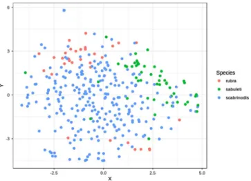
To visualise species differences in the pattern of infection over the body of ants we used the Barnes-Hut implementation of t-distributed stochastic neighbour embedding (t-SNE [72,73]) with the packageRtsne [42]. We chose this method over implementing a Principle Component Analysis approach, because in t-SNE we could explicitly specify the number of dimensions onto which to reduce the original data. Therefore, we were able to plot infection patterns (both of absolute and relative thallus number) on a 2D scatterplot. For t-SNE we used the square root-transformed values for both absolute and relative thallus numbers.
Potential origin of infection on the body
It has been suggested that infection withR. wasmanniistarts from the ant head [15,32,48]. If so, one would expect to see that, in the early stages of infection, only (or mostly) the head is parasitised. Therefore, the relative number of thalli should be high during thefirst stages of infection (= when total number of thalli is small). Consequently, if the infection spreads from the head to other body parts, we should see a decrease in the relative thallus number on the head simultaneously with the increase of total number of thalli on the whole body.
First, we checked the range (minimum and maximum val- ues) of the relative thallus number on each body part of infected ants, separately for the three ant species. We selected those body parts to be of interest in which one) the minimum value of relative thallus number was larger than zero and two) the maximum value was the largest in comparison to other body parts. InM. rubra, the frontal (or dorsal) side of the head and the gaster tergites satisfied our criteria. In M. sabuleti, only the frontal side of the head had a minimum relative thallus num- ber value larger than zero, and it had the largest maximum value among all body parts. InM. scabrinodis, there was no body part on which the minimum value of relative thallus num- ber was larger than zero, and so we selected the body part with the largest maximum value, which was the gaster tergites. Nota- bly, on the gaster tergites of M. scabrinodis, we observed the lowest incidence of zero values in relative thallus number as well. As a result, we decided to use this body part as a starting point to test our hypothesis about the infection’s spread.
To test whether there is indeed a significant negative asso- ciation between total thallus number and relative thallus number on selected body parts (frontal side of the head, gaster tergites), we used quasi-binomial generalised linear regression models.
Using these, we were able to reliablyfit models on a numeric scale ranging from zero to one (i.e. on the scale of the data) and to control for over-dispersion in the data. Two models were fitted. We specified the response variables to be the relative thallus number on the frontal side of the head and on the gaster tergites in the first and second model, respectively. In both models, predictor variables were total number of thalli on the
Figure 3. Phylogenetic reconstruction of Rickia species using a combined ITS + 28S dataset. The topology is the result of maximum likelihood inference. For each node, ML and MP bootstraps are presented above and below the branch leading to that node. For eachR.
wasmanniiisolate, isolate name,Myrmicahost epithet, and country code (AT, Austria; BE, Belgium; HU, Hungary; NL, The Netherlands) are presented. To the right of the phylogeny, results of species delimitation methods are summarised, from left to right: ABGD of the aligned ITS
& 28S datasets under most parameters combinations; ABGD of the 28S dataset forP= 0.001–0.001668 andX= 0.1–1.0 (asterisk *); bPTP of the ITS and 28S topologies; and GMYC of the ITS and 28S ultrametric trees, respectively. Hatching implies lack of support, whereas the dashed rectangle under ABGD 28S* means that four putative species were found withinR. wasmannii.
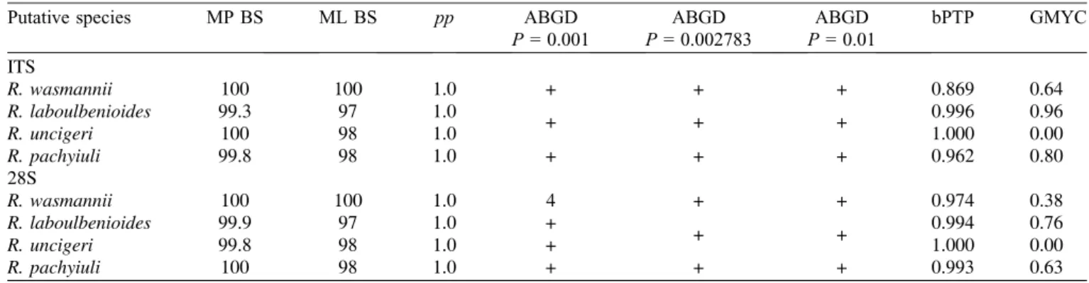
Table 2.Summary of results of MP, ML, BI, and species delimitation analyses (ABGD, bPTP, GMYC). Explanation of symbols and values used:pp= posterior probability; + under ABGD represents supported clades; 4 under ABGD means that the analysis found support for 4 species hypotheses (fide[55]) withinR. wasmanniiunder prior maximum distance (P) = 0.001, 0.001292, and 0.001668; numbers under bPTP and GMYC are Bayesian support values for delimited species hypotheses.
Putative species MP BS ML BS pp ABGD ABGD ABGD bPTP GMYC
P= 0.001 P= 0.002783 P= 0.01 ITS
R. wasmannii 100 100 1.0 + + + 0.869 0.64
R. laboulbenioides 99.3 97 1.0
+ + + 0.996 0.96
R. uncigeri 100 98 1.0 1.000 0.00
R. pachyiuli 99.8 98 1.0 + + + 0.962 0.80
28S
R. wasmannii 100 100 1.0 4 + + 0.974 0.38
R. laboulbenioides 99.9 97 1.0 +
+ + 0.994 0.76
R. uncigeri 99.8 98 1.0 + 1.000 0.00
R. pachyiuli 100 98 1.0 + + + 0.993 0.63
whole body, a factor variable generated by specifying species name and country of origin, and the interaction term between these two variables. The factor variable had four levels:
(1)M. rubrafrom Austria, (2)M. sabuletifrom the Netherlands, (3)M. scabrinodisfrom Hungary, and (4)M. scabrinodisfrom the Netherlands.
In both models, we used square root-transformed response and predictor variables. Also, to be able to infer on mean spe- cies-level differences using the intercept estimates of the mod- els, we centred the square root-transformed predictor variable at zero by subtracting the mean of the variable from each of its values. Furthermore, in the Results section we report actual regression coefficients for the slopes from each factor level, using the packagejtools[47].
Results
Phylogenetic analyses, and species delimitation The ITS dataset comprised 258 characters, of which 182 were constant and 70 were parsimony-informative. A total of 16 isolates were included (Table 1):Rickia wasmannii(10 iso- lates as ingroup),R. laboulbenioides De Kesel (two isolates), R. pachyiuli M. Bechet & I. Bechet (three isolates), and
R. uncigeri Scheloske (one isolate). Rickia wasmannii was retrieved as a monophyletic clade with maximum support from MP, ML, and BI (not shown). In this clade were includedR. was- manniiisolates of thalli removed fromM. rubra(two isolates), M. sabuleti(four isolates), and M. scabrinodis (four isolates).
The 28S dataset comprised 547 characters, of which 423 were constant and 113 were parsimony-informative. A total of 11 iso- lates were included (Table 1):Rickia wasmannii(five isolates as ingroup),R. laboulbenioides(two isolates),R. pachyiuli(three isolates), and R. uncigeri(one isolate).Rickia wasmanniiwas retrieved as a monophyletic clade with maximum support from MP, ML, and BI (not shown), including isolates from Table 4. Contrasts acquired from the model estimating between-
species differences in total thalli number.
Contrasts ofMyrmicaspp. Estimate SE z-ratio P M. rubra–M. sabuleti 0.070 0.155 0.45 0.893 M. rubra–M. scabrinodis 0.046 0.128 0.36 0.932 M. sabuleti–M. scabrinodis 0.024 0.105 0.23 0.971
Figure 4. Total number of thalli on worker bodies of the three examined Myrmica species (n = 34 for M. rubra, n = 50 for M. sabuleti,n= 270 forM. scabrinodis).
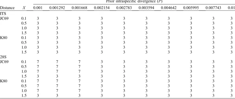
Table 3.Results of the Automatic Barcode Gap Discovery (ABGD) analyses.X, relative gap width; JC69, Jukes-Cantor substitution model;
K80, Kimura 2-parameter substitution model.
Prior intraspecific divergence (P)
Distance X 0.001 0.001292 0.001668 0.002154 0.002783 0.003594 0.004642 0.005995 0.007743 0.01 ITS
JC69 0.1 3 3 3 3 3 3 3 3 3 3
0.5 3 3 3 3 3 3 3 3 3 3
1.0 3 3 3 3 3 3 3 3 3 3
1.5 3 3 3 3 3 3 3 3 3 3
K80 0.1 3 3 3 3 3 3 3 3 3 3
0.5 3 3 3 3 3 3 3 3 3 3
1.0 3 3 3 3 3 3 3 3 3 3
1.5 3 3 3 3 3 3 3 3 3 3
28S
JC69 0.1 7 7 7 3 3 3 3 3 3 3
0.5 7 7 7 3 3 3 3 3 3 3
1.0 7 7 7 3 3 3 3 3 3 3
1.5 3 3 3 3 3 3 3 3 3 3
K80 0.1 7 7 7 3 3 3 3 3 3 3
0.5 7 7 7 3 3 3 3 3 3 3
1.0 7 7 7 3 3 3 3 3 3 3
1.5 3 3 3 3 3 3 3 3 3 3
M. rubra(one isolate),M. sabuleti(two isolates), andM. scabrin- odis(two isolates). The concatenated ITS + 28S dataset com- prised 804 characters, of which 604 were constant and 183 were parsimony-informative. A total of 16 isolates were included. Once again,R. wasmanniiwas retrieved as a mono- phyletic clade with maximum support from MP and ML (Fig. 3).
Results of the species delimitation methods are summarised inTables 2and3andFigure 3. The number of putative species
of Rickia was three in the ITS dataset with ABGD: Rickia laboulbenioides + uncigeri, R. pachyiuli, and R. wasmannii.
In the 28S dataset, this number varied from three to seven, depending on the prior intraspecific divergence parameter, whereas other parameters (relative gap width, distance metrics employed) had no influence on the results (Table 3). The bPTP analysis of both ITS and 28S topologies resulted in four highly supported species: Rickia laboulbenioides, R. pachyiuli, Table 5.Results of Conover-Iman tests on the species differences of body parts in absolute thallus density. The column“Largest thallus density”shows which host species had highest number of thalli on a given body part; species names in parentheses indicate that the difference between the specified species and one of the other two species is not significant;“none”means that the three species did not differ significantly from one another.
Body part Comparisons ofMyrmicaspp. t P(adjusted) Largest thallus density
Antennae M. rubra–M. sabuleti 5.28 <0.001 M. sabuleti
M. rubra–M. scabrinodis 1.56 1.000
M. sabuleti–M. scabrinodis 5.78 <0.001
Head (frontal) M. rubra–M. sabuleti 3.15 0.029 (M. sabuleti)
M. rubra–M. scabrinodis 3.09 0.034
M. sabuleti–M. scabrinodis 0.90 1.000
Head (ventral) M. rubra–M. sabuleti 0.52 1.000 (M. sabuleti)
M. rubra–M. scabrinodis 2.26 0.254
M. sabuleti–M. scabrinodis 3.43 0.012
Pronotum M. rubra–M. sabuleti 4.54 <0.001 M. sabuleti
M. rubra–M. scabrinodis 2.96 0.048
M. sabuleti–M. scabrinodis 3.06 0.036
Mesonotum M. rubra–M. sabuleti 4.64 <0.001 (M. sabuleti)
M. rubra–M. scabrinodis 3.48 0.011
M. sabuleti–M. scabrinodis 2.59 0.127
Propodeum M. rubra–M. sabuleti 2.65 0.110 None
M. rubra–M. scabrinodis 2.80 0.076
M. sabuleti–M. scabrinodis 0.52 1.000
Petiole M. rubra–M. sabuleti 3.29 0.018 (M. scabrinodis)
M. rubra–M. scabrinodis 4.05 0.001
M. sabuleti–M. scabrinodis 0.03 1.000
Postpetiole M. rubra–M. sabuleti 2.46 0.173 None
M. rubra–M. scabrinodis 2.30 0.242
M. sabuleti–M. scabrinodis 0.83 1.000
Gaster tergites M. rubra–M. sabuleti 0.96 1.000 (M. scabrinodis)
M. rubra–M. scabrinodis 2.35 0.224
M. sabuleti–M. scabrinodis 4.17 0.001
Gaster sternites M. rubra–M. sabuleti 3.48 0.011 (M. rubra)
M. rubra–M. scabrinodis 0.07 1.000
M. sabuleti–M. scabrinodis 4.95 <0.001
Procoxa M. rubra–M. sabuleti 3.39 0.014 M. rubra
M. rubra–M. scabrinodis 4.74 <0.001
M. sabuleti–M. scabrinodis 0.70 1.000
Profemur M. rubra–M. sabuleti 1.22 1.000 None
M. rubra–M. scabrinodis 0.60 1.000
M. sabuleti–M. scabrinodis 1.06 1.000
Mesocoxa M. rubra–M. sabuleti 2.67 0.107 (M. rubra)
M. rubra–M. scabrinodis 3.80 0.003
M. sabuleti–M. scabrinodis 0.64 1.000
Mesofemur M. rubra–M. sabuleti 0.25 1.000 None
M. rubra–M. scabrinodis 1.53 1.000
M. sabuleti–M. scabrinodis 1.44 1.000
Metacoxa M. rubra–M. sabuleti 3.30 0.018 M. rubra
M. rubra–M. scabrinodis 3.79 0.003
M. sabuleti–M. scabrinodis 0.29 1.000
Metafemur M. rubra–M. sabuleti 1.48 1.000 None
M. rubra–M. scabrinodis 1.28 1.000
M. sabuleti–M. scabrinodis 0.63 1.000
R. uncigeri, andR. wasmannii. The GMYC analysis of the ITS resulted in the recognition of four species, all with moderate to high support except forR. uncigeri(of which only a single iso- late was included). The GMYC analysis of the 28S led to com- parable results, but in this analysis also theR. wasmanniiclade received low support (pp= 0.38).
Absolute and relative thallus numbers
There were no significant differences between Myrmica species in total thallus number (Table 4, Fig. 4). In the Conover-Iman tests comparing absolute thallus number of each body part between species, we found 20 significant differences after Bonferroni’s P-value adjustment (Table 5). Overall, M. sabuleti specimens were more heavily infected on their antennae, head, pronotum, and mesonotum compared to workers ofM. rubraandM. scabrinodis, whereasM. scabrin- odisants appeared to have the highest thallus density on their petiole and gaster tergites in comparison to the other host species.Myrmica rubraworkers showed highest thallus density on their gaster sternites, procoxa, mesocoxa, and metacoxa (Fig. 5).
In the Conover-Iman tests comparing relative thallus num- ber of each body part between species, we found 29 significant differences after Bonferroni’sP-value adjustment (Table 6). In comparison toM. rubraandM. scabrinodis, values of relative thallus number were highest in M. sabuleti on the antennae, head, pronotum, mesonotum, propodeum, and petiole. On the gaster tergites,M. scabrinodishad larger proportions of thalli compared to the other host species. Furthermore, M. rubra workers had highest proportions of thalli on their gaster sternites, procoxa, mesocoxa, mesofemur, metacoxa, and metafemur, compared to the other host species (Fig. 6).
Potential origin of infection on the body
The total number of thalli was significantly negatively asso- ciated with relative thallus number on the frontal side of the head
in M. rubra (Estimate = 0.054, SE = 0.014, t = 3.81, P < 0.001) and in M. sabuleti (Estimate = 0.046, SE= 0.011,t=3.97,P< 0.001). In the case ofM. scabrinodis, the regression coefficients differed between ants from the Netherlands and Hungary: in the Netherlands, the association between total number of thalli and relative thallus number on the frontal side of the head was negative and relatively strong (Estimate =0.076,SE= 0.029,t=2.57,P= 0.011), whereas in Hungary, the regression coefficient was positive and weaker than in the other groups (Estimate = 0.013, SE = 0.006, t= 2.27,P= 0.024). These associations are shown inFigure 7.
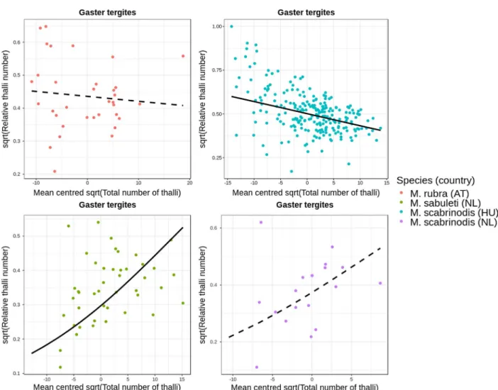
In the model in which wefitted the relative thallus number of gaster tergites on total thallus number, the regression coeffi- cient was significantly negative for M. scabrinodis ants col- lected in Hungary (Estimate = 0.042, SE = 0.005, t=9.02,P< 0.001), but it was not significant in workers from the Netherlands (Estimate = 0.039, SE = 0.026, t = 1.52, P = 0.129). The association was not significant in M. rubra either (Estimate = 0.006, SE = 0.010, t = 2.57, P= 0.011). However, we found a significantly positive effect in M. sabuleti (Estimate = 0.024, SE = 0.011, t = 2.16, P= 0.031). These associations are shown inFigure 8.
Discussion
Rickia wasmanniiwas described in the 19th century, based on morphological characters only [9].Myrmicahosts ofR. was- manniibelong in two phylogenetically distinct species groups (rubra-group and scabrinodis-group). The genus Myrmica quickly diversified around the Eocene–Oligocene transition.
The scabrinodis-group is among the oldest species groups (21.46 ± 4.00 Mya), whereas the estimated crown age for the rubra-group is 10.88 ± 2.12 Mya, in the Late Miocene [39].
Our results demonstrate thatR. wasmanniidoes not encompass divergent genetic lineages segregated by host. In all molecular phylogenetic reconstructions, R. wasmannii isolates formed a monophyletic clade with maximum support. Infected workers ofMyrmicaspp. were collected in Austria, Belgium, Hungary, and the Netherlands. Even so, there is no geographic signal.
The ITS sequences ofR. wasmannii are all identical, whereas there are 0, 1, or 2 nucleotide differences among LSU sequences. We conclude that, contrary to species of Gloean- dromycesandHesperomyces, inR. wasmanniineither geogra- phy nor host species are drivers of divergent evolution. The absence of host specificity in R. wasmannii is quite different from what has been observed in a Myrmica-associated group of endosymbiotic bacteria; Spiroplasma species co-diverged with their hosts over evolutionary time [1].
Most species of Laboulbeniales have been described based on morphological characters. In fact, only four species and four formaehave been described based on combined morphological and molecular data [27,34,35]. In addition, for only a handful of species the taxonomic status has been assessed using molecular phylogenetic data following description. For exam- ple, Corethromyces bicolor Thaxt., after having been transferred to another genus, was re-installed in the genus Corethromyces Thaxt. based on DNA studies [76], and using sequence data from three loci, distinct clades within Figure 5. Visualisation of infection patterns based on absolute
thallus number on 16 body parts, using t-SNE to reduce the number of dimensions of the data set (n= 354).
Hesperomyces virescens were found, each corresponding to host-specific species [30].
Species in the genusCoreomyces Thaxt. do not show host specificity–similar toR. wasmannii.Coreomyces corixae(green clade in [68]), for example, occurs on water boatmen (Heteroptera, Corixidae) in the generaCallicorixaWhite 1873,
Hesperocorixa Kirkaldy 1908, and Sigara Fabricius, 1775 [67]. As more examples of Laboulbeniales fungi are explored, we can start linking speciation patterns to presence and absence of fungal traits. One candidate trait that may have an influence in host-dependent speciation in Laboulbeniales is the presence of a haustorium. Haustoria are rhizoidal structures that can be simple Table 6.Results of Conover-Iman tests on the species differences of body parts in relative thallus number. The column“Largest proportion of thalli”shows which host species had highest proportion of thalli on a given body part; species names in parentheses indicate that the difference between the specified species and one of the other two species is not significant;“none”means that the three species did not differ significantly from one another.
Body part Comparisons ofMyrmicaspp. t P(adjusted) Largest proportion of thalli
Antennae M. rubra–M. sabuleti 6.77 <0.001 M. sabuleti
M. rubra–M. scabrinodis 1.77 0.542
M. sabuleti–M. scabrinodis 7.68 <0.001
Head (frontal) M. rubra–M. sabuleti 3.59 0.005 M. sabuleti
M. rubra–M. scabrinodis 1.68 0.616
M. sabuleti–M. scabrinodis 3.20 0.017
Head (ventral) M. rubra–M. sabuleti 0.15 1.000 (M. sabuleti)
M. rubra–M. scabrinodis 4.49 <0.001
M. sabuleti–M. scabrinodis 5.53 <0.001
Pronotum M. rubra–M. sabuleti 7.40 <0.001 M. sabuleti
M. rubra–M. scabrinodis 4.04 0.001
M. sabuleti–M. scabrinodis 5.91 <0.001
Mesonotum M. rubra–M. sabuleti 7.33 <0.001 M. sabuleti
M. rubra–M. scabrinodis 4.36 <0.001
M. sabuleti–M. scabrinodis 5.43 <0.001
Propodeum M. rubra–M. sabuleti 4.23 <0.001 (M. sabuleti)
M. rubra–M. scabrinodis 3.29 0.013
M. sabuleti–M. scabrinodis 2.22 0.231
Petiole M. rubra–M. sabuleti 4.03 0.001 M. sabuleti
M. rubra–M. scabrinodis 4.94 <0.001
M. sabuleti–M. scabrinodis 0.03 1.000
Postpetiole M. rubra–M. sabuleti 2.00 0.368 None
M. rubra–M. scabrinodis 1.32 0.934
M. sabuleti–M. scabrinodis 1.34 0.934
Gaster tergites M. rubra–M. sabuleti 3.73 0.003 (M. scabrinodis)
M. rubra–M. scabrinodis 2.38 0.160
M. sabuleti–M. scabrinodis 8.20 <0.001
Gaster sternites M. rubra–M. sabuleti 5.26 <0.001 (M. rubra)
M. rubra–M. scabrinodis 0.58 1.000
M. sabuleti–M. scabrinodis 6.90 <0.001
Procoxa M. rubra–M. sabuleti 5.11 <0.001 M. rubra
M. rubra–M. scabrinodis 7.41 <0.001
M. sabuleti–M. scabrinodis 1.38 0.934
Profemur M. rubra–M. sabuleti 1.85 0.489 None
M. rubra–M. scabrinodis 1.64 0.616
M. sabuleti–M. scabrinodis 0.73 1.000
Mesocoxa M. rubra–M. sabuleti 3.49 0.007 M. rubra
M. rubra–M. scabrinodis 5.00 <0.001
M. sabuleti–M. scabrinodis 0.88 1.000
Mesofemur M. rubra–M. sabuleti 0.80 1.000 (M. rubra)
M. rubra–M. scabrinodis 3.01 0.028
M. sabuleti–M. scabrinodis 2.41 0.155
Metacoxa M. rubra–M. sabuleti 4.18 0.001 M. rubra
M. rubra–M. scabrinodis 5.34 <0.001
M. sabuleti–M. scabrinodis 0.27 1.000
Metafemur M. rubra–M. sabuleti 3.07 0.025 M. rubra
M. rubra–M. scabrinodis 3.17 0.018
M. sabuleti–M. scabrinodis 0.68 1.000
or branched and penetrate the host’s integument to provide additional holdfast and to increase surface area, presumably for nutrient uptake. Benjamin [6] believed that all Laboulbeniales produce haustoria. This is contrary to Tragustet al. [71] who, based on light and electron microscopy techniques, found no evidence for penetration in four species of Laboulbeniales:
Laboulbenia camponoti S.W.T. Batra, L. formicarum Thaxt., Rickia lenoiriiSantam., andR. wasmannii.
A note about millipede-associated Laboulbeniales
The sequences of R. laboulbenioides, R. pachyiuli, and R. uncigeri were generated for this study and are the first published ones for millipede-associated Laboulbeniales.
Laboulbeniales on millipedes occur infive genera:Diplopodo- myces W. Rossi & Balazuc, Rickia, the recently described Thaxterimyces Santam., Reboleira & Enghoff, Triainomyces Figure 6. Visualisation of infection patterns based on relative
thallus number on 16 body parts, using t-SNE to reduce the number of dimensions of the data set (n= 354).
Figure 7. Association between total number of thalli and relative number of thalli on the frontal side of the head (n = 354). Black curves are regression lines from the fitted model: solid and dashed lines represent significant and non-significant associations, respectively.
W. Rossi & A. Weir, andTroglomycesS. Colla [15,22,58–61].
Similar to ourfindings with batfly-associated Laboulbeniales fungi [33], we expect that parasitism of millipedes by
Laboulbeniales arose several times independently. Some species ofRickiaon millipedes are known to parasitise several millipede hosts. For example,R. candelabriformis Santam., Enghoff &
Figure 8. Association between total number of thalli and relative number of thalli on the gaster tergites (zeros excluded,n= 336). Black curves are regression lines from thefitted model: solid and dashed lines represent significant and non-significant associations, respectively.
Table 7.Distance matrix of the aligned ITS sequences.
Isolate Species GenBank acc. no. 1 2 3 4 5 6 7 8 9 10 11 12 13 14 15 16
1 SR4s Rickia laboulbenioides MH040592
2 SR5s Rickia laboulbenioides MK500060 0
3 SR8s Rickia pachyiuli MK500058 16 16
4 SR1s Rickia pachyiuli MH040593 16 16 0
5 SR13s Rickia pachyiuli MK500059 16 16 0 0
6 SR12s Rickia uncigeri MK500061 6 6 20 20 20
7 Újléta2014 Rickia wasmannii MK500053 24 24 25 25 25 25
8 DE_Rak4 Rickia wasmannii KT800050 24 24 25 25 25 25 0
9 Wien2015-1 Rickia wasmannii MK500055 24 24 25 25 25 25 0 0
10 D. Haelew. 1234a Rickia wasmannii MH040595 24 24 25 25 25 25 0 0 0
11 ADK6272a Rickia wasmannii MK500050 24 24 25 25 25 25 0 0 0 0
12 ADK6274c Rickia wasmannii MK500051 24 24 25 25 25 25 0 0 0 0 0
13 Wien2016-1 Rickia wasmannii MK500056 24 24 25 25 25 25 0 0 0 0 0 0
14 Wiensabuleti2016-1 Rickia wasmannii MK500057 24 24 25 25 25 25 0 0 0 0 0 0 0 15 Bükkszentkereszt2016 Rickia wasmannii MK500052 24 24 25 25 25 25 0 0 0 0 0 0 0 0
16 Újléta2015-4 Rickia wasmannii MK500054 24 24 25 25 25 25 0 0 0 0 0 0 0 0 0
Reboleira, R. gigas Santam., Enghoff & Reboleira, and R. lophophoraSantam., Enghoff & Reboleira [60] are potential next targets to study intraspecific diversity, to assess whether our current observations forR. wasmanniihold for the entire genus.
Species delimitation analyses
The ABGD analysis of the 28S dataset found different numbers of putative species depending on the prior intraspecific divergence (P), which is in line with previous work. Puillandre et al. [55] put forward to useP= 0.01, because under this set- ting, ABGD results in the same number of putative species found using different approaches. In our analyses of both the ITS and 28S datasets, ABGD found three species under this setting: R. laboulbenioides+uncigeri, R. pachyiuli, and R. wasmannii. Checking the distance matrices, we found that the lowest number of inter-species nucleotide differences was observed betweenR. laboulbenioidesandR. uncigeri(Tables 7 and8). For example, in the ITS dataset,R. laboulbenioidesdif- fered in six nucleotides fromR. uncigeri, whereas it differed in 16 nucleotides from R. pachyiuliand in 24 nucleotides from R. wasmannii (details in Table 7). Apparently, ABGD was not able to identify the divergence amongR. laboulbenioides andR. uncigeriisolates as a“barcode gap”(fide[55]), which will likely be resolved once we generate and include more sequences of R. uncigeri. The GMYC results are congruent with the results from the other species delimitation methods.
One clade lacks support, the singleton cladeR. uncigeri, and this is no surprise because GMYC looks at intraspecific branch- ing versus interspecific branching.
The lack of phylogenetic structuring amongR. wasmannii populations may be attributed to two different but not mutually exclusive scenarios: (1) intermittent gene flow homogenising populations and (2) recent spread of the fungus starting from a small founder population. The first scenario is possible because co-occurring arthropods may share Laboulbeniales par- asites. Interspecific ascospore transmission in sympatric species has been observed for R. wasmannii parasitising M. scabrin- odis, mites, and aMicrodon myrmicaelarva (Diptera, Syrphi- dae) in ant nests [53]. The second scenario can best be illustrated with the following example. Laboulbenia formi- carum Thaxt. is thought to have spread from North America to Europe on an unknown ant host [23], followed by host shifts to European-native and invasive ant host species during its
rapid spread in recent years [24,28]. It was shown forM. rubra that it survived the last glacial period in multiple refugia and expanded its distribution along different routes [46]. It might be possible thatR. wasmanniihas undergone postglacial spread with its host, followed by multiple host shifts to otherMyrmica species. Microsatellite studies are required to assess population- wide genetic differences, e.g., to answer the question whether incipient sympatric speciation is taking place.
Habitat specificity and host spectrum
Rickia wasmannii is a single species, clearly shared by a number of Myrmica hosts and with a vast distribution area.
The species is non-penetrating [71] and compared to taxa with a haustorium such asH. virescens, it has several hosts but only if these occupy a similar habitat (Myrmicanests). This habitat specificity– preference forMyrmicanests and habitat choices –can explain the wide distribution on multipleMyrmicaspecies and ant nest inquilines [53]. Moreover, the fact that there is overlap and even contact betweenMyrmicapopulations of dif- ferent species [78] implies that regular or at least sufficient interspecific transfer ofR. wasmanniioccurs between host taxa.
It also means that the different host taxa, their specific habitat choices, and the nature of their nests, allow the development of the fungus population. Considering the high thallus densities observed, we doubt there is enough reason to consider one Myrmicaspecies as a main host (fide[62]) over other species.
In this context, we propose thatR. wasmanniiis a true eurytopic species with a wide ecological amplitude. It is expected that other species ofMyrmicamay also carry this parasite. However, absence ofR. wasmanniion a givenMyrmicaspecies does not necessarily mean that this ant species, its nests, and/or its habitat selection are unsuitable for this fungus. Indeed, in areas where several infected nests ofM. scabrinodis occur, some adjacent nests can be entirely free ofR. wasmannii [15]. This has also been observed for M. sabuleti, where infection frequency of workers can vary from 0 to 100% among nests that are only a few meters apart (P. Boer, unpublished data).
Distribution of thalli on worker bodies
The original morphological description [8] holds to the phylogenetic species concept. This implies that differences in thallus numbers of different body parts between ant species Table 8.Distance matrix of the aligned 28S rDNA sequences.
Isolate Species GenBank acc. no. 1 2 3 4 5 6 7 8 9 10 11
1 SR4s Rickia laboulbenioides MH040592
2 SR5s Rickia laboulbenioides MK500060 0
3 SR8s Rickia pachyiuli MK500058 39 39
4 SR1s Rickia pachyiuli MH040593 39 39 0
5 SR13s Rickia pachyiuli MK500059 39 39 0 0
6 SR12s Rickia uncigeri MK500061 19 19 42 42 42
7 Újléta2014 Rickia wasmannii MK490857 79 79 67 67 67 76
8 DE_Rak4 Rickia wasmannii KT800021 79 79 68 68 68 77 1
9 Wien2015-1 Rickia wasmannii MK490858 80 80 68 68 68 77 1 2
10 D. Haelew. 1234a Rickia wasmannii MH040595 79 79 67 67 67 76 0 1 1
11 ADK6272a Rickia wasmannii MK500050 79 79 67 67 67 76 0 1 1 0
and populations must be explained by behaviour, cuticular chemical profiles, and/or environmental stresses [7, 20, 21, 65]. In our dataset, there was no evidence for differences between host species in total number of thalli on worker bodies.
If the ant species in the study area are of the same body size, this might suggest that the overall number of thalli on a worker’s body is simply a factor of the worker’s age, irrespec- tive of host species; older workers show heavier infection by R. wasmannii[3].
We did observe differences betweenMyrmicaspecies in the pattern of infection over the body. Tests on both absolute and relative thallus number indicate that M. sabuleti workers are more heavily infected on thefirst few body segments compared to other hosts (Tables 5and6).Myrmica rubraworkers show highest thallus densities on the coxa and femur, whereas in M. scabrinodishighest thallus densities were found on the gaster tergites. These results might indicate differences among host species in how ants come into contact with the fungus, or even differences in grooming efficacy. Which body parts are workers able to groom (and thus stop ascospores from adhering and developing) more effectively? Another possibility involves differences in the cuticle itself [20,65]; surface characteristics may have a fundamental impact on the success of an ascospore to adhere to the cuticle and develop to a mature thallus.
Based on our statistical analyses, it is likely that inM. rubra and M. sabuleti the locus of infection (= the area where the infection originates) is the frontal side of the head. For M. scabrinodis workers from the Netherlands, the locus of infection also appears to be the frontal side of the head.
However, forM. scabrinodis specimens from Hungary, infec- tion likely starts from the gaster tergites. These results indicate differences among populations of the same species in a wide geographical range. Different Myrmica species display diver- gent foraging, allo-grooming, and secretion emission activities [7]. This likely leads to differences in how workers enter into contact with ascospores, which should be investigated with behavioural studies of their hosts.
Conflict of interest
The authors declare that they have no conflicts of interest in relation to this article.
Authors’ contributions
D.H. coordinated the study, performed molecular phyloge- netic and species delimitation analyses, and wrote the manu- script with input from all co-authors; P.B., F.B., A.T., and A.D.K. collected ants; P.B. and F.B. counted thalli on worker bodies; Z.R. performed statistical analyses; D.H., A.S.P.S.R., and W.P.P. extracted DNA and generated sequence data;
A.D.K. provided the photograph forFigure 1and the drawing included inFigure 3; A.D.K. and O.N. provided expertise at all stages of research.
Acknowledgements. D.H. received graduate student funding from the Department of Organismic and Evolutionary Biology at Harvard University. F.B. was supported by the NKFI KH 130338 project.
A.S.P.S.R. was supported by a research grant (no. 15471) from the VILLUM FONDEN and by a Harvard University Herbaria travel grant, allowing her to visit the Farlow Herbarium in 2016. A.T. was supported by the “AntLab”Marie Curie Career Integration Grant, within the 7th European Community Framework Programme, a Bolyai János Research Scholarship of the Hungarian Academy of Sciences (MTA), and the ÚNKP-18-4 New National Excellence Program of the Ministry of Human Capacities. DNA sequences of millipede- associatedRickiaspecies were obtained under the Danish Council for Independent Research project ref. DFF–FNU 4002-00269.
References
1. Ballinger MJ, Moore LD, Perlman SJ. 2018. Evolution and diversity of inheritedSpiroplasmasymbionts inMyrmicaants.
Applied and Environmental Microbiology, 84, e02299–17.
2. Báthori F, Csata E, Tartally A. 2015. Rickia wasmannii increases the need for water inMyrmica scabrinodis(Ascomy- cota: Laboulbeniales; Hymenoptera: Formicidae). Journal of Invertebrate Pathology, 126, 78–82.
3. Báthori F, Pfliegler WP, Radai Z, Tartally A. 2018. Host age determines parasite load of Laboulbeniales fungi infecting ants:
implications for host-parasite relationship and fungal life history. Mycoscience, 59, 166–171.
4. Báthori F, Pfliegler WP, Zimmerman C-U, Tartally A. 2017.
Online image databases as multi-purpose resources: discovery of a new host ant ofRickia wasmannii Cavara (Ascomycota, Laboulbeniales) by screening AntWeb.org. Journal of Hyme- noptera Research, 61, 85–94.
5. Báthori F, Rádai Z, Tartally A. 2017. The effect of Rickia wasmannii(Ascomycota, Laboulbeniales) on the aggression and boldness ofMyrmica scabrinodis(Hymenoptera, Formicidae).
Journal of Hymenoptera Research, 58, 41–52.
6. Benjamin RK. 1971. Introduction and supplement to Roland Thaxter’s contribution towards a monograph of the Laboulbe- niaceae. Bibliotheca Mycologica, 30, 1–155.
7. Cammaerts M-C, Cammaerts R. 1980. Food recruitment strategies of the antsMyrmica sabuletiandMyrmica ruginodis.
Behavioural Processes, 5, 251–270.
8. Capella-Gutiérrez S, Silla-Martínez JM, Gabaldón T. 2009.
TrimAl: a tool for automated alignment trimming in large-scale phylogenetic analyses. Bioinformatics, 25, 1972–1973.
9. Cavara F. 1899. Di una nuova Laboulbeniacea: Rickia was- mannii, nov. gen. et nov. spec. Malpighia, 13, 173–188.
10. Chernomor O, von Haeseler A, Minh BQ. 2016. Terrace aware data structure for phylogenomic inference from supermatrices.
Systematic Biology, 65, 997–1008.
11. Csata E, Bernadou A, Rákosy-Tican E, Heinze J, Markó B.
2017. The effects of fungal infection and physiological condi- tion on the locomotory behaviour of the antMyrmica scabrin- odis. Journal of Insect Physiology, 98, 167–172.
12. Csata E, Erős K, Markó B. 2014. Effects of the ectoparasitic fungusRickia wasmanniion its ant hostMyrmica scabrinodis:
changes in host mortality and behavior. Insectes Sociaux, 61, 247–252.
13. Csata E, Timusß N, Witek M, Casacci LP, Lucas C, Bagnères AG, Sztencel-Jabłonka A, Barbero F, Bonelli S, Rákosy L, Markó B. 2017. Lock-picks: fungal infection facilitates the intrusion of strangers into ant colonies. Scientific Reports, 7, 46323.
14. Darriba D, Taboada GL, Doallo R, Posada D. 2012. jModelTest 2: more models, new heuristics and parallel computing. Nature Methods, 9, 772.
15. De Kesel A, Haelewaters D, Dekoninck W. 2016. Myrme- cophilous Laboulbeniales (Ascomycota) in Belgium. Sterbeeck- ia, 34, 3–6.
16. Dinno A. 2017. conover.test: Conover-Iman test of multiple comparisons using rank sums. R package version 1.1.5.
Accessed January 25, 2019. https://CRAN.R-project.org/pack- age=conover.test
17. Drummond AJ, Suchard MA, Xie D, Rambaut A. 2012.
Bayesian phylogenetics with BEAUti and the BEAST 1.7.
Molecular Biology and Evolution, 29, 1969–1973.
18. Edgar RC. 2004. MUSCLE: multiple sequence alignment with high accuracy and high throughput. Nucleic Acids Research, 32, 1792–1797.
19. Egger KN. 1995. Molecular analysis of ectomycorrhizal fungal communities. Canadian Journal of Botany, 73, S1415–S1422.
20. Elmes G, Akino T, Thomas J, Clarke R, Knapp J. 2002.
Interspecific differences in cuticular hydrocarbon profiles of Myrmicaants are sufficiently consistent to explain host specificity byMaculinea(large blue) butterflies. Oecologia, 130, 525–535.
21. Elmes GW, Thomas JA, Wardlaw JC, Hochberg ME, Clarke RT, Simcox DJ. 1998. The ecology ofMyrmicaants in relation to the conservation ofMaculineabutterflies. Journal of Insect Conservation, 2, 67–78.
22. Enghoff H, Santamaria S. 2015. Infectious intimacy and contaminated caves –three new species of ectoparasitic fungi (Ascomycota: Laboulbeniales) from blaniulid millipedes (Diplopoda: Julida) and inferences about their transmittal mechanisms. Organisms Diversity & Evolution, 15, 249–263.
23. Espadaler X, Lebas C, Wagenknecht J, Tragust S. 2011.
Laboulbenia formicarum (Ascomycota, Laboulbeniales), an exotic parasitic fungus, on an exotic ant in France. Vie &
Milieu, 61, 41–44.
24. Espadaler X, Santamaria S. 2003. Laboulbenia formicarum Thaxt. (Ascomycota, Laboulbeniales) crosses the Atlantic.
Orsis, 18, 97–101.
25. Ezard T, Fujisawa T, Barraclough TG. 2009. splits: SPecies’ LImits by Threshold Statistics. R package version 1.0-14/r31.
Accessed January 23, 2019. http://RForge.R-project.org/pro- jects/splits/.
26. Gardes M, Bruns TD. 1993. ITS primers with enhanced specificity for Basidiomycetes–application to the identification of mycorrhizae and rusts. Molecular Ecology, 2, 113–118.
27. Goldmann L, Weir A, Rossi W. 2013. Molecular analysis reveals two new dimorphic species ofHesperomyces(Ascomy- cota, Laboulbeniomycetes) parasitic on the ladybird Coleome- gilla maculata (Coleoptera, Coccinellidae). Fungal Biology, 117, 807–813.
28. Gómez K, Espadaler X, Santamaria S. 2016. Ant-fungus interactions: Laboulbenia camponoti Batra in Italy and a new host for L. formicarum Thaxter (Fungi: Ascomycota, Laboul- beniales). Sociobiology, 63, 950–955.
29. Haelewaters D, Boer P, Noordijk J. 2015. Studies of Laboul- beniales (Fungi, Ascomycota) on Myrmica ants: Rickia was- mannii in the Netherlands. Journal of Hymenoptera Research, 47, 39–47.
30. Haelewaters D, De Kesel A, Pfister DH. 2018. Integrative taxonomy reveals hidden species within a common fungal parasite of ladybirds. Scientific Reports, 8, 15966.
31. Haelewaters D, Gorczak M, Pfliegler WP, Tartally A, Tischer M, Wrzosek M, Pfister DH. 2015. Bringing the Laboulbeniales to the 21st century: enhanced techniques for extraction and PCR amplification of DNA from minute ectoparasitic fungi. IMA Fungus, 6, 363–372.
32. Haelewaters D, Gort G, Boer P, Noordijk J. 2015. Studies of Laboulbeniales (Fungi, Ascomycota) on Myrmica ants (II):
variation of infection by Rickia wasmannii over habitats and time. Animal Biology, 65, 219–231.
33. Haelewaters D, Page RA, Pfister DH. 2018. Laboulbeniales hyperparasites (Fungi, Ascomycota) of bat flies: independent origins and host associations. Ecology and Evolution, 8, 8396–
8418.
34. Haelewaters D, Pfister DH. 2019. Morphological species of Gloeandromyces(Ascomycota, Laboulbeniales) evaluated using single-locus species delimitation methods. Fungal Systematics and Evolution, 3, 19–33.
35. Haelewaters D, Pfliegler WP, Gorczak M, Pfister DH. 2019. Birth of an order: comprehensive molecular phylogenetic study excludesHerpomyces(Fungi, Laboulbeniomycetes) from Laboul- beniales. Molecular Phylogenetics and Evolution, 133, 286–301.
36. Hall TA. 1999. BioEdit: a user-friendly biological sequence alignment editor and analysis program for Windows 95/98/NT.
Nucleic Acids Symposium Series, 41, 95–98.
37. Hoang DT, Chernomor O, von Haeseler A, Minh BQ, Vinh LS.
2017. UFBoot2: improving the ultrafast bootstrap approxima- tion. Molecular Biology and Evolution, 35, 518–522.
38. Hopple JS Jr, Vilgalys R. 1994. Phylogenetic relationships among coprinoid taxa and allies based on data from restriction site mapping of nuclear rDNA. Mycologia, 86, 96–107.
39. Jansen G, Savolainen R, Vepsäläinen K. 2010. Phylogeny, divergence-time estimation, biogeography and social parasite- host relationships of the Holarctic ant genus Myrmica (Hymenoptera: Formicidae). Molecular Phylogenetics and Evo- lution, 56, 294–304.
40. Jukes TH, Cantor CR. 1969. Evolution of protein molecules, in Mammalian protein metabolism, Munro NH, Editor. Academic Press: New York. p. 21–132.
41. Kimura M. 1980. A simple method for estimating evolutionary rates of base substitutions through comparative studies of nucleotide sequences. Journal of Molecular Evolution, 16, 111– 120.
42. Krijthe JH. 2015. Rtsne: T-distributed stochastic neighbor embedding using Barnes-Hut implementation. R package ver- sion 0.13. Accessed January 25, 2019. https://github.com/
jkrijthe/Rtsne.
43. Kumar S, Stecher G, Tamura K. 2016. MEGA7: molecular evolutionary genetics analysis version 7.0 for bigger datasets.
Molecular Biology and Evolution, 33, 1870–1874.
44. Kurtzman CP, Robnett CJ. 1998. Identification and phylogeny of ascomycetous yeasts from analysis of nuclear large subunit (26S) ribosomal DNA partial sequences. Antonie van Leeuwen- hoek, 73, 331–371.
45. Lenth RV. 2016. Least-squares means: the R package lsmeans.
Journal of Statistical Software, 69, 1–33.
46. Leppänen J, Vepsäläinen K, Savolainen R. 2011. Phylogeog- raphy of the antMyrmica rubraand its inquiline social parasite.
Ecology and Evolution, 1, 46–62.
47. Long JA. 2018. jtools: analysis and presentation of social scientific data. R package version 1.1.1. Accessed January 25, 2019.https://cran.r-project.org/package=jtools.
48. Markó B, Csata E, Erős K, Német E, Czekes Z, Rózsa L. 2016.
Distribution of the myrmecoparasitic fungusRickia wasmannii (Ascomycota: Laboulbeniales) across colonies, individuals, and body parts of Myrmica scabrinodis. Journal of Invertebrate Pathology, 136, 74–80.
49. Michonneau F, Bolker B, Holder M, Lewis P, OMeara B. 2018.
rncl: an interface to the nexus class library. R package version 0.8.3. Accessed January 25, 2019.http://CRAN.R-project.org/
package=rncl.
50. Miller MA, Pfeiffer W, Schwartz T. 2010. Creating the CIPRES science gateway for inference of large phylogenetic trees.
Proceedings of the gateway computing environments workshop (GCE), 14 Nov. 2010, New Orleans, Louisiana. p. 1–8.
51. Nguyen L-T, Schmidt HA, von Haeseler A, Minh BQ. 2015.
IQ-TREE: a fast and effective stochastic algorithm for estimat- ing maximum likelihood phylogenies. Molecular Biology and Evolution, 32, 268–274.
52. Pech P, Heneberg P. 2015. Benomyl treatment decreases fecundity of ant queens. Journal of Invertebrate Pathology, 130, 61–63.
53. Pfliegler WP, Báthori F, Haelewaters D, Tartally A. 2016.
Studies of Laboulbeniales on Myrmica ants (III): myrme- cophilous arthropods as alternative hosts of Rickia wasmannii.
Parasite, 23, 50.
54. Pons J, Barraclough T, Gomez-Zurita J, Cardoso A, Duran D, Hazell S, Kamoun S, Sumlin W, Vogler A. 2006. Sequence- based species delimitation for the DNA taxonomy of unde- scribed insects. Systematic Biology, 55, 595–609.
55. Puillandre N, Lambert A, Brouillet S, Achaz G. 2012. ABGD, automatic barcode gap discovery for primary species delimita- tion. Molecular Ecology, 21, 1864–1877.
56. Radchenko AG, Elmes GW. 2010.Myrmica ants (Hymenop- tera: Formicidae) of the old world. Warsaw, Poland: Natura optima dux.
57. Rambaut A, Suchard MA, Xie D, Drummond AJ. 2014. Tracer v1.6. Accessed December 12, 2018.http://tree.bio.ed.ac.uk/soft- ware/tracer/.
58. Reboleira ASPS, Enghoff H, Santamaria S. 2018. Novelty upon novelty visualized by rotational scanning electron micrographs (rSEM): Laboulbeniales on the millipede order Chordeumatida.
Plos One, 13, e0206900.
59. Santamaria S, Enghoff H, Reboleira ASPS. 2014. Laboulbe- niales on millipedes: the genera Diplopodomycesand Troglo- myces. Mycologia, 106, 1027–1038.
60. Santamaria S, Enghoff H, Reboleira ASPS. 2016. Hidden biodiversity revealed by collections-based research –Laboul- beniales in millipedes: genusRickia. Phytotaxa, 243, 101–127.
61. Santamaria S, Enghoff H, Reboleira ASPS. 2018. New species ofTroglomycesandDiplopodomyces(Laboulbeniales, Ascomy- cota) from millipedes (Diplopoda). European Journal of Tax- onomy, 429, 1–20.
62. Scheloske H-W. 1969. Beiträge zur Biologie, Ökologie und Systematik der Laboulbeniales (Ascomycetes) unter besondere Berücksichtigung des Parasit-Wirt-Verhältnisses. Parasitologis- che Schriftenreihe, 19, 1–176.
63. Schoch CL, Seifert KA, Huhndorf S, Robert V, Spouge JL, Levesque CA, Chen W, Fungal Barcoding Consortium. 2012.
Nuclear ribosomal internal transcribed spacer (ITS) region as a universal DNA barcode marker for Fungi. Proceedings of the National Academy of Sciences of the United States of America, 109, 6241–6246.
64. Seifert B. 1988. A taxonomic revision of theMyrmicaspecies of Europe, Asia Minor and Caucasia (Hymenoptera, Formicidae).
Abhandlungen und Berichte des Naturkundemuseums Görlitz, 62, 1–75.
65. Seifert B. 2018. The ants of Central and North Europe. Tauer, Germany: lutra Verlags- und Vertriebsgesellschaft.
66. Stadler T. 2009. On incomplete sampling under birth-death models and connections to the sampling-based coalescent.
Journal of Theoretical Biology, 261, 58–66.
67. Sundberg H. 2018. Contributions to the understanding of diversity and evolution in the genus Coreomyces. Ph.D.
dissertation. Sweden: Uppsala University.
68. Sundberg H, Kruys Å, Bergsten J, Ekman S. 2018. Position specificity in the genus Coreomyces (Laboulbeniomycetes, Ascomycota). Fungal Systematics and Evolution, 1, 217–228.
69. Swofford DL. 1991. PAUP: phylogenetic analysis using parsimony, version 3.1. Champaign, Illinois: Computer program distributed by the Illinois Natural History Survey.
70. Thaxter R. 1908. Contribution toward a monograph of the Laboulbeniaceae. Part II. Memoirs of the American Academy of Arts and Sciences, 13, 217–469. Plates XXVIII-LXXI.
71. Tragust S, Tartally A, Espadaler X, Billen J. 2016. Histopathol- ogy of Laboulbeniales (Ascomycota: Laboulbeniales): ectopara- sitic fungi on ants (Hymenoptera: Formicidae). Myrmecological News, 23, 81–89.
72. van der Maaten L. 2014. Accelerating t-SNE using tree-based algorithms. Journal of Machine Learning Research, 15, 3221–3245.
73. van der Maaten L, Hinton G. 2008. Visualizing data using t-SNE. Journal of Machine Learning Research, 9, 2579–2605.
74. Vilgalys R, Hester M. 1990. Rapid genetic identification and mapping of enzymatically amplified ribosomal DNA from several Cryptococcus species. Journal of Bacteriology, 172, 4238–4246.
75. Walker MJ, Dorrestein A, Camacho JJ, Meckler LA, Silas KA, Hiller T, Haelewaters D. 2018. A tripartite survey of hyperpar- asitic fungi associated with ectoparasitic flies on bats (Mammalia: Chiroptera) in a neotropical cloud forest in Panama. Parasite, 25, 19.
76. Weir A, Hughes M. 2002. The taxonomic status of Corethro- myces bicolorfrom New Zealand, as inferred from morphological, developmental, and molecular studies. Mycologia, 94, 483–493.
77. White TJ, Bruns TD, Lee SB, Taylor JW. 1990. Analysis of phylogenetic relationships by amplification and direct sequenc- ing of ribosomal RNA genes, in PCR protocols: a guide to methods and applications, Innis MA, Gelfand DH, Sninsky JJ, White TJ, Editors. Academic Press: New York. p. 315–322.
78. Witek M, Casacci LP, Barbero F, Patricelli D, Sala M, Bossi S, Maffei M, Woyciechowski M, Balletto E, Bonelli S. 2013.
Interspecific relationships in co-occurring populations of social parasites and their host ants. Biological Journal of the Linnean Society, 109, 699–709.
79. Zhang J, Kapli P, Pavlidis P, Stamatakis A. 2013. A general species delimitation method with applications to phylogenetic placements. Bioinformatics, 29, 2869–2876.
Cite this article as: Haelewaters D, Boer P, Báthori F, Rádai Z, Reboleira AS, Tartally A, Pfliegler WP, De Kesel A & Nedvěd O. 2019.
Studies of Laboulbeniales onMyrmicaants (IV): host-related diversity and thallus distribution patterns ofRickia wasmannii. Parasite26, 29.
An international open-access, peer-reviewed, online journal publishing high quality papers on all aspects of human and animal parasitology
Reviews, articles and short notes may be submitted. Fields include, but are not limited to: general, medical and veterinary parasitology;
morphology, including ultrastructure; parasite systematics, including entomology, acarology, helminthology and protistology, and molecular analyses; molecular biology and biochemistry; immunology of parasitic diseases; host-parasite relationships; ecology and life history of parasites; epidemiology; therapeutics; new diagnostic tools.
All papers in Parasite are published in English. Manuscripts should have a broad interest and must not have been published or submitted elsewhere. No limit is imposed on the length of manuscripts.
Parasite(open-access) continuesParasite(print and online editions, 1994-2012) andAnnales de Parasitologie Humaine et Comparée (1923-1993) and is the official journal of the Société Française de Parasitologie.
Editor-in-Chief: Submit your manuscript at
Jean-Lou Justine, Paris http://parasite.edmgr.com/