Chapter 1
A N A T O M Y , M O R P H O L O G Y , A N D D E V E L O P M E N T
Η. M. JAHNS
I. Introduction 3 II. Anatomy of the Thallus 4
A. Cells 4 B. Tissues 5 C. Structure of the Thallus 9
D . Attachment of the Thallus to the Substrate 18
III. Morphology of the Thallus 20 A. Color of the Thallus 20 B. Growth Forms of the Thallus 20 C. Development of the Thallus 32 D . Individuality of the Thallus 36 IV. Vegetative Structures and Their Development 37
A. Aeration Pores 37 B. Vegetative Diaspores 39
C. Cephalodia 49 V. Influence of Fungus and Alga on the Habit of the Lichen 53
VI. Influence of Fruiting Bodies on the Development of Thallus Tissues 55
References 57
I. Introduction
The thallus of the lichenized ascomycetes exhibits such a complexity of form and color that the inexperienced observer may be forgiven for failing to realize what diverse organisms belong to this group of plants. At the begin
ning of the nineteenth century botanists were making their first attempts to classify the lichens, and in order to systematize the immense variety of growth habit they first divided the class on the basis of several major growth forms. For example, lichens covering rocks, trees, or soil with a thin, more or less well-developed crust were separated from leaflike species which adhere more or less firmly to the substrate and from upright, branching,
3
4 Η. Μ. JAHNS
bushlike forms. It will become obvious that this principle of classification is more or less arbitrary accentuating only the most striking stages on a scale of continuous development from a primitive to a highly differentiated organism. There are numerous intermediates between the three basic growth forms.
Certainly the growth form of lichen thalli cannot be considered as a principal characteristic on which taxonomy can be based. The foliose lichens, for example, do not form a taxonomic group of related species but only a morphological unity. The lichens of one family, even of one genus, may belong to the crustose, foliose, and fruticose growth form.
A theory proposed by Reinke (1894-1896) suggests that the lichens of different taxonomic groups have developed independently from crustose species to foliose forms and finally to fruticose plants. Fruticose lichens are regarded as the most highly developed peaks of several parallel lines of evolution. This theory, although attractive, is not acceptable. As will be seen, the structure of foliose thalli is not less complicated than that of fruticose thalli. On the contrary, some highly differentiated organs such as cyphellae are confined to foliose genera. For this reason, the foliose and fruticose lichens must be regarded as forms equal in their level of development in a line of evolution leading from a crustose organization to a more highly dif
ferentiated habit. Crustose lichens can, with some certainty, be considered as primitive or secondarily derived.
In spite of the above-mentioned objections it is still convenient to divide the lichens into growth forms. The habit being often the most obvious char
acteristic for distinguishing lichen species, the growth form is the most use
ful starting point in the construction of artificial keys for their determination.
The habit of a lichen is due not only to the overall growth form, but is often the result of special anatomical characteristics. For example, the shape of the thallus surface depends on the anatomy of the cortex. External ap
pearance and internal structure are interdependent.
The tissues of the thallus consist of certain cell types, which are derived from the simple cells of the fungal hyphae. For a better understanding of the habit and structure of the lichen thallus it is, therefore, convenient to de
scribe the original fungal cell and to follow its development into the special
ized cells of which the various tissues and thalli are composed.
II. Anatomy of the Thallus
A. Cells
The spores of lichenized and nonlichenized fungi germinate and produce hyphae which are divided into cells by means of cross walls called septa.
These cells are characterized by their basic cylindrical form and thin walls
1. ANATOMY, MORPHOLOGY, AND DEVELOPMENT 5 (Fig. 4). Cells of different hyphae may become secondarily connected. This happens at points where adjacent hyphae touch one another; where their cell walls fuse interconnecting pores are developed. These points of contact between two cells are referred to as anastomoses (Fig. 17). Cells which are connected by pores are chiefly confined to lichens with highly differentiated thalli. In Peltigera, pores can be found in the cross walls between the cells as well as in the anastomoses of the adjacent longitudinal walls. The hyphae of Parmelia are often swollen at the cross septa thereby appearing bone- shaped. The cross septa in this genus are thickened and perforated by pores (Fig. 16).
The fungal cell retains its cylindrical form in loosely organized tissues. In modified lichen tissues the shape of the cells changes because of their special growth and differentiation. Adjoining cells may also influence the shape. For example, cells may be flattened or become angular by the mutual pressure of adjoining developing cells.
In cell differentiation, one of the most important features is the distance between the septa of the hyphae. If the septa are close together, the cells are nearly square in longitudinal section; if the septa are far apart, the cells will appear more rectangular. Many of these cells continue to grow and they begin to swell. The square cells tend to become spherical (Fig. 2) while the rectangular cells take on a more ellipsoid form (Fig. 3). If only one end of the cell swells, it becomes clavate. These asymmetrical cells are often found in the fruiting body at the tips of the paraphyses. In Aspicilia and Coenogonium, for example, several club-shaped cells are formed successively (Fig. 19).
In most lichen tissues the cells show a less regular form than in the ultimate cells of the paraphyses. Usually the lumina are irregularly enlarged and multiangular with thin protuberances (Fig. 1). In cross section these cells are three- to many-cornered and only a part of the cell retains a rounded form.
The shape of the lumen can be influenced by changes in the structure of the wall. Substances may be deposited on the walls or they may become gelatin- ized and swollen. In this way the cell lumen may be reduced to a thin, often attenuated cavity. In some tissues the cell walls become indistinct and form a homogeneous substance around the lumina (Figs. 9, 13, and 14).
B. Tissues
1. DEVELOPMENT OF TISSUES
The structure and development of the tissues depends on the form of the cells and on the particular type of contact between them. This is achieved either by the mutual adherence of the cell walls, by the formation of anasto- moses, or by the gelatinization of the cell walls. Other important factors are the direction of growth and the orientation of the hyphae to the surface of the thallus and to each other (Figs. 5-8).
6 Η. Μ. JAHNS
5 6 7 8
13 14 15
FIGS. 1-15. Hyphae and tissues. Fig. 1, hyphae with multi-angular cells; Fig. 2, hyphae with globose cells; Fig. 3, hyphae with ellipsoid cells; Fig. 4, branched hyphae with cylindrical cells; Fig. 5, interwoven hyphae in anticlinal arrangement; Fig. 6, interwoven hyphae in periclinal arrangement; Fig. 7, parallel oriented hyphae in periclinal arrangement; Fig. 8, parallel oriented hyphae in anticlinal arrangement; Fig. 9, prosoplectenchymatous tissue
1. ANATOMY, MORPHOLOGY, AND DEVELOPMENT 7 In the tissues the hyphae are either parallel, resulting in a fastigiate arrangement (Figs. 7 and 8), or they are irregularly bent to produce a tissue of interwoven threads (Figs. 5 and 6). Only rarely do the hyphae branch at right angles; usually they branch at an acute angle and form a fan- shaped tissue (Fig. 11). In lichens the most characteristic tissue arrangement is a netlike structure composed of branched, anastomosing hyphae (Figs.
12-14). The cells of this tissue usually have angular or irregular lumina (Fig.
13). This netlike tissue is rarely found in unlichenized ascomycetes and is therefore absent in the description of the textura types given by Korf (1958).
The other tissue arrangements described here correspond to the types given by Korf.
2. TYPES OF TISSUES
The development of a true parenchymatous tissue in lichens is rare. Such a tissue is formed by cells dividing in three planes. This kind of cell division, which is characteristic for higher plants, is found in the stroma of some ascolocular fungi and in the muriform ascospores of some lichens, for example, in Polyblastia and Staurothele.
With the exception of these special cases, all lichen tissues are plectenchy- matous in origin. The cells divide in only one plane forming cellular hyphal threads. In plectenchyma, the hyphae are loosely interwoven, intercon- nected by anastomoses, or firmly glued together. The secondary contact between different hyphae can be so close and united that the individual hyphae may be indistinguishable. Some plectenchyma are similar to tissues of higher plants and accordingly are given names that express this resem- blance. If the cellular structure of a plectenchyma, consisting of closely packed cells, resembles the parenchyma of higher plants, the tissue is called pseudoparenchymatous or paraplectenchymatous. If the walls of the cells are strongly gelatinized, so that the tissue is similar to prosenchyma (col- lenchyma) of the higher plants, it is called prosoplectenchymatous.
These two types of compacting tissues can develop from different basic cell forms. For example, a pseudoparenchyma may arise from short, rounded thin-walled cells of different hyphae, which are pressed together, finally forming an unbroken tissue of angular isodiametric cells (Fig. 15). It may be impossible to recognize that this tissue really consists of individual
developed from anticlinal hyphae with strongly gelatinized walls; Fig. 10, pseudoparenchy- matous tissue formed from thin-walled anticlinal hyphae; Fig. 11, fan-shaped arrangement of hyphae; Fig. 12, branched hyphae in a netlike arrangement; Fig. 13, cell lumina of netlike hyphae lying in the homogeneous substance of the gelatinized walls; Fig. 14, prosoplectenchy- matous tissue formed by hyphae in a netlike arrangement with gelatinized walls; Fig. 15, pseudoparenchymatous tissue. (Figs. 1-15 from Henssen and Jahns, 1973.)
8 Η . Μ. JAHNS
1. ANATOMY, MORPHOLOGY, AND DEVELOPMENT 9 hyphae. In other pseudoparenchymatous lichen tissues the individual hyphae are still discernible (Fig. 10). This kind of tissue develops from elongated swollen cells of loosely interwoven hyphae. In pseudoparenchyma the cell walls may become gelatinized.
Most prosoplectenchymatous tissues develop from plectenchyma with a netlike structure of multiangular or irregularly shaped cells. The walls of the cells gelatinize and become a homogeneous mass in which it is no longer possible to distinguish individual hyphae. Frequently, the shape of the cell lumen changes during the growth of the tissue and accordingly the appear- ance of the tissue can vary considerably in detail. Not only short-celled hyphae, but also long-celled hyphae in parallel orientation, can form proso- plectenchyma. The cell walls, already connected by anastomoses, may be- come gelatinized and firmly cemented together.
The hyphae of loosely interwoven plectenchymatous tissues are either irregularly bent or parallel. In tissues with a fastigiate arrangement, the hyphae lie parallel or perpendicular to the surface of the thallus (Figs. 7, 8, 18, 20, and 21). The second type is referred to as a palisade tissue while the perikline structure has no special name.
In some gelatinous lichens, for example in Leptogium, the hyphae are compacted at the surface of the thallus into a pseudoparenchymatous layer which is only one cell thick. Seen from above, this tissue consists either of isodiametric cells pressed together in an unbroken layer or of loosely organ- ized irregular cells (Fig. 22).
C. Structure of the Thallus
The habit of some primitive lichens, especially of those species where the process of lichenization is not far advanced and the relation between mycobiont and phycobiont is not yet definitely stabilized, resembles the thallus of free-living fungi or algae. In some species, the primitive lichen thalli consist of a loose fungal mycelium enclosing scattered groups of algae, which spreads over the substrate, while other thalli resemble a gelati- nous algal colony penetrated and interwoven by fungal hyphae. An example of a thin mycelium with loosely associated algae is the genus Lepraria, a lichen that grows on soil, rocks, or tree bark. Alternatively, the thallus of
FIGS. 16-21. Fig. 16, hyphae from the medulla of Parmelia cetrarioides with bone-shaped cross-septa perforated by pores; Fig. 17, hyphae from a rhizine of Peltigerapraetextata showing anastomoses and pores; Fig. 18, palisade tissue in the cortex of Roccella phycopsis; Fig. 19, ascus and paraphyses of Coenogonium with club-shaped cells; Figs. 2 0 - 2 1 , longitudinal and vertical sections through the thallus of Darbishirella gracillima showing the algal layer and arrangement of cortical hyphae. (Figs. 16-21 from Henssen and Jahns, 1973.)
FIGS. 22-26. Fig. 22, types of cortex in the Collemataceae. Above: cortex of isodiametric cells in Leptogium sinuatum seen in cross section (left) and from above (right). Middle: pri- mitive cortex of Leptogium apalachense; the cell lumen lies inside a gelatinous substance and forms an irregular pattern when seen from above (right). Below: cortex of Physma byrsinwn
1. ANATOMY, MORPHOLOGY, AND DEVELOPMENT 11 some species of Collema consists largely of the blue-green alga Nostoc and resembles a colony of this alga (Fig. 66). The hyphae of the mycobiont of Collema grow inside the gelatinous matrix of the phycobiont.
The lichen thalli described above, characterized by a simple and undif- ferentiated thallus with irregularly distributed algae, are termed homoiomer- ous. Only a few lichen genera have this type of thallus. Though the thallus of most lichens is separated into several distinct layers, there exists one other group with an unstratified thallus, which nevertheless is not usually referred to as homoiomerous; these are lichens with extremely short and hairlike thalli consisting of strands of filamentous alga closely wrapped in fungal hyphae (Figs. 23-25).
Most lichens are more complex in structure. The algae are restricted to a particular layer in the thallus and besides the algal zone there is at least one other defined layer, the medulla, which contains no algae. Other layers, a cortex for example, may also be developed. These thalli with a stratified organization are called heteromerous (Fig. 93).
1. ALGAL LAYER
Within the algal layer the contact between the partners of the symbiosis is established. The relations between algae and hyphae vary considerably.
Mycobiont and phycobiont are either without direct contact or the hyphae of the fungus more or less completely clasp and surround the algae. In some lichens, cells of the mycobiont are pressed against the algal cells and are called appressoria. In other genera haustoria penetrate the algal cell mem- brane. Haustoria that penetrate the living cells may kill the algae. In some lichens algae and attacking haustoria divide simultaneously. The two daughter cells of the alga are clasped by two branches of the divided haus- torium.
In the algal layer the algae multiply by mitotic cell division and by aplano- spores. In Trebouxia, for example, the protoplast of an algal cell divides into several protoplasts, each of which subsequently secretes a cell wall. These aplanospores are freed by the rupture of the wall of the mother cell. The stages of this process can easily be observed in sections of the thallus. Sexual reproduction by zoospore formation has not been observed within the lichen
which is several cells deep in some places. Fig. 23, hairlike thalli of Coenogoniwn sp. with Trentepohlia as phycobiont; in two places the hyphae gather to form a fruiting body; Fig. 24, hairlike or granular thallus of Coenogoniwn moniliforme with Physolinum as phycobiont;
Fig. 25, hairlike thallus of Cystocoleus niger with Trentepohlia as phycobiont; the alga is completely covered by hyphae; Fig. 26, young perithecium of Porina nucula within a thallus granule; the thallus contains large crystals of calcium oxalate. (Figs. 22-26 from Henssen and Jahns, 1973.)
FIGS. 2 7 - 3 1 . Fig. 27, part of a cross section of the radial thallus of Sphaerophorusglobosus showing a strongly gelatinized cortex; Fig. 28, cross section of the thallus of Sphaerophorus melanocarpus showing cortical hyphae with gelatinized walls in a netlike arrangement; Fig. 29,
1. ANATOMY, MORPHOLOGY, AND DEVELOPMENT 13 thallus, although the algae produce these motile stages in pure cultures of the phycobiont.
The thickness of the algal layer varies in different lichen genera and the position of the algal zone in the thallus is not invariable. The algae are situated in that part of the thallus where the hyphae are sufficiently loosely interwoven to leave enough space for the algae and where they have an optimum light intensity. The algae are therefore seldom located at the surface or deep within the thallus. The fact that the algae are not strictly confined to a specific layer of the thallus becomes apparent if, by chance, the
position of a lichen thallus is changed in nature. If a lobe of a foliose thallus is reversed, the algal layer, which usually lies near the upper surface of the thallus, migrates to the new upper surface and establishes itself inside a tissue which was originally part of the medulla in the lower part of the thallus before reversal (Jahns, 1970).
2. MEDULLA
The medulla consists of loosely interwoven hyphae in a periclinal arrange- ment. The hyphae are in general only weakly gelatinized and often have a fibrous or a cottony appearance. The medulla has a greater water-holding capacity than any of the other tissues and is a region of food storage. The individual hyphae are not easily moistened and this, together with their loose interweaving, facilitates gas exchange within the thallus. Many lichen substances are deposited extracellularly in the medulla and other layers of the thallus. Besides the deposits of typical lichen substances the thallus may be interspersed with large crystal clusters of calcium oxalate. This occurs, for example, in species of Cladonia, Porina, and Usnea (Fig. 26).
In some fruticose lichens, such as Usnea which has a radial arrangement of the tissues, a central axial strand can be distinguished internal to the medulla (Fig. 46). The structure of the central axis is dense and consists of paraplectenchymatous or prosoplectenchymatous tissue giving considerable tensile or skeletal strength to the thallus. In other genera, i.e., Alectoria,
Cladonia, and Ramalina, the central axis is absent. Its place can be taken by a central hollow or by gelatinous or spongy tissues. In Letharia a central cord is formed by fusion of several smaller strands (Fig. 29).
In fruticose thalli which are held upright by the tube-shaped cortex, the cortical hyphae are either arranged netlike or periklin or they form a palisade tissue (Figs. 18, 27, and 28).
cross section of the thallus of Letharia vulpina with a central cord formed by several smaller strands; Fig. 30, cross section of the isolateral thallus of Ramalina siliquosa. The algal cells are either situated in the medulla or in the cortex; Fig. 31, the double cortex of Ramalina siliquosa. (Figs. 27-31 from Henssen and Jahns, 1973.)
FIGS. 3 2 - 3 8 . Fig. 3 2 , hairs on the lower side of the thallus of Leptogium americanum resembling a string of pearls; Fig. 3 3 , development of the cortex in Collema occultatum; Fig. 3 4 , thallus of Peltigera horizontalis with a smooth cortex; Fig. 3 5 , thallus of Peltigera scabrosa
1. ANATOMY, MORPHOLOGY, AND DEVELOPMENT 15
3. CORTEX
Lichens which consist only of medulla and algal layer have a granular or powdery appearance. Most lichens are protected by a cortical layer which is sometimes pigmented and always covers the upper side of the thallus and sometimes also the lower surface. The cortical layers are comparable to the epidermis of a green leaf. The thickness of the cortex varies in different lichen genera and the layer does not always form a continuous stratum. For example, in Ramalina and Solorina, the cortex can be broken by clefts or can be thinner in places thereby allowing the algae to penetrate into the covering layer (Figs. 30 and 115).
All tissue types can build a lichen cortex. Occasionally, two different tissues form a cortex which then appears as a two-layered structure. For example, in Ramalina siliquosa the outer part of the cortex is formed by a few parallel to reticulately orientated hyphae, connected by anatomoses.
The rest of the thick cortex is formed of hyphae with gelatinized walls with a fastigiate arrangement. The hyphae of the two tissues lie at right angles to one another (Fig. 31).
The homoiomerous thalli of some gelatinous lichens show a few of the phylogenetic steps by which a cortex has been formed. Most species of Collema have a simple uncorticated thallus, but in some species the first stages in the development of a primitive cortex can be observed. Hyphae growing from the inner part of the thallus towards the surface bend at right angles and continue their growth parallel to the surface but still inside the gelatinous substance of thallus (Fig. 33). The species of Leptogium show all steps of development from a cortex formed by loosely organized irregular cells to a layer of isodiametric cells pressed together forming an unbroken stratum. In Leptogium the cortex is always formed outside the gelatinous substance of the thallus. In this genus the cortex is usually one cell thick (Fig. 22) but in some related genera it is several cells deep.
VEGETATIVE STRUCTURES OF THE CORTEX. The anatomy of the lower cortex of the thallus can differ from that of the upper cortex even in the same species. The shape of the outermost cells of the cortex has an important influence on the habit of the lichen. The surface is often covered with a thin homogenous cuticle, but in a number of lichens the outermost cells
with a granular cortex; Fig. 36, podetia of Cladonia showing dispersal of soredia; the upper part of the podetia is ecorticate and completely sorediate (S, soredia; C, cortex); Fig. 37, thallus of Peltigera canina covered with hyaline hairs; Fig. 38, bushlike group of podetia of Cladonia showing distribution by fragmentation of the thallus (a, zone of growth; b, older parts of the podetia which do not grow; c, decaying podetia; f, fragmentation). (Figs. 32-35 and 37 from Henssen and Jahns, 1973; Figs. 36 and 38 from Hennipman, 1969.)
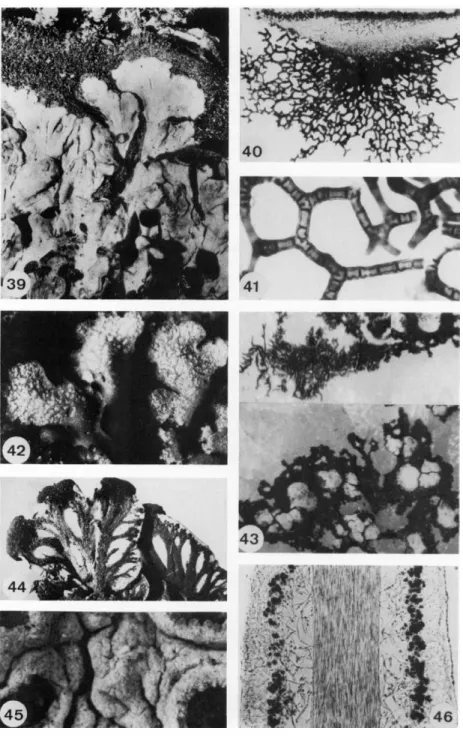
FIGS. 39-46. Fig. 39, thallus of Parmeliella plumbea with dark, hairy prothallus (2 x ) ; Figs. 4 0 - 4 1 , netlike tomentum on the underside of Anzia ornata (65 χ and 330 x ) ; Fig. 42, thallus of Physconia grisea covered with white pruina (9 x ) ; Fig. 43, black prothallus of
1. ANATOMY, MORPHOLOGY, AND DEVELOPMENT 17 become necrotic and give the thallus a scurfy appearance. These tiny granules are called pruina. They may also be an accumulation of carbonates
and oxalates. For example, the margins of foliose thalli of Physconia and the disks of the apothecia in particular are covered with a whitish dust (Figs.
42 and 45).
Some cortical cells may continue their growth and develop into thin, hyaline hairs (Fig. 72), each consisting of one or more cells. The hairs are long and pointed or branched and connected by anastomoses (Fig. 37). In some species the cells of the hairs are globose and resemble a string of pearls (Fig. 32). Hairs may form a felted, hirsute, or cottony mat called a tomentum (Figs. 83, 85, and 123). Many foliose species, especially those without a cortex or with only a poorly developed one, are characterized by a tomentum on the lower surface of the thallus. Peltigera and Lobariazxz well-known examples of this type. The tomentum can become a thick, spongy layer of netlike branched hairs as in Anzia (Figs. 40 and 41). The blackened hypothallus of Parmeliella and Pannaria also resembles a tomen
tum (Fig. 39). The thalli of some crustose lichens bear bristly hairs.
The form of the marginal cells of the cortex, and as a result the habit of the thallus, may vary in closely related species. In Peltigera horizontalis all marginal cells of the cortex end in an unbroken layer, covered by a gelatinous cuticle (Fig. 34). The thallus appears smooth and glossy (Fig. 121). The cortex of Peltigera scabrosa is characterized by little granules (Fig. 122), each consisting of several cells equivalent to abbreviated bundles of hyphae which have grown above the general surface of the thallus (Fig. 35). In Peltigera canina the cells of the cortex end in interwoven, unorientated hyaline hairs (Fig. 37) giving the thallus its felted appearance (Fig. 123).
The upper surface and the lower surface of foliose lichens can be covered by netlike veins (Fig. 44). For example, the underside of Peltigera always has veins of a spongy and felted appearance. In this genus the veins are formed by the medullary hyphae multiplying at certain places. In Hydro- thyria, an aquatic lichen belonging to the same family as Peltigera, the veins consist of single medullary hyphae with strongly enlarged cell lumina.
The layers of the thallus—upper cortex, algal layer, medulla, and lower cortex—are more or less present in all heteromerous lichens. Those lichens with a radially organized thallus are no exception. Here the only difference is
Rhizocarpon geographicum growing on white quartz stone; at some places squamules of the thallus have developed (15 χ ); Fig 44, lower surface of the thallus of Peltigera venosa showing black veins (3 x ) ; Fig. 45, thallus of Diploschistes covered with whitish pruina and bearing apothecia (18 x ) ; Fig. 46, longitudinal section through the thallus of Usnea ceratina showing the central cord (140 x ) . (Figs. 3 9 - 4 2 and 4 4 - 4 6 from Henssen and Jahns, 1973).
18 Η. Μ. JAHNS
that the medulla lies in the center of the thallus and is enclosed by a cylindrical algal layer and cortex. The medulla may have a hollow center or an axial strand.
Foliose lichen thalli resemble the leaves of higher plants. The cortex of the lichen corresponds to the epidermis and the algal layer to the palisade layer. The cyphellae and pseudocyphellae of lichens have the same function as the stomata of the leaf, while their anatomical structure resembles the lenticels of higher plants. There are significant differences in thallus thick
ness between lichen specimens growing in the shade and others exposed to the sun (Scott, 1971). For example, thalli of Xanthoria parietina growing on shaded tree trunks are thinner than thalli growing on exposed rocks, a feature corresponding to the sun and shade leaves of angiosperms. Not only the thickness of a lichen thallus as a whole, but also the relative thickness of the different layers, varies under different circumstances. The cortex and medulla of lichens growing in the shade are thinner while the algal layer is thicker than in specimens exposed to the sun.
D. Attachment of the Thallus to the Substrate
The thallus of homoiomerous lichens is fastened to the substrate by the basal hyphae. The same simple way of attachment also is found in some heteromerous lichens. The rhizoidal hyphae, which anchor the thallus by clasping little particles of the substrate, are like the tomental hyphae.
Lichens growing on soil incorporate grains of sand between the hyphae of the lower part of the thallus.
Rhizines have the same function as rhizoidal hyphae. They are composed of bundles of more or less parallel aligned hyphae and develop in three dif
ferent ways. In some lichens, i.e., in the Parmeliaceae, the hyphae of the rhizines are cemented together as soon as they start to develop. They form a direct elongation of the cortex tissue. The walls of the hyphae are glued together by gelatinization. When the tip of the rhizines reaches the substrate the growing hyphae spread and form a disklike holdfast attaching the lichen to the substrate (Fig. 51). In this disk the hyphae and particles of the substrate are glued together. In other groups of lichens, for example, in the Pelti- geraceae and Stictaceae, young rhizines consist of loosely associated hyphae, which later become closely connected by anastomoses. The mature rhizines of this type may spread at their tip and become brushlike (Fig. 53). In Leptogium the rhizines are formed by a tuft of individual hyphae not con
nected with one another.
To what extent rhizines can transport dissolved mineral or organic meta
bolites from the substrate to the thallus has not yet been established. Prob
ably there is some correlation between the type of rhizine and its ability to transport water which varies considerably. For example, the compact
FIGS. 4 7 - 5 3 . Fig.41, underside of Umbilicariapustulata with central holdfast (1.5x );Fig.48, bulbate cilia at the margin of apothecia of Parmelia abstrusa (25 χ ); Fig. 49, squarrose rhizines of Parmelia ecuadoriensis (25 χ ); Fig. 50, thallus of Heterodermia leucomela with long dark cilia (8 χ ); Fig. 51, rhizine of Parmelia sulcata showing point of attachment to the substrate (50 χ );
Fig. 52, dichotomous rhizines of Parmelia revoluta (25 χ ); Fig. 53, rhizines of Peltigera aphthosa (12 χ ) . (Figs. 47, 4 9 - 5 1 , and 53 from Henssen and Jahns, 1973.)
20 Η. Μ. JAHNS
rhizines of Parmelia are not quickly wetted by water while the treelike rhizines of Peltigera function like a wick.
Rhizines always grow from the underside of the thallus. Vegetative structures emerging from the margin of the thallus and closely resembling the rhizines are called cilia. The habit of rhizines and cilia varies indifferent genera. The simplest type is an unbranched strand of hyphae (Figs. 50 and 51). Branched rhizines and cilia are of a squarrose or dichotomous type (Figs. 49 and 52). Short cilia can have a bulbate inflated base (Fig. 48).
Unbranched cilia, for example in Usnea, are sometimes called fibrillae (Fig. 76) and short pin-shaped protuberances are named papillae. As it is unknown whether there are any fundamental differences between these vegetative structures, it is really impossible to give a meaningful definition of the different names.
Some lichens, especially those growing on rocks or tree bark, are attached by a disklike holdfast (Figs. 47 and 74).
III. Morphology of the Thallus
A. Color of the Thallus
Most lichens are gray or brown when dry. In wet thalli the color of the algae can be seen more distinctly through the cortex and these lichens become more or less green. Many species are brightly colored by incrusta
tions of special lichen pigments in the cortex. For example, the orange or yellow substance called parietin is found in the Teloschistaceae. Green and yellow tints are common in lichens while red, blue, and violet colors are rare. Many of these pigments are not confined to a single genus but often occur widely throughout the lichens. They are absent in most gelatinous lichens and rare in pyrenocarpic species.
B. Growth Forms of the Thallus
The segregation of lichens into the large groups of crustose, foliose, and fruticose lichens has already been mentioned. However, the hairlike or filamentous lichens with their short, thin branches and the gelatinous lichens form two extra groups which are not satisfactorily incorporated into one of the three main types. The gelatinous lichens are cartilaginous when dry, but immediately swell and become gelatinized in wet conditions. To a certain extent all lichens, which are hard and brittle when dry, may swell when wet and become soft and flexible. When the traditional classification into growth forms is applied to all the different species of lichen, intermediate forms are usually arbitrarily placed in that group to which the majority of closely related species belong.
1. ANATOMY, MORPHOLOGY, AND DEVELOPMENT 21
1. CRUSTOSE LICHENS
Crustose lichens never possess a lower cortex. They are attached to soil, rock, or tree bark by the hyphae of the medulla and the contact is so intimate that they are practically inseparable from the substrate. A patch of crusta- ceous lichen may belong to one species and yet be composed of many individuals which have fused together. Simple crustose lichens are homoio- merous. They lack a cortex and are therefore granular in structure. The mycelium spreads over the substrate in a thin filamentous mat enclosing the algae.
The thallus of most crustaceous lichens consists of little scales called areoles (Fig. 54). The lower hyphae of the areoles usually grow faster than the main part of the thallus and form a thin spreading layer around it. This mat of hyphae is usually dark in color and is called a prothallus (Fig. 43). The name hypothallus, also previously used for this structure, is more correctly applied to the thin filaments which link the areoles of the inner part of the thallus. A cracked surface in lichens may develop in one of two ways. In many species the thallus is initiated evenly and becomes cracked, often incompletely, at a later stage. In other groups of lichens, small defined areas of the thallus develop on an advancing prothallus appearing as separate entities and gradually becoming more closely compacted towards older parts of the thallus (Fig. 43).
Lichens with very small areoles are frequently homoiomerous. Bigger areoles begin to show the first signs of a differentiation into layers. The algae are accumulated in the upper part of the thallus, and at the surface a kind of cortex is formed by necrotic, gelatinized cells. This layer of dead cells is continuously sloughed off, but is always reformed by the growth of the thallus. An example of this type is Acarospora. Other crustose lichens, especially the intermediate forms between the crustose and the foliose type, have a true heteromerous thallus. The cortex may cover only the upper sur- face of the thallus or it also may include the margin of the individual areoles.
An extreme example of the crustose type are lichen thalli which grow completely inside their substrate, whether it be wood or stone. Species grow- ing inside rock are called endolithic, and those penetrating wood are termed endophloeodic. Sometimes the thallus of these lichens can be seen as a discoloring of the substrate (Fig. 55), but frequently only the ascocarps in pits or on the surface of rock or bark indicate the presence of a lichen.
The hyphae of endolithic lichens appear to excrete lichen substances which are able to dissolve the stone and thus make it possible for hyphae and algae to penetrate several millimeters into the rock. Species growing inside lime- stone develop special oil cells that are intercalated along the hyphae. The irregularly swollen cells appear clustered with oil drops.
FIGS. 5 4 - 6 0 . Fig. 5 4 , areolate thallus of Lecanorafrustulosa ( 2 χ ) ; Fig. 5 5 , endolithic thallus of Lecidea; the black apothecia emerge from the stone ( 0 . 6 χ ) ; Fig. 5 6 , thallus of Acarospora oxytona with effigurated margin ( 3 x ) ; Figs. 5 7 - 5 8 , placoid thallus of Xanthopeltis rupicola seen from above (with apothecia) and from below (with umbilicus) ( 3 χ and 3 x ) ; Fig. 5 9 ,
1. ANATOMY, MORPHOLOGY, AND DEVELOPMENT 23 2. INTERMEDIATE FORMS BETWEEN CRUSTOSE AND FOLIOSE LICHENS
In some crustose lichens elongated, small lobes replace the areoles.
These lobes can be fastened to the substrate by the entire lower surface or the margin of the thallus can be free and ascending. Different combinations of these characteristics lead to a variety of described growth types.
If the margin of the thallus consists of small, elongated lobes while the inner part is composed of small areoles, the lichen is said to have an effigura- ted margin (Fig. 56). If the whole thallus is formed by elongated lobes, the lichen belongs to the placoid type. Thalli of the effigurate and placoid type are closely appressed to the substrate by their whole lower surface.
The squamulose thallus of some species of Heppia, Lecanora, Lecidea, and Placynthium consists of little scales. At one side their margin separates from the substrate and bends upwards (Fig. 59). Several scales can be arranged in a rosette. The squamules may overlap like the tiles of a roof and are then imbricate.
Further development of the squamulose thallus is seen in peltate lichens.
In this type only the central part of the scales is fastened to the substrate, the whole margin becoming free. Lichens of this type have more or less the same habit as the umbilicate foliose genera (Figs. 57, 58, and 60). The scales and lobes of some squamulose and peltate thalli are bent upwards and begin to resemble fruticose growth forms. For example, some species of Peltula with upright lobes and with a corresponding radial anatomy could be classified as having a fruticose growth habit (Fig. 65).
3. FOLIOSE LICHENS
The thallus of foliose lichens is formed by flattened lobes, which are heteromerous and dorsoventral in structure. Two principal types, the laci- niate and the umbilicate growth form, can be distinguished. Laciniate thalli adhere more or less firmly to the support on which they grow. Either the whole lower surface is in contact with the substrate or the margin of the lobes becomes free and bends upwards. The thalli are usually attached by rhizines or rhizoidal hyphae. The umbilicate lichens are platelike and attached by a central discoid holdfast called the umbilicus (Fig. 47).
a. LACINIATE FOLIOSE LICHENS. The laciniate lichens form an extremely polymorphous group. Their habit and the mode of attachment of the thallus vary and their anatomy is the most complex of all lichens. Some are
squamulose thallus of Lecidea scalaris, the margin of the squamules becoming sorediate (8 x ) ; Fig. 60, umbilicate thallus of Glypholecia scabra{\ x). (Figs. 54and 57-60from Henssen and Jahns, 1973.)
FIGS. 6 1 - 6 3 . Lobaria pulmonaria; note on the right side an inverted part of the thallus (I χ ); Fig. 62, foliose, laciniate thallus of Parmelia quercina (2.5 χ ). Fig. 63, foliose laciniate thallus of Peltigera canina (\ χ ). (Figs. 61 and 62 from Henssen and Jahns, 1973.)
1. ANATOMY, MORPHOLOGY, AND DEVELOPMENT 25 very large plants. Lobes of Lobaria pulmonaria (Fig. 61) may reach a length of 30 cm and multilobed thalli of Parmelia (Fig. 62) reach \ m in diameter (Fig. 64). The thalli of Lobaria are covered on both sides by a cortex, while in Peltigera (Fig. 63) the cortex is restricted to the upper side, the underside being tomentose with veins. In Parmelia the whole lower surface of the thallus or only part of it is attached to the substrate by rhizines. Rhizines, cilia, veins on the thallus surface, and other vegetative structures are com- mon in foliose lichens. The cortex layers are derived from different types of tissue.
b. UMBILICATE FOLIOSE LICHENS. Umbilicate lichens have a disklike thallus that is attached to the substrate with a central holdfast. The holdfast causes a small depression in the surface of the thallus. All species of Umbili- caria (Fig. 67) have this type of thallus, as the name of the genus indicates.
An umbilicate thallus occurs in other lichens which are not closely related to Umbilicaria, as, for example, in the pyrenocarpous genus Dermatocarpon (Fig. 68) and in the gymnocarpous genera Glypholecia, Omphalodium, and Xanthopeltis (Figs. 57,58, and 60). The thallus of Umbilicaria is heteromerous and fully corticated. Many species also are characterized by a veined or rugose thalline surface.
4. INTERMEDIATE FORMS BETWEEN FOLIOSE AND FRUTICOSE LICHENS
The thalli of foliose lichens of the laciniate type, for example, those of Cetraria (Fig. 73), are sometimes nearly erect so that they are often con- sidered to be fruticose. The lobes are fastened by the lower surface and in older thalli the base begins to rot. This further proves the arbitrariness of classification based on thallus types.
5. FRUTICOSE LICHENS
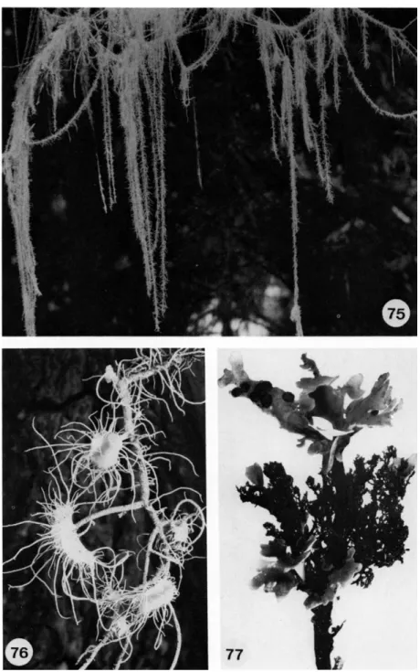
The lobes of fruticose lichens are strap-shaped or threadlike with a radial or dorsiventral thallus. Ramalina (Fig. 70) and Roccella (Fig. 79) are good examples of strap-shaped, radial thalli, while Usnea (Fig. 75) consists of thin strands up to 5 m long. Evernia and Pseudevernia have strap-shaped, dorsi- ventral thalli.
Many strap-shaped and radiate thalli are attached to the substrate by a holdfast (Fig. 74). Some long, pendulous, threadlike strands of certain species of Usnea hang from the branches of trees, without any organized attachment to the bark. Of these, Usnea longissima may reach a length of several meters. Other fruticose lichens that grow on soil form little cushions which consist of separated upright lobes. Frequently, they are not attached
FIGS. 6 4 - 6 8 . Fig. 64, zoned thalli of Parmelia centrifuga: new thalli are continuously formed in the center of the outer growing zones χ ) ; Fig. 65, fruticose thallus of Peltula (4 χ ); Fig. 66, Gelatinous thallus of Collema subfurvum (1 χ ). Fig. 67, umbilicate thallus of Vmbilicaria rigida (1 χ ); Fig. 68, umbilicate thallus of Dermatocarpon miniatum; theostioli of the perithecia can be seen as blackpoints (3 χ ). (Figs. 64, 67 and 68 from Henssen and Jahns, 1973; Fig. 65 from A. Henssen.)
FIGS. 6 9 - 7 4 . Fig. 69, fruticose thallus of Cladonia impexa (1 χ ); Fig. 70, fruticose thallus of Ramalina fraxinea (1 χ ); Fig. 71, fruiting bodies of Baeomyces placophyllus (9 χ ) ; Fig. 72, hairy thallus of Teloschistes flavicans (10 x ) ; Fig. 73, thallus of Cetraria cucullata bearing apothecia (1 χ ); Fig. 74, thallus showing holdfast of Ramalina curnowii (1 χ ); (Figs. 71 and 74 from Henssen and Jahns, 1973.)
FIGS. 75-77. Fig. 75, thallus of Usnea longissima χ ); Fig. 76, thallus of Usnea ceratina;
note fibrillae on the apothecial margins (2.5 χ ); Fig. 77, Sticta filix; the dark fruticose thallus with Nostoc as phycobiont bears the foliose thallus which has a green phycobiont (2 χ ). (Figs.
75-77 from Henssen and Jahns, 1973.)
1. ANATOMY, MORPHOLOGY, AND DEVELOPMENT 29 to the soil. Some species degenerate at the base and become completely free.
They may be dislodged by the wind and blown over the ground. Good exam
ples of this type are species of Cladonia, sect. Cladina, and Cornicularia.
The stiffness of fruticose lobes is achieved by two different types of basic construction. In some lichens the hyphae of the cortex serve as supporting tissue. They form a cylindrical tube at the lateral edge of the thallus, while the center of the lichen is hollow or filled with a cottony medulla. This type of construction serves to keep the plant upright and to withstand lateral pressure. The supporting tissue is a prosoplectenchyma or pseudoparen- chyma with the hyphae being closely cemented. In the second type of fruticose lichen the supporting tissue is situated in the center of the medulla.
A central cord or axial strand is constructed from thick-walled, perpendi
cular, agglutinated hyphae. Usnea has a single threadlike elastic cord, while other lichens develop several individual strands which later fuse. This central axial strand gives the requisite tensile and skeletal strength to pendulous lichens.
6. LICHEN THALLI WITH A TWOFOLD CHARACTER
In some lichens the thallus consists of a horizontal part lying on the sub
strate and of a vertical, fruticose part, bearing the fruiting bodies. The horizontal thallus can be crustose, as in some species of Βaeomyces (Fig. 71) or foliose, as in Cladonia (Fig. 78). The horizontal thallus of a lichen maybe evanescent, being only found in a very young specimen, disappearing as it matures. The adult lichen consists only of a vertical fruticose thallus (Fig.
69).
The twofold thallus has been independently developed in several lichen families and can be of different origin. In Cladonia the thallus verticalis is formed from the generative tissue, a tissue which surrounds the sexual organs and usually gives rise to the ascocarp. Thus, the thallus verticalis of this genus is ontogenetically a part of the fruiting body. This kind of fruticose stalk is called a podetium. The podetia may be simple or richly branched with pointed apices or apical cups.
The development of the thallus verticalis in Stereocaulon (Fig. 80) and Pilophorus (Fig. 81) is different. A part of a squamule of the thallus horizon- talis or a complete granule of the thallus grows vertically upwards and develops into a simple or branched more or less erect thallus verticalis. The primordium of the fruiting body is formed only at the top of this stalk. The generative tissue builds only the ascocarp while the thallus verticalis is dif
ferentiated from vegetative thallus tissue. This kind of stipe is called a pseudopodetium.
30 Η. Μ. JAHNS
1. ANATOMY, MORPHOLOGY, AND DEVELOPMENT 31
7. HAIRLIKE THALLI
The habit of hairlike lichens, with their threadlike thalli, resembles that of fruticose lichens. These lichens, however, are much smaller and usually not more than a few millimeters high. In contrast to most lichens, the habit is principally determined by the phycobiont. Filamentous algae, belonging to the Chlorophyceae or the Cyanophyceae, are more or less closely ensheathed by hyphae of the mycobionts (Figs. 23-25).
8. GELATINOUS LICHENS
The consistency and growth form of gelatinous lichens are for the most part determined by the blue-green phycobiont. The characteristic swelling of the wet thallus is due to the gelatinous sheath of the phycobiont. Fre
quently, the structure of the thallus is homoiomerous but the anatomy is different from the homoiomerous crustaceous lichens. In crustose lichens the algae are scattered in a mycelium of loosely interwoven hyphae, while the hyphae of gelatinous lichens usually grow inside the gelatinous algal substance that fills the thallus. The hyphae often do not touch the algae.
The lateral margin of the thallus is also sometimes formed by the gelatinous sheath of the algal cells.
In some genera the fungus provides a cortex at the surface of the thallus.
A gradation of differentiation can be found among genera of the Collema- taceae. Most species of Collema are noncorticate, but in some species vertically oriented hyphae reach the surface of the thallus, bend at right angles, and spread parallel to the surface (Fig. 33). Leptogium has a cortex which is one cell layer thick. It consists of irregularly formed cells which are arranged in either a broken pattern or in a regular layer of isodiametric cells when viewed from above (Fig. 22). The cortex of other lichens belong
ing to the Collemataceae is several cell layers thick.
All types of growth forms are found in the gelatinous lichens. Most species are very small and only the foliose thalli reach a diameter greater than 10 cm (Fig. 66). The color of the thallus is olive-green, blackish, or gray. The bright colors of lichen substances or pigments are not present in this group.
Some species are red or violet when wet, but this color is due to the gela
tinous sheath of certain blue-green phycobionts such as Gloeocapsa.
FIGS. 7 8 - 8 1 . Fig. 78, cup-shaped podetia of Cladonia chlorophaea growing from a foliose primary thallus; the older podetia bear apothecia (3 x ) ; Fig. 79, thallus of Roccella fuci- formis with soralia (1 χ ); Fig. 80, pseudopodetia of Stereocaulon alpinum with phyllocladia
and apothecia (7 χ ) ; Fig. 81, Pilophorus strumaticus; upright pseudopodetium bears black apothecia; note black cephalodium on the thallus (25 χ ) ; (Figs. 7 8 - 8 0 from Henssen and Jahns, 1973.)
32 Η. Μ. JAHNS
C. Development of the Thallus
A review of lichen development has been presented by Steiner (1965), and only selected aspects of this subject will be described in this chapter.
New lichen thalli can only develop in a favorable environment. If the biotope is unsuitable for the lichen, the development stops at an early stage and only a more or less homoiomerous layer of sorediate appearance is formed. These half-developed thalli can frequently be observed in humid and shadowy places. But even under the most favorable conditions a lichen thallus can only evolve if both partners of the symbiosis are present at the same time. Therefore, those methods of reproduction which disperse both partners in close union are the most valuable to the lichen. It is to the advantage of the lichens that in many species the vegetative diaspores, such as isidia, soredia, and thallus fragments have taken the place of sexual spores. If the mycobiont is dispersed by means of ascospores, basidiospores, conidia, or gemmae the germinating mycelium has to find suitable free- living algae for a new lichenization.
The hyphae emerging from the germinating spores can be of different shapes. In Xanthoria parietina they consist either of short, rounded, or of long, thin cells (Werner, 1931). Those hyphae which make contact with the algae become hook-shaped and clasp the algal cells. The fungus is the active partner in the development of the primary stages of the thallus, although the hyphae do not grow towards the algae.
Probably only very few germinating spores succeed in forming a new lichen (Scott, 1971). This difficulty leads to some adaptations, by which the chance of success is increased. The most successful method is the form
ing of hymenial algae that occur, for example, in Endocarpon. These algae lie in the fruiting body between the asci and are ejected together with the spores. Both partners are together at the time of germination. The spores of some lichens after being ejected are dormant until contact with algae is made. In other genera the spores germinate directly but the mycelia seem to live as saprophytes for a short time. The spores of Verrucaria margacea germinate inside the perithecium and only the young mycelium is ejected (Tobler, 1925). Perhaps this mycelium, being larger than germinating spores, can make contact with algae more easily.
If lichenization is achieved, the young mycelium and the acquired algae at first form an undifferentiated lump. In Xanthoria parietina it takes nearly a month before any signs of different layers can be observed. The same undifferentiated mass of algae and hyphae can be observed if the develop
ment starts from a vegetative diaspore. The first hyphae growing from a vegetative propagule function as hapteres, attaching the diaspore to the
1. ANATOMY, MORPHOLOGY, AND DEVELOPMENT 33 substrate. The subsequent differentiation of thallus layers is as slow a process as in thalli developed from germinating ascospores. In Xanthoria parietina it takes two months before cortex, algal layer, and medulla are differentiated. After eight months the rosette-shaped thallus has reached 2-6 mm in diameter. Apothecia are formed after 1 or 2 years.
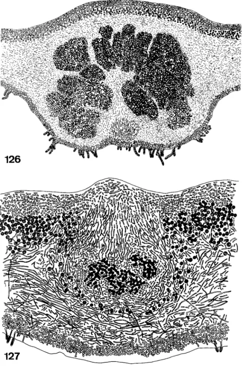
The mycobiont is responsible for the growth of the thallus. At the margin of the thallus the growth of the fungal hyphae may be so vigorous that an undifferentiated zone free of algae is formed around the lichen. This zone is called a prothallus and is hairy or netlike in Parmeliella, Placynthium, and other genera and cartilagenous in Pertusaria. The hyphae growing ahead of the thallus naturally need algae to build a complete lichen, but as the phycobionts of the lichens do not form motile stages inside the thallus they are not able to move to the new parts of the lichen by themselves. There seem to be two fundamentally different means by which growth and development are promoted. Either algae from the old part of the thallus are pushed passively towards the prothallus by the fungal hyphae or free-living algae are incorporated into the prothallus. The first of the two methods can be observed in Cladonia. The primordium of the podetium growing vertically from the primary thallus carries with it algal cells from the algal layer. In the thallus of Pertusaria the algae are pushed into the new parts of the thallus by special bundles of hyphae showing a fastigiate arrangement (Schiebehyphen) (Nienburg, 1926). The same process of differentiation can be found during the development of internal cephalodia in Lobaria (Fig.
127).
The incorporation of free-living algae during the growth of the lichen is also well known. In principle it resembles a new lichenization from spores and algae. The trapping of algae by the mycobiont has been observed as the first stage of development of cephalodia and of certain isidia. It has been claimed that the algal layer of the podetia of Cladonia also develops from free-living algae coming into contact with the stalk (Weise, 1937). This seems questionable as Trebouxia, the phycobiont of Cladonia, is probably not sufficiently abundant in the free-living state. Perhaps the conditions of the experiments leading to the observation differed too much from the natural environment.
In Placynthium nigrum the trapping of new algae and the transport of algae from old parts of the thallus to younger areas has been combined in an interesting manner (Geitler, 1933-1938). The squamules of the thallus, which contains a symbiotic blue-green Rivulariaceae, are surrounded by a hairlike prothallus. Hormogonia of the alga are freed from the margin of the scales by water and come to rest on the prothallus. Here they are enmeshed by the hyphae. Where they are incorporated into the tissue new squamules
34 Η. Μ. JAHNS
are formed on the prothallus. These continue to grow and finally reach the older squamules, forming a continuous system of scales.
Although lichens never stop growing, the typical morphological habit of the species is preserved as the growth is limited in some directions. For example, the vertical growth of foliose lichens is so slow that it only replaces the uppermost cells of the cortex which are continuously sloughed off.
Fruticose lichens, on the other hand, show an unlimited vertical growth.
They develop either from foliose stadia growing upwards and secondarily becoming fruticose and erect or the primordia of the thallus directly becom
ing fruticose. A good example of the first method of development is Lichina (Henssen, 1969). Most frutescent species of this genus are foliose when young. The other type of development is represented by Stereocaulon sub
genus Holostelidium (Lamb, 1951). The pseudopodetia of these lichens develop from granular primordia that directly grow into vertical, branched stalks. In Cladonia subgenus Cladina the annual growth can be extremely regular. Every year a new nodium with lateral branches is formed at the top of the treelike thallus.
The mechanisms regulating and controlling the growth are mostly un
known. Humidity, light, and gravity seem to influence the vertical orienta
tion of frutescent lichens. The growth of hyphal tissues sometimes seems to be stimulated by the presence of algae (Tobler, 1928).
In lichens growth is usually restricted to the tip of the thallus. The zone of growth does not exceed a few millimeters a year and intercalary growth is negligible. In fruticose lichens the proportion of newly developed to dead parts of the thallus becomes increasingly unfavorable as only the upper parts of the lichen continue to grow, while the lower, unproductive parts accumulate. In crustose lichens the annual increase of thallus surface mea
sured as the percentage of the actual size of the thallus is smaller in old thalli than it is in young ones (Steiner, 1965). The inner parts of the thallus have no means of transporting photosynthetic products to the growing outer squamules, so that only the photosynthetic products of the marginal parts of the thallus can be used for the growth process.
In many foliose lichens the old parts in the middle of the thallus die and disappear while the young marginal parts of the lichen continue to grow outward. The result is a ring-shaped thallus as found in Parmelia centrifuga (Fig. 64). The rings may reach a diameter of 1 meter. As the lichen grows about 2 mm each year such a thallus began its development some 500 years ago, but as the living, ring-shaped part of the thallus measures only about 6-10 cm in diameter the oldest living part of the lichen is 30-50 years old.
Crustose lichens may become much older. Thalli of Rhizocarpon are said to reach an age of 1000 years or more.
1. ANATOMY, MORPHOLOGY, AND DEVELOPMENT 35
82
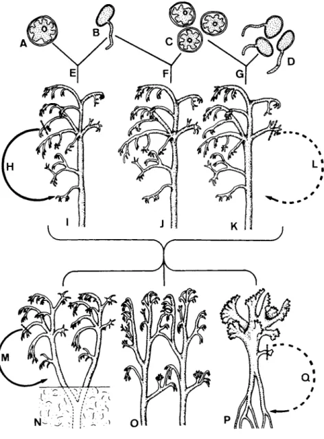
FIG. 82. Diagrammatic representation of possible origins of lichen thalli, i.e., (I) from one spore and one algal cell (J) from one spore and several algal cells (K) from several spores and several algal cells. The question of what constitutes individuality of these thalli is dis- cussed in the text. (From Henssen and Jahns, 1973.)
36 Η. Μ. JAHNS
D. Individuality of the Thallus
The growth forms of lichens and some special characteristics of their ontogeny make it difficult to define the term "individual" in this group of plants. The schematic drawing in Fig. 82 gives a detailed review of some of the problems using the fruticose lichens as an example. In the most simple case, one germinating spore (B) contacts (E) one alga (A). From these two bionts the whole thallus descends (1). The thallus does not fuse with other thalli. This plant certainly is an individual, which either forms spores or reproduces by vegetative diaspores (H). In most cases the germinating ascospore (B) will achieve lichenization (F) by including more than one alga (C) into the mycelium. The developing lichen (J), therefore, contains algae of different genetic origin; but as the my cobiont dominates these lichen thalli the individuality of the plant is not questioned. In many fruticose lichens and frequently in crustose lichens the thallus not only is formed by several algae (C) but mycelium from more than one germinating fungal spore (D) is incorporated (G). The tissue of this lichen (K), therefore, is not genetically uniform. Theoretically, it is possible that a vegetative diaspore of this plant does not contain all of the genetically different hyphae which have descended from the different spores. These diaspores would not contain all the genetic information which determined the habit of the mother plant and vegetative reproduction may lead to a certain variability (L).
The thalli of frutescent lichens grow at the top and die away at the base.
As a result branched thalli may split and become separate plants (N). These are genetically identical if the mother plant was uniform. The individuality of the two daughter plants, which can reproduce by vegetative diaspores (M), is evident. The process of division by growth is known from other plants, for example, from the moss Sphagnum where it occurs regularly.
Lichen thalli tend to fuse, especially in the dense cushions of certain fruticose lichens such as Cladonia. In most lichen species only the branches of different plants grow together at points of contact (O). This type of con
nection should be clearly distinguished from the complete fusion achieved by other lichens (P). The second type of contact is typical for Cladia (Jahns,
1972). In this genus nearly all fructificating branches arise from the complete fusion of small branches belonging to different plants. In these lichens it is quite probable that different parts of the big branches contain genetically different hyphae. Therefore, in Cladia the reproduction by vegetative dia
spores in connection with the fusion of branches leads to a recombination of morphological characteristics (Q). The lichens of the genus Cladia cannot be called individuals in the strict sense of the definition.
1. ANATOMY, MORPHOLOGY, AND DEVELOPMENT 3 7 IV. Vegetative Structures and Their Development
Lichens have unique vegetative structures that are found frequently in foliose and fruticose lichens and sometimes in crustose forms. Some of these structures serve as vegetative diaspores for the dispersal of the lichen and may have special physiological functions. All vegetative structures are of vital importance in taxonomic studies of lichen speciation.
A. Aeration Pores
The cortex of the larger heteromerous foliose lichens with its closely agglutinated hyphae seems to be a serious obstacle to the exchange of gases.
Recently, investigations with the scanning electron microscope have shown that some lichens have small pores between the cortical hyphae (Peveling,
1970). Some genera of lichens are characterized by special openings in the cortex which are believed to assist in the aeration of the thallus. They are macroscopically visible. It has never been experimentally demonstrated, however, that these openings really have a function in promoting the exchange of gases.
1. CYPHELLAE
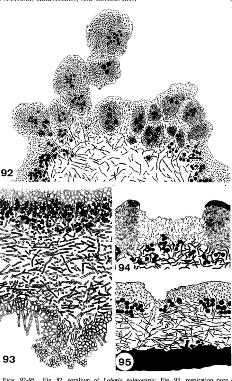
Cyphellae are rounded pores that are characteristic of the foliose genus Sticta (Fig. 83). They develop as little rounded pits in the lower surface of the thallus. The lower cortex extends as an encircling protruding rim, enclosing a small craterlike depression. At the bottom of the crater the texture of the tissue is looser, the hyphae becoming separated to form a layer of globose cells (Fig. 90). This structure is similar to that of the lenticels of higher plants. The cyphellae may enlarge and become much bigger and irregular in outline. Old cyphellae lose their function as the rounded cells adhere and are embedded in the gelatinized remnants of the dying outer cells (Fig. 91).
Only the aerating organs of Sticta are called cyphellae in the strict sense, although the pores of other genera are not fundamentally different in struc- ture. The openings, called tubercles (Fig. 85), in the lower side of Nephroma are also raised above the thallus but exhibit no central depression as those in Sticta. They too are filled with short separating cells (Fig. 93). Protuber- ances on the upper side of Parmelia aspidota are synonymous with cyphellae or pseudocyphellae. They have an elevated central opening where the cortex is reduced and loose hyphae of the medulla penetrate through the
algal layer to reach the surface (Fig. 86). The difference between cyphellae
3 8 Η. Μ. JAHNS
1. ANATOMY, MORPHOLOGY, AND DEVELOPMENT 39 and pseudocyphellae is not great and the distinction is arbitrary. The variety of differentiation is far greater than the two names indicate.
2. PSEUDOCYPHELLAE
Pseudocyphellae are formed on the upper and lower surface of several foliose and fruticose lichens. In Pseudocyphellaria they form irregular warts. The openings are filled by a network of short cells (Fig. 89). In very old and abnormally large pseudocyphellae algal cells may be deposited between the loose hyphae. These pseudocyphellae resemble soralia but the similarity is misleading as no soredia are produced.
The pseudocyphellae of the fruticose lichen Cornicularia divergens (Fig.
84) are either shallow depressions in the cortex or they form pores that penetrate to the medulla (Fig. 94). The pseudocyphellae on the upper side of Cetrelia cetrarioides show a similar development (Fig. 95).
In some lichens, i.e., Parmelia exasperatula and Placopsis cribellans, the remnants of isidia serve as aerating organs. When the isidia break off, they leave on the thallus little warts which have a central opening filled by loosely interwoven hyphae.
The size of pseudocyphellae is quite constant in most species and has considerable taxonomic significance (Hale, 1967). This fact recently has helped to separate populations of Cetraria chicitae and Parmelia olivetorum, two vegetatively extremely similar foliose lichens in eastern North America.
At first thought to be indistinguishable when sterile except by chemical tests (olivetoric and alectoronic acids, respectively), these two species were found to differ significantly in pore size, the average maximum size in P.
olivetorum being 0.48 mm and in C. chicitae 1.20 mm.
B. Vegetative Diaspores
Many lichens develop vegetative organs of dispersal, called isidia, soralia, and hormocystangia. It is open to question whether isidia always act as vegetative propagules, but it is interesting that lichens producing isidia or soredia do not freely produce ascocarps. In some lichens isidia probably only increase the surface area. In the soralia, vegetative diaspores—the soredia—are produced. Lichenized hormocysts are the diaspores formed in hormocystangia of some lichens with blue-green phycobionts. Soredia
FIGS. 83-88. Fig. 83, cyphellae on the tomentous underside of Sticta (20 χ ) ; Fig. 84, pseudocyphellae of Cornicularia divergens (16 x ) ; Fig. 85, tubercles on the underside of the tomentous thallus of Nephroma (8 χ ); Fig. 86, areation pore of Parmelia aspidota (200 χ ) ; Fig. 87, young isidia of Collema flaccidum in section (350 χ ) ; Fig. 88, hormocystangium of Lempholemma versiculiferum (60 χ ) . (Figs. 83, 84, and 8 6 - 8 8 from Henssen and Jahns, 1973.)
40 Η. Μ. JAHNS
FIGS. 8 9 - 9 1 . Fig. 89, pseudocyphellae of Pseudocyphellaria; Figs. 9 0 - 9 1 , young and old cyphellae of Sticta sylvatica. (Figs. 89-91 from Henssen and Jahns, 1973.)
1. ANATOMY, MORPHOLOGY, AND DEVELOPMENT 4 1
FIGS. 92-95. Fig. 92, soralium of Lobaria pulmonaria; Fig. 93, respiration pore of Nephroma resupinatum; Fig. 94, pseudocyphellae of Cornicularia divergens; Fig. 95, pseudo- cyphellae of Cetrelia cetrarioides (Figs. 9 2 - 9 4 from Henssen and Jahns, 1973.)