Received 17 Jan 2014|Accepted 12 Jan 2015|Published 3 Mar 2015
Disruption of STAT3 signalling promotes KRAS-induced lung tumorigenesis
Beatrice Grabner1, Daniel Schramek2,w, Kristina M. Mueller1,3, Herwig P. Moll4, Jasmin Svinka5, Thomas Hoffmann6, Eva Bauer1, Leander Blaas7, Natascha Hruschka1, Katalin Zboray1, Patricia Stiedl1, Harini Nivarthi1,w, Edith Bogner1, Wolfgang Gruber8, Thomas Mohr5, Ralf Harun Zwick9, Lukas Kenner1,10,11, Valeria Poli12, Fritz Aberger8, Dagmar Stoiber1,4, Gerda Egger10, Harald Esterbauer13, Johannes Zuber6, Richard Moriggl1,3, Robert Eferl5, Bala´zs Gy+orffy14,15,16, Josef M. Penninger2, Helmut Popper17
& Emilio Casanova1,18
STAT3 is considered to play an oncogenic role in several malignancies including lung cancer;
consequently, targeting STAT3 is currently proposed as therapeutic intervention. Here we demonstrate that STAT3 plays an unexpected tumour-suppressive role inKRASmutant lung adenocarcinoma (AC). Indeed, lung tissue-specific inactivation of Stat3 in mice results in increased KrasG12D-driven AC initiation and malignant progression leading to markedly reduced survival. Knockdown of STAT3 in xenografted human AC cells increases tumour growth. Clinically, low STAT3 expression levels correlate with poor survival and advanced malignancy in human lung AC patients with smoking history, which are prone to KRAS mutations. Consistently, KRAS mutant lung tumours exhibit reduced STAT3 levels.
Mechanistically, we demonstrate that STAT3 controls NF-kB-induced IL-8 expression by sequestering NF-kB within the cytoplasm, thereby inhibiting IL-8-mediated myeloid tumour infiltration and tumour vascularization and hence tumour progression. These results elucidate a novel STAT3–NF-kB–IL-8 axis in KRAS mutant AC with therapeutic and prognostic relevance.
DOI: 10.1038/ncomms7285 OPEN
1Ludwig Boltzmann Institute for Cancer Research (LBI-CR), Vienna 1090, Austria.2Institute of Molecular Biotechnology of the Austrian Academy of Sciences (IMBA), Vienna 1030, Austria.3Institute of Animal Breeding and Genetics, University of Veterinary Medicine, Vienna 1210 and Medical University Vienna, Vienna 1090, Austria.4Institute of Pharmacology, Medical University of Vienna, Vienna 1090, Austria.5Institute of Cancer Research & Department of Internal Medicine I, Comprehensive Cancer Center, Medical University of Vienna, Vienna 1090, Austria.6Research Institute of Molecular Pathology (IMP), Dr. Bohr Gasse 7, Vienna 1030, Austria.7Department of Biosciences and Nutrition, Center for Innovative Medicine, Karolinska Institutet, Novum, Huddinge 141 83, Sweden.8Department of Molecular Biology, Paris-Lodron University of Salzburg, Salzburg 5020, Austria.9Department of Internal Medicine, Karl Landsteiner University, 3430 Tulln, Austria.10Clinical Institute of Pathology, Medical University of Vienna, Vienna 1090, Austria.11Unit of Pathology of Laboratory Animals (UPLA), University of Veterinary Medicine Vienna, 1210 Vienna, Austria.12Molecular Biotechnology Center (MBC), Department of Genetics, Biology and Biochemistry, University of Turin, Turin 10126, Italy.13Department of Laboratory Medicine, Medical University of Vienna, Vienna 1090, Austria.14MTA TTK Lendu¨let Cancer Biomarker Research Group, Budapest 1117, Hungary.152nd Department of Pediatrics, Semmelweis University, Budapest 1094, Hungary.16MTA-SE Pediatrics and Nephrology Research Group, Budapest 1083, Hungary.17Institute of Pathology, Research Unit Molecular Lung and Pleura Pathology, Medical University of Graz, Graz 8036, Austria.18Department of Physiology, Center of Physiology and Pharmacology, Comprehensive Cancer Center, Medical University of Vienna, Vienna 1090, Austria.wPresent addresses: Howard Hughes Medical Institute, Laboratory of Mammalian Cell Biology and Development, Rockefeller University, 10065 New York, USA (D.S.); CeMM Research Center for Molecular Medicine of the Austrian Academy of Sciences, 1090 Vienna, Austria (H.N.). Correspondence and requests for materials should be addressed to E.C. (email: emilio.casanova@lbicr.lbg.ac.at).
L
ung cancer is still the leading cause of cancer deaths worldwide1. The most frequent genetic alterations found in lung adenocarcinomas (ACs) are missense mutations and amplifications of kirsten rat sarcoma viral oncogene (KRAS)and epidermal growth factor receptor(EGFR)in about 30% and 20%of all Caucasian cases, respectively2,3. Although several targeted anti-EGFR therapies are effective in patients with EGFR mutations, oncogenic KRAS is still considered as an undruggable target. To elucidate further treatment strategies, the focus has therefore shifted towards KRAS-cooperating and downstream signalling pathways. Signal transducer and activator of transcription 3 (STAT3) represents one important transcription factor thought to cooperate withrat sarcoma viral oncogene (RAS) mutations during tumorigenesis4,5. STAT3 is activated in response to several cytokines and growth factors, such as interleukin-6 (IL-6), oncostatin-M (OSM) and epidermal growth factor (EGF). Upon binding of the ligand to its cognate receptor, STAT3 becomes tyrosine-phosphorylated and forms homodimers or heterodimers before translocating into the nucleus to induce the transcription of several target genes implicated in cell cycle regulation, apoptosis, angiogenesis, tumour invasion and metastasis6. Depending on the oncogenic driver mutation or on the cell type, STAT3 has been reported to play either pro-oncogenic4,5,7or tumour-suppressive rolesin vivo andin vitro8–12. Interestingly, STAT3 was shown to be activated in 54% of lung AC patient samples and human lung cancer cell lines13–15. Furthermore, based onin vitroand xenograft models, STAT3 is thought to play a tumour-promoting role in non-small- cell lung cancer (NSCLC) and during acquired drug resistance13,14,16–18.
Here, we investigated the role of STAT3 during oncogenic Kras-driven lung tumorigenesis using the Cre-inducible Lox- Stop-Lox-KrasG12D/þ knock-in lung cancer mouse model19and a human xenograft model. Surprisingly, our results show that STAT3 functions as tumour suppressor in KrasG12D/þ lung tumours as well asKRASmutant human AC cell lines. Deletion of STAT3 inKrasG12D/þ lung tumours and human AC resulted in an increased tumour growth, higher tumour grade, increased vascularization, changes in the tumour microenvironment and significantly reduced survival. Mechanistically, we show that genetic ablation of Stat3 in murine as well as knockdown of STAT3 in human cells resulted in an increase of nuclear factor- kappa B (NF-kB)-induced expression of the pro-angiogenic chemokine ligand 1 (CXCL1; murine orthologue to the human IL-8), thereby promoting tumour growth. Pharmacological inhibition of CXCL1’s cognate receptor CXCR2 normalizes tumour vascularization and microenvironment and reduces tumour burden. Thus, STAT3 functions as tumour suppressor by sequestering NF-kB in the cytoplasm and thereby reducing NF-kB-dependentCXCL1transcription.
Results
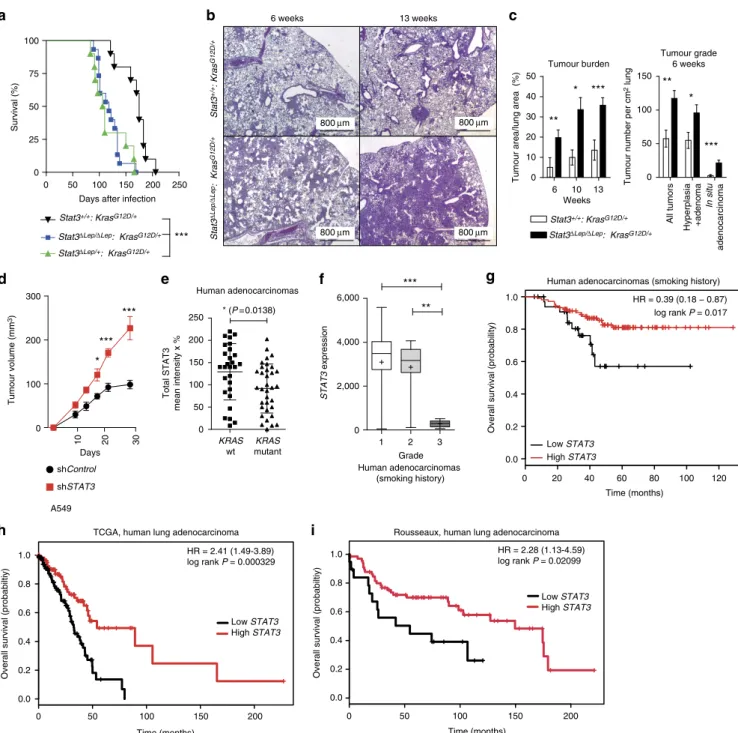
STAT3 suppressesKras-induced lung tumorigenesis. First, we investigated the activation status of STAT3 within murine KrasG12D-driven lung tumours. In all, 20–30% of tumour cells showed active STAT3 (tyrosine 705 phosphorylated STAT3) throughout tumour development. ELISA analysis revealed increased expression of STAT3-activating cytokines such as EGF, OSM and IL-6 in lungs harbouring KrasG12D-driven tumours compared with healthy control lungs (Supplementary Fig. 1a,b).
To functionally test the role of STAT3 duringKrasG12D-driven lung tumour formation, we crossed the KrasLSL-G12D/þ strain19 with conditionalStat3fl/flmice20. Lung epithelial-specific activation of oncogenic KrasG12D and concomitant deletion of Stat3 was achieved by adenoviral delivery of Cre-recombinase (AdCre) through inhalation of 8- to 10-week-old Stat3fl/fl:KrasLSL-G12D/þ
mice (Stat3DLep/DLep:KrasG12D/þ hereafter; DLep: deleted in the lung epithelium). Efficient deletion ofStat3in lung tumour cells was confirmed by immunohistochemistry (IHC) of total STAT3 (Supplementary Fig. 1c). Notably, genetic ablation of Stat3 resulted in a markedly shortened survival. Although Stat3DLep/
DLep:KrasG12D/þ males showed a median survival of 118 days post AdCre infection, the controlStat3þ/þ:KrasG12D/þ male littermates survived 175 days (Fig. 1a). Interestingly, heterozygous loss of STAT3 within the tumours (Stat3DLep/þ:KrasG12D/þ) led to the same drastic phenotype as observed in Stat3DLep/DLep:KrasG12D/þ animals with a median survival of 108 days. Of note, female Stat3DLep/DLep:KrasG12D/þ mice showed a less pronounced survival disadvantage when compared with their female littermate controls (Supplementary Fig. 1d).
We performed tumour analysis in male mice at three different stages: 6, 10 and 13 weeks post AdCre-induction.Stat3DLep/DLep: KrasG12D/þ mice revealed a dramatically increased tumour burden compared with littermate controls at all time points (Fig. 1b). Already 6 weeks after tumour induction,Stat3DLep/DLep: KrasG12D/þ mice showed an increased number of hyperplasia (including multifocal pneumocyte hyperplasia and adenoma) as well as malignantin situACs compared with controlKrasG12D/þ animals (Fig. 1c and Supplementary Fig. 1e). At 13 weeks post AdCre-induction, the size of ACs spreading throughout the whole lung precluded analysis of individual tumours. Therefore, we assessed tumour area/lung area and observed significantly increased areas of multifocal hyperplasia and in situ ACs in Stat3DLep/DLep:KrasG12D/þ mice compared with control animals (Supplementary Fig. 1e). Of note, small areas of invasive ACs were observed in both experimental groups at 13 weeks post AdCre inhalation (Supplementary Fig. 1e). In line with these observations, Stat3-deficient multifocal hyperplasia at 6 and 13 weeks as well as in situ ACs at 6 weeks post AdCre infection displayed significantly enhanced cell proliferation compared with tumours fromStat3-proficient control littermates (Supplementary Fig. 1f), whereas the low amount of apoptosis observed in the KrasG12D-driven lung tumours was not altered upon genetic ablation of STAT3 (refs 21,22; Supplementary Fig. 1f). In addition, xenograft experiments using the human AC cell line A549, which carries aKRASmutation in the same amino acid as our mouse lung cancer model (G12S), showed increased tumour growth upon STAT3 knockdown, thus confirming the observa- tions made in the mouse model (Fig. 1d and Supplementary Fig. 2a). Together, these data show that STAT3 suppresses tumorigenesis of KrasG12D-driven lung tumours in mouse and human xenograft models.
Given these results, we hypothesized that human AC with a molecular signature similar toStat3-deficient tumours might have a worse prognosis. To test this idea, microarray data were used to identify differentially expressed genes between Stat3-proficient and Stat3-deficient tumours isolated 13 weeks post induction.
Subsequent gene set enrichment analysis (GSEA) revealed that Stat3DLep/DLep:KrasG12D/þ tumours indeed showed significant positive enrichment for genes associated with poor prognosis in human AC lung cancers and, conversely, significant negative enrichment for genes associated with good prognosis in humans (Supplementary Fig. 1g; ref. 23).
These data show that Stat3-deficient tumours share a transcriptional profile with advanced progression and poor prognosis observed in human AC. In addition, the gene expression profile of murineKrasG12Dtumours devoid of STAT3 signalling significantly overlapped with human KRAS mutant lung AC, and, conversely, murine KrasG12Dtumours competent of STAT3 signalling showed a significant enrichment for genes usually downregulated in human KRAS mutant lung AC (Supplementary Fig. 1h; ref. 24). This result led us to test
whetherSTAT3 signalling is perturbed in human lung ACs and we analysed four different patient cohorts. Interestingly, we found a significant decrease inSTAT3expression levels inKRASmutant
human tumours compared with lung tumours with wild-type KRAS in the first cohort of smoking patients (Fig. 1e). STAT3 activation status was significantly reduced when compared with
Tumour grade 6 weeks
Hyperplasia +adenoma In situ adenocarcinoma
All tumors
0 50 100 150 **
*
***
Tumour number per cm2 lung d
13 weeks 6 weeks
Tumour burden
0 10 20 30 40 50
* ***
**
Tumour area/lung area (%)
6 10 13 Weeks
Human adenocarcinomas
KRAS wt
KRAS mutant Total STAT3 mean intensity x %
0 50 100 150 200
250 * (P=0.0138)
shControl shSTAT3 0
100 200 300
10 20 30
Days Tumour volume (mm3)
*
***
***
A549
Human adenocarcinomas (smoking history)
0 20 40 60 80 100 120
0.0 0.2 0.4 0.6 0.8 1.0
Time (months)
Overall survival (probability)
Low STAT3 High STAT3
HR = 0.39 (0.18 − 0.87) log rank P = 0.017
Human adenocarcinomas (smoking history)
1 2 3
0 2,000 4,000 6,000
***
Grade
STAT3 expression
**
***
0 50 100 150 200 250 0
25 50 75 100
Days after infection
Survival (%)
0 50 100 150 200
0.0 0.2 0.4 0.6 0.8 1.0
Low STAT3 High STAT3
Overall survival (probabiltiy)
Time (months) TCGA, human lung adenocarcinoma
HR = 2.41 (1.49-3.89) log rank P = 0.000329
0 50 100 150 200
0.0 0.2 0.4 0.6 0.8 1.0
Overall survival (probabiltiy)
Time (months)
HR = 2.28 (1.13-4.59) log rank P = 0.02099 Rousseaux, human lung adenocarcinoma
Low STAT3 High STAT3 Stat3+/+: KrasG12D/+
Stat3+/+: KrasG12D/+
Stat3ΔLep/ΔLep: KrasG12D/+
Stat3+/+: KrasG12D/+
Stat3ΔLep/ΔLep: KrasG12D/+
Stat3ΔLep/ΔLep: KrasG12D/+
Stat3ΔLep/+: KrasG12D/+
800 μm 800 μm
800 μm 800 μm
Figure 1 | STAT3 suppressesKras-induced lung tumorigenesis.(a) Kaplan–Meier plot showing overall survival of male mice with the indicated genotypes infected with AdCre (nZ10 male mice per genotype; log-rank test). (b) Representative haematoxylin and eosin stainings and quantification of tumour area/lung area in mice at indicated time points. Data were analysed by Student’st-test and are shown as mean±s.d.,n¼6–10. Scale bar, 800mm.
(c) Tumour grade quantification at 6 weeks post AdCre infection inStat3DLep/DLep:KrasG12D/þ compared withStat3þ/þ:KrasG12D/þ mice. Data were analysed by Student’st-test and are shown as mean±s.e.m.,nZ7 mice per genotype. (d) Human A549 lung AC cells were transducted with scrambled shRNA (shControl) or shRNA againstSTAT3(shSTAT3) and 2106cells were injected into male nude mice (n¼5 per group). Data were analysed by two-way analysis of variance with Bonferroni correction and are shown as mean±s.e.m. (e) IHC evaluation of total STAT3 in human mucinous adenocarcinomas with or withoutKRASmutation. Intensity of staining (0/þ1/þ2/þ3) was multiplied by percentage of stained tumour cells. Data are shown as mean±s.d.nZ28 per group. (Mann–WhitneyU-test,P¼0.0138) (f)STAT3mRNA expression of lung adenocarcinoma patients harbouring smoking history at different tumour grades is shown (n¼139; grade I, low metastatic potential; grade II, intermediate metastatic potential; grade III, high metastatic potential25). (Kruskal–Wallis test with Dunn’s multiple comparison testingPo0.0001) (g) Kaplan–Meier plot showing overall survival of lung adenocarcinoma patients with smoking history stratified by high or lowSTAT3mRNA expression (lower quartile, log-rank)nZ35–104 per group.
Kaplan–Meier plot showing overall survival of the (h) TCGA cohort or the (i) Rousseaux cohort stratified by high or low STAT3 mRNA expression (log-rank test). For all graphs: *Po0.05; **Po0.01; ***Po0.001.
an EGFRmutant cohort (Supplementary Fig. 2b). Furthermore, analysis of the second cohort containing 139 patients with known smoking history showed that STAT3 expression is significantly reduced in grade III tumours (with high metastatic potential25; Fig. 1f, P¼o0.0001)26. Furthermore, in this patient cohort we found that low STAT3 expression levels correlate with reduced overall survival (Fig. 1g, P¼0.017)26. Of note, it is well established that smoking is strongly associated with KRAS mutations3. In this line,STAT3expression levels do not stratify AC patients without smoking history, a subgroup less prone to harbourKRASmutations3(Supplementary Fig. 2c). The third and fourth cohort containing 255 and 85 lung AC patient samples, respectively, confirmed our initial findings thatSTAT3expression levels are significantly reduced in the high-risk population (Supplementary Fig. 2d and e)27. Furthermore, low STAT3 expression correlates with poor overall survival outcome in lung AC patients (Fig. 1h,P¼0.000329; Fig. 1i,P¼0.002099). Thus, these results indicate that high-grade tumours as well as lung AC are selected for low STAT3expression presumably to overcome STAT3-mediated tumour suppression, which is consistent with poor survival observed in patients with low levels ofSTAT3and a smoking history.
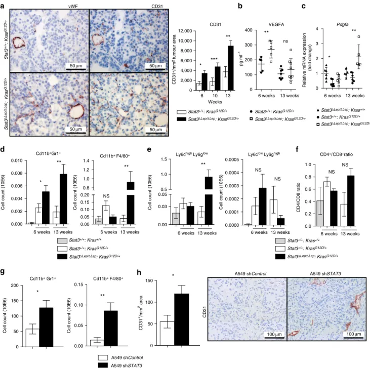
STAT3 alters tumour microenvironment and angiogenesis.
Detailed histopathological analysis revealed that Stat3-deficient murine tumours were better vascularized at all investigated time points, which was corroborated by increased CD31þ and von Willebrand Factor staining of vessel walls (Fig. 2a). Tumour angiogenesis is mainly driven by vascular endothelial growth factor alpha (VEGFA) and platelet-derived growth factor alpha (PDGFA) in response to certain stimuli derived from the tumour cells directly or from the surrounding microenvironment28. ELISA revealed significantly increased VEGFA levels in lungs from Stat3DLep/DLep: KrasG12D/þ mice compared with their littermate controls at 6 weeks, whereas at 13 weeks post AdCre infection no difference could be observed (Fig. 2b). However, at that later time point,Pdgfaexpression was significantly elevated in Stat3DLep/DLep:KrasG12D/þ mice, which might contribute to enhanced tumour vascularization indirectly through the recruitment of angiogenic stroma cells29,30(Fig. 2c).
It has been shown that tumour cell-specific Stat3 blockade leads to changes in the tumour microenvironment and increased immune cell infiltration31. Qualitative IHC analysis of the tumour microenvironment revealed that several cell types are present within the lung tumours (NK-cells, T cells, dendritic cells, fibroblasts and macrophages. Supplementary Fig. 3a). As macrophages and granulocytes contribute to tumour angiogenesis32, we performed quantitative assessment of these cell types via flow cytometry analysis of bronchoalveolar lavage (BAL) and observed significantly more CD11bþGr1þ granulocytes in Stat3DLep/DLep:KrasG12D/þ lungs compared with controls at 6 and 13 weeks post AdCre-induction (Fig. 2d).
Furthermore, infiltrating CD11bþF4/80þmacrophages were sig- nificantly elevated within tumours ofStat3DLep/DLep:KrasG12D/þ mice at 13 weeks post AdCre in BAL, which was confirmed by IHC (Fig. 2d and Supplementary Fig. 3b). Further analysis of myeloid-derived suppressor cells (MDSCs), which are known to play an important role in suppressing the antitumour immune response33, showed that MDSCs from Stat3-deficient mice were more differentiated towards a monocytic phenotype (CD11bþLy6ChighLy6Glow, M-MDSCs) than to a granulocytic phenotype (G-MDSCs, CD11bþLy6ClowLy6Ghigh) at later stages of tumour development (13 weeks; Fig. 2e). M-MDSCs have been postulated to be more potent in suppressing T-cell responses33. In line with this, we found that Stat3DLep/DLep: KrasG12D/þ
animals showed an increased CD4þ/CD8þratio compared with control mice at the latest time point (13 weeks), indicating a suppression of the cytotoxic CD8þ T cells response within the tumours (Fig. 2f).
We further investigated tumour microenvironment and angiogenesis within our patient cohort. Correlations between CD31 or VEGF and P-STAT3 or STAT3 did not reveal any significant differences betweenKRASwild-type and mutant cases (Supplementary Fig. 3c,d). Qualitative assessment of infiltrating immune cells revealed that only 3% of tumours were infiltrated with macrophages and we did not observe any differences between both genotypes (Supplementary Fig. 3e). However, the number of tumours scoring positive for infiltrating granulocytes was higher in KRAS mutant compared with KRAS wild-type tumours (18% within theKRASmutant versus 10% withinKRAS wild-type cohort); in addition, less cases of KRAS mutant tumours scored positive for lymphocyte infiltration compared with KRAS wild-type tumours (31% within the KRAS mutant versus 48% in KRASwild-type cohort; Supplementary Fig. 3e).
In order to substantiate the findings of our mouse model, we analysed the human AC xenograft samples and found that tumours lackingSTAT3showed significantly increased numbers of infiltrating CD11bþGr1þ and CD11bþ F4/80þ cells (Fig. 2g) and were significantly more vascularized (Fig. 2h), thus confirming our observations in the murine model. Together, these results implicate that STAT3 ablation results in a considerably different tumour microenvironment composed of more myeloid cells, increased CD4þ/CD8þ lymphocyte ratio and enhanced angiogenesis in our murine as well as in the human AC xenograft models.
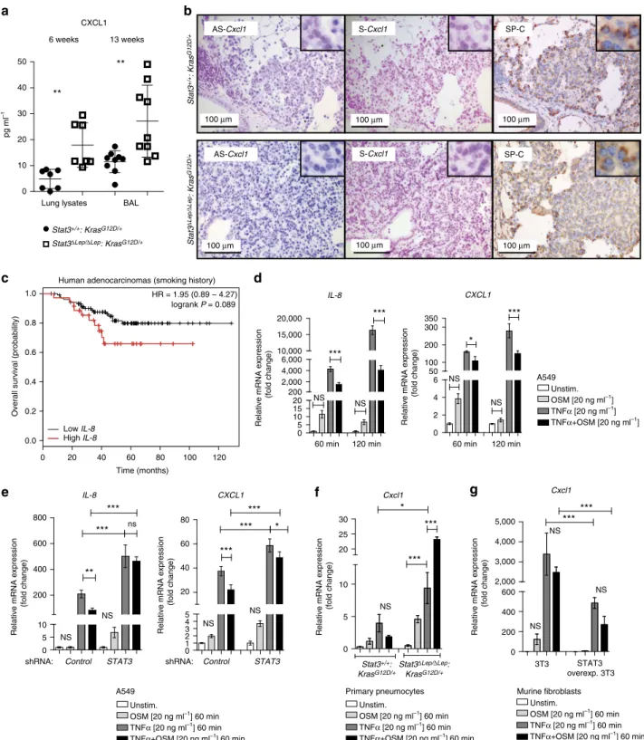
STAT3 regulates chemoattractive CXCL1 expression. To test if the increased recruitment of myeloid cells is a result of changes within the local cytokine and growth factor milieu in Stat3DLep/DLep:KrasG12D/þ tumours, we performed ELISA of several key cytokines, which may modulate the infiltration of myeloid cells into tumours. IL-2, IL-12p40, IL-4 and IL-17 were not detectable and we did not find differences in IL-5, interferon-g, IL-10, IL-1a, EGF, OSM expression between the two genotypes (Supplementary Figs 4a and 1b). Granulocyte–
macrophage colony-stimulating factor and IL-6 expression were upregulated in Stat3-deficient tumours only at the latest time point (Supplementary Figs 4a and 1b). Interestingly, CXCL1 was the only cytokine, which showed a significant increase in lungs from Stat3DLep/DLep:KrasG12D/þ mice compared with their con- trol littermates at all time points (Fig. 3a). Cxcl1(murine ortho- logue to humanCXCL1and IL-8(ref. 34)) is a well-known and potent chemo-attractant for macrophages and granulocytes and can induce angiogenesis35,36. In situ hybridization not only corroborated the increased expression ofCxcl1inStat3-deficient tumours but also identified the tumour cells as the main cellular origin of Cxcl1as confirmed by staining for the alveolar type 2 cell-specific marker SP-C (surfactant protein C) in serial sections (Fig. 3b and Supplementary Fig. 4b). Together, these data suggest that STAT3 negatively regulates the angiogenic and pro- inflammatory cytokine CXCL1 in the murine model.
Next, we analysed the patient cohorts forIL-8expression, the human orthologue of Cxcl1 (ref. 34). Interestingly, STAT3 and IL-8 expression tended to inversely correlate in KRAS mutant samples, whereas KRAS wild-type samples showed a trend towards a positive correlation between STAT3 and IL-8 expression (Supplementary Fig. 4c). Furthermore, high IL-8 expression levels have been associated with poor prognosis in smoking patients37, a finding which we could confirm within the second patient cohort used in this study (Fig. 3c)26.
Relative mRNA expression (fold change)
Pdgfa
**
*
0 1 2 3 4
13 weeks 6 weeks
13 weeks
6 weeks 6 weeks 13 weeks 6 weeks 13 weeks 6 weeks 13 weeks 6 weeks 13 weeks
13 weeks 6 weeks VEGFA
0 100 200 300
400 **
ns
pg ml–1
CD31
0 2,000 4,000 6,000 8,000 10,000 12,000
* ***
**
Weeks
6 10 13
0.000 0.002 0.004 0.006 0.008 0.010
*
**
Cell count (10E6)
0.00 0.05 0.10 0.15 0.20 0.8 1.0 1.2 1.4
**
NS
Cell count (10E6)
**
0.00 0.05 0.10 0.15
Cell count (10E6)
*
0 50 100 150 200
Cell count (10E6)
0 50 100
150 *
CD31+/mm2 area
A549 shControl A549 shSTAT3
0.00 0.03 0.5 1.0 1.5
0.05
Cell count (10E6)
0.0000 0.0001 0.0002 0.0003 0.0004 0.0005
Cell count (10E6)
0.0 0.2 0.4 0.6 0.8 1.0
CD4/CD8 ratio
A549 shControl A549 shSTAT3
CD31
CD31
vWF
**
NS
NS NS NS
Stat3+/+: KrasG12D/+Stat3ΔLep/ΔLep: KrasG12D/+
Cd11b+Gr1+
Cd11b+ F4/80+ Ly6chigh Ly6glow Ly6clow Ly6ghigh
Stat3+/+: Kras+/+
Stat3+/+: KrasG12D/+
Stat3ΔLep/ΔLep: KrasG12D/+
Stat3+/+: KrasG12D/+
Stat3+/+: KrasG12D/+
Stat3ΔLep/ΔLep: KrasG12D/+
Stat3ΔLep/ΔLep: KrasG12D/+
Stat3ΔLep/ΔLep: Kras+/+
Stat3+/+: KrasG12D/+
Stat3ΔLep/ΔLep: KrasG12D/+
Stat3ΔLep/ΔLep: KrasG12D/+
Stat3+/+: Kras+/+
Stat3+/+: KrasG12D/+
CD4+/ CD8+ratio
Cd11b+ Gr1+ Cd11b+ F4/80+
50μm 50μm
50μm 50μm
100μm 100μm
CD31+/mm2 tumour area
Figure 2 | STAT3 alters tumour microenvironment and angiogenesis.(a) IHC analysis of von Willebrand Factor (vWF) and CD31 is shown in consecutive sections of murine lung tumours of the indicated genotype. On right, CD31þ counts per tumour area (mm2) quantification at indicated time points.
Data were analysed by Student’st-test and displayed as mean±s.e.m.nZ5 tumours per mouse withnZ4 mice per genotype and time point. Scale bar, 50mm. (b) ELISA of VEGFA levels in lung tumour lysates at indicated time points. Data were analysed by Student’st-test and displayed as mean±s.d., nZ6 mice per genotype and time point. (c) Expression levels ofPdgfain total lungs were measured by quantitative real-time PCR at indicated time points.
Values are presented as fold change of relative mRNA expression compared withStat3DLep/DLep:Krasþ/þmice. Data were analysed by one-way analysis of variance (ANOVA) with Tukey’s multiple comparison test and are shown as mean±s.d.,nZ5 animals/genotype. (d) Flow cytometric analysis of Cd11bþGr1þ granulocytes and Cd11bþ F4/80þ macrophages in bronchoalveolar lavage (BAL) at 6 and 13 weeks post AdCre. Data were analysed by one-way ANOVA with Tukey’s multiple comparison test and are shown as mean±s.e.m. (e) Flow cytometric analysis of myeloid-derived suppressor cell (MDSC) subsets at 6 and 13 weeks post AdCre. Data were analysed by one-way ANOVA with Tukey’s multiple comparison test and are shown as mean±
s.e.m. (f) Ratio of CD4þ/CD8þ T-cell counts in BAL are shown. Data were analysed by Kruskal–Wallis test with Dunn’s multiple comparison testing and are shown as mean±s.e.m. Data displayed ind–farenZ6 mice per genotype and time point, 13-week group represents two independent experiments. (g) Flow cytometric analysis of Cd11bþGr1þ and Cd11bþ F4/80þ cells of A549 shControlversus A549-shSTAT3xenograft tumours. Data were analysed by Student’st-test and are displayed as mean±s.e.m. (nZ8 tumours;Z4 mice per group). (h) IHC analysis and representative pictures of CD31þcounts per xenograft tumour area (mm2). Scale bar, 100mm (n¼8 tumours/Z4 mice). Data were analysed by Student’st-test and are shown as mean±s.e.m. For all graphs: *Po0.05; **Po0.01; ***Po0.001.
CXCL1 6 weeks
Lung lysates
13 weeks
BAL 0
10 20 30 40 50
**
**
pg ml–1
S-Cxcl1 SP-C
AS-Cxcl1 AS-Cxcl1
S-Cxcl1 SP-C
Unstim.
TNFα [20 ng ml–1] OSM [20 ng ml–1] TNFα+OSM [20 ng ml–1] A549
IL-8
60 min 120 min Relative mRNA expression (fold change)
0 5 10 15 20 2,000 4,000 6,000 10,000 15,000 20,000
200
***
NS NS
***
0 2 4 6 100 200 300
50 350
*
***
NS NS CXCL1
Relative mRNA expression (fold change)
60 min 120 min
Primary pneumocytes Unstim.
TNFα [20 ng ml–1] 60 min OSM [20 ng ml–1] 60 min TNFα+OSM [20 ng ml–1] 60 min 0
5 10 20 25 30
*
***
***
Relative mRNA expression (fold change) NS
Cxcl1
Unstim.
TNFα [20 ng ml–1] 60 min OSM [20 ng ml–1] 60 min TNFα+OSM [20 ng ml–1] 60 min
Cxcl1
3T3 STAT3
overexp. 3T3 Relative mRNA expression (fold change)
Murine fibroblasts 0
200 400 600 2,000 3,000 4,000 5,000
NS
NS
***
NS
***
Unstim.
TNFα [20 ng ml–1] 60 min OSM [20 ng ml–1] 60 min TNFα+OSM [20 ng ml–1] 60 min A549
0 5 10 200 400 600 800
**
NS
*** ns
***
NS
STAT3 Control shRNA:
Relative mRNA expression (fold change)
0 1 2 3 4 5 20 40 60
80 *
NS
***
***
***
NS
STAT3 Control
shRNA:
Relative mRNA expression (fold change)
CXCL1 IL-8
Low IL-8 High IL-8
HR = 1.95 (0.89 − 4.27) logrank P = 0.089
0 20 40 60 80 100 120
0.0 0.2 0.4 0.6 0.8 1.0
Overall survival (probability)
Time (months)
Human adenocarcinomas (smoking history)
100 μm 100 μm
100 μm 100 μm 100 μm
100 μm
Stat3ΔLep/ΔLep: KrasG12D/+ ΔLep/ΔLepG12D/+Stat3: Kras Stat3+/+: KrasG12D/+
Stat3ΔLep/ΔLep: KrasG12D/+
Stat3+/+: KrasG12D/+
Stat3+/+: KrasG12D/+
Figure 3 | STAT3 regulates chemoattractive CXCL1 expression.(a) ELISA of CXCL1 levels in lung lysates and BAL at 6 and 13 weeks post AdCre.
Data were analysed by Student’st-test and shown as mean±s.d.,nZ7 mice per genotype and time point. (b) Representativein situhybridization with probes specific for murineCxcl1(AS) in lung tumours at 13 weeks post AdCre. Sense probes (S) were used as negative control and surfactant protein C (SP-C) as lung epithelial maker. Scale bar, 100mm. (c) Kaplan–Meier plot showing overall survival of lung adenocarcinoma patient samples harbouring smoking history with high or lowIL-8mRNA expression (expression values of upper quartile, log-rank test)nZ35–104 per group. (d) Human A549 lung cancer cell line (KRASG12Smutated) was stimulated with designated cytokines for indicated time points.IL-8orCXCL1expression was analysed via qRT–PCR. Data were analysed by one-way ANOVA with Tukey’s multiple comparison test and shown as mean±s.d. (e) A549 cells transducted with lentiviral vectors expressing nonspecific scrambled small hairpin (sh)RNA or shRNA againstSTAT3were stimulated with indicated cytokines for 60 min.
Data were analysed by one-way ANOVA with Tukey’s multiple comparison test and shown as mean±s.d. (f) Primary pneumocytes were isolated and infected with AdCre. 120 h post infection cells were stimulated with indicated cytokines for 60 min.Cxcl1expression was analysed by qRT–PCR. Data are shown as mean±s.e.m. (g) Murine 3T3 fibroblasts and STAT3 overexpressing 3T3 fibroblasts were stimulated as inf.Cxcl1expression was analysed by qRT–PCR. Data were analysed by one-way ANOVA with Tukey’s multiple comparison test and shown as mean±s.d. Values ind–gare presented as fold change of relative mRNA expression compared with each unstimulated individual cell line. At least two independent experiments with three individual plates per stimulation were performed. For all graphs: *Po0.05; **Po0.01; ***Po0.001.
We next performed various cytokine stimulation experiments in vitro to corroborate the Stat3-mediated repression of Cxcl1 observed in the murine model. First, we confirmed that the human KRAS mutant AC cell line A549 activates STAT3 in response to cytokines such as OSM, IL-6 and EGF found to be expressed in our murine lung tumours in vivo (Supplementary Figs 1b and 4d). We used tumour necrosis factor-a(TNFa) to stimulate NF-kB induced IL-8 and CXCL1 expression in A549 cells and analysed the effect of co-stimulating STAT3 by OSM or IL-6. As expected, TNFastimulation induced a markedIL-8and CXCL1 expression, which was significantly inhibited by co- treatment with OSM (Fig. 3d). Interestingly, OSM treatment alone also led to a very small and transient increase ofIL-8and CXCL1. Similar results were obtained with IL-6 co-stimulation (Supplementary Fig. 4e). Importantly, small hairpin-mediated knockdown of STAT3 confirmed that the observed effects on CXCL1 and IL-8 repression were at least in part mediated by STAT3 (Fig. 3e and Supplementary Fig. 4f).
Next, we isolated primary mouse alveolar type 2 pneumocytes from the lungs of Stat3flox/flox:KrasLSL-G12D/þ and Stat3þ/þ: KrasLSL-G12D/þ animals (Supplementary Fig. 4g). Complete deletion of STAT3 was observed 120 h post AdCre infection in vitro(Supplementary Fig. 4h). In these primary cells, TNF a treatment again triggeredCxcl1expression albeit less pronounced compared with A549 cells (Fig. 3f). Importantly,Stat3-deficient KrasG12D/þ-mutant pneumocytes expressed significantly increased levels of Cxcl1 upon TNF a stimulation compared with syngeneic Stat3-proficient cells. Upon co-stimulation with OSM, Stat3-deficient pneumocytes not only failed to repress Cxcl1 expression but even showed vastly increased Cxcl1 expression levels (Fig. 3f). Conversely, experiments using murine 3T3 fibroblasts overexpressing STAT3 (ref. 10) showed that STAT3 is not only required but is sufficient to repress Cxcl1 expression (Fig. 3g). These results suggest that STAT3 represses Cxcl1expressionin vivoandin vitro.
CXCL1 inhibiton reverts oncogenic effects of STAT3 ablation.
Next, we tested whether CXCL1 is responsible for the increased tumour growth supported by vascularization and infiltration. We used the SB225002 compound to inhibit CXCL1’s cognate receptor CXCR2, which we found to be expressed on epithelial, endothelial and myeloid cells within the lung tumours (Supplementary Fig. 5a)36,38. CXCR2 antagonist treatment was conducted in two ways: first, we treated the mice 1 week after AdCre tumour induction (Supplementary Fig. 5b). After 5 weeks of continuous CXCR2 blockade, we could not only observe a marked reduction in tumour vascularization but importantly tumour incidence and overall tumour burden were reduced, suggesting that CXCL1 is crucial for tumour development at early stages inStat3DLep/DLep:KrasG12D/þ mice (Fig. 4a,b). Second, we tried to mimic a therapeutic setting and treated tumour-bearing mice (Supplementary Fig. 5b). After 7 weeks of treatment, we could observe a significant reduction of vascularization and of infiltrating F4/80þ macrophages in the tumours ofStat3DLep/DLep: KrasG12D/þ mice (Supplementary Fig. 5c), but not in overall tumour burden (Supplementary Fig. 5d). To further confirm these findings in a human cell line, we performed a small hairpin- mediated double knockdown of IL-8and STAT3 in A549 cells (Supplementary Fig. 5e). Indeed, enhanced tumour growth of shSTAT3cells was significantly reduced upon knockdown ofIL-8 (Fig. 4c). Furthermore, vascularization as well as macrophage and granulocyte infiltration was reduced in the IL-8/STAT3 double knockdown tumours compared withSTAT3knockdown tumours (Fig. 4d,e). Interestingly, knockdown of IL-8 alone showed an increased tumour growth in nude mice compared with shControl
cells for unknown reasons (Fig. 4c). Taken together, these data suggest that loss of STAT3 in the tumour cells results in increased CXCL1/IL-8 expression triggering infiltration of myeloid cells as well as augmented vascularization, and identifies CXCL1/IL-8 as an essential mediator for the accelerated development and progression ofStat3-deficient tumours.
STAT3 retains p65 in the cytoplasm to reduce NF-jB activity.
Having demonstrated the importance of STAT3-mediated repression ofCXCL1in tumour cellsin vitroandin vivo, we set out to mechanistically interrogate this pathway further. As NF-kB subunit p65 is an important regulator of CXCL1expression, we first examined the p65 activation39,40. Increased levels of activated p65 were observed, which was mirrored by increased expression ofNFkB-p65target genes such asTnfa,c-Myc, Il-6 andCxcl1in Stat3-deficient lungs, whereasBcl2, Bcl2l1, Ccnd1and Ccl5were comparably expressed (Figs 3a,5a,b; Supplementary Figs 1b and 6b)41. These data demonstrate thatStat3-deficient tumours show increasedNF-kBactivity.
We examined the association ofNF-kBandSTAT3expression in our patient sample archive. We found a significant positive correlation of NF-kBand STAT3 expression inKRAS wild-type samples. On the contrary, we found a trend towards an inverse correlation between NF-kB and STAT3 expression in KRAS mutant samples (Supplementary Fig. 6c).
As STAT3 has been shown to compete with NF-kB on various promoters42,43, we further analysed the crosstalk of STAT3 and NF-kB binding on the human CXCL1 and the mouse Cxcl1 promoter. Within the mouse Cxcl1promoter, we found several putative STAT3-binding sites—two of them overlapping with putative NF-kB-binding sites and conserved in humans (hereafter termed responsive element, RE1and 2; Supplementary Fig. 6d).
However, electrophoretic mobility shift assays of all putative binding sites (Supplementary Fig. 6e) as well asin vivochromatin immunoprecipitation experiments could not reveal strong endogenous binding of STAT3 on RE1 orRE2 (Supplementary Fig. 6f). We next tested the human A549 cell line focusing on the RE2 site (Fig. 5c). Upon TNF a treatment, we could detect efficient recruitment of p65 to the CXCL1 promoter and co-stimulation of STAT3 signalling by OSM treatment indeed markedly reduced p65 recruitment. However, we failed to detect direct STAT3 binding at the CXCL1 promoter site (Fig. 5c), indicating that STAT3 regulates p65-induced CXCL1 expression by means other than competing for promoter-binding sites.
As STAT3 and the NF-kB subunit p65 have been reported to interact at the protein level43–45, we next tested if STAT3 may bind and control subcellular localization of p65. Co- immunoprecipitation experiments in A549 cells showed that the majority of STAT3 and NF-kB–p65 interact in the cytoplasm under basal conditions as well as upon stimulation with OSM, TNF aor a combination thereof (Fig. 5d). We next quantified nuclear p65 levels normalized to poly (ADP-ribose) polymerase family, member 1 (PARP). Upon OSM stimulation, a reduction of nuclear p65 compared with unstimulated conditions was observed. Increased levels of nuclear p65 were obtained upon TNFastimulation, which was reduced to basal levels upon co- stimulation with OSM (Fig. 5d). To further confirm this, we analysed the subcellular localization of p65 using immunofluorescence imaging of STAT3-proficient A549 cells and compared it to the STAT3 knockdown cell line (Fig. 5e).
Under basal conditions, A549-shSTAT3 cells already showed increased levels of nuclear p65 compared with controls.
Interestingly, we observed a reduction of nuclear p65 in OSM- stimulated A549-shSTAT3 cells compared with unstimulated controls, indicating that additional OSM-induced effectors other
than STAT3 are also regulating the cytoplasmic-nuclear trafficking of p65. As expected, both cell lines showed increased levels of nuclear p65 upon TNF a stimulation. Importantly, co-stimulation with OSM resulted in a significant reduction of TNF a-induced nuclear p65 accumulation in A549 scrambled control cells but not in shSTAT3-infected cells (Fig. 5e). In summary, our results suggest that STAT3 represses NF-kB-
dependent CXCL1 expression by sequestering NF-kB in the cytoplasm and mechanistically delineate a novel tumour- suppressive pathway governed by the STAT3–NF-kB–CXCL1 axis.
Discussion
Personalized treatment decisions based on the genetics of the individual tumour will be paramount to combat malignancies in
0 50 100 150 200
shSTAT3 shControl
***
20
***
10 Tumour volume (mm3)
Days
shControl; shIL-8 shSTAT3; shIL-8
0 50 100
150 *
*
CD31+ count/area (mm2) 0.0000
0.0005 0.0010 0.0015 0.0020 0.0025
Cell count (10E6)
0.0000 0.0005 0.0010 0.0015 0.0020
Cell count (10E6)
shSTAT3 shControl shControl; shIL-8 shSTAT3; shIL-8
0 2,000 4,000 6,000
8,000 *
*
CD31 count/area (mm2)
Vehicle SB225002
shSTAT3 shControl
shControl; shIL-8 shSTAT3; shIL-8
CD31CD31
VehicleSB225002CD31
0 10 20 30 40
**
*
Tumour area/lung area (%)
Vehicle SB225002
0 24 6 20 40 60 80 100
*
*** ***
** **
Tumour number per cm2 lung
Vehicle SB225002 Vehicle SB225002 }
}
Hyperplasia +adenoma
In situ adeno carcinoma All tumours
shSTAT3 shControl shControl; shIL-8 shSTAT3; shIL-8
VehicleSB225002
100μm
100μm 100μm
100μm 100μm
100μm 100μm
100μm
CD11b+ F4/80+ CD11b+ Gr1+ Stat3ΔLep/ΔLep: KrasG12D/+
Stat3+/+: KrasG12D/+
Stat3+/+: KrasG12D/+ Stat3ΔLep/ΔLep: KrasG12D/+
Stat3+/+: KrasG12D/+
Stat3+/+: KrasG12D/+
Stat3ΔLep/ΔLep: KrasG12D/+
Stat3ΔLep/ΔLep:
KrasG12D/+ Stat3+/+:
KrasG12D/+
Stat3ΔLep/ΔLep: KrasG12D/+
the near future. STAT3 has been implicated in several malignancies5,7,13,14,46 and therefore, clinical studies are currently evaluating the efficacy of STAT3 inhibition in various kinds of cancer46. However, although STAT3 traditionally has been described as an oncogene7,9, recent reports have shown that STAT3 can also behave as a tumour suppressor in the very same organ systems9,11,12,42. In addition, other reports validated high STAT3 expression as a potential marker for good prognosis in human colorectal carcinoma and breast cancer47,48. Therefore, the idea emerged that STAT3 function is context-dependent either with regards to the oncogenic driver mutation, cancer type or the specific tumour microenvironment.
Specifically in lung cancer, STAT3 has been shown to function as one of the main downstream transcription factors in EGFR mutant ACs13and to act as an oncogene in a chemically induced mouse model of lung cancer49. In addition, several studies of human lung cancer specimens have shown the activation of STAT3 in those tumours13,14,16. These findings made STAT3 an attractive drug target to treat NSCLC patients. We hypothesized that lung AC patients, especially those with activating KRAS mutations, might also benefit from STAT3 inhibition. AsKRASis still considered as an undruggable target and is responsible for 30% of all Caucasian AC cases, we investigated whether STAT3 inhibition has a potential survival benefit in a preclinical lung cancer mouse model and in a human cell line xenograft model.
However, our results clearly show that STAT3 behaves as a haploinsufficient tumour suppressor during KrasG12D-induced lung tumorigenesis. Mice lacking Stat3 signalling specifically withinKrasG12D-mutant tumour cells not only display increased tumour initiation and tumour cell proliferation but also accelerated malignant progression and ultimately markedly reduced survival compared to mice with intact Stat3signalling.
We could confirm these observations in human lung AC cell- derived xenografts. Of note, others have shown that reducing STAT3 activity by using STAT3 inhibitors or STAT3 decoy oligonucleotides suppressed tumour growth in xenograft-derived NSCLC cell lines18,50,51. However, by using a powerful short hairpin RNA (shRNA)-mediated loss-of-function approach, we found that STAT3 suppresses tumorigenesis in A549 cell-derived xenografts, thus supporting our findings in the murine model.
Despite the fact that Stat3-deficient tumours had a growth advantage, they did not progress to become more invasive than Stat3-proficient tumours or metastatic. Tumour invasion and metastasis are a multistage development in which, in addition to neo-angiogenesis, malignant tumour cells have to undergo EMT, detach from the primary tumour, migrate and pass mechanical barriers. This is a complex process driven by multiple somatic aberrations. Thus, most likely, Stat3-deficient tumours need additional hits to become invasive and to metastasize.
Interestingly, the observed survival disadvantage is more pronounced in male mice than in female mice reminiscent of observations made in the clinic: although women have a higher incidence rate in developing lung cancer, they have a better survival outcome compared with men52. Further on, STAT3 already has been linked to gender-specific diseases in liver and lung cancer53,54. Detailed analysis of this gender-specific difference associated with STAT3 will be of high interest. One of the main pro-inflammatory and neo-angiogenesis promoting cytokines responsible for our observations in Stat3-deficient tumours is Cxcl1.Cxcl1, the murine orthologue to human IL-8 andCXCL1, is known to be RAS dependent40, to directly attract myeloid cells and to stimulate endothelial cells to promote angiogenesis35,36. Clinically, IL-8 expression is associated with a tendency for poor prognosis in human lung cancer37 and expression of CXCR2 also correlates with smoking and poor prognosis55. In agreement with this, we could confirm thatIL-8 expression tends to correlate with poor survival in smoking patients (who frequently acquire KRAS mutations) within the publically available AC patient cohort used in this study.
Interestingly, we could also show that low STAT3 expression has a tendency to correlate with increasedIL-8mRNA expression in KRASmutant patient samples.
To investigate whether CXCL1 is the central angiogenesis and tumour-promoting factor in our model and a potential therapeutic target, we treated the mice with SB225002, a small- molecule antagonist of CXCR2. TreatingStat3-deficient mice at tumour initiation resulted in a reduction of tumour growth as well as a decrease in vascularization compared with vehicle- treated control mice. This result corroborates that CXCL1 signalling indeed accelerates tumour growth at early stages. In addition, we cannot rule out that CXCL1 also acts as an autocrine tumour-promoting factor in lung cancer cells56–58. We also tried to validate CXCL1 as a therapeutic target by blocking CXCL1 signalling in established tumours. By using this approach, we could show that tumour vascularization and infiltration of macrophages were reduced at the end stage of the treatment;
however, we did not observe any effect on tumour growth. This result indicates that other effectors in Stat3-deficient tumours, like elevated NF-kB activity and/or increasedc-Myc expression, may contribute to tumour proliferation at late stages.
Nevertheless, although late-stage Stat3-deficient tumours failed to respond to an anti-CXCL1 monotherapy, the use of CXCL1 blockers combined with standard chemotherapy or as maintenance therapy could be a reasonable alternative to treat these tumours and it deserves further investigation. To substantiate these findings, we inhibited IL-8 signalling in STAT3 knockdown human lung AC cells. Short hairpin- mediated IL-8/STAT3 double knockdown resulted in a
Figure 4 | CXCL1 inhibiton reverts oncogenic effects of STAT3 ablation.(a) Mice were treated with the CXCR2 antagonist SB225002 or vehicle control starting 1 week after tumour induction and euthanized after 5 weeks (treatment 1 shown in Supplementary Fig. 5b). Tumour area/lung area was quantified within each group and at least two sections of each lung were stained with haematoxylin and eosin and analysed in a blinded manner. Tumour grading is shown in the right panel (n¼4–7 mice per genotype). Data in both panels were analysed by one-way ANOVA with Tukey’s multiple comparison test and shown as mean±s.e.m. Scale bar, 2 mm. (b) Tumour vascularization was quantified by CD31þ counts per tumour area (mm2). At least 3 tumours per mouse were analysed withn¼4–7 mice per genotype and treatment. Data were analysed by one-way ANOVA with Tukey’s multiple comparison test and shown as mean±s.e.m. Scale bar, 100mm. (c) Short hairpin-mediated knockdown of IL-8 (shIL-8) in either shControlor shSTAT3A549 NSCLC cells were performed and 2106cells were injected in both flanks of male nude mice (n¼5 per group). Xenograft tumour growth was determined at indicated time points. Data were analysed by Two-way ANOVA with Bonferroni multiple comparison test and shown as mean±s.e.m. (d) IHC analysis of CD31þ counts per tumour area (mm2) showed reduced vascularization of A549-shSTAT3;shIL-8xenograft tumours compared with controls. Data were analysed by one-way ANOVA with Tukey’s multiple comparison test and shown as mean±s.e.m. Scale bar, 100mm. (e) Flow cytometric analysis of Cd11bþGr1þ granulocytes and Cd11bþF4/80þmacrophages displayed reduced myeloid infiltration in A549-shSTAT3;shIL-8xenograft tumours compared with controls (nZ8 tumours; 5¼mice per group). At least 6 tumours per mouse were analysed withnZ7 mice per genotype and treatment. Data were analysed by Kruskal–Wallis test with Dunn’s multiple comparison testing and shown as mean±s.e.m. For all graphs: *Po0.05; **Po0.01; ***Po0.001.