Prognostic impact of progesterone receptor expression in HER2- negative Luminal B breast cancer
Tímea Selmeci1+, Anna-Mária Tőkés2+, Ágnes Róna1, Béla Ákos Molnár3, István Kenessey1, Borbála Székely1, Lilla Madaras, A. Marcell Szász1, Janina Kulka1
Abstract
Aim: The new classification of breast cancer is based on microarray studies. Within the estrogen receptor (ER) positive breast carcinoma subtype further subgroups could be identified. In the present study, we analyzed the Her2 negative, highly proliferative subgroup (Luminal B1-like, LUMB1) with emphasis on their clinicopathological characteristics and progesterone receptor (PR) expression.
Patients and methods: Our retrospective study concerned the period between 2000 and 2010. 158 patients were selected with ER positive, Her2 negative, Ki67>15% breast cancer. The pathological and clinical data were collected and analyzed. Age, tumor grade and stage, ER, PR, Her2 and Ki67 expression were recorded. The clinicopathological variables were correlated to PR expression.
Results: The mean age of the patients was 57.5 (28-75) years. The ratio of patients younger than 40, was 8.86%. Shorter metastasis-free survival was observed in this young age group (P=0.044). The majority of our cases belonged to the pT1-pT2 stages (41.28% and 44.95%, respectively) whereas pT3 and T4 stage was detected in 5.50% and 8.25% of the cases, respectively. Almost half of the cases had no axillary lymph node metastasis (pN0: 48.91%), 1-3 lymph node metastases were detected in 38.04% (pN1), 4-10 metastatic lymph nodes were identified in 9.78% (pN2) and pN3 stage was found in 3.26% of the cases.
Most commonly the tumors were either grade 2 or 3 (44.16% and 45%, respectively). The median value of Ki67 labeling index was 30%. Disease progression was detected in 36.19% of the patients. According to PR expression, a tendency to better prognosis (i.e. longer disease free- and overall survival) was detected in
1 2nd Department of Pathology, Semmelweis University, Budapest, Hungary
2 MTA-SE Tumour Progression Research Group, 2nd Department of Pathology, Semmelweis University, Budapest, Hungary
3 1st Department of Surgery, Semmelweis University, Budapest, Hungary + contributed equally
Corresponding author:
Janina Kulka
2nd Department of Pathology, Semmelweis University, Budapest, Hungary tel: +36-1-2157300/53430
e-mail: kulka.janina@med.semmelweis.-univ.hu
cases showing >10% PR positivity. However, no difference was found regarding tumor size, axillary stage, grade and age when comparing lower and higher PR expressing tumors.
Conclusions: LUMB1 breast carcinomas are typically grade 2 and grade 3, the Ki67 labeling index is often 30% or higher. Distant metastases occur in more than one third of the cases. Within this subgroup, those cases with low PR expression represent a poor prognostic cohort. These findings require further investigations in larger number of LUMB1 breast cancer cases.
Introduction
One of the reasons behind the heterogeneity of breast cancer is the phenotypical reflection of their different gene expression profiles. Cancers with different expression profiles show different clinical and prognostic features. Since the advent of microarray based classifications, various molecular classes of breast cancer have been described. The first and most robust molecular classification was published in 2000, when Perou and his group published their work on cDNA microarray studies of human breast cancers and described five major subtypes [24]. Hormone receptor positive breast cancers are treated with endocrine therapy. Although, in the majority of patients the likelihood of recurrence and disease related death decreased considerably due to administration of endocrine therapy, a number of patients are not responding or develop resistence to the treatment with time and therefore have a poorer prognosis. [1, 2]. It was, thus, a logical conclusion that there are subclasses within the group of ER positive breast cancers. Sorlie and co- workers [3] classified the ER-positive breast cancers into Luminal A, B and C subgroups.
Expression of ERα was highest in Luminal A cancers. Expression of ER regulated genes were lower in Luminal B és C subgroups. Most recently, the St. Gallen International Consensus Conference [4-8] suggested the following definitions for subclassifying breast cancers based on estrogen (ER), progesterone receptor (PR), Her2 and Ki67 protein expression. Luminal A-like (LUMA): ER-positive and PR-positive, low Ki67 index, Her2-negative, "recurrence risk" low, Her2-
negative Luminal B-like (LUMB1): ER-positive and Her2-negative, and at least one of the followings:
high Ki67, PR-negative/low, "recurrence risk"
high; Her2-positive Luminal B-like (LUMB2): ER- positive and Her2-positive, any PR, any Ki67, Her2-positive (non Luminal): ER-negative, PR- negative, Her2-positive, Triple-negative: ER- negative, PR-negative, Her2-negative.
The prognostic and possible predictive role of low PR expression has been studied more recently.
Within the luminal group of breast cancers, low expression of PR characterizes a more agressive, less endocrine therapy sensitive subgroup: lower ER levels, higher proliferation rate, larger tumor size, more positive axillary lymph nodes, aneuploid DNA content and increased expression of epidermal growth factor receptor were found [9]. Cancello and co-workers [10] investigated the role of PR in relation with recurrence in Lumial B breast cancers, considered a less favourable prognostic group. They divided the Luminal B breast cancers into 4 subgroups based on Her2 and PR expression: ER+/PR+/Her2-; ER+/PR- /Her2-; ER+/PR-/Her2+ és ER+/PR+/Her2+. They concluded that in both the Her2 positive and Her2 negative groups, low expression or lack of PR is related to shorter metastasis-free- and overall survival. Considering the difficulties in treating Luminal B breast cancers, many ongoing research aim at identifying newer targets for therapy. In our present study, we investigated LUMB1 breast carcinoma cases from the point of view of PR expression, among others. We
compared the level of PR expression with clinico- pathological and follow-up data.
Patients and methods
ER-positive, Her2-negative breast carcinoma cases with Ki67 index ≥15% diagnosed between the period 2000-2010 were selected and the clinico-pathological data were collected. Follow- up data of the patients were retrieved from the University’s database (MedSolution), the pathological data were collected from the files of the 2nd Department of Pathology following approval by the Institutional Review Board (SE- IKEB 77/2007). Overall survival data were provided by the Central Office for Administrative and Electronic Public Services. In our study we considered age of the patients, grade, TNM stage, ER, PR, Her2 and Ki67 expression of the tumors.
Age groups were created according to patient’s age at the time of the primary diagnosis of breast cancer. In order to elucidate the suspected prognostic significance of age at the primary diagnosis, we analyzed distant metastasis-free survival data at 35, 40 and 45 age thresholds.
Tumor grade was defined according to the Nottingham grading system [11], TNM stage was recorded according to the 7th Edition of the American Joint Committee on Cancer and the International Union for Cancer Control (AJCC- UICC) manual [12]. We calculated the distant metastasis-free survival (DMFS) as the period in months elapsed between the diagnosis of the primary tumor and the occurance of the first distant metastasis. Overall survival (OS) was also calculated in months: time elapsed between the diagnosis of the primary tumor to the time of disease related death. In some cases, in the cohort not all of the clinical or pathology data were available, but we didn’t exclude these cases from the statistical analyses upon this fact had no interference with the result of the calculation.
Details of the immunohistochemical reactions for
ER, PgR, Her2 and Ki67 are summarized in Table 1.
Table 1. Antibodies, dilutions and providers used in the study
Antigen Provider Clone Dilution
ER Novocastra 6F11 1:200
PgR Novocastra 312 1:200
HER2 Novocastra CB11 1:150
Ki67 DAKO MIB1 1:100
Hormone receptor positivity was recorded if >1%
of the tumor cells showed positive nuclear reaction, in line with the ASCO/CAP guideline [25]. Concerning PR status we divided our cases into „PR low” and „PR high” subgroups. Since only scattered recent literature data are available regarding the prognostic meaning of low PR expression in breast cancer, we tested our cases at 5, 10 and 20% limits of PR expression. Ki67 labeling index was estimated by eye-balling on a representative tumor slide. Any intensity of positive reaction was considered and the percentage of positive tumor cells was recorded.
Her2 status was defined according to the ASCO/CAP recommendations valid at the time of the period under investigation [13, 26]. The study group comprised of 158 patients.
For survival analysis Kaplan-Meier method was used; log-rank statistics was used to characterize the differences between prognostic groups.
Categorical data were analysed using Chi-square test and Fischer exact test. Results were regarded statistically significant at p<0.05. Statistica 11.0 software was used for each analysis (StatSoft, Tulsa, OK, USA).
Results
Clinical and pathological characteristics of the cases are shown in Table 2.
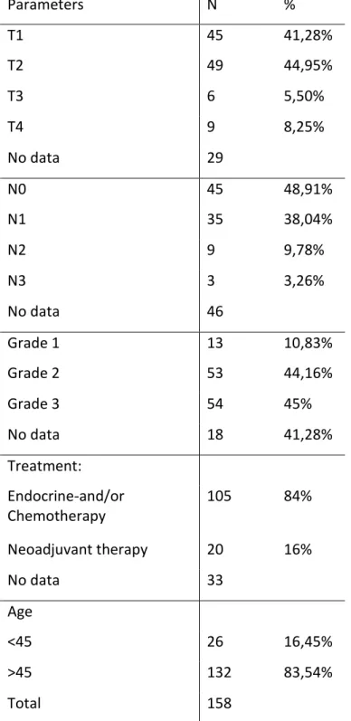
Table 2. Clinical and pathological characteristics of LUMB1 cancers included in this study
Parameters N %
T1 45 41,28%
T2 49 44,95%
T3 6 5,50%
T4 9 8,25%
No data 29
N0 45 48,91%
N1 35 38,04%
N2 9 9,78%
N3 3 3,26%
No data 46
Grade 1 13 10,83%
Grade 2 53 44,16%
Grade 3 54 45%
No data 18 41,28%
Treatment:
Endocrine-and/or Chemotherapy
105 84%
Neoadjuvant therapy 20 16%
No data 33
Age
<45 26 16,45%
>45 132 83,54%
Total 158
Mean age of the patient at the time of the primary diagnosis was 57.51 (range: 25-75).
Twenty six of 158 patients (16.45%) were <45, 14/158 (8.86%) were <40 and 5/158 (3.16%) were
<35 years. Age, as an adverse prognostic factor was significant regarding DMFS in patients <40 when compared to DMFS of patients >40 (p=0.044) (Figure 1).
Figure 1. Distant metastasis-free survival according to patients’ age (p=0.044).
We had treatment data from 125/158 patients.
Neoadjuvant oncological treatment was used in 20/125 cases (16%). In these patients, ER, PR, Ki67, Her2 immunohistochemistry results of the core biopsies were considered, while pT and pN stage was known from the surgical resection specimens. These cases were not considered further in the statistical analyses: we analysed data of 138 patients.
Tumor size was known in 109/138 cases. The majority of the known cases belonged to the pT1 and pT2 stage category (41.28% and 44.95%
respectively), 5.50% were pT3. pT4 cases occured in 8.25%. Regarding axillary lymph nodes we could identify 92/138 cases with known regional lymph node status. Negative axillary lymph nodes were present in 48.91% of the cases, 1-3 metastatic lymph nodes were recorded in 38.04%, more than 10 metastatic lymph nodes were present in 9.78% and pN3 was diagnosed in
3.26% of the cases. Tumor grade was known in 120 cases. The majority of the cancers were of grade 2 or grade 3 (53/120, 44.16% and 54/120, 45%, respectively). The median Ki67 labeling index was 30%.
In the adjuvant treated patient group we had follow up data in 105/138 patients. During the period examined 38/105 (36.19%) patients developed distant metastasis, among them 21 patients (55.27%) presented with dissemination to multiple organs. More than half of the solitary metastases (52.94%) localized to the skeletal system.
Relationship between PR expression and prognostic factors
Clinical and pathological variables were analyzed in relation to PR expression. Since there are few data related to the exact role of PR positivity and its extent, we performed analyses with different cutpoints at 5, 10 and 20% PR positivity. Very low PR expression (0-5%) was detected in 53 cases (38.40%), 59 cases showed 0-10% PR positive tumor cells, and 69 cases (50%) had 0-20% PR positivity.
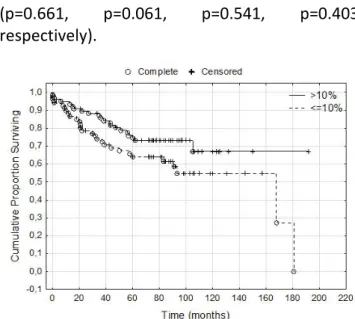
When considering all cases, including the 20 core biopsies, the number of cases in the above three PR expression categories was the following: 63 cases (39.87%) 0-5% PR positive tumor cells, 71 cases (44.93%) 0-10% PR positive tumor cells, 73 cases (46.20%) 0-20% PR positive tumor cells. In our cohort, the cutpoint of PR positivity that divided the patients into a better and a worse prognosis group was at 10%, but only at the level of tendency: cancers that have >10% PR positive cell population show better DMFS (p=0.07) (Figure 2). Regarding age groups, pT categories, tumor grade categories and pN categories, lower and higher (0-10% vs. >10%) PR expressing tumors didn’t show any specific distribution.
(p=0.661, p=0.061, p=0.541, p=0.403, respectively).
Figure 2. Distant metastasis-free survival according to PR expression at 10% positivity cut- off (p=0.07). A tendency of shorter survival could
be detected in low-PR expressing tumors.
Discussion
According to our results, grade 2 and grade 3 cases predominate within the group of Luminal B1 breast cancers. Similar to these results, Park et al. found that among Luminal B subtype breast cancers, higher grade tumors are more common [14]. Many recent studies aim at clarifying the exact prognostic significance of Ki67 labeling index, although the cut-off value has been a subject of discussion: some authors use 10%, 14%, or 20%, yet others suggest the use of the mean or median percentage of Ki67 positive tumor cells as threshold [15]. Despite the reported inter-observer differences in evaluating Ki67 labeling index, it is obvious that higher Ki67 labeling index predicts poorer prognosis.
Azambuja and co-workers [16] in a large meta- analysis using 68 studies confirmed the higher risk of relapse and shorter survival in cases with higher Ki67 labeling index. Cheang and co- workers [15] identified 13,25% Ki67 labeling
index as cut-off value for the differentiation of Luminal A from Luminal B breast cancers. The median value of Ki67 labeling index in ER- positive, Her2-negative breast cancer cases was 14% in a multicenter study performed by Cserni and his group [17], but no subtyping of luminal cases was performed. In our present study, we used the 15% cut-off as it was also shown in a study by Cserni and co-workers that approximation of the value of Ki67 labeling index to the closest 0 or 5 is an adequate approach [17].
In our study group of Luminal B1 breast cancer cases the median value of Ki67 labeling index was 30%.
In our patient cohort, more than half (52.94%) of the solitary metastases occured in the skeletal system. Kennecke and co-workers [18] by studying the metastatic pattern of the different molecular subtypes found that in Luminal B breast cancers the most common distant metastatic site was in bones. More recently even more studies are focused on the significance of PR expression that show increased activity between ER and Her2 pathways behind low PR expression. The interaction between ER and Her2 pathways probably downregulates PR [19].
However, there is no consensus in the literature regarding the cut point of relevant PR expression level. Prat and co-workers, based on statistical calculations, defined 20% PR positivity as relevant [20]. In our study, we found that at 10% cut-off of PR expression, a tendency of longer DMFS occured in those patients having >10% PR positivity in the primary tumor. Bardou and co- workers reported longer 5-year survival in ER+/PR+ tumors (82,5%) than in ER+/PR- tumors (73,8%). The same tendency was observed regarding OS. In multivariate analysis, the relative risk of relapse and disease related death was lower in ER+/PR+ and ER+/PR- tumors than in ER-/PgR- tumors. However, they
only could show a tendency and not statistically significant difference between ER+/PR+ and ER+/PR- tumors. [21].
Ciriello and co-workers in 2013 [22], by investigating a large cohort of Luminal A breast cancers by means of genetic profiling found at least 4 prognostically different subtypes within the Luminal A subgroup. The prognosis of LUMB1 cancers is regarded poor by many studies’ results, therefore any feature that could help to identify a better and a poorer prognostic subgroup could be clinically relevant (9, 15). According to one study, mutation of the TP53 gene is an independent adverse prognostic factor in Luminal B type breast cancer [23]. According to the results of our present study LUMB1 breast cancers show poor prognostic features (the vast majority belongs to grade 2 or 3 category, one third develops distant metastasis, the median Ki67 labeling index is high). These adverse features must encourage further research of this group of breast cancer.
LUMB1 breast cancers expressing low levels of PR probably represent a poor prognostic group, that could be proved in much larger cohorts of breast cancer patients. Identification of poorer prognosis in hormone receptor positive breast carcinoma cases may allow more effective oncological approaches.
Conclusions
LUMB1 breast carcinomas are mainly grade 2 and grade 3, the Ki67 labeling index is often 30% or higher. Distant metastases occur in more than one third of the cases. Within this subgroup of breast carcinoma, those cases with low PR expression represent a poorer prognostic cohort presenting with shorter distant metastasis free survival. Further investigations are necessary in larger number of LUMB1 breast cancer cases to elucidate the exact role of PR lacking or low level expression in this subgroup of breast cancer.
References
1. Bianchini G, Pusztai L, Karn T, Iwamoto T, Rody A, Kelly C, et al. Proliferation and estrogen signaling can distinguish patients at risk for early versus late relapse among estrogen receptor positive breast cancers. Breast Cancer Res 2013; 15(5): R86.
2. Zhang MH, Man HT, Zhao XD, Dong N, Ma SL. Estrogen receptor-positive breast cancer molecular signatures and therapeutic potentials (Review). Biomed Rep 2014; 2(1): 41-52.
3. Sorlie T, Perou CM, Tibshirani R, Aas T, Geisler S, Johnsen H, et al. Gene expression patterns of breast carcinomas distinguish tumor subclasses with clinical implications. PNAS 2001 ; 98(19): 10869-10874.
4. Untch M, Gerber B, Harbeck N, Jackisch C, Marschner N, Mobus V, et al. 13th St. Gallen international breast cancer conference 2013: primary therapy of early breast cancer evidence, controversies, consensus - opinion of a German team of experts (Zurich 2013). Breast Care (Basel) 2013; 8(3): 221- 229.
5. Yanagawa M, Ikemot K, Kawauchi S, Furuya T, Yamamoto S, Oka M, et al. Luminal A and luminal B (HER2 negative) subtypes of breast cancer consist of a mixture of tumors with different genotype.
BMC Research Notes 2012; 5: 376.
6. Guiu S, Michiels S, Andre F, Cortes J, Denkert C, Di Leo A, et al. Molecular subclasses of breast cancer:
how do we define them? The IMPAKT 2012 Working Group Statement. Annals of Oncol 2012;23(12):2997-3006.
7. Goldhirsch A, Winer EP, Coates AS, Gelber RD, Piccart-Gebhart M, Thurlimann B, et al. Personalizing the treatment of women with early breast cancer: highlights of the St Gallen International Expert Consensus on the Primary Therapy of Early Breast Cancer 2013. Annals of Oncol 2013; 24(9): 2206- 2223.
8. Prat A, Parker JS, Fan C, Perou CM. PAM50 assay and the three-gene model for identifying the major and clinically relevant molecular subtypes of breast cancer. Breast Cancer Res Treat 2012; 135(1):
301-306.
9. Creighton CJ. The molecular profile of luminal B breast cancer. Biologics: Targets & therapy. 2012; 6:
289-297.
10. Cancello G, Maisonneuve P, Rotmensz N, Viale G, Mastropasqua MG, Pruneri G, et al. Progesterone receptor loss identifies Luminal B breast cancer subgroups at higher risk of relapse. Annals of Oncol 2013; 24(3): 661-668.
11. Elston CW, Ellis IO. Pathological prognostic factors in breast cancer. I. The value of histological grade in breast cancer: experience from a large study with long-term follow-up. Histopathology. 2002;
41(3A): 151-161.
12. Edge SB, Compton CC. The American Joint Committee on Cancer: the 7th edition of the AJCC cancer staging manual and the future of TNM. Annals of Surg Oncol 2010; 17(6): 1471-1474.
13. Hanna WM, Ruschoff J, Bilous M, Coudry RA, Dowsett M, Osamura RY, et al. HER2 in situ hybridization in breast cancer: clinical implications of polysomy 17 and genetic heterogeneity.
Modern Pathol 2014; 27(1): 4-18.
14. Park S, Koo JS, Kim MS, Park HS, Lee JS, Lee JS, et al. Characteristics and outcomes according to molecular subtypes of breast cancer as classified by a panel of four biomarkers using immunohistochemistry. Breast 2012; 21(1): 50-57.
15. Cheang MC, Chia SK, Voduc D, Gao D, Leung S, Snider J, et al. Ki67 index, HER2 status, and prognosis of patients with luminal B breast cancer. JNCI 2009; 101(10): 736-750.
16. de Azambuja E, Cardoso F, de Castro G, Jr., Colozza M, Mano MS, Durbecq V, et al. Ki-67 as prognostic marker in early breast cancer: a meta-analysis of published studies involving 12,155 patients. Br J Cancer 2007; 96(10): 1504-1513.
17. Cserni G, Voros A, Liepniece-Karele I, Bianchi S, Vezzosi V, Grabau D, et al. Distribution pattern of the Ki67 labelling index in breast cancer and its implications for choosing cut-off values. Breast 2014;
23(3): 259-263.
18. Kennecke H, Yerushalmi R, Woods R, Cheang MC, Voduc D, Speers CH, et al. Metastatic behavior of breast cancer subtypes. J Clin Oncol 2010; 28(20): 3271-3277.
19. Cui X, Schiff R, Arpino G, Osborne CK, Lee AV. Biology of progesterone receptor loss in breast cancer and its implications for endocrine therapy. J Clin Oncol 2005; 23(30): 7721-7735.
20. Prat A, Cheang MC, Martin M, Parker JS, Carrasco E, Caballero R, et al. Prognostic significance of progesterone receptor-positive tumor cells within immunohistochemically defined luminal A breast cancer. J Clin Oncol 2013; 31(2): 203-209.
21. Bardou VJ, Arpino G, Elledge RM, Osborne CK, Clark GM. Progesterone receptor status significantly improves outcome prediction over estrogen receptor status alone for adjuvant endocrine therapy in two large breast cancer databases. J Clin Oncol 2003; 21(10): 1973-1979.
22. Ciriello G, Sinha R, Hoadley KA, Jacobsen AS, Reva B, Perou CM, et al. The molecular diversity of Luminal A breast tumors. Breast Cancer Res Treat 2013; 141(3): 409-420.
23. Silwal-Pandit L, Moen Vollan HK, Chin SF, Rueda OM, McKinney SE, Osako T, et al. TP53 mutation spectrum in breast cancer is subtype specific and has distinct prognostic relevance. Clin Cancer Res 2014; 20(13): 3569-3580.
24. Perou CM, Sørlie T, Eisen MB, van de Rijn M, Jeffrey SS, Rees CA et al. Molecular portraits of human breast tumours. Nature 2000 Aug 17;406(6797):747-52.
25. Hammond ME et al. on behalf of the American Society of Clinical Oncology/College of American Pathologists guideline recommendations for immunohistochemical testing of estrogen and progesterone receptors in breast cancer. J Clin Oncol 2010; 28:2784 – 2795.
26. Wolf AC et al. on behalf of the American Society of Clinical Oncology/College of American Pathologists guideline recommendations for human epidermal growth factor receptor 2 testing in breast cancer. Arch Pathol Lab Med 2007;131: 18-43.