N
ervouss
ystem aNdo
there
ffects aNd thec
orrespoNdiNgm
etall
evels iNr
atst
reated withN
aNoparticulate aNdd
issolvedl
eadEDINA HORVÁTH1, ZSUZSANNA MÁTÉ1, PÉTER PUSZTAI2, ANDRÁS SÁPI2, ZOLTÁN KÓNYA2, EDIT PAULIK1, ANDRÁS PAPP1
1Department of Public Health, University of Szeged Faculty of Medicine. Szeged, Hungary
2Department of Applied and Environmental Chemistry, University of Szeged Faculty of Scence and Informatics, Szeged, Hungary
abstract: Lead (Pb) is, due to past and present uses, an abundant neurotoxic xenobiotic.
Population-level exposure is typically oral while occupational exposure is frequently by in- halation of metal fumes. To replicate exposure from these sources in rats, the animals were treated orally with Pb acetate (80 and 320 mg /kg b.w.) for 3 or 6 weeks, followed by intratra- cheal (it.) application of PbO nanoparticles (2 mg/kg b.w.) for another 3 or 6 weeks. Parallel controls were vehicle treated. During the 3, 6 or 12 weeks of treatment, body weight gain was daily observed. The effect of oral Pb on that was slight but nanoparticles applied it. caused a marked drop in weight gain. Open field motility test at the end of treatment showed decreased rearing but increased ambulation and local activity. Finally, somatosensory cortical evoked potentials were recorded from the rats. Evoked potentials showed increased latency; where the increase, together with certain open field changes, was in significant linear relationship with brain Pb levels. Comparison to the applied doses showed that nanoparticulate Pb, ad- ministered it., caused disproportionately strong brain Pb level increase, and, consequently, strong open field and electrophysiological alterations. The results underlined the importance of the physicochemical form of Pb in its toxicity and the extra risk from complex, oral and inhaled, exposure.
Keywords: Lead, nanoparticle, combined exposure, open field, cortical evoked potential, rat
Corresponding author: András Papp
Department of Public Health
University of Szeged Faculty of Medicine.
H-6720 Szeged, Dóm tér 10., Hungary Phone +36-62-342-870, Fax +36-62-545-120 Email: papp.andras@med.u-szeged.hu
Received : 5th March 2019 Accepted :19th April 2019
INTRODUCTION
Due to its extensive uses in the past and today, lead (Pb) is one of the most common environ- mental xenobiotics, present in soil, groundwater, air and foodstuffs. Roots, leafy vegetables, meats, dairy products or fishes may carry significant amounts of Pb, and represent major sourc- es of population-wide oral Pb intake, together with drinking water (ATSDR, 1999). Emissions of lead processing/reprocessing industries may cause elevated local environmental levels which may lead to higher, sometimes life-threatening, food/waterborne intake (Haelfiger et al., 2009;
Rosner and Markowitz, 2007). Ingested Pb is absorbed to 10-15 % (but in children to 50%);
absorption of inhaled Pb is, in contrast, ca. 50% (Järup, 2003).
Inhalation of Pb is first of all an occupational hazard. Heated to 550-600°C, above its melting point of 327.5°C, Pb emits a lot of fumes, consisting mostly of lead oxide particles in the micron and submicron range. Inhalation of these particles results (due to efficient absorption mentioned above) in significant internal exposure both in humans and in experimental animals (Griffin et al., 1975a; 1975b). Submicron grains (so-called nanoparticles, NPs; with diameter less than 0.1 µm) represent a small mass fraction in dust or fume samples, but the high number and large surface area of the NPs results in increased biological – including toxicological – activity. Com- pared to microscopic particles, NPs have higher mobility within the organism, including direct access to the CNS, by penetrating tissue boundaries like the alveolar wall, capillary wall or the blood-brain barrier (BBB) (Oberdörster et al., 2000).
Absorbed Pb is accumulated in the CNS, first of all in the cortex and hippocampus. The thresh- old of causing encephalopathy was earlier supposed to be at blood Pb levels of 1000-1200 μg/L in adults and 800-1000 μg/L in children (Chisolm, 1965). The adults’ threshold is still valid, but it has been realized that in children exposure even to low levels of Pb is associated with behav- ioural abnormalities, learning impairment, decreased hearing, and impaired cognitive functions (Lidsky and Schneider, 2003). Accordingly, children’s blood Pb already at ≥ 100 μg/L justifies community-wide intervention (Rogan et al., 2001). Moreover, recent studies deny the existence of any “safe threshold” (Jakubowski, 2011).
In workers suffering from chronic Pb exposure, the documented neurologic effects included headache, lethargy, dizziness, lengthened reaction time, worsened cognitive and visuomotor performance, and slowed nerve conduction (Araki et al., 2000; Lille et al., 1998). Impaired postural balance was seen in workers exposed to airborne Pb (Yokoyama et al., 2002). Em- ployees with inhalational and dermal exposure to inorganic and organic Pb had significant alterations of peripheral nerve conduction velocities and somatosensory evoked potentials (Jeyaratnam et al., 1985).
Real life exposure seldom comes from a single source or in a single form. Hence, the present study was based on combined Pb exposure, using oral and intratracheal administration (in analogy to food/waterborne environmental and inhaled occupational Pb load, respectively).
The general and neurotoxic effects of Pb were investigated by behavioural and electrophysi- ological methods.
MATERIALS AND METHODS
Animals and treatment
Young adult male Wistar rats (200 ± 20 g) obtained from the university’s breeding centre were housed in a GLP-rated animal house (22 ± 1ºC, 30-60% relative humidity, 12 h light/dark cycle with light on at 06:00) with 2 or 3 rats in one cage (polypropylene, 27 x 39 cm, height 19 cm).
The animals had free access to tap water (with 0.5 mg/L Pb content, as stated by the local wa- terworks) and standard rodent chow.
To realize the combined experimental exposure mentioned above, various treatments were ap- plied (summarized, with doses and group coding, in Table I). Orally treated rats received Pb acetate [Pb(CH3COO)2 3H2O; Sigma-Aldrich Hungary] daily for 3 and 6 weeks, dissolved in distilled water to 80 (low dose) and 320 (high dose) mg/ml concentration and administered by gavage in 1 ml/kg b.w. volume (groups PbL3, PbH3, PbL6, PbH6). In other groups, the same oral administration was followed by intratracheal application (daily for further 3 and 6 weeks) of a suspension of PbO NPs (2.0 mg/ml) instilled into the trachea of the rats (1 ml/kg b.w.) in brief ether anaesthesia (groups PbL3+3, PbH3+3, PbL6+6, PbH6+6). Vehicle control groups for oral (CW3, CW6) and oral + intratracheal (CWV3+3, CWV6+6) treatments, and an un- treated control group (C), were also included, so that there were altogether 13 groups of 8 rats each. The doses and the time scheme were based on previous experience (oral dose: Pecze et al., 2005; intratracheal dose: Oszlánczi et al., 2011, this paper also gives more detailed information on the administration procedure; time scheme: Horváth et al., 2012). The solution and nanosus- pension used for treating the rats was freshly made every week and stored at 4°C.
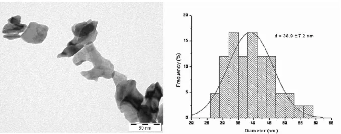
The NPs of PbO (diameter 38.9±7.2 nm, Figure 1) were synthesized at the Department of Ap- plied Chemistry, University of Szeged. A mixture of Pb(CH3COO)2 and NaOH was milled in a ball mill [reaction 1: Pb(CH3COO)2 + 2 NaOH → 2 Na(CH3COO) + Pb(OH)2]. After cal- cination [reaction 2: Pb(OH)2 → PbO + H2O] the mixture was washed and filtered. Chemical purity of the NPs was determined by X-ray diffraction, and their particle size, by X-ray dif-
Figure 1. TEM image (left) and size histogram (right) of the PbO NPs.
fraction and transmission electron microscopy. The NPs were suspended in a slightly viscous vehicle in order to prevent sedimentation (1% hydroxyethyl cellulose in PBS, pH 7.4; HEC) and the nanosuspension was mechanically resuspended immediately before each application.
During the whole study, the principles of the Ethical Committee for the Protection of An- imals in Research of the University were strictly followed. The methods used in the ex- periments were licensed by the authority competent in animal welfare issues under No.
XXI./02039/001/2006.
General toxicological investigation
Before every administration, body weight of the animals was measured and their general state was observed, noting all signs of toxicity (aggression, lethargy, rough fur, etc.). Following elec- trophysiological recording (see below), the animals were sacrificed by an overdose of urethane and were dissected. Organs – brain, liver, lungs, kidneys, spleen, femur – were removed and weighed, and relative organ weights, on the basis of brain weight, were calculated (brain weight was chosen because of the body weight effect of the treatments).
From the groups PbH3, PbH3+3, PbH6, PbH6+6 and the corresponding vehicle controls (CW3, CWV3+3, CW6, CWV6+6) 4 rats each were chosen randomly. Of these, whole blood (from the abdominal vein right after opening the abdominal cavity), brain and femoral bone samples were shock-frozen in liquid nitrogen and stored at -20°C, for later tissue Pb level determination (see below). Dissection and sample taking was done on separate days for each group which helped in avoiding cross-contamination.
To see the relationship of external dose, internal dose, and toxicological effects, rat-by-rat summed dose was calculated as the sum of daily amounts of Pb; based on actual body weight, the mg/ kg b. w. dose (Table 1) and the formula of the Pb compound used.
Behavioural investigation
The rats’ spontaneous locomotor behaviour pattern was assessed in an open field (OF) instru- ment at the end of the treatment period, 2 days after the last Pb application, always between 08:00 and 12:00. The OF box used was of 48x48x40 cm size and was equipped with two arrays of infrared beam gates at 1.5 and 12 cm above floor level, with 15 mm spacing between the beams (Conducta 1.0 System; Experimetria Ltd., Budapest, Hungary). After 20–30 min habitu- ation in the dimly lit test room, the animals were put into the centre of the box one by one for a 10 min session. From the beam interruptions, event counts and summed time of the basic activ- ity forms (ambulation, local activity, rearing, immobility), as well as run length of ambulation, were computed as follows: more than 40 mm shift in the location of interrupted beams at the floor level during a time unit of 1 s was interpreted as horizontal activity; less shift, as local activity; and no shift at all, as immobility. Rearing was recorded if beams at floor level and at the higher level were interrupted simultaneously. In earlier works of us (Vezér et al., 2007) this method was found sensitive to metal-induced changes of motor activity.
Electrophysiological investigation
Electrophysiological recording was done on the day following the OF test. Preparation for recording, and the recording itself, was performed in urethane anaesthesia (1000 mg/kg ip:
Maggi and Meli, 1986). The left hemisphere was exposed, and a ball-tipped silver electrode was placed on the projection site of the contralateral whisker pad (barrel field: Waite, 2004) for recording evoked potentials (EPs). A pair of stimulating needle electrodes, delivering square electric pulses (3-4 V, 0.05 ms) was inserted among the whiskers. The punctum maximum of the EP was determined by single pulses, then one train of 50 pulses each at 1, 2 and 10 Hz fre- quency was applied. The EPs were amplified (1000x, 1.6 and 1000 Hz filtering limits) and fed, with 8 kHz rate of digitizing, in a PC running the software Neurosys 1.11 for electrophysiologi- cal recording and analysis (Experimetria Ltd., Budapest, Hungary). The 50 EPs per train were averaged, and onset latency measured, by means of the software.
From individual latency data, group means with standard deviation were calculated; these were further normalized to the corresponding vehicle control mean at 1 Hz stimulation frequency (measured in groups CW3, CWV3+3, CW6, CWV6+6) to make all group data comparable despite differences in the control values.
Determination of Pb level
Level of Pb was determined in the blood, brain and femoral bone samples mentioned above.
Approximately 1 g of the samples was dried at 80°C to constant weight, and digested in 4 ml 65% HNO3 at 90°C for 90 min. The digested matter was washed quantitatively into 100 ml measuring flasks and Pb determination was done from this diluted sample by inductively cou- pled plasma mass spectrometry. The measurements were done at the laboratory of the MOL Hungarian Oil and Gas Company, on an Agilent 7500 ce instrument, according to the Hungar- ian standard MSZ EN ISO 17294-2:2005.
Statistical analysis
The distribution of data was checked for normality by means of the Kolmogorov-Smirnov test.
Data analysis was done by one-way ANOVA. Post hoc analysis of group differences was per- formed by LSD test, setting the probability level at p > 0.05.
table i.
Treatment scheme with group codes, doses and treatment times.
Groups and treatment Group codes Doses
Untreated control C ---
Vehicle control, 3 weeks CW3, Distilled water, oral
Vehicle control, 6 weeks CW6 Distilled water, oral
Oral Pb, low dose, 3 weeks PbL3 Pb(CH3COO)2 80 mg /kg b.w., oral1
Oral Pb, high dose, 3 weeks PbH3 Pb(CH3COO)2 320 mg /kg b.w., oral2
Oral Pb, low dose, 6 weeks PbL6 Pb(CH3COO)2 80 mg /kg b.w., oral
Oral Pb, high dose, 6 weeks PbH6 Pb(CH3COO)2 320 mg /kg b.w., oral Combined vehicle control,
3+3 weeks CWV3+3 Distilled water, oral; then HEC intratracheal (it.) Combined vehicle control,
6+6 weeks CWV6+6 Distilled water, oral; then HEC it.
Combined Pb, low dose,
3+3 weeks PbL3+3 Pb(CH3COO)2 80 mg /kg b.w. oral;
then PbO NPs 2 mg /kg b.w. it.3 Combined Pb, high dose,
3+3 weeks PbH3+3 Pb(CH3COO)2 320 mg /kg b.w. oral;
then PbO NPs 2 mg /kg b.w. it.
Combined Pb, low dose,
6+6 weeks PbL6+6 Pb(CH3COO)2 80 mg /kg b.w. oral;
then PbO NPs 2 mg/kg b.w. it.
Combined Pb, high dose,
6+6 weeks PbH6+6 Pb(CH3COO)2 320 mg /kg b.w. oral;
then PbO NPs 2 mg/kg b.w. it.
1 equal to 51 mg/kg b.w. lead
2 equal to 204 mg/kg b.w. lead
3 equal to 1.73 mg/kg b.w. lead
table ii.Summed external Pb dose and tissue (blood, brain and femoral bone)Pb levels in the various groups after 3, 6 and 12 weeks of Pb exposure according to Table I.
Summed external dose (mg Pb/rat) Possibly absorbed amount (mg Pb/rat)1 Tissue Pb levels (µg/kg) GroupsBloodBrainFemur2
CW3--588.94 ± 100.41546.28 ± 78.35--
PbH3710.29±50.5571.03±5.062739.29 ± 622.963136.99 ± 315.61 ***259042.87 ± 40559.55 ***
CWV3+3----520.44 ± 96.79714.40 ± 484.34--
PbH3+3796.41±57.8196.57±7.029616.74 ± 1979.04 ***5894.15 ± 548.82 ***###339026.55 ±79372.43 ***
CW6--856,69±145,82659.34 ± 145.97--
PbH61675.23±67.67167.52±6.777464.30 ± 3076.79 **##5654.01 ± 909.01 ***###263573.42 ± 15850.43 ***$
CWV6+6--644.76 ± 138.62515.98 ± 29.42437.75 ± 18.96
PbH6+61819.80±83.51197.66±70.2415229.64 ± 4848.66 ***###♦°7182.00 ± 1342.53 ***###♦°399829.06 ± 65925.23 ***#♦♦Mean±SD, n=4.*, **, ***: p < 0.05, 0.01, 0.001 PbH3 vs. CW3; PbH3+3 vs. CWV3+3; PbH6 vs. CW6; PbH6+6 vs. CWV6+6#,##, ###: p < 0.05,0.01, 0.001 PbH3+3, PbH6, PbH6+6 vs. PbH3;♦: p < 0.05 PbH6+6 vs. PbH6°: p < 0.05 PbH6+6 vs. PbH3+3$: p <0.05 PbH3+3 vs. PbH6
1Calculated with the absorption percentages in Järup (2003): 10% for ingested and 50% for inhaled lead.2Femur control Pb levels were measured only in samples of the 6+6 weeks vehicle control (CWV6+6), due to financial limitations, and all data of treated rats’ femur Pb were compared to that. As, however, rats in this group had the longest period to accumulate Pb from the environmental background, Pb level in CWV6+6 was an obvious acceptable upper estimate of other vehicle treated rats’ levels.
table iii.
Relative change of the latency of somatosensory evoked potentials in the various treatment groups.
Groups Stimulation frequency
1Hz 2Hz 10Hz
CW3 1.00 ± 0.05 0.96 ± 0.04 1.04 ± 0.06
PbL3 0.99 ± 0.06 1.02 ± 0.08 1.08 ± 0.05 °
PbH3 1.03 ± 0.06 1.06 ± 0.04 1.12 ± 0.04 °°
CWV3+3 1.00 ± 0.04 1.03 ± 0.06 1.05 ± 0.03
PbL3+3 1.01 ± 0.05 1.03 ± 0.04 1.13 ± 0.04 *°°
PbH3+3 1.05 ± 0.04 1.10 ± 0.03 *° 1.15 ± 0.04 **°°
CW6 1.00 ± 0.03 1.01 ± 0.03 1.05 ± 0.02
PbL6 1.04 ± 0.03 1.06 ± 0.03 1.11 ± 0.04 °
PbH6 1.07 ± 0.02 1.13 ± 0.02 1.15 ± 0.02 *°°
CWV6+6 1.00 ±0.03 1.02 ± 0.03 1.05 ± 0.02 °
PbL6+6 1.06 ± 0.03 1.08 ± 0.03 * 1.13 ± 0.03 **°°
PbH6+6 1.07 ± 0.03 1.11 ± 0.03 **° 1.17 ± 0.05 **°°
Mean±SD, n=8.
*,**,***: p < 0.05, 0.01, 0.001 PbL3+3, PbH3+3 vs. CWV3+3; PbH6 vs. CW6; PbL6+6, PbH6+6 vs. CWV6+6
°,°°: p < 0.05, 0.01 vs. latency with 1 Hz stimulation within a treatment group.
RESULTS
General toxicity
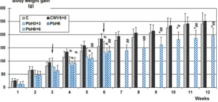
In Figure 2, body weight gain of the groups C, CWV6+6, PbH3+3, PbH6 and PbH6+6 is com- pared (the data of CWV3+3 were practically identical to those of CWV6+6 and were left out from the graph to avoid crowding). Weight gain deficit vs. controls on 6 weeks oral Pb treat- ment (PbH6 and the first part of PbH6+6) and on the 6 weeks combined treatment (PbH3+3) was similar although the daily amount of Pb administered it. was much lower than the amounts given orally. And, the onset of it. application (marked by arrows at the corresponding groups in Figure 2) caused an abrupt drop in body weight gain, first of all in group PbH6+6. Later on, the slope of weight gain in the combined treatment groups (PbH3+3, PbH6+6) was again as before the onset of it. application, that is why the plot of daily weight gain (total weight increase divid- ed by the numbers of days between start of treatment and sacrifice) against calculated summed dose (external dose) shown in Figure 3 gave a fair linear relationship.
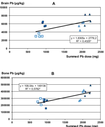
All Pb-treated groups showed significantly increased Pb levels in blood, brain and bone (fe- mur) samples at the end of the experiment (Table II). Brain Pb levels (Figure 4A) as a meas- ure of the internal dose likely to induce neuro-functional effects showed, besides a basic lin- earity, clear dependence on the physicochemical form: a little plus Pb administered in form of PbO NPs caused disproportionately high elevation of brain Pb. Bone Pb levels, representing whole-body internal dose, depended much more on the summed dose (Figure 4B).
Due to the effect of the treatments on body weight, relative weight data based on brain weight were evaluated. Relative weight of the lungs increased significantly in each group receiving Pb NPs (CWV3+3: 1.21 ± 0.29, PbL3+3: 2.52 ± 0.30, PbH3+3: 2.38 ± 0.32; p < 0.001 vs. CWV for both treated groups; CWV6+6:1.42 ± 0.11, PbL6+6: 2.87 ± 0.35, PbH6+6: 3.02 ± 0.56;
p < 0.001 vs. CWV for both treated groups). Relative kidney weight increased significantly vs.
the corresponding vehicle control in PbH3 (1.36 ± 0.19 vs. 1.18 ± 0.04, p < 0.05) PbH3+3 (1.54
± 0.18 vs. 1.34 ± 0.12, p < 0.05), PbH6 (1.76 ± 0.46 vs. 1.29 ± 0.18, p < 0.05), as well as in PbL6+6 (1.59 ± 0.16) and PbH6+6 (1.63 ± 0.12) vs. CWV6+6 (1.32 ± 0.16, p < 0.001 for both treated groups). Other organs showed no significant alteration of relative weight.
Figure 2. Time course of the rats’ body weight gain. Mean values, n=8. The arrows denote the onset of intratracheal application of PbO NPs. For group codes, see Table I.
*: p < 0.05 vs. C; #, ##: p < 0.05, 0.01 vs. CWV6+6
Figure 3: Relationship of individual treated rats’ calculated daily weight gain to their calculated summed Pb dose. The data plotted are of the same 4 animals per group whose data are also shown in Figs. 4 and 6.
Light squares, PbH3; filled squares, PbH3+3; light triangles, PbH6; filled triangles, PbH6+6. Insert:
equation and R2 of the fitted line; *: p < 0.05 (F-test for the linear fit).
y = -0,0012x + 6,1437 R2= 0,5743*
0 1 2 3 4 5 6 7
0 500 1000 1500 2000 2500
Summed Pb dose (mg) Daily weight gain (g)
y = 1,8305x + 2779,2 R2= 0,4325*
0 2000 4000 6000 8000 10000
0 500 1000 1500 2000 2500
A
Summed Pb dose (mg) Brain Pb (µg/kg)
y = 108,54x + 149136 R2= 0,5782*
0 100000 200000 300000 400000 500000 600000
0 500 1000 1500 2000 2500
B
Summed Pb dose (mg) Bone Pb (µg/kg)
Figure 4. Relationship of brain (A) and femoral bone (B) Pb level in individual treated rats to their calculated summed Pb dose. Insert and data point marking as in Fig. 3.
Neurotoxicity
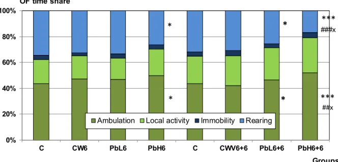
Open field motility: The time spent with immobility, local activity and ambulation was in- creased in the treated groups, significantly only in PbH6 and PbH6+6. In these groups, dimin- ished rearing was also significant (Figure 5). The dependence of OF motility on internal (brain) Pb doses is shown in Figure 6D.
Cortical evoked potentials: The latency of SS EPs was significantly lengthened in each treat- ment group vs. their controls (Table III). The data suggested that EP latency had a similar rela- tionship to external Pb dose as the brain Pb levels did (see Figure 6A-C). Plotting the individual latency values against the same rat’s brain Pb level indeed showed fair linear correlation (R2
> 0.25) but the data points representing rats of group PbH3+3 (filled squares) and PbH6 (light triangles) were in overlap; indicating that a lower overall dose, if it was partly in NP form, had ca. the same effect as a higher dose of dissolved Pb given orally. The same holds true also for OF motility and brain Pb levels (Figure 6D).
Figure 5. Data of open field motility in rats treated with Pb by the 6 and 6+6 weeks scheme and the corresponding controls. Mean+SD, n=8.
*, **, ***: p < 0.05, 0.01, 0.001 PbH6 vs. C; PbL6+6 vs. C; PbH6+6 vs. C (Rats in group C had an open field session after 6 weeks, and another one after 12 weeks).
#, ### p < 0.05, 0.001 PbH6+6 vs. CWV6+6
X: p < 0.05 PbH6+6 vs. PbL6+6
Figure 6. Relationship of the latency of somatosensory evoked potential (A, B, C) and open field ambulation distance (D) of individual treated rats and their brain Pb level. Insert and data point marking as in Fig. 3.
0%
20%
40%
60%
80%
100%
C CW6 PbL6 PbH6 C CWV6+6 PbL6+6 PbH6+6
OF time share
Groups Ambulation Local activity Immobility Rearing
***###x
***##x
*
*
*
*
DISCUSSION
The physicochemical form of Pb (dissolved or nanoparticulate) seemed to exert a substantial effect on several of the investigated toxicological parameters, and thus showed that the assump- tion which the experiments were based on was correct.
Tissue Pb levels in blood and brain, indicating internal dose, were higher in group PbH3+3 than in PbH6, although the overall treatment time was the same and the calculated summed applied dose and the possibly absorbed amount was approximately twice as high in PbH6 than in PbH3+3 (applied dose: 1,675.23 ± 67.67 mg Pb/rat vs. 796.41 ± 57.81 mg Pb/rat; absorbed amount: 167.52 ± 6.77 mg Pb/rat vs. 96.57 ± 7.02 mg Pb/rat; see Table II). Similarly, the differ- ence in blood and brain levels of PbH3+3 vs. PbH3, and PbH6+6 vs. PbH6, was much higher than the difference of the corresponding total amounts of Pb (summed external dose, mg Pb / rat) given orally and intratracheally. In parallel with that, latency lengthening of SS EP (Table III) in PbH3+3 and PbH6 was nearly equal, and in PbL3+3 was somewhat more than in PbL6 – even though the summed external dose was higher in the groups with oral application only.
The correlation of SS EP latency (Figure 6A-C) and OF ambulation distance (Figure 6D) with brain Pb level (i.e. internal dose) also supported that the NP form of Pb had a disproportionately strong influence on central nervous functions. Other toxicological parameters, however, i.e. Pb level in the femoral bone and daily body weight gain, were simply dependent on the summed external dose, irrespective of the form.
The more-than-additive interaction of dissolved and nanoparticulate Pb in the neuro-functional effects, as described above, could be due to weakened BBB (a known effect of Pb: Goldstein et al., 1974). After weeks of effect by dissolved Pb, BBB may have been less able to exclude NPs.
The extreme mobility and biological activity of NPs (Oberdörster et al., 2005) could in itself result in higher metal levels in the central nervous system, but comparison with one of our pre- vious experiments (Oszlánczi et al., 2011) suggested that the preceding effect of dissolved Pb (which was important in examining the interaction of two different forms and exposure modes of the same environmental heavy metal, see Introduction) was of importance. In the cited ex- periment, PbO NPs, highly similar to those used this time, was applied it. for 6 weeks in 4 mg/
kg b.w. dose and caused a relative increase of SS EP latency of ca. 1.05 (at 1 Hz stimulation frequency) while in the present work 3 weeks of 2 mg/kg b.w. were enough to cause a similar EP lengthening.
Oxidative stress induced by Pb, with ROS generation and membrane lipid peroxidation, was reported both in animals (Adonaylo and Oteiza, 1999; Patra et al., 2001) and humans (Ahamed and Siddiqui, 2007). Damaged lipids lead to changes in fluidity and other properties of the cell membrane, disturb- ing membrane- and receptor-bound phenomena (Coyle and Puttfarcken, 1993). The activity of as- trocytic glutamate transporter is reduced both by membrane lipid peroxidation (Willmore and Ueda, 2009) and direct effect of Pb2+ (Struzynska et al., 2005) which may result first in overexcitation and finally in desensitization of postsynaptic glutamate receptors, and this way, in increased latency of cortical EPs. Mitochondrial damage caused by Pb (Lidsky and Schneider, 2010) may have contrib- uted to the SS EP latency lengthening directly by energy shortage and indirectly by generating ROS.
Motivation, determining spontaneous open field locomotor activity, is regulated by mesolimbic and mesocortical dopaminergic structures (Alexander et al., 1990). Dopaminergic neurons are especially vulnerable to oxidative stress due to the auto-oxidizing tendency of dopamine and to the presence of monoamine oxidase producing hydrogen peroxide (Alexi et al., 2000), and oxidative stress is a likely consequence of Pb load in the CNS (as outlined above). Vertical mo- tility is an especially sensitive indicator of striatal dopaminergic activity (Sedelis et al., 2001), and significantly diminished rearing was observed in groups PbH6, PbL6+6 and PbH6+6 (with anomalous dose-dependence similar to that seen in EP latency). In the same groups, increased locomotion was also observed, which was similar to what was reported by Ma et al. (1999) from rats following Pb exposure during intra-and extrauterine development, and was explained there by decreased cortical D2 receptor level. A possible human analogue is “attention defi- cit hyperactivity disease” found to be more frequent among children with elevated blood Pb (Needleman and Gatsonis, 1990).
CONCLUSION
As xenobiotic heavy metals are found both in the occupational and residential environment and in commodities of environmental origin (here: drinking water and food), the health effects in individuals exposed to both sources, and in particular the effects on sensitive systems like the nervous system, are of especial concern. The more-than-additive neurotoxic interaction of nanoparticulate and dissolved Pb demonstrated in the presented work underlines the health rel- evance of such combined exposures and the necessity of investigations in this field.
ACKNOWLEDGEMENT
The authors are thankful to Mr. József Koszta and Ms. Edit Pálinkás at the laboratory of the MOL Hungarian Oil and Gas Company for the Pb level measurements. The authors acknowl- edge the financial support by the Hungarian grant TÁMOP-4.2.2./B-10/1-2010-0012.
REFERENCES
ADONAYLO V.N. and OTEIZA I. (1999). Lead intoxication: antioxidant defences and oxida- tive damage in rat brain. Toxicology 135:77–85.
ATSDR – AGENCY FOR TOXIC SUBTANCES AND DISEASE REGISTRY. (1999).
Toxicological Profile for Lead. Department of Health and Human Services, Atlanta, USA.
AHAMED M. and SIDDIQUI M.K.J. (2007). Low level lead exposure and oxidative stress:
Current opinions. Clin. Chim. Acta 383:57–64.
ALEXANDER G.E., CRUTCHER M.D., and DELONG M.R. (1990). Basal ganglia-thalam- ocortical circuits: parallel substrates for motor, oculomotor, “prefrontal” and “limbic” func- tions. Prog. Brain Res. 85:119-146.
ALEXI T., BORLONGAN C.V., FAULL R.L.M, et al. (2000). Neuroprotective strategies for basal ganglia degeneration: Parkinson’s and Huntington’s diseases. Prog. Neurobiol. 60:409–470.
ARAKI S., SATO H., YOKOYAMA K., and MURATA K. (2000). Subclinical neurophysi- ological effects of lead: A review on peripheral, central, and autonomic nervous system effects in lead workers. Am. J. Ind. Med. 37:193–204.
CHISOLM J.J. (1965). Chronic lead intoxication in children. Dev. Med. Child Neurol. 7:529–36.
COYLE J.T. and PUTTFARCKEN P. (1993). Oxidative stress, glutamate and neurodegenera- tive disorders. Science 262:689-695.
GOLDSTEIN G.W., ASBURY A.K., and DIAMOND I. (1974). Pathogenesis of lead encepha- lopathy. Uptake of lead and reaction of brain capillaries. Arch. Neurol. 31:382–389.
GRIFFIN T.B., COULSTON F., WILLIS H., and RUSSELL J.C. (1975a) Biologic effects of airborne particulate lead on continuously exposed rats and rhesus monkeys. Environ. Qual.
Saf. Suppl. 2:202–220.
GRIFFIN T.B., COULSTON F., WILLIS H., and RUSSELL J.C. (1975b) Clinical studies on men continuously exposed to airborne particulate lead. Environ. Qual. Saf. Suppl. 2:221–240.
HAELFIGER P., MATHIEU-NOLF M., LOCICIRO S., et al. (2009). Lead intoxication from informal used lead-acid battery recycling in Dakar, Senegal. Environ. Health Persp. 117:1535–1540.
HORVÁTH E., MÁTÉ ZS., TAKÁCS SZ., et al. (2012). General and electrophysiological toxic effects of manganese in rats following subacute administration in dissolved and nanopar- ticle form. Sci. World J. Article ID 520632.
JAKUBOWSKI M. (2011). Low-level environmental lead exposure and intellectual impairment in children – The current concepts of risk assessment. Int. J. Occup. Med. Environ. Health 24:1-7.
JÄRUP L. (2003). Hazards of heavy metal contamination. Br. Med. Bull. 68:167–182.
JEYARATNAM J., DEVATHASAN G., ONG N., et al. (1985). Neurophysiological studies on workers exposed to lead. Br. J. Ind. Med. 42:173–177.
LIDSKY T.I. and SCHNEIDER J.S. (2010). Lead neurotoxicity in children: basic mecha- nisms and clinical correlates. Brain 126:5–19.
LILLE F., HAZEMANN P., GARNIER R., and DALLY S. (1988) Effects of lead and mer- cury intoxications on evoked potentials. J.Toxicol. Clin. Toxicol. 26:103–116.
MA T., CHEN H.H., and HO I.K. (1999). Effects of chronic lead (Pb) exposure on neurobehav- ioral function and dopaminergic neurotransmitter receptors in rats. Toxicol. Lett. 105:111–121.
MAGGI C.A. and MELI A. (1986). Suitability of urethane anesthesia for physiopharmacolog- ical investigations in various systems. Part 1: General considerations. Experientia 42:109-114.
NEEDLEMAN H.L. and GATSONIS C.A. (1990). Low-level lead exposure and the IQ of chil- dren. A meta-analysis of modern studies. JAMA 263:673–678.
OBERDÖRSTER G., FINKELSTEIN J.N., JOHNSTON C., et al. (2000). Acute pulmonary effects of ultrafine particles in rats and mice. Res. Rep. Health Eff. Inst. 96:5–74; disc. 75-86.
OBERDÖRSTER G., OBERDÖRSTER E., and OBERDÖRSTER J. (2005). Nanotoxicol- ogy: An emerging discipline evolving from studies of ultrafine particles. Environ. Health Persp.
113:823–839.
OSZLÁNCZI G., PAPP A., SZABÓ A., et al. (2011). Nervous system effects in rats on suba- cute exposure by lead-containing nanoparticles via the airways. Inhal. Toxicol. 23:173–181.
PATRA R.C., SWARUP D., and DWIVEDI S.K. (2001). Antioxidant effects of alpha-tocoph- erol, ascorbic acid and L-methionine on lead induced oxidative stress to the liver, kidney and brain in rats. Toxicology 162:81–88.
PECZE L., PAPP A., INSTITÓRIS L., et al. (2005). Acute and subchronic effects of lead on the central and peripheral nervous system in rats in combination with alcohol. Ecotoxicol. En- viron. Saf. 61:139–144.
ROGAN W.J., DIETRICH K.M., WARE J.H., et al. (2001) The effect of chelation therapy with succimer on neuropsychological development in children exposed to lead. N. Engl. J.
Med. 344:1421–1426.
ROSNER D. and MARKOWITZ D. (2007). The Politics of Lead Toxicology and the Devas- tating Consequences for Children. Amer. J. Ind. Med. 50:740–756.
RUFF H.A., MARKOWITZ M.E., BIJUR P.E., and ROSEN J.F. (1996). Relationship among blood lead levels iron deficiency, and cognitive development in two year old children. Environ.
Health Persp. 104:180–185.
SEDELIS M., SCHWARTING R.K., and HUSTON J.P. (2001). Behavioral phenotyping of the MPTP mouse model of Parkinson’s disease. Behav. Brain Res. 125:109–125.
STRUZYNSKA L., CHALIMONIUK M., and SULKOWSKI G. (2005). The role of astroglia in Pb-exposed adult rat brain with respect to glutamate toxicity. Toxicology 212:185–194.
VEZÉR T., KURUNCZI Á., NÁRAY M., et al. (2007). Behavioral effects of subchronic inor- ganic manganese exposure in rats. Am. J. Ind. Med. 50:841–852.
WAITE P.M. (2004). Trigeminal sensory system. In: The Rat Nervous System (G.Paxinos, ed.), Academic Press, New York, pp.817-851.
WILLMORE L.J. and UEDA Y. (2009). Posttraumatic epilepsy: Hemorrhage, free radicals and the molecular regulation of glutamate. Neurochem. Res. 34:688–697.
YOKOYAMA K., ARAKI S., YAMASHITA K., et al. (2002). Subclinical cerebellar anterior lobe, vestibulocerebellar and spinocerebellar afferent effects in young female lead workers in Chi- na: computerized posturography with sway frequency analysis and brainstem auditory evoked potentials. Ind. Health 40:245–253.