PLASMID COPY NUMBER AND QNR GENE EXPRESSION IN SELECTION OF FLUOROQUINOLONE-RESISTANT
ESCHERICHIA COLI
DÁNIEL GULYÁS*, BÉLA KOCSIS and DORA´ SZAB ´O
Institute of Medical Microbiology, Semmelweis University, Budapest, Hungary
(Received: 21 September 2018; accepted: 19 October 2018)
Fluoroquinolone resistance in Enterobacteriales is developed by chromosomal and plasmid-mediated mechanisms. Plasmids play an important role in dissemination of resistant genes and they carry genes that protect bacteria in different stress-induced situations. In this study, we studiedEscherichia colistrains, each carried one plasmid- mediated quinolone resistance determinant namely,qnrA1, qnrB1, qnrC1, andqnrD1.
We exposed 0.5 McFarland density of each strain to 0.5 mg/L ciprofloxacin from the period of 30, 60, 90, and 120 min over 24 h. All treated strains were further exposed to a constantly increasing 1, 2, 4, and 8 mg/L ciprofloxacin solution through 24, 48, and 120 h. In given timepoints, RNA was extracted from all treated strains. Expression of qnrA1, qnrB1, qnrC1, andqnrD1was investigated by quantitative PCR. Mutations in gyrAand parCgenes were analyzed by PCR and nucleic acid sequencing. In this study, during 0.5 mg/L ciprofloxacin exposition, the following expression levels were detected: 1.2 forqnrA1, 1.47 forqnrD1, 12.44 forqnrC1, and 80.63 forqnrB1. In case of long-term study, we selected a resistant strain inqnrB1-positiveE. coli, and its expression increased from 105.91 to 212.31. On the contrary, plasmid copy number increased in time from 1 to 4.13. No mutations ingyrAor inparCchromosomal genes of treated strains were detected. Our results show thatqnrB1-positiveE. colistrain was able to developfluoroquinolone resistance by upregulatedqnrB1expression that was linked to a minor increase in plasmid copy number but no mutations occurred ingyrA orparC.
Keywords:fluoroquinolone resistance,qnrdeterminants,qnrgene expression Introduction
Fluoroquinolones are broad-spectrum antibiotics with bactericidal effect on Gram-negative and Gram-positive bacteria [1]. They bind to bacterial gyrase and
*Corresponding author; E-mail:dnlglys@gmail.com
topoisomerase IV enzymes, thereby inhibit DNA synthesis. Fluoroquinolones have a good penetration in various human tissues; therefore, they are used asfirst- line therapy of complicated urinary tract infections (UTIs) [2] and community- acquired pneumonia [3]. Furthermore, tuberculosis and infections of abdominal cavity, skin and soft tissue, bone, and joint can also be treated with fluoroquinolones [4].
Fluoroquinolone-resistant strains show a raising tendency worldwide and it causes new challenges for effective treatment. According to data of Nature Reviews Urology, global prevalence rate in Gram-negative bacteria demonstrated 75% in China, India, and Central America, whereas in Hungary, proportion of these resistant bacteria is about 15%–20% [5]. Based on ECDC database, fluoroquinolone resistance in Escherichia coli and Klebsiella pneumoniae is about 30%–40% [6].
Fluoroquinolone resistance is caused by chromosomal and plasmid-mediated mechanisms. High-level fluoroquinolone resistance with minimum inhibitory concentration (MIC) of 1 mg/L or higher values is developed by chromosomal mutations in quinolone-resistance determining regions (QRDRs), namely in gyrase enzyme coding gyrA and gyrB genes and in topoisomerase IV coding parC and parE genes. Plasmid-mediated quinolone resistance (PMQR) confers reduced susceptibility and low-level fluoroquinolone resistance [7,8]. Plasmids are double-stranded, superhelical extrachromosomal DNA molecules with vari- able size from 1,000 to 100,000 base pairs. Plasmids are not essential elements for bacteria; therefore, microbes do not carry and express plasmid-coded genes under non-stressed conditions, because it would cause an unnecessary energy burden [9, 10]. Plasmids can replicate autonomously from the cell cycle’s replication phase, but they consume ATP and ezymes of host cell. Plasmid copy number is an amount of plasmid DNA compared to that of housekeeping genes [10]. Plasmids carry various genes [11–13], and the most important of them are antibiotic resistance genes. Expression of these genes can protect bacteria in stress-induced conditions, e.g., in an environment containing antibiotic. PMQRs include three groups of determinants: Qnr-protective proteins, enzymatic modification of bifunctional aminoglycoside acetyltransferase-Ib-cr [ACC(6′)-Ib-cr], and efflux pumps (QepA and OqxAB) [14]. Each PMQR determinant is able to maintain 0.125–0.5 mg/L ciprofloxacin MIC value and facilitate selection of fluoroquino- lone resistance, as frequency of chromosomal mutations will be increased [7,8].
Thefirst PMQR gene,qnrA1, was detected in 1998 inK. pneumoniae[7]. Since then, several determinants were described and have been identified worldwide in Enterobacteriaceae, mainly in K. pneumoniae, Enterobacter spp., E. coli, andSalmonella entericaboth in community- and nosocomial-acquired infections [15]. PMQR genes are associated with other resistance determinants in
Enterobacteriaceae, often with extended-spectrum beta-lactamases (ESBLs) [16].
Conjugative plasmid that carry qnrB19, blaKPC-3, blaSHV-11, blaTEM-1, and aac(6′)-Ib was detected in K. pneumoniae [17]. Furthermore, armA, qnrS1, aac(6′)-Ib-cr, blaCTX-M-15, blaTEM-1, and blaNDM-1 were also detected on trans- ferable plasmid in K. pneumoniae [18]. In E. coli, qepA, armA, and blaTEM-1 resistance determinants were found on a single conjugative plasmid [19].
The global prevalence of qnr determinants andaac(6′)-Ib-cr ranges from 0.2% to 94% depending on studied strains [19–21]. In Northwest Iran, prevalence of PMQRs in Enterobacteriaceae isolated from UTIs showed a very high prevalence about 89.1% and among themaac(6′)-Ib-crwas the most frequently detected [22]. In Hungary, prevalence of PMQR-positive Enterobacteriaceae isolated from urine clinical samples showed 17.7% [23]. In the past 8 years, incidence of PMQRs among ESBL-producing E. coli andK. pneumoniaefrom bloodstream infection increased in Hungary [24].
Materials and Methods Strains
In this study, we included E. coli TG1 control strains, where each were transformed by plasmids carrying qnrA1 (GenBank accession number:
AY070235),qnrB1(GenBank accession number: DQ351241),qnrC1(GenBank accession number: EU917444), and qnrD1 (GenBank accession number:
FJ228229) [25]. The ciprofloxacin MIC of tested strains was 0.5 mg/L.
Antimicrobial susceptibility testing was performed according to EUCAST 2016 protocol.
Ciprofloxacin exposure
In our investigation, we performed short-term study as we analyzed time dependence offluoroquinolone resistance development. Each strain was adjusted to 0.5 McFarland density and was exposed to 0.5 mg/L ciprofloxacin from the period of 30, 60, 90, and 120 min over 24 h. After the strains adapted to ciprofloxacin, we performed long-term study to analyze concentration dependence offluoroquinolone resistance. The 0.5 McFarland solutions of treated strains were exposed in constantly increasing 1, 2, 4, and 8 mg/L ciprofloxacin solutions through 24, 48, and 120 h. All investigations of ciprofloxacin exposition were performed in Mueller–Hinton broth.
Bacterial RNA extraction
At given timepoints, total RNA of treated strains was extracted by Qiagen RNeasy Mini Kit (Hilden, Germany). Briefly, samples were placed into an Eppendorf tube, and were centrifuged at 5,000×g over 10 min and supernatants were removed. Tris-EDTA pH 8 buffer containing 20μl proteinase K and 200 μl lyzozime was added to the pellet of each strain. It was followed by incubation at 15–25 °C and samples were vortexed and RLT buffer was added. An amount of 700μl from the solution was pipetted to RNeasy Mini spin columns and centrifuged at 8,000×gfor 15 s. An amount of 700μl RW1 buffer was added and centrifuged at 8,000×gthrough 15 s, 500μl RPE buffer was added and centrifuged at 8,000×g over 15 s, and this step was repeated. An additional 500μl RPE-buffer was added and centrifugation at 8,000×g for 2 min. After the last centrifugation step, the sample was transfered into a new tube, and eluation of RNA was performed into 50μl RNase-free water by centrifugation at 8,000×gover 1 min.
RT-real-time PCR
Gene expressions of qnrA1, qnrB1, qnrC1, and qnrD1 and plasmid copy numbers were quantified. Extracted RNA of each tested strain was applied in RT-real-time PCR in a Step One Real-Time PCR System (Applied BioSystems, Thermo Fisher Scientific, Foster City, CA, USA) using PCR protocol: 60 °C for 30 s, 50 °C for 5 min, 95 °C for 10 min, and (95 °C for 15 s and 60 °C for 1 min)×40 cycles, and 60 °C for 30 s. Oligonucleotid primers and probes were designed by Primer Express 3.0 software (Applied BioSystems, Thermo Fisher Scientific, Foster City, CA, USA). Eachqnrdeterminant and each plasmid backbone were targeted. Chromosomal housekeepingicdgene was used as internal control.
We normalized each targeted sequence to icd gene according to CT values with formula 2−ΔΔCT, where ΔΔCT=(CTgene of interest−CTinternal control)studied strain− (CTgene of interest−CTinternal control)control strain.
Mutations ofgyrA andparC genes
Mutations ingyrAandparCgenes were analyzed by PCR and nucleic acid sequencing. Each treated strain was suspended in 500 μl bidestillated water (Millipore Merk, Darmstadt, Germany) and each was incubated at 100 °C for 15 min, and centrifuged at 13,000 rpm for 10 min at 4 °C. Supernatant was applied as DNA template in PCR, and mixture contained 1 Unit DNA Taq polymerase (Sigma-Aldrich, St. Louise, MO, USA) and 10 pmol from each primer namely, gyrA F-5′-CAG CCC TTC AAT GCT GAT-3′ and gyrA R-5′-CGC TTT TAC
TCC TTT TCT GTT C-3′and parC F-5′-CTC AAT CAG CGT AAT CGC C-3′ and parC R-5′-AAT CCT CAG CCG ATC TCA C-3′. Oligonucleotid primers of this study were designed by Eurofins Genomics online tools. PCR protocol was programmed as follows: 96 °C for 3 min, (95 °C for 1 min, 52 °C for 1 min, and 72 °C for 1 min) for 30 cycles, and itfinished 72 and 4 °C for 5 min each asfinal step. PCR amplicons were purified by Qiagen PCR purification Kit (Hilden, Germany) and samples were sent to be sequenced (BIOMI Kft, Gödöllo).˝ Obtained sequences were analyzed against NCBI GenBank database.
Results
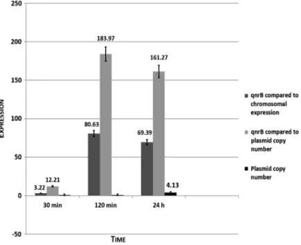
In short-term study, during exposure to 0.5 mg/L ciprofloxacin solution from 30 min to 24 h, we detected the following results:qnrA1andqnrD1showed 1.2 and 1.47 level expressions,qnrC1was 12.44. Compared to these three studied qnrdeterminants,qnrB1demonstrated a 3.22–80.63 expression. We also studied copy numbers of plasmids carryingqnrA1, qnrB1, qnrC1, andqnrD1 genes. In qnrA1 andqnrD1plasmids, 1–1.4-folds were detected, in qnrC1plasmid, copy number changed from 3.1 to 4.42, while it reached fold change of 4.13 inqnrB1 plasmid. Data are shown in Figure1.
Figure 1.Results of short-term study in case ofqnrB1
We implicated the treated strains into long-term study. Treated strains were adjusted to 0.5 McFarland and were kept in constantly increasing 1, 2, 4, and 8 mg/L ciprofloxacin solutions. According to this model, resistant strain was selected in qnrB1carrierE. coli. In course of long-term study, 105.91 and 212.31qnrB1level expressions were observed in 4 and 8 mg/L, respectively (Figure2). On the other hand,qnrBplasmid copy number reached 4.13-fold increase. In this study, we did notfind mutations in QRDRs.
Discussion
The selection of fluoroquinolone resistance in E. colistrains was investi- gated. In the case of qnrB1-positive E. coli, resistant strain was obtained by 0.5–8 mg/L ciprofloxacin exposure. Plasmid copy number change had minor role in this process, so our results show that selection of resistant strains inqnrB1-positive E. coli was mainly caused by increase of qnrB expression. Furthermore, in this study, no mutations in QRDR were detected. In the case of qnrA-, qnrC-, and qnrD-positive E. coli, in this study, these strains could adapt to 0.5 mg/L ciprofloxacin withqnrexpressions ranging from 1.2 to 12.44, but further selection was not possible.
Figure 2.Results of long-term study in case ofqnrB1
The importance of detectedqnrBexpression is that this PMQR determinant plays a role in SOS-response regulation system [26,27]. This response is activated when bacteria gets into a stress-induced condition, such as a DNA damage by UV, oxidative stress, metabolic pH-change or when it senses antibiotic in its environment. Consequently, the SOS-response is triggered by a factor that can be the increasing ciprofloxacin concentration. Importance of SOS-response is to protect bacterial DNA from harmful effects by enhancing mutation frequence and by production of protective proteins. Two regulator proteins play key role in SOS-reponse, namely LexA transcription repressor and RecA coprotease. The interactions between these two proteins help bacteria to survive [28].
The function of these proteins is as follows. Promoter ofqnrBgene includes CTGT binding site of LexA-protein. If bacteria do not sense any damaging effect, e.g., in ciprofloxacin-free condition, then LexA binds to CTGT region, so synthesis of RNA is blocked, and RecA protein is inactive and expression ofqnrB takes a basic level (Figure3). When antibiotic concentration shows an increase, it means a warning signal for bacteria, and RecA coprotease activates (with ssDNA arising) and aids autoproteolysis of LexA transcription repressor. Autoproteolized LexA leaves CTGT region, so RNA synthesis disengages from inhibition and synthesis of QnrB protein will be upregulated. QnrB is a pentapeptide-repeat protein that binds to gyrase through protein–protein interactions and protects it from ciprofloxacin. Consequently, MIC rises and susceptibility decreases (Figure4) [26–28].
Figure 3.SOS-response in ciprofloxacin-free environment
Our studies were performed according to EUCAST protocol from 2016, but this was changed in January 2017. According to this new protocol, ciprofloxacin resistance breakpoint was revised from 1 to 0.5 mg/L [29]. Our results correlate with this change, as we demonstrated that each testedE. coli could survive in 0.5 mg/L ciprofloxacin concentration by an increased qnrexpression.
The clinical relevance of our results is that ciprofloxacin is commonly sub- scribed to treat UTIs. It is estimated that around 150 million UTI cases are diagnosed each year and about 75% are caused byE. coli[22]. Ciprofloxacin concentrations used in this study demonstrate tissue concentrations during a per os therapy [30]. Thus, E. coli that carries a qnr determinant can develop resistance by increased qnr expression during a ciprofloxacin therapy that can lead to therapy failure.
Furthermore, the most common PMQR resistance gene isqnrBin the world and it has the highest number of variants (almost 100) amongqnrdeterminants [25].
The reason of this abundance can have evolutionary advantage against other qnr determinants. This theory is supported by the fact thatqnrBis often associated with other resistance genes, such as blaCTX-M-15, blaCTX-M-14, and aac(6′)-Ib-cr [22], which can cause multiresistance and facilitate dissemination of resistant strains [9].
Acknowledgements
The authors would like to thank Giuseppe Cornaglia, (Università degli Studi di Verona) for providing qnrA1-, qnrB1-, qnrC1-, and qnrD1-positive E. coli
Figure 4.SOS-response in an environment with raising ciprofloxacin concentrations
control strains. This study was financially supported by OTKA Hungarian Scientific Fund, grant number 108481.
Conflict of Interest There are no conflicts of interest.
References
1. Redgrave, L. S., Sutton, S. B., Webber, M. A., Piddock, L. J. V.: Fluoroquinolone resistance: Mechanisms, impact on bacteria, and role in evolutionary success. Trends Microbiol22, 438–445 (2014).
2. Grabe, M., Bartoletti, M., Bjerklund Johansen, T. E., Cai, T., Cek, M., Köves, B., Naber, K. G., Pickard, R. S., Tenke, P., Wagenlehner, F., Wullt, B.: Guidelines on Urological Infections. European Association of Urology, Arnhem, 2015, 24–25.
3. Van Berge Henegouwen, J. M., Groeneveld, G. H., de Boer, M. G. J., Visser, L. G.: A more restrictive use of quinolones in patients with community acquired pneumonia is urgently needed. Neth J Med75, 462–463 (2017).
4. Naeem, A., Badshah, S. L., Muska, M., Ahmad, N., Khan, K.: The current case of quinolones: Synthetic approaches and antibacterial activity. Molecules21, 268 (2016).
5. Zowawi, H. M., Harris, P. N., Roberts, M. J., Tambyah, P. A., Schembri, M. A., Pezzani, M. D., Williamson, D. A., Paterson, D. L.: The emerging threat of multidrug-resistant Gram-negative bacteria in urology. Nat Rev Urol12, 570–584 (2015).
6. ECDC database: Surveillance Atlas of Infectious Diseases. Available athttp://atlas.ecdc.
europa.eu/public/index.aspx
7. Martínez-Martínez, L., Pascual, A., Jacoby, G. A.: Quinolone resistance from a transferable plasmid. Lancet351, 797–799 (1998).
8. Rodríguez-Martínez, J. M., Machuca, J., Cano, M. E., Calvo, J., Martínez-Martínez, L., Pascual, A.: Plasmid-mediated quinolone resistance: Two decades on. Drug Resist Updat 29, 13–29 (2016).
9. Carattoli, A.: Resistance plasmid families in Enterobacteriaceae. Antimicrob Agents Chemother53, 2227–2238 (2009).
10. Thomas, C. M., Nielsen, K. M.: Mechanisms of, and barriers to, horizontal gene transfer between bacteria. Nat Rev Microbiol3, 711–721 (2005).
11. Watson, R., Rowsome, W., Tsao, J., Visentin, L. P.: Identification and characterization of Col plasmids from classical colicin E-producing strains. J Bacteriol147, 569–577 (1981).
12. Shintani, M., Takahashi, Y., Yamane, H., Nojiri, H.: The behavior and significance of degradative plasmids belonging to Inc groups inPseudomonaswithin natural environments and microcosms. Microbes Environ25, 253–265 (2010).
13. Johnson, T. J., Nolan, L. K.: Pathogenomics of the virulence plasmids ofEscherichia coli.
Microbiol Mol Biol Rev73, 750–774 (2009).
14. Yanat, B., Rodríguez-Martínez, J. M., Touati, A.: Plasmid-mediated quinolone resistance in Enterobacteriaceae: A systematic review with a focus on Mediterranean countries. Eur J Clin Microbiol Infect Dis36, 421–435 (2017).
15. Rodríguez-Martínez, J. M., Cano, M. E., Velasco, C., Martínez-Martínez, L., Pascual, A.:
Plasmid-mediated quinolone resistance: An update. J Infect Chemother 17, 149–182 (2011).
16. García-Fulgueiras, V., Bado, I., Mota, M. I., Robino, L., Cordeiro, N. F., Varela, A., Algorta, G., Gutkind, G., Ayala, J. A., Vignoli, R.: Extended-spectrumβ-lactamases and plasmid-mediated quinolone resistance in enterobacterial clinical isolates in the paediatric hospital of Uruguay. J Antimicrob Chemother 66, 1725–1729 (2011).
17. Endimiani, A., Carias, L. L., Hujer, A. M., Bethel, C. R., Hujer, K. M., Perez, F., Hutton, R. A., Fox, W. R., Hall, G. S., Jacobs, M. R., Paterson, D. L., Rice, L. B., Jenkins, S. G., Tenover, F. C., Bonomo, R. A.: Presence of plasmid-mediated quinolone resistance in Klebsiella pneumoniaeisolates possessingblaKPCin the United States. Antimicrob Agents Chemother52, 2680–2682 (2008).
18. Wei, D. D., Wan, L. G., Yu, Y., Xu, Q. F., Deng, Q., Cao, X. W., Liu, Y.: Characterization of extended-spectrum beta-lactamase, carbapenemase and plasmid quinolone determinants in Klebsiella pneumoniaeisolates carrying distinct types of 16s rRNA methylase genes and their association with mobile genetic elements. Microb Drug Resist21, 186–193 (2015).
19. Perichon, B., Bogaerts, P., Lambert, T., Frangeul, L., Courvalin, P., Galimand, M.:
Sequence of conjugative plasmid pIP1206 mediating resistance to aminoglycosides by 16s rRNA methylation and to hydrophilicfluoroquinolones by efflux. Antimicrob Agents Chemother52, 2581–2592 (2008).
20. Strahilevitz, J., Jacoby, G. A., Hooper, D. C., Robicsek, A.: Plasmid-mediated quinolone resistance: A multifaceted threat. Clin Microbiol Rev22, 664–689 (2009).
21. Robicsek, A., Strahilevitz, J., Jacoby, G. A., Macielag, M., Abbanat, D., Park, C. H., Bush, K., Hooper, D. C.: Fluoroquinolone-modifying enzyme: A new adaptation of a common aminoglycoside acetyltransferase. Nat Med12, 83–88 (2006).
22. Azargun, R., Sadeghi, M. R., Soroush, B, Arhaghi, M. H., Samadi Kafil, H., Yeganeh, F., Ahangar Oskouee, M., Ghotaslou, R.: The prevalence of plasmid-mediated quinolone resistance and ESBL-production inEnterobacteriaceaeisolated from urinary tract infec- tions. Infect Drug Resist11, 1007–1014 (2018).
23. Szab´o, O., Gulyás, D., Szab´o, N., Krist´of, K., Kocsis, B., Szab´o, D.: Plasmid-mediated quinolone resistance determinants inEnterobacteriaceaefrom urine clinical samples. Acta Microbiol Immunol Hung65, 255–265 (2018).
24. Domokos, J., Krist´of, K., Szab´o, D.: Plasmid-mediated quinolone resistance among extended-spectrum beta-lactamase producing Enterobacteriaceae from bloodstream infections. Acta Microbiol Immunol Hung63, 313–323 (2016).
25. http://www.lahey.org/qnrStudies/.
26. Wang, M., Jacoby, G. A., Mills, M. D., Hooper, D. C.: SOS regulation of qnrB expression.
Antimicrob Agents Chemother53, 821–823 (2009).
27. Da Re, S., Garnier, F., Guérin, E., Campoy, S., Denis, F., Ploy, M. C.: The SOS-response promotes qnrB quinolone-resistance determinant expression. EMBO Rep10, 929–933 (2009).
28. Janion, C.: Inducible SOS response system of DNA repair and mutagenesis inEscherichia coli. Int J Biol Sci4, 338–344 (2008).
29. http://www.eucast.org/clinical_breakpoints/.
30. Sanford, J. P., Moellering, C. R., Gilbert, N. D., Eliopoulos, M. G., Sande, A. M.:
The Sanford Guide to Antimicrobial Therapy, 47thEdition. Antimicrobial Therapy Inc., Sperryville, 2017, p. 91.