Stromal Matrix Protein Expression Following Preoperative Systemic Therapy in Breast Cancer
Anna-MariaTokes,1Attila Marcell Szasz,1Andrea Farkas,1Adrienn IldikoToth,1Magdolna Dank,2
Laszlo Harsanyi,3Bela Akos Molnar,3Istvan Arthur Molnar,3Zsolt Laszlo,4Zoltan Rusz,5and Janina Kulka1
Abstract Purpose:Stromal alterations are observed following preoperative systemic therapy in breast cancer. The aim of the present study was to analyze the qualitative and quantitative changes of representative tumor stroma proteins in the context of neoadjuvant therapy and the response of patients undergoing preoperative systemic therapy.
Experimental Design:Fifty women receiving preoperative systemic therapy were evaluated for clinical and pathologic parameters. Clinical response was defined according to International Union against Cancer (UICC) criteria, whereas pathologic responses to preoperative systemic therapy were defined according to the Chevallier and Sataloff classifications. The expression of tenascin-C, syndecan-1, collagen IV, and smooth muscle actin proteins was investigated using morphometric analysis of immunohistochemical reactions. Quantitative reverse transcription- PCR was done to evaluate the mRNA expression level of syndecan-1 and tenascin-C. The data were compared with 20 breast cancer samples of patients not treated with preoperative systemic therapy.
Results:According to UICC criteria, the expression levels of collagen IV were up-regulated in all preoperative systemic therapy ^ treated patients (P= 0.002). Collagen IV was up-regulated in the preoperative systemic therapy group in both Chevallier and Sataloff classifications compared with the control cases (P= 0.025 andP= 001, respectively). There were no significant differences in the expression of smooth muscle actin between the treated and nontreated groups. The synde- can-1 proteoglycan level was significantly down-regulated in the preoperative systemic therapy group (Chevallier classesP= 0.015, Sataloff classesP= 0.015). Tenascin-C was up-regulated in women with progressive disease (P= 0.005).
Conclusion:We have observed that the stromal component of breast carcinomas following pre- operative systemic therapy differs from the nontreated tumors, which can be evaluated with the analysis of the above mentioned proteins.
O
ver the past 15 to 30 years preoperative systemic therapy has been used to treat locally advanced and inflammatory breast carcinomas to make surgical therapy possible and to improve patient survival. Although long-term follow-up data do not support increased overall patient survival due to preoperative systemic therapy (1 –6), this strategy has been extended and isrecently being used for the treatment of patients with operable breast carcinomas to increase the rate of breast conservation.
There are many controversial discussions on the benefits and risks of preoperative systemic therapy. It is generally accepted that preoperative systemic therapy results in clinical response in 60% to 90% of patients, whereas pathologic complete response, the predictor of survival, occurs only in 3% to 16%
of patients (7, 8).
Following preoperative systemic therapy there is a significant change in the appearance of the tumor tissue; most strikingly the stromal alterations are visible during standard pathologic assessment. Once the surgical resection is done in a patient giving complete pathologic response, thus not giving evidence of the resection being done at the right location, there is need for a biological marker that can state the site of the original tumor in an efficient manner.
Despite the multitude of clinical trials and basic research concerning preoperative systemic therapy for breast cancer patients, there are still fundamental unanswered questions from a biological perspective. Which genetic and epigenetic factors influence the results of preoperative systemic therapy? Is there an expression change of stromal matrix proteins after preoperative systemic therapy or, for example, are distinct tumors foredoomed to fail to express proteins related to good
Authors’ Affiliations:1Second Department of Pathology,2Department of Diagnostic Radiology and Oncotherapy, and3First Department of Surgery, Semmelweis University ;4MaMMa Healthcare Institute; and5Department of Surgery, Schopf-Merei Hospital, Budapest, Hungary
Received 6/24/08; revised 7/25/08; accepted 8/16/08.
Grant support:Hungarian Society of Medical Oncology.
The costs of publication of this article were defrayed in part by the payment of page charges. This article must therefore be hereby markedadvertisementin accordance with 18 U.S.C. Section 1734 solely to indicate this fact.
Note:Supplementary data for this article are available at Clinical Cancer Research Online (http://clincancerres.aacrjournals.org/).
A-M. Tokes and A.M. Szasz contributed equally to this work.
Requests for reprints:Anna-Maria Tokes, Second Department of Pathology, Semmelweis University, 93 Ulloi ut, 1091 Budapest, Hungary. Phone: 36-1- 2157300, ext. 3430; Fax: 36-1-2156921; E-mail: tokesa1972@yahoo.co.uk.
F2009 American Association for Cancer Research.
doi:10.1158/1078-0432.CCR-08-1523
response/behavior? From a pathologic perspective, it is not clear to what extent the published results depend on the different classification systems of pathologic complete re- sponse. From a clinical perspective, it is still a matter of debate how the response to preoperative systemic therapy should be predicted.
To date, the classification of tumors being responsive or nonresponsive to preoperative systemic therapy remains controversial. The pathologic markers such as estrogen receptor and progesterone receptor status, human epidermal growth factor receptor 2/neu, p53, and Ki-67 immunohistochemistry are not useful in predicting the tumor responsiveness to preoperative systemic therapy (9, 10). As the response rate of breast cancers to preoperative systemic therapy is a short-term marker but with a long-term outcome and severe influence on patient lives, it is important to identify new and reliable factors that may predict the response to preoperative systemic therapy.
Up to now, the influence of the microenvironment on tumor cell survival and on the success of chemotherapy has been the subject of intense studies. The extracellular matrix is a complex structure of structural proteins, adhesive proteins, and proteo- glycans that provide essential factors for tumor development, detachment of cells, and migration (11). Recent research studies and publications point out the important role of extracellular matrix components in the effectiveness of chemo- therapy as well (12, 13).
The extracellular matrix plays an important role in modulating breast cancer cell behavior: structural proteins, e.g., basal membrane proteins (collagen IV), represent a barrier which preserves the structural integrity of ductal epithelial cell layers, whereas cell surface proteoglycans (such
as syndecan-1) modulate tumor cell adhesion, proliferation, and angiogenesis. Collagen IV is the most abundant protein of basal membranes, thus being very durable against mechanical and proteolytic insults (14). Collagen IV contributes to the proliferation of mammary epithelium, and is a major target for matrix metalloproteinase –mediated disruption of basal membrane during tumor cell invasion as well (15). The a- smooth muscle actin can be found in the myoepithelial cells of the breast ducts and in the smooth muscle cells and pericytes of vessels. In fibrocytes the up-regulated expression of smooth muscle actin can be the result of the activation of transforming growth factor-h(TGF-h) signaling in carcinomas (16). Syndecan-1 is a transmembrane-anchored heparan sulfate proteoglycan that can be converted into a soluble effector via matrix metalloproteinase –mediated shedding (17). It modulates a multitude of biological processes relevant to tumor progression, acting as a receptor for interstitial matrix proteins, and as a coreceptor that facilitates signal transduction through receptors regulating cell proliferation, chemokine activity, and protease activities (18). Studies in a xenograft breast cancer model indicate a modulation of tumor angiogenesis by stromal syndecan-1 (19), conforming with clinical correlation studies showing coexpression of syndecan- 1 and markers of angiogenesis (20, 21). Syndecan-1, a transmembrane heparan sulfate proteoglycan, is in the plasma membrane and regulates cell behavior through regulatory proteins. Although early studies provided evidence that heparan sulfate acts to suppress the malignant phenotype, evidence is mounting that heparan sulfate can also promote tumor growth and metastasis. Another adhesion modulatory extracellular matrix molecule, tenascin-C is a factor in the tumor-specific microenvironment that is highly expressed in most solid tumors. Tenascin-C actions promote malignant transformation, uncontrolled proliferation, metastasis, angio- genesis, and escape from tumor immunosurveillance (22, 23).
Preliminary results have shown that syndecan-1 –expressing carcinomas show decreased response to chemotherapy (5).
Ilunga et al. (24) described the association of tenascin-C expression with disease progression and poor prognosis.
Considering the above-mentioned information on the role of syndecan-1 and tenascin-C, these two extracellular matrix members are analyzed in our study in detail. The induction of changes in matrix protein expression as a consequence of preoperative systemic therapy can have a profound influence on breast cancer pathobiology, and potentially on clinical outcome. In the present study we analyzed the qualitative and quantitative changes of representative tumor stroma proteins following preoperative systemic therapy in the context of the response rate of breast cancer patients undergoing neo- adjuvant therapy, because only very few studies in this area on preoperative systemic therapy patient collectives are currently available.
Patients and Methods
Clinical records and surgical samples of patients receiving preoper- ative systemic therapy were evaluated for clinical and pathologic parameters. Fifty patients had available preoperative and postoperative immunhistochemical evaluation among the cases diagnosed in our institute from 2001 to 2007. The median age of the patients was 54.9 y (range, 29-73 y). Originally the diagnosis of carcinoma was established
Translational Relevance
Preoperative systemic therapy in operable breast cancer allows the increase of breast conservation rates and offers the ability to discerntreatment effectivityinvivo.TheNation- al Adjuvant Breast and Bowel Project trial established path- ologic complete response as a prognostic marker. Recent results showed that stromal enhancement ratio detected by dynamic contrast-enhanced magnetic resonance imaging is a potential indicator of response to preoperative systemic therapyandofoveralloutcomeinpatients withbreastcancer.
We have identified three stromal components (collagen IV, tenascin-C, and syndecan-1) that differentially expressed in tumors of preoperative systemic therapy ^ treated patients compared with nontreated cases. Recent studies have shown that patients with breast cancer have elevated serum levels of collagen IVcompared withhealthy women, and col- lagen IV levels increase further during chemotherapy. There are encouraging results of a pilot study seeking the feasibility and assess the efficacy among malignant glioma patients, of administering131I-labeled murine anti-tenascin monoclonal antibody. Syndecan-1 ^ expressing breast carcinomas show decreased response to chemotherapy.We believe that our data and the results mentioned above indicate that stromal protein level or quality changes may be important biological and clinicalindicators of tumor response topreoperativesys- temic therapy.
by fine-needle aspiration (in 38 cases) or core-needle biopsy (24 cases) and by both methods (in 17 cases) of the primary tumor and/or the metastasis in the palpable axillary lymph nodes. Distant metastases were screened and their absence was confirmed by chest X-ray, abdominal sonography, and bone scan. Patients were treated by different cycles of anthracyclin-based or taxane-based regimens accord- ing to the following protocol: All 50 patients had locally advanced disease (large tumor compared with breast size, or breast carcinoma with axillary metastasis at the time of the diagnosis). Those patients with axillary metastasis were treated with taxanes, whereas those without regional spread of the tumor underwent anthracyclin-based regimens. The pathologic response to preoperative systemic therapy was defined by the use of the Chevallier classification (25) and the Sataloff classification for the primary tumor (26). The clinical response to preoperative systemic therapy was classified according to the UICC criteria (27). Supplementary Table S1 presents detailed descriptions of the classification systems.
The expression levels of the above mentioned proteins were compared with 20 breast cancer samples of patients not treated with preoperative systemic therapy. The clinicopathologic parameters of patient cohorts are presented in Table 1. A general assessment was done comparing the two cohorts to see whether this latter group—diagnosed with invasive carcinoma from 2001 to 2003—can be used as a control population. The control tumors had available immunohistochemical data and were selected to represent matching tumor-node-metastasis status to the preoperative systemic therapy –treated patients.
Immunohistochemistry. With the use of immunohistochemistry, the expression of the following extracellular matrix proteins were analyzed:
structural proteins (collagen IV, smooth muscle actin), a proteoglycan (syndecan-1), and an adhesion molecule (tenascin-C).
The immunohistochemical reactions were done on 5-Am-thick paraffin sections. The deparaffinization steps were carried out for all the slides in the same way: after treatment in xylene (twice for 10 min), an ethanol gradient (96%, 75%, 50%) was used, each for 5 min followed by washing in PBS. Different antigen retrieval methods were used. For collagen IV and smooth muscle actin, antigen unmasking solution (Vector Laboratories) was used for 6 min. For syndecan-1, the slides were treated in microwave oven in a pH6.0 antigen retrieval solution (DAKO North America, Inc.) for 30 min. For tenascin-C, antigen retrieval was done in two steps: the slides were treated in antigen retrieval solution first (DAKO) for 30 min, followed by digestion with pepsin for 6 min. Monoclonal antibodies from DAKO were applied (Supplementary Table S2). Visualization was done with a standard three-step streptavidin-peroxydase system using 3,3-diamino- benzidine as chromogen. The reactions were carried out in a Ventana ES automatic immunostainer (Ventana Medical Systems, Inc.) using the reagents provided by the manufacturer. The sections were counter- stained with hematoxylin. Positive control cases with known reactivity or tissues recommended by the manufacturer were included in every run. A negative control with omission of the primary antibody was included for all of the analyzed proteins.
The immunohistochemical analysis was done in two ways: semi- quantitatively and by morphometric software. Ten representative digital photos were taken from each case at magnification of200 (1,320 1,02424 b in pixels) using an Olympus BX50F-3 microscope and DP70 digital CCD camera (Olympus Co.).
The images of the immunohistochemical reactions were analyzed with Leica Qwin V3.1.0 morphometric software (Leica Microsystems Imaging Solutions Ltd.) enabling an objective quantification of the immunohistochemical results. Binary transformation was done, and threshold levels were selected on the positive control tissues (collagen IV,R= 247,G= 188,B= 135; smooth muscle actin,R= 229,G= 182, B= 145; syndecan-1,R = 180,G = 116,B= 82; tenascin-C,R= 217, G = 182, B = 169, respectively). Evaluation was done on each digitalized image after correction for possible errors occurring from inhomogene tissue samples: rejecting the lumen of vessels and ducts with manual selection. The units of expression were based on pixels.
Real-time reverse transcription-PCR mRNA detection for syndecan-1 and tenascin-C. The formalin-fixed and paraffin-embedded blocks previously used for the immunohistochemical analyses were used for the evaluation. Depending on the size of the dissected area, two to eight 5-Am-thick sections were cut from each tissue block and placed in 1.5 mL RNase-free centrifuge tubes. RNA was extracted using the High Pure RNA Paraffin kit (Roche) according to the manufacturer’s protocol. After total RNA isolation, samples were kept at -80jC until further use. Total RNA integrity was verified as described previously (28). Five hundred nanograms of total RNA were reverse-transcribed at 37jC for 120 min with High Capacity cDNA Reverse Transcription Kit (Applied Biosystems-ABI) in the presence of RNase inhibitor (ABI).
The real-time reverse transcription-PCR (qPCR) reaction was done for syndecan-1, tenascin-C, and referenceGAPDH genes using 2 AL complementary DNA template in a total volume of 25AL using Power SYBR Green PCR Master Mix (ABI) and ABI Prism 7000 sequence detection system followed by melting curve analysis from 55jC to 95jC. The qPCR was done in duplicates in 96-well plates with the following running conditions: initial denaturation at 95jC for 10 min, then 40 cycles at 95jC for 15 s, at 60jC for 1 min, and finally at 72jC for 1 min. No primer-dimer formations were observed during the 40 qPCR amplification cycles. Tenascin mRNA expression was analyzed with two specially designed primer pairs. Alternative splicing of fibronectin-like repeats of tenascin-C generates a number of splice variants. As the material used for this study is paraffin-embedded, only the small variants (f200 bp) should be reliably detected. Melting analyses as well as gel electrophoresis were done after PCR reactions.
The primer pairs used were as follows:
Syndecan-1 (GI: 55749479): forward 5¶-GCCGCAAATTGTGGC- TACT-3¶; reverse 5¶-GCT GCG TGT CCT TCC AAG T-3
Tenascin-C (GI: 89161216): forward 5¶-CAATCCAGCGACCAT- CAACG-3¶; reverse 5¶-CGTCCACAGTTACCATGGAG-3
ten 9/14: forward 5¶-GGCATCCACTGCCAAAGAAC-3¶; ten 14/16 reverse 5¶-TTCGGCTTCTGTCGTGGC-3
GAPDH(GI: 7669491): forward 5¶-CATTGACCTCAACTACATGG-3¶; reverse 5¶-GAAGATGGTGATGGGATTTC-3
Statistical analysis. We analyzed the correlation between high or low expression of the four examined proteins and the clinical and pathologic response by using independent-samples t test between groups. On the protein level, all individual numerical values of the distinct measurements of the image analysis were included in the statistical evaluation, which was done using SPSS 15.0 version. The PCR data analysis and statistical evaluation were accomplished with the REST tool6using pairwise fixed reallocation randomization test (29).
This is a relative expression analysis based on the expression ratio of a target gene versus a reference gene (GAPDH). Clinical response was assessed as ‘‘responding’’ (complete or partial response) or ‘‘non- responding’’ (stable disease or progression). The pathologically complete response was represented in the groups of class I and II of the Chevallier classification or T-A and T-B classes of the Sataloff classification. P values <0.05 were considered to be statistically significant.
Results
According to the Chevallier classification, 7 patients were classified as class I, 6 as class II, 18 as class III, and 19 as class IV. Classes I and II represent the complete pathologic response group (the group of patients in whom no invasive tumor was further found in the surgical specimen) with 13 patients.
6http://www.wzw.tum.de/gene-quantification
According to the Sataloff classification, there were 7 patients classified as class A, 11 as class B, 17 as class C, and 15 as class D. According to the UICC clinical response criteria, 10 patients had complete response, 7 reached partial response, 22 had stable disease, and in 11 cases progression was observed.
Table 2 shows the results of the different classification methods.
Among all the 50 preoperative systemic therapy treated cases there were 17 triple-negative carcinomas, among which 4 tumors were positive for CK5/6 immunostaining as well. These cases were noted to contribute to 58% of the clinical complete response group.
Increased collagen IV expression was detected in the extracellular matrix of the treated tumors compared with the previously untreated carcinomas. Collagen was noted along basal membranes of healthy mammary ducts, around in situ carcinomas, and responding tumor cell nests as well (Supple- mentary Fig. S1).
Smooth muscle actin was present in myofibroblasts, myoe- pithelial cells, and in blood vessels (Supplementary Fig. S2). In 11 of 50 cases (22%), high smooth muscle actin expression was detected in the stromal myofibroblasts.
Moderate to strong syndecan-1 expression was observed at least focally in stromal fibroblasts in 23 (46%) of the preoperative systemic therapy cases. Syndecan-1 expression of the carcinoma cells was variable, ranging from completely negative to strongly positive (Supplementary Fig. S3). In
contrast to the carcinoma, no or only weak stromal syndecan- 1 staining was detected in normal breast tissue stroma surrounding breast carcinoma.
Tenascin-C staining was noted periductally and in the stroma if present (Supplementary Fig. S4). Tenascin-C was not present either in the stroma or periductally in 5 (10%) tumors irrespective of the pathologic response. Normal adjacent tissue surrounding tumors showed weaker staining or no tenascin-C expression.
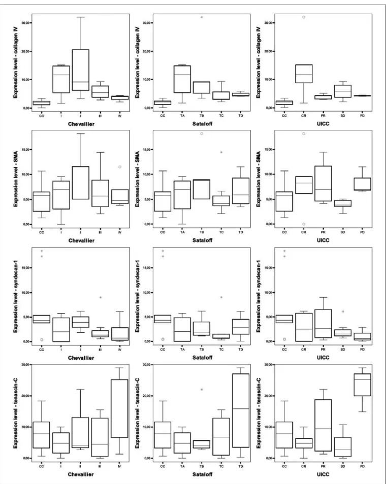
The collagen IV, smooth muscle actin, syndecan-1, and tenascin-C protein expression levels of the Chevallier and Sataloff pathologic response classification groups were com- pared with the samples of breast cancer patients who did not receive preoperative systemic therapy (CC group). Among the structural proteins, the expression of collagen IV was signifi- cantly up-regulated in all four groups both in the Chevallier and Sataloff classifications (P = 0.025 and P < 0.001, respectively). No significant differences were observed concerning the smooth muscle actin expression between the different groups, but there was an up-regulation of smooth muscle actin in the Chevallier class II group compared with the control group. The syndecan-1 proteoglycan levels were down- regulated in all four groups (P = 0.015 and P = 0.015, respectively) compared with the nontreated group. The adhesion molecule tenascin-C was significantly up-regulated in the Chevallier IV class of patients (P = 0.008) and the according to the Sataloff D class of patients (P = 0.003).
According to the UICC criteria, the expression levels of collagen IV were up-regulated in all the preoperative systemic therapy –treated patients (P= 0.002). There were no significant differences in the expressions of smooth muscle actin and syndecan-1 between the treated and the nontreated groups.
Tenascin-C was up-regulated in women with progressive disease (P = 0.005). Figure 1 contains the graphs showing the results of the morphometric evaluation. Supplementary Table S3 shows the results of morphometric analysis of the immunohistochemical reactions according to the classification systems, respectively.
Next, we analyzed the data according to their possible prognostic value. The expression of collagen IV was significantly up-regulated in the responding group versus the nonresponding group based on all the classification methods (Chevallier, Sataloff, and UICC at P = 0.022,P = 0.035, andP = 0.029, respectively; see Fig. 2 for the graphs showing the prog- Table 1.Clinicopathological parameters of the
evaluated patient groups
n%
Patient group PST Control
Age, y*, mean (range) 54.9 (29-73) 60.3 (43-85) Histologyc
IDC 43/50 (86) 20/20 (100)
ILC 5/50 (10) 0
Other 2/50 (4) 0
Immunohistochemistryb
Estrogen receptor 20/50 (40) 16/19 (84) Progesterone receptor 9/50 (18) 5/19 (26)
HER2/neu 21/50 (42) 1/19 (5.26)
CK5/6 4/50 (8) 0
pTc
pT0 6/50 (12) 0
pT1 20/50 (40) 16/20 (80)
pT2 15/50 (30) 1/20 (5)
pT3 4/50 (8) 0
pT4 5/50 (10) 3/20 (15)
Lymph node metastasisc
Not present (pN0) 22/50 (44) 9/20 (45) Present (pN0<) 28/50 (56) 11/20 (55) Histologic gradec
Grade 1 4/44 (9.1) 7/20 (35)
Grade 2 17/44 (38.6) 8/20 (40)
Grade 3 23/44 (52.3) 5/20 (25)
Abbreviations: PST, preoperative systemic therapy; IDC, invasive ductal carcinoma; ILC, invasive lobular carcinoma; HER2, human epidermal growth factor receptor 2; CK5, cytokeratin 5.
*Mean (range).
cNumber of cases/ all cases (%) in the PST group ypT is given histologic grade after surgery, following neoadjuvant therapy in PST group.
bNumber of positive cases/all cases (%).
Table 2. Results of the evaluation of the pathological and clinical response to primary systematic therapy according to the Chevallier and Sataloff classifications and UICC criteria
Chevallier Sataloff UICC
Class I 7T-A 7 cCR 10
Class II 6 T-B 11 cPR 7
Class III 18 T-C 17cSD 22
Class IV 19 T-D 15 cPD 11
Abbreviations: cCR, clinical complete response; cPR, clinical partial response; cSD, clinical stable disease; cPD, clinical progressive disease.
Fig. 1. Box-plot diagrams showing the results of the morphometric evaluation of the analyzed stromal proteins. Collagen IV (1st line), smooth muscle actin (2nd line), syndecan-1 (3rd line), and tenascin-C (4th line) expression in the preoperative systemic therapy (PST) groups compared with the control (CC) group. Classification:
pathologic Chevallier (1st column) and Sataloff classifications (2nd column), and clinical assessment according to UICC criteria (3rd column).
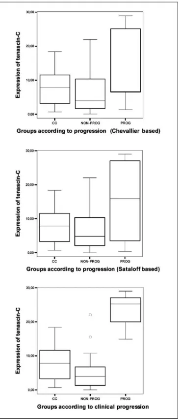
nostic groups separately). The expression of tenascin-C was up-regulated in the progressive patient group (Fig. 3). Taken either in the pathologic (Chevallier or Sataloff) or the clinical (UICC) classification method, this protein was detected to be increased in the progressing carcinomas (P= 0.012,P= 0.004, andP < 0.001, respectively). Although syndecan-1 expression was statistically significantly down-regulated in the preoperative systemic therapy –treated group compared with the nontreated patients, from the point of view of response the evaluated groups did not differ from each other significantly.
According to the final qPCR data, syndecan-1 was down- regulated in the preoperative systemic therapy group compared with nontreated cases, but there were no statistically significant differences between the analyzed response classes (Fig. 4).
Tenascin-C was up-regulated in all treated cases, but statistically significant up-regulation (11.86 fold; P = 0.012) was observed in the progressive disease group only, in the Chevallier class IV, and the according Sataloff class D patients (Fig. 4). PCR data are presented in Supplementary Table S4.
Discussion
Several studies have analyzed the role of biological markers to predict response to preoperative systemic therapy in breast carcinomas. At present, no biological markers are useful enough to predict response to chemotherapy or preoperative systemic therapy (30). Previous studies have examined the role of certain biological characteristics and markers of tumors (involved in proliferation, cell cycle, hormonal pathways) in predicting the likelihood of response to preoperative systemic therapy (31 –33). However, their exact role in prediction is uncertain. The aim of our study was to analyze the expression levels of representative extracellular matrix components follow- ing preoperative systemic therapy, and to evaluate these changes in the light of clinical and pathologic response.
The triple-negative breast carcinomas with or without CK5/
6-positive immunostaining contributed to the majority of the clinical complete response group, similar to the finding of Leivonen et al. (34). Thus, this patient group may be the appropriate target for primary chemotherapy. To date the biological classification of tumors as responsive or nonre- sponsive to treatment remains controversial. Several mecha- nisms involved in chemoresistance (multidrug resistance associated proteins, alteration in pathways that influence DNA repair, apoptosis or cell cycle) have been extensively studied (35, 36). Recent results point out the important role of the extracellular matrix components in the success of chemotherapy as well (12).
The extracellular matrix can interact with tumor cell integrins and affect tumor cell sensitivity to chemotherapeutic drugs (37). Rintoul and Sethi suggested that the tumor cells may condition the neighboring stroma to synthesize protective extracellular matrix proteins (38, 39).
In our study we found that the expression of collagen IV was significantly up-regulated in all four groups according to both pathologic classification systems. Increased expression of collagen IV may implicate improved structural integrity of the breast epithelium. Further, the expression of collagen IV was found to be even more increased in the responding carcinomas in this study. Also, degradation or loss of the basement membrane collagen IV was found to promote tumor metastasis
(40). Thus, an increase in collagen IV expression is very likely to be of beneficial nature, as seen in the responder group.
There were no significant differences in smooth muscle actin protein expression in the analyzed groups, which may result from no differences after chemotherapy affecting the tissues containing smooth muscle actin (e.g., microvessel density). A very recent study found no changes in nuclear pleomorphism
Fig. 2. Box-plot diagrams showing the significant up-regulation of collagen IV in the pathologically (Chevallier I and II; Sataloff A and B) and clinically responsive (UICC complete response and partial response) groups compared with the nonresponding carcinomas (Chevallier III and IV; Sataloff C and D, UICC stable disease and progressive disease).
and hormone receptor status of tumors comparing their state before and after the chemotherapeutic treatment (41).
Syndecan-1 is an important prognostic marker in breast cancer, and its biological functions include a coreceptor role for
mitogenic growth factor signaling, a role in modulating tumor angiogenesis, and modulation of cell adhesion and motility (42). Syndecan-1 –deficient mice are resistant to breast cancer (43). Here we found that syndecan-1 was decreased in the preoperative systemic therapy –treated patients compared with nontreated ones, more likely in nonresponding carcinomas, but the difference was not significant compared with the responder group.
Little is known about the role and predictive value of proteoglycans according to the response to neoadjuvant chemotherapy. We found decreased syndecan-1 expression in all treated tumors compared with the control group. Go¨tte et al.
showed that the pathologic response to chemotherapy was decreased in syndecan-1 –positive patients and that no synde- can-1 –positive patient showed complete remission (5). Taking into consideration the aforementioned results and those observed by Leivonen et al. that concomitant expression of syndecan-1 in both tumor cells and stroma may be a predictor of unfavorable prognosis in breast cancer, whereas the loss of epithelial syndecan-1 is associated with a more favorable prognosis (34), it seems that the loss of syndecan-1 expression might be considered as a predictive factor for response to preoperative systemic therapy.
It is known that tenascin-C up-regulates the expression of growth-associated genes (cyclin-1, c-myc) in mammary epithelial cells and tenascin-C increases endothelial cell migration and growth factor –dependent endothelial cell sprouting (22, 44).
High tenascin-C expression in breast cancer was shown to have an adverse effect on survival. Tenascin-C is an extracellular matrix protein that is expressed at low levels in normal adult tissue but is highly expressed around poor prognosis breast carcinomas (44). In this study syndecan-1 was down-regulated in all four groups as opposed to tenascin-C, which was significantly up-regulated in the Chevallier IV and the Sataloff D class of patients. Tsanou et al. have shown a negative correlation between syndecan-1 protein expression and collagen IV and tenascin-C in breast cancer, which is similar to our findings (45). Tenascin-C has also been shown to modulate integrin, but not endothelin receptor signaling via interactions with a different member of the syndecan family, syndecan-4 (46, 47). There is still a question if preoperative systemic therapy does
Fig. 3. Box-plot diagrams showing the significant up-regulation of tenascin-C in the pathologically (Chevallier IV; Sataloff D) and clinically progressive groups compared with the nonprogressing carcinomas (Chevallier I to III; Sataloff A to C, UICC complete response, partial response, and stable disease).
Fig. 4.Graph showing the results of the PCR analysis of syndecan-1and tenascin C expression. The syndecan-1was down-regulated in the treated carcinomas, whereas tenascin C expression was elevated with resistance (stable disease) and progression (progressive disease;P= 0.012), correspondingly.
change distinct cells’ individual properties, or from the aspect of tenascin-C if it facilitates the contraselection of the more malignant tumor cell populations not responding to neo- adjuvant therapy. One could suggest that tenascin-C –expressing cells may be more resistant to preoperative systemic therapy, or those cells surviving are capable of changing their expression and producing more tenascin-C as this molecule is suggested to contribute to the tenacious attribute against chemotherapy.
In a study by Sethi et al. the elevated expressions of collagen IV, fibronectin, and tenascin-C have been found in most cases of small cell lung cancers examined. These proteins were found to protect small cell lung cancers from chemotherapy-induced apoptosis (39). In addition, syndecan-1 may be implicated in the regulation of apoptosis, as a recent report has shown inhibition of n-3 polyunsaturated fatty acids induced apoptosis in syndecan-1 –silenced breast cancer cells (48).
Studies by Miyamoto et al. suggested that long-term exposure of cancer patients to antitumor drugs is associated with the up- regulation of extracellular matrix genes in tumor samples (49).
Understanding how the tumor stroma influences adhesion and tissue architecture to modulate tumor cells survival should help to better analyze the events that regulate resistance or response to preoperative systemic therapy. It seems that tumor cells may produce their own extracellular matrix molecules and the corresponding growth factors may influence stromal produc- tion of extracellular matrix.
Conclusion
The choice of treatment in preoperative systemic therapy is based on assessments of hormone receptor status, nuclear grade, human epidermal growth factor receptor 2 status, and
not ultimately on the microvessel density. The composition of the extracellular matrix and its receptors are not routinely considered as prognostic or predictive factors. Our results suggest that stromal changes occur in breast carcinoma during and/or after preoperative systemic therapy, which may be helpful in diagnostics, and the changes may also differ according to the response of the tumor. A study with a large number of patients could support our findings. Up-regulation of the basement membrane protein collagen IV is presumed, and decreased expression of the negative prognostic marker syndecan-1 in all treated cases suggests a beneficial effect on tumors during and/or after preoperative systemic therapy. Also, tenascin-C deserves further investigations because it is found to be up-regulated in the nonresponding group, and thus may be a marker for the worse prognosis group in the context of neoadjuvant therapy. Recent studies emphasize the biological importance of its gene in the aspect of relapse and development of metastasis as well (50). Evaluation on a larger patient cohort is desired to establish or reject a classification system based on stromal matrix protein expression, and to investigate the consequences of anthracyclin- or taxane-based regimen affect- ing the tumor stroma.
Disclosure of Potential Conflicts of Interest
No potential conflicts of interest were disclosed.
Acknowledgments
We thank Azumah Francisne and Bornemissza Ilona for the immunohistochemi- cal reactions and Andras Kiss and Rigone Kale Elvira for the careful reading of the article.
References
1.Fisher B, Bryant J, Wolmark N, et al. Effect of pre- operative chemotherapy on the outcome of women with operable breast cancer. J Clin Oncol 1998;16:
2672 ^ 85.
2.Hattangadi J, Park C, Rembert J, et al. Breast stromal enhancement on MRI is associated with response to neoadjuvant chemotherapy. AJR Am J Roentgenol 2008;190:1630 ^ 6.
3.Mazouni C, Arun B, Andre F, et al. Collagen IV levels are elevated in the serum of patients with primary breast cancer compared to healthy volunteers. Br J Cancer 2008;99:68 ^ 71. Epub 2008 Jun 17.
4.Reardon DA, Zalutsky MR, Akabani G, et al. A pilot study: 131I-antitenascin monoclonal antibody 81c6 to deliver a 44-Gy resection cavity boost. Neuro Oncol 2008;10:182 ^ 9.
5.Gotte M, Kersting C, Ruggiero M, et al. Predictive val- ue of syndecan-1 expression for the response to neo- adjuvant chemotherapy of primary breast cancer.
Anticancer Res 2006;26:621 ^ 7.
6.Broet P, Scholl SM, de la Rochefordiere A, et al. Short and long-term effects on survival in breast cancer patients treated by primar y chemotherapy : an updated analysis of a randomized trial. Breast Cancer ResTreat 1999;58:151 ^ 6.
7.Cameron DA, Anderson ED, Levack P, et al. Primary systemic therapy for operable breast cancer-10-year survival data after chemotherapy and hormone thera- py. Br J Cancer 1997;76:1099 ^ 105.
8.Shannon C, Smith I. Is there still a role for neoadju- vant therapy in breast cancer? Crit Rev Oncol Hematol 2003;45:77 ^ 90.
9.Buzdar AU, Valero V, Ibrahim NK, et al. Neoadjuvant therapy with paclitaxel followed by 5-fluorouracil, epirubicin, and cyclophosphamide chemotherapy
and concurrent trastuzumab in human epidermal growth factor receptor 2-positive operable breast cancer: an update of the initial randomized study population and data of additional patients treated with the same regimen. Clin Cancer Res 2007;13:
228 ^ 33.
10.Sachelarie I, Grossbard ML, Chadha M, Feldman S, Ghesani M, Blum RH. Primary systemic therapy of breast cancer. Oncologist 2006;11:574 ^ 89.
11.Yip GW, Smollich M, Gotte M. Therapeutic value of glycosaminoglycans in cancer. Mol CancerTher 2006;
5:2139 ^ 48.
12.Morin PJ. Drug resistance and the microenviron- ment: nature and nurture. Drug Resist Updat 2003;6:
169 ^ 72.
13.Pupa SM, Giuffre S, Castiglioni F, et al. Regulation of breast cancer response to chemotherapy by fibu- lin-1. Cancer Res 2007;67:4271 ^ 7.
14.LeBleu VS, Macdonald B, Kalluri R. Structure and function of basement membranes. Exp Biol Med May- wood 2007;232:1121 ^ 9.
15.Ioachim E, Charchanti A, Briasoulis E, et al. Immu- nohistochemical expression of extracellular matrix components tenascin, fibronectin, collagen type IV and laminin in breast cancer: their prognostic value and role in tumour invasion and progression. Eur J Cancer 2002;38:2362 ^ 70.
16.Barth PJ, Moll R, Ramaswamy A. Stromal remodel- ing and SPARC (secreted protein acid rich in cysteine) expression in invasive ductal carcinomas of the breast.
Virchows Arch 2005;446:532 ^ 6.
17.Sanderson RD. Heparan sulfate proteoglycans in in- vasion and metastasis. Semin Cell Dev Biol 2001;12:
89 ^ 98.
18.Vanhoutte D, Schellings MW, Gotte M, et al. In-
creased expression of syndecan-1 protects against cardiac dilatation and dysfunction after myocardial in- farction. Circulation 2007;115:475 ^ 82.
19.Maeda T, Desouky J, Friedl A. Syndecan-1 expres- sion by stromal fibroblasts promotes breast carcinoma growthin vivoand stimulates tumor angiogenesis.
Oncogene 2006;25:1408 ^ 12.
20.Baba F, Swartz K, van Buren R, et al. Syndecan- 1 and syndecan-4 are overexpressed in an estrogen receptor-negative, highly proliferative breast carci- noma subtype. Breast Cancer Res Treat 2006;98:
91 ^ 8.
21.Gotte M, Kersting C, Radke I, Kiesel L,Wulfing P. An expression signature of syndecan-1 (CD138), E-cad- herin and c-met is associated with factors of angio- genesis and lymphangiogenesis in ductal breast carcinomain situ. Breast Cancer Res 2007;9:R8.
22.Jones FS, Jones PL. The tenascin family of ECM glycoproteins: structure, function, and regulation dur- ing embryonic development and tissue remodeling.
DevDyn 2000;218:235 ^ 59.
23.Tokes AM, Paku S,Toth S, et al. Tenascin expression in primary and recurrent breast carcinomas and the ef- fect of tenascin on breast tumor cell cultures. Pathol Oncol Res 2000;6:202 ^ 9.
24.Ilunga K, Nishiura R, Inada H, et al. Co-stimulation of human breast cancer cells with transforming growth factor-hand tenascin-C enhances matrix met- alloproteinase-9 expression and cancer cell invasion.
Int J Exp Pathol 2004;85:373 ^ 9.
25.Chevallier B, Roche H, Olivier JP, Chollet P, Hurteloup P. Inflammatory breast cancer. Pilot study of intensive induction chemotherapy (FEC-HD) results in a highhistologic response rate. AmJClin Oncol1993;
16:223 ^ 8.
26.Sataloff DM, Mason BA, Prestipino AJ, Seinige UL, Lieber CP, Baloch Z. Pathologic response to induction chemotherapy in locally advanced carcinoma of the breast : a determinant of outcome. J Am Coll Surg 1995;180:297 ^ 306.
27.Hayward JL, Carbone PP, Heusen JC, Kumaoka S, Segaloff A, Rubens RD. Assessment of response to therapy in advanced breast cancer. Br J Cancer 1977;
35:292 ^ 8.
28.Paska C, Bogi K, Szilak L, et al. Effect of formalin, acetone, and RNAlater fixatives on tissue preservation and different size amplicons by real-time PCR from paraffin-embedded tissue. Diagn Mol Pathol 2004;
13:234 ^ 40.
29.Pfaffl MW, Horgan GW, Dempfle L. Relative ex- pression software tool (REST) for group-wise com- parison and statistical analysis of relative expression results in real-time PCR. Nucleic Acids Res 2002;
30:e36.
30.Sjostrom J. Predictive factors for response to che- motherapy in advanced breast cancer. Acta Oncol 2002;41:334 ^ 45.
31.Dressman HK, Hans C, Bild A, et al. Gene expres- sion profiles of multiple breast cancer phenotypes and response to neoadjuvant chemotherapy. Clin Cancer Res 2006;12:819 ^ 26.
32.Jones RL, Smith IE. Neoadjuvant treatment for ear- ly-stage breast cancer: opportunities to assess tu- mour response. Lancet Oncol 2006;7:869 ^ 74.
33.MacGrogan G, Mauriac L, Durand M, et al.
Primary chemotherapy in breast invasive carcino- ma: predictive value of the immunohistochemical detection of hormonal receptors, p53, c-erbB-2, MiB1, pS2 and GST pi. Br J Cancer 1996; 74:
1458 ^ 65.
34.Leivonen M, Lundin J, Nordling S, von Boguslawski K, Haglund C. Prognostic value of syndecan-1 expres- sion in breast cancer. Oncology 2004;67:11 ^ 8.
35.Sarkadi B, Homolya L, Szakacs G,Varadi A. Human multidrug resistance ABCB and ABCG transporters:
participation in a chemoimmunity defense system.
Physiol Rev2006;86:1179 ^ 236.
36.Minchinton AI,Tannock IF. Drug penetration in solid tumours. Nat RevCancer 2006;6:583 ^ 92.
37.Weaver VM, Lelievre S, Lakins JN, et al.h4 Integrin- dependent formation of polarized three-dimensional architecture confers resistance to apoptosis in normal and malignant mammary epithelium. Cancer Cell 2002;2:205 ^ 16.
38.Rintoul RC, Sethi T. Extracellular matrix regulation of drug resistance in small-cell lung cancer. Clin Sci Lond 2002;102:417 ^ 24.
39.Sethi T, Rintoul RC, Moore SM, et al. Extracellular matrix proteins protect small cell lung cancer cells against apoptosis: a mechanism for small cell lung cancer growth and drug resistancein vivo. Nat Med 1999;5:662 ^ 8.
40.Nerlich AG, Lebeau A, Hagedorn HG, Sauer U, Schleicher ED. Morphological aspects of altered base- ment membrane metabolism in invasive carcinomas of the breast and the larynx. Anticancer Res 1998;18:
3515 ^ 20.
41.Adams AL, Eltoum I, Krontiras H, Wang W, Chhieng DC. The effect of neoadjuvant chemotherapy on histo- logic grade, hormone receptor status, and Her2/neu status in breast carcinoma. Breast J 2008;14:141 ^ 6.
Epub 2008 Jan 31.
42.Bernfield M, Gotte M, Park PW, et al. Functions of cell surface heparan sulfate proteoglycans. Ann Rev Biochem 1999;68:729 ^ 77.
43.Alexander CM, Reichsman F, Hinkes MT, et al.
Syndecan-1 is required for Wnt-1-induced mamma- ry tumorigenesis in mice. Nat Genet 2000;25:
329 ^ 32.
44.Jones PL, Jones FS. Tenascin-C in development and disease: gene regulation and cell function. Matrix Biol 2000;19:581 ^ 96.
45.Tsanou E, Ioachim E, Briasoulis E, et al. Clinicopath- ological study of the expression of syndecan-1in inva- sive breast carcinomas. correlation with extracellular matrix components. J Exp Clin Cancer Res 2004;23:
641 ^ 50.
46.Lange K, Kammerer M, Hegi ME, et al. Endothelin receptor type B counteracts tenascin-C-induced endothelin receptor type A-dependent focal adhesion and actin stress fiber disorganization. Cancer Res 2007;67:6163 ^ 73.
47.Huang W, Chiquet-Ehrismann R, Moyano JV, Garcia-Pardo A, Orend G. Interference of tenascin-C with syndecan-4 binding to fibronectin blocks cell adhesion and stimulates tumor cell proliferation.
Cancer Res 2001;61:8586 ^ 94.
4 8.Sun H, Berquin IM, Owens RT, O’Flaherty JT, Edwards IJ. Peroxisome proliferator-activated recep- torg-mediated up-regulation of syndecan-1 by n-3 fatty acids promotes apoptosis of human breast can- cer cells. Cancer Res 2008;68:2912 ^ 9.
49.Miyamoto H, Murakami T, Tsuchida K, Sugino H, Miyake H,Tashiro S.Tumor-stroma interaction of human pancreatic cancer: acquired resistance to anticancer drugs and proliferation regulation is dependent on ex- tracellular matrix proteins. Pancreas 2004;28:38 ^ 44.
50.Tavazoie SF, Alarcon C, Oskarsson T, et al. Endoge- nous human microRNAs that suppress breast cancer metastasis. Nature 2008;451:147 ^ 52.
2009;15:731-739.
Clin Cancer Res
Anna-Maria Tokes, Attila Marcell Szasz, Andrea Farkas, et al.
Systemic Therapy in Breast Cancer
Updated version
http://clincancerres.aacrjournals.org/content/15/2/731
Access the most recent version of this article at:
Material Supplementary
http://clincancerres.aacrjournals.org/content/suppl/2009/01/11/15.2.731.DC1.html
Access the most recent supplemental material at:
Cited Articles
http://clincancerres.aacrjournals.org/content/15/2/731.full.html#ref-list-1
This article cites by 50 articles, 15 of which you can access for free at:
Citing articles
http://clincancerres.aacrjournals.org/content/15/2/731.full.html#related-urls
This article has been cited by 1 HighWire-hosted articles. Access the articles at: