cancers
Review
Targeted Cancer Therapy Using Compounds Activated by Light
Petra Dunkel1 and Janez Ilaš2,*
Citation: Dunkel, P.; Ilaš, J. Targeted Cancer Therapy Using Compounds Activated by Light.Cancers2021,13, 3237. https://doi.org/10.3390/
cancers13133237
Academic Editors: Neha Kaushik and Petra Wilder-Smith
Received: 23 April 2021 Accepted: 24 June 2021 Published: 29 June 2021
Publisher’s Note:MDPI stays neutral with regard to jurisdictional claims in published maps and institutional affil- iations.
Copyright: © 2021 by the authors.
Licensee MDPI, Basel, Switzerland.
This article is an open access article distributed under the terms and conditions of the Creative Commons Attribution (CC BY) license (https://
creativecommons.org/licenses/by/
4.0/).
1 Department of Organic Chemistry, Semmelweis University, 1092 Budapest, Hungary;
dunkel.petra@pharma.semmelweis-univ.hu
2 Faculty of Pharmacy, University of Ljubljana, 1000 Ljubljana, Slovenia
* Correspondence: janez.ilas@ffa.uni-lj.si; Tel.: +386-1-4769-576
Simple Summary:Cancer is among the leading causes of death, and cancer therapy suffers from many drawbacks, the lack of selectivity being most noteworthy. In this review, we present innovative approaches in the discovery of novel anticancer compounds, which can use light activation to achieve more potent cancer therapy with fewer side effects. We describe recent approaches to prepare photocages and photoswitches and obstacles that photopharmacology must overcome to achieve clinical use.
Abstract: Cancer chemotherapy is affected by a modest selectivity and toxic side effects of phar- macological interventions. Among novel approaches to overcome this limitation and to bring to therapy more potent and selective agents is the use of light for selective activation of anticancer compounds. In this review, we focus on the anticancer applications of two light-activated approaches still in the experimental phase: photoremovable protecting groups (“photocages”) and photoswitches.
We describe the structural considerations behind the development of novel compounds and the plethora of assays used to confirm whether the photochemical and pharmacological properties are meeting the stringent criteria for an efficient in vivo light-dependent activation. Despite its immense potential, light activation brings many challenges, and the complexity of the task is very demanding.
Currently, we are still deeply in the phase of pharmacological tools, but the vivid research and rapid development bring the light of hope for potential clinical use.
Keywords:photocages; photoswitches; photopharmacology; light-activatable materials; photore- sponsive drug delivery systems; azobenzenes
1. Introduction
Cancer chemotherapy is affected by a modest selectivity and toxic side effects of pharmacological interventions [1]. Therefore, besides finding novel, efficient, targeted antitumor agents, developing innovative solutions for endowing cancer chemotherapeutics with a more selective, localized effect is of paramount importance. Applying various internal (e.g., pH, redox environment, enzymatic processes) or external triggers (e.g., ultrasound, magnetic field, light irradiation) for on-site activation of stimuli-sensitive drug molecules or drug delivery systems (as, e.g., liposomes, micelles, polymeric or metal nanoparticles, dendrimers) [2–4] has long been in the forefront of research interest. Of the potential physical stimuli, light is particular from several aspects, from its orthogonality with biological systems to the precise control over the wavelength and the irradiation dose. Light activation is independent of the properties of the tumor environment as well, allowing to target a variety of tumors. Light-activated approaches made their way into clinical use already in the 1990s, with the approval of the first photodynamic therapy regimes [5]. Photodynamic therapy (PDT) is based on the interaction of photosensitizer agents with light to produce singlet oxygen and superoxide in the presence of molecular oxygen and the local cytotoxic effect of these species. The discussion of PDT is beyond the scope of the present review, that will focus on the anticancer applications of two further
Cancers2021,13, 3237. https://doi.org/10.3390/cancers13133237 https://www.mdpi.com/journal/cancers
Cancers2021,13, 3237 2 of 49
light-activated approaches still in the experimental phase: photoremovable protecting groups (“photocages”) and photoswitches (illustrated in Figure1by the examples of a photocaged vs. a photoswitchable agent). The concept of photopharmacology was recently introduced to describe the area dealing with photocontrollable molecules for therapy.
In this review, we focus on small-molecule approaches, where a parent drug itself is rendered photoactivatable via structural modifications, and the approach is backed up by in cellulo assays. However, more complex drug delivery systems could also be rendered light-responsive by judiciously designing photoactivatable units into their structure [6,7].
For such approaches and designs where photoactivation is used for controlling targeting or cell penetration [8,9] (i.e., not directly the drug activity), we refer to the respective reports and reviews (e.g., (upconversion) nanoparticles [10–15], organic nanoparticles [16], chitosan nanoparticles [17], gold nanoparticles [18], photocaged folate nanoconjugates [19], two-photon nanoimpellers [20], photoresponsive antibody-drug conjugates [21,22]).
Cancers 2021, 13, x FOR PEER REVIEW 2 of 50
oxygen and the local cytotoxic effect of these species. The discussion of PDT is beyond the scope of the present review, that will focus on the anticancer applications of two further light-activated approaches still in the experimental phase: photoremovable protecting groups (“photocages”) and photoswitches (illustrated in Figure 1 by the examples of a photocaged vs. a photoswitchable agent). The concept of photopharmacology was re- cently introduced to describe the area dealing with photocontrollable molecules for ther- apy. In this review, we focus on small-molecule approaches, where a parent drug itself is rendered photoactivatable via structural modifications, and the approach is backed up by in cellulo assays. However, more complex drug delivery systems could also be rendered light-responsive by judiciously designing photoactivatable units into their structure [6,7].
For such approaches and designs where photoactivation is used for controlling targeting or cell penetration [8,9] (i.e., not directly the drug activity), we refer to the respective re- ports and reviews (e.g., (upconversion) nanoparticles [10–15], organic nanoparticles [16], chitosan nanoparticles [17], gold nanoparticles [18], photocaged folate nanoconjugates [19], two-photon nanoimpellers [20], photoresponsive antibody-drug conjugates [21,22]).
Figure 1. Photocaged vs. photoswitchable pharmacological agents, with the respective photoactivated transformations.
2. Irreversible Activation with Light: Photoremovable Protecting Groups (“Photo- cages”) for Antitumor Applications
The first application of photoremovable protecting groups (PPGs) as experimental tools for biological studies dates back to the 1970s. The photocage terminology coined at the advent of the field might be now ambiguous; therefore, nowadays, the photoremova- ble/photolabile/photoactivatable protecting group is commonly used in the literature. The design rationale behind PPGs is to mask the biological activity of a given substrate by covalently binding the protecting group to a moiety critical for the action (“caging”). The activity can be restored on demand in a spatiotemporally controlled manner by removing the PPG with light irradiation, i.e., the absorbed energy is translated into a photocleavage reaction (“uncaging”) [23]. The activation is irreversible, which is often considered as a disadvantage of the approach, as the released agents might still lead to unwanted effects upon diffusion or excretion. On the other hand, several PPG families of different proper- ties are available, and temporary deactivation of the parent drug by adding a PPG is typ- ically more straightforward due to the often significant structural differences upon caging.
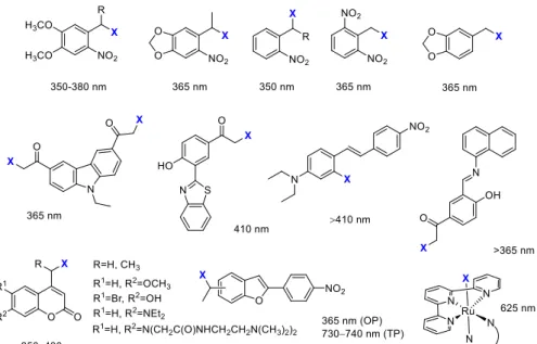
Moreover, with appropriate designs, dual or sequential, the wavelength-selective release of different agents could be envisaged as well (“chromatic orthogonality”) [24–28]. The PPGs used in the discussed studies with the respective uncaging wavelengths are sum- marized in Figure 2.
Figure 1.Photocaged vs. photoswitchable pharmacological agents, with the respective photoactivated transformations.
2. Irreversible Activation with Light: Photoremovable Protecting Groups (“Photocages”) for Antitumor Applications
The first application of photoremovable protecting groups (PPGs) as experimental tools for biological studies dates back to the 1970s. The photocage terminology coined at the advent of the field might be now ambiguous; therefore, nowadays, the photoremov- able/photolabile/photoactivatable protecting group is commonly used in the literature.
The design rationale behind PPGs is to mask the biological activity of a given substrate by covalently binding the protecting group to a moiety critical for the action (“caging”). The activity can be restored on demand in a spatiotemporally controlled manner by removing the PPG with light irradiation, i.e., the absorbed energy is translated into a photocleavage reaction (“uncaging”) [23]. The activation is irreversible, which is often considered as a disadvantage of the approach, as the released agents might still lead to unwanted effects upon diffusion or excretion. On the other hand, several PPG families of different properties are available, and temporary deactivation of the parent drug by adding a PPG is typically more straightforward due to the often significant structural differences upon caging. More- over, with appropriate designs, dual or sequential, the wavelength-selective release of different agents could be envisaged as well (“chromatic orthogonality”) [24–28]. The PPGs used in the discussed studies with the respective uncaging wavelengths are summarized in Figure2.
CancersCancers 2021, 13, x FOR PEER REVIEW 2021,13, 3237 3 of 493 of 50
Figure 2. PPGs used in the discussed studies with their respective activation wavelengths.
Initially, PPGs have found widespread application as experimental tools for studying dynamic processes, particularly in the field of neurophysiology [29,30]. Moreover, the number of studies on PPG-based light-activatable prodrug designs is steadily growing [31]. An optimal PPG-substrate construct should comply with several criteria already for in vitro applications, such as proper solubility, stability of the molecule to be released by light irradiation, efficient masking of the activity, clean and rapid photorelease, and cel- lular tolerance to the products of the photoreaction and the applied irradiation (dose-in- tensity, wavelength). The typical workflow for the rational design and evaluation of pho- toactivatable antitumor agents consists of the following steps: (i) determining the key pharmacophore for introducing the PPG unit, so that either the key ligand-target interac- tions are disturbed, or spatial conflicts arise rendering the caged drug inactive, (ii) as- sessing the photostability of the parent drug, so that its activity remains intact upon the light irradiation used for the uncaging step, (iii) assessing the hydrolytic stability of the caged prodrug at physiological pH, so that the parent drug is released exclusively upon light irradiation, (iv) determining the pharmacological activity of the parent and the caged drug molecule, assessment of the efficiency of masking (e.g., target enzyme inhibition, cellular assay), (v) studying the photolysis of the caged molecule, determining the condi- tions necessary for uncaging (wavelength and intensity of light irradiation, irradiation time necessary for complete conversion), (vi) determining the pharmacological activity of the caged molecule in the absence of light and following light irradiation, assessment of the efficiency of restoring the pharmacological activity, and (vii) verifying the tolerance of the experimental system to the light irradiation applied. Novel probes are typically char- acterized by UV/VIS absorption, hydrolytic stability, photocleavage rates, and quantum yields. Despite the complexity of the task [32], there is rapid development, and novel re- sults on caged anticancer agents are discussed in the following sections.
2.1. Kinase Inhibitors
Kinase inhibitors affecting cell signaling pathways implicated in oncogenesis offer targeted therapy against specific malignant diseases. Introduced into therapy in the 2000s (2001—imatinib), the number of approved small-molecule kinase inhibitors was steadily growing. Although kinase inhibitors are the flagship of targeted therapy, their application is not without its challenges (e.g., development of resistance); therefore, they are a prime choice for further developments. The design of photoactivatable protein kinase inhibitors Figure 2.PPGs used in the discussed studies with their respective activation wavelengths.
Initially, PPGs have found widespread application as experimental tools for studying dynamic processes, particularly in the field of neurophysiology [29,30]. Moreover, the number of studies on PPG-based light-activatable prodrug designs is steadily growing [31].
An optimal PPG-substrate construct should comply with several criteria already for in vitro applications, such as proper solubility, stability of the molecule to be released by light irradi- ation, efficient masking of the activity, clean and rapid photorelease, and cellular tolerance to the products of the photoreaction and the applied irradiation (dose-intensity, wave- length). The typical workflow for the rational design and evaluation of photoactivatable antitumor agents consists of the following steps: (i) determining the key pharmacophore for introducing the PPG unit, so that either the key ligand-target interactions are disturbed, or spatial conflicts arise rendering the caged drug inactive, (ii) assessing the photostability of the parent drug, so that its activity remains intact upon the light irradiation used for the uncaging step, (iii) assessing the hydrolytic stability of the caged prodrug at physiological pH, so that the parent drug is released exclusively upon light irradiation, (iv) determin- ing the pharmacological activity of the parent and the caged drug molecule, assessment of the efficiency of masking (e.g., target enzyme inhibition, cellular assay), (v) studying the photolysis of the caged molecule, determining the conditions necessary for uncaging (wavelength and intensity of light irradiation, irradiation time necessary for complete conversion), (vi) determining the pharmacological activity of the caged molecule in the absence of light and following light irradiation, assessment of the efficiency of restoring the pharmacological activity, and (vii) verifying the tolerance of the experimental system to the light irradiation applied. Novel probes are typically characterized by UV/VIS absorption, hydrolytic stability, photocleavage rates, and quantum yields. Despite the complexity of the task [32], there is rapid development, and novel results on caged anticancer agents are discussed in the following sections.
2.1. Kinase Inhibitors
Kinase inhibitors affecting cell signaling pathways implicated in oncogenesis offer targeted therapy against specific malignant diseases. Introduced into therapy in the 2000s (2001—imatinib), the number of approved small-molecule kinase inhibitors was steadily growing. Although kinase inhibitors are the flagship of targeted therapy, their application is not without its challenges (e.g., development of resistance); therefore, they are a prime choice for further developments. The design of photoactivatable protein kinase inhibitors has been extensively studied by Peifer and coworkers (Figure3). They designed a photoactivatable prodrug forimatinib[33], the Bcr-Abl tyrosine kinase inhibitor, which was
Cancers2021,13, 3237 4 of 49
approved for treating Philadelphia chromosome-positive chronic myelogenous leukemia, but started to be also used for other malignancies. Docking studies ofimatinibin the ATP binding pocket of PDGF-Rβshowed two potential positions for attaching a PPG group to mask bioactivity: the NH in the benzanilide and in theN-arylpyrimidine-2-amine moieties, of which the first design was pursued in the study. As PPG, two groups were tested: a 4,5-dimethoxy-2-nitrobenzyl (DMNB) (1) and a coumarylmethyl scaffold (2); however, only the former provided efficient photorelease ofimatinibin vitro at 365 nm (1 mM DMSO solution, 5.4 W, 10 min). In an enzymatic PDGF-Rβassay, a 100-fold difference was detected between the parentimatinib(IC50= 0.059µM) and its caged prodrug (IC50= 5.8µM). The residual activity was assumed to originate from uncagedimatinibcontaminating the sample.
Following 365 nm irradiation, the bioactivity ofimatinibcould be nearly totally restored in vitro in the PDGF-Rβassay, experimentally confirming a successful caging approach, while further bioactivity studies were not reported.
Cancers 2021, 13, x FOR PEER REVIEW 5 of 50
Figure 3. Photocaged kinase inhibitors, the PPG indicated in blue.
The same group also reported photoactivatable caged prodrugs for 3,4-diarylmalei- mide antiangiogenic VEGFR-2 kinase inhibitors [35,36]. Based on the analysis of key lig- and-target interactions by molecular docking, the hinge binder region was masked by a PPG unit (DMNB), potentially rendering the molecules inactive also toward other protein kinases. Docking of the caged prodrugs verified that protection disrupts key H-bond in- teractions to the hinge region as well as leads to steric hindrance hampering the access to the binding pocket. Uncaging was studied with 365 nm irradiation, confirming efficient photolysis of 7 (365 nm LED reactor, 5.4 W, 1 mM solution in DMSO, >90% conversion in 5 min) while the irradiation of compound 6 leads to a 1,6-π-electrocyclization not followed by oxidation to carbazole, making this derivative a less suitable candidate for further stud- ies. VEGFR-2 kinase inhibitory assays confirmed a significant difference (300-fold) in the activity of the caged and the parent compounds. The same tendency was also observed in the in vitro antiproliferative assays (VEGFR-2 dependent PC-3 cells); however, in both studies, the caged inhibitors still showed some activity, ascribed to the presence of resid- ual non-caged parent compound (GI50: 6—not reached (parent drug—6.4 µ M); 7–35 µ M (parent drug—0.2 µ M)). Dose-response curves were recorded for both the parent and the caged compounds before and after irradiation (365 nm, 110 mW/cm2, 5 min). The activity of the parent drug was restored for compound 7, whereas interestingly an increased ac- tivity was observed for compound 6 (GI50 after irradiation: 6—0.1 µ M (parent drug—2.9 µ M); 7—0.2 µ M (parent drug—0.4 µ M)). This was presumed to result from several factors:
restored activity of the kinase inhibitor, the activity of the cyclized intermediate formed upon irradiation, the antiproliferative effect of the cleaved cage itself (confirmed by con- trol experiments with Boc-Ala and DMNB-Boc-Ala model compounds, DMNB-Boc-Ala:
GI50 = 50 µ M) and a synergistic effect of the UV irradiation. Although tolerable in the stud- ied case, the pharmacological effect or toxicity of the PPG itself might be a serious concern
Figure 3.Photocaged kinase inhibitors, the PPG indicated in blue.
Vemurafenib(a BRAFV600Eserine/threonine kinase inhibitor) was approved for late- stage melanoma in 2011. Besides its therapeutic importance, vemurafenibtolerates the 365 nm UV irradiation relatively well, and chemical modifications could be straightfor- wardly carried out on its structure; therefore, it is an ideal candidate for the photoactivatable prodrug approach [34]. Photocaging would allow reaching higher target concentrations and thus avoid typical side effects, a high incidence of arthralgia (joint pain), skin rash, and the development of squamous cell carcinoma, which appear with higher dosage. Key pharmacophoric moieties for efficiently masking the activity ofvemurafenibwere deter- mined by docking studies. In the subsequent work, two potential NH protection sites were assessed usingo-nitrobenzylic PPGs (DMNB and 4,5-dimethoxy-2-nitrophenylethyl
Cancers2021,13, 3237 5 of 49
(DMNPE)), protection on the azaindole (3,4), and protection on the sulfonamide moiety (5) ofvemurafenib. The former was expected to diminish hinge binding and therefore diminish activity on other kinases as well, whereas the latter was expected to allow residual activity on further kinases. AsN-heterocycles are rarely described leaving groups, the authors studied the minimal structural requirements for the photoreaction as well, irradiating caged fragments of the parent compound besides the fully armed prodrugs (365 nm LED reactor, 5.4 W, 10µM solutions in PBS buffer containing 10% DMSO, >90% conversion in 1 min). Regarding the photoreaction (i.e., rapid and quantitative photorelease), both protection sites proved to be feasible.
Next, the BRAFV600E binding activity ofvemurafeniband its caged prodrugs was determined, confirming a lower binding affinity for the caged inhibitors (Kd:vemurafenib—
10 nM,3—440 nM,4—77 nM,5—79 nM, overall ~8–40-fold difference). The selectivity profile was determined on a panel of 140 kinases, and in line with the modeling results, the azaindole-protected derivative3proved to be the most selective (activity on 2 kinases, vs. 13 for the sulfonamide-protected derivative5and 32 forvemurafenib). Antiprolifer- ative activities were assessed in cellular growth assays (SKMel13, SKMel28, M14, and UACC62 melanoma cell lines), the caged compounds showing cytostatic activity at higher concentrations vs. the nanomolar cytotoxicity of the parentvemurafenib(GI50(SKMel13):
vemurafenib—0.17µM,3—no cell growth inhibition,4—4.3µM,5—2.6µM,). Irradiating the cells (SKMel13, 1.8 W, 5 min) at 365 nm after compound incubation, the cytotoxic activ- ity ofvemurafenibcould be restored, although not totally, presumably due to incomplete photorelease (GI50:vemurafenib—0.19µM,3—1.5µM,4—0.46µM,5—0.35µM). In control experiments using Boc-Ala and Boc-Ala-DMNB model compounds, also the cleaved PPG itself was found to have antiproliferative activity, however at much higher concentrations thanvemurafenib(GI50= 34µM). As further proof of restoringvemurafenibactivity, the effect of caged molecules on ERK phosphorylation (SKMel13 cells) following UV irradiation was studied by Western blot analysis (complete suppression of ERK phosphorylation above 0.1µM concentrations, dose dependency correlating with that ofvemurafenib).
The same group also reported photoactivatable caged prodrugs for 3,4-diarylmaleimide antiangiogenic VEGFR-2 kinase inhibitors [35,36]. Based on the analysis of key ligand- target interactions by molecular docking, the hinge binder region was masked by a PPG unit (DMNB), potentially rendering the molecules inactive also toward other protein kinases. Docking of the caged prodrugs verified that protection disrupts key H-bond interactions to the hinge region as well as leads to steric hindrance hampering the access to the binding pocket. Uncaging was studied with 365 nm irradiation, confirming efficient photolysis of7(365 nm LED reactor, 5.4 W, 1 mM solution in DMSO, >90% conversion in 5 min) while the irradiation of compound6leads to a 1,6-π-electrocyclization not followed by oxidation to carbazole, making this derivative a less suitable candidate for further studies. VEGFR-2 kinase inhibitory assays confirmed a significant difference (300-fold) in the activity of the caged and the parent compounds. The same tendency was also observed in the in vitro antiproliferative assays (VEGFR-2 dependent PC-3 cells); however, in both studies, the caged inhibitors still showed some activity, ascribed to the presence of residual non-caged parent compound (GI50: 6—not reached (parent drug—6.4µM);
7—35µM (parent drug—0.2µM)). Dose-response curves were recorded for both the parent and the caged compounds before and after irradiation (365 nm, 110 mW/cm2, 5 min).
The activity of the parent drug was restored for compound7, whereas interestingly an increased activity was observed for compound6(GI50after irradiation:6—0.1µM (parent drug—2.9µM);7—0.2µM (parent drug—0.4µM)). This was presumed to result from sev- eral factors: restored activity of the kinase inhibitor, the activity of the cyclized intermediate formed upon irradiation, the antiproliferative effect of the cleaved cage itself (confirmed by control experiments with Boc-Ala and DMNB-Boc-Ala model compounds, DMNB-Boc-Ala:
GI50= 50µM) and a synergistic effect of the UV irradiation. Although tolerable in the studied case, the pharmacological effect or toxicity of the PPG itself might be a serious
Cancers2021,13, 3237 6 of 49
concern in therapeutic applications. On the positive side, caging did not hamper the cellular uptake of the inhibitors (studied on live PC-3 cells by fluorescence microscopy).
While looking at the photocaged kinase inhibitors, it is also worth noticing that several kinase inhibitors intrinsically possess interesting photochemical properties. Phototoxic- ity was reported forvemurafenib[37], whiledabrafenibcan be photodegraded to a novel fluorescent BRAFV600Einhibitor [38].
2.2. Anthracyclines
Anthracyclines have broad use in cancer chemotherapy, with a wide activity spectrum (e.g., solid tumors, hematological malignancies, soft tissue sarcomas). Regarding their activity, intercalation with DNA, affecting DNA replication and transcription and interac- tion with topoisomerase II have been studied. However, a very effective anticancer agent, the therapeutic use ofdoxorubicinis limited notably by its cardiotoxicity. For constructing a photoactivatable prodrug, Esener and coworkers bound a PPG unit (ano-nitrophenyl group) to an active amine on the sugar moiety ofdoxorubicin, a modification already demon- strated to decrease its toxicity [39]. Additionally, the construct was armed with a biotin group, linked via a water-soluble glycol spacer, for enhanced membrane interaction (8) (Figure4). Biotin was intended to increase the clearance rate of the PPG-drug-construct and as well PPG fragment from the circulating blood using clearing agents after UV ex- posure. First, the release and release kinetics ofdoxorubicinin vitro upon UV irradiation was measured by LC-MS and NMR studies (in DMSO/water 1/9, 350 nm, 120 or 240 s of 2.19 mW/cm2irradiation; kinetics: in DMSO/water 2/8, 350 nm, 0 to 10 min, 1.8 mW/cm2 irradiation: 1.8µM/mindoxorubicin release). The uptake and intracellular localization of the PPG construct vs. freedoxorubicin(in 5 vs. 50µM concentration) was studied in live PTK2 cells, using the intrinsic red fluorescence ofdoxorubicin(free or photocaged).
Unlike the parent drug, the PPG construct was not entering the nucleus before light ex- posure, however following UV irradiation (60 s), the released freedoxorubicinbehaved similarly to the parent drug. Cytotoxicity was measured on A549 human lung cancer cells, showing a significant difference between the parent and the photocaged drug (doxorubicin:
IC50= 1.2µM, doxorubicin-PPG construct: IC50= 250µM); however, the activity could be efficiently restored by UV irradiation in a light dose-dependent manner (samples were UV- irradiated in the media before incubation of the cells, i.e., cells were not irradiated directly.
Maximal effect at 60 min: IC50= 3.5µM). To assess the feasibility of future applications, in a control experiment, A549 cells were irradiated (1.8 mW/cm2) but retained cell viability after 20 min of irradiation (after 60 min, 90% cell viability was recorded). To ascertain that drug release occurs only upon UV irradiation, the metabolic stability of thedoxorubicin-PPG construct was verified using human liver microsomes. One major metabolite was identified, however, in small quantities, and its effect was not studied. Importantly, further in vivo studies were conducted [40]. First, the penetration of 365 nm light through ex vivo tumor tissue (irradiation from the center, penetrating light intensity measured at different angles) and the release ofdoxorubicin was measured using a LED/fiber-optic system (efficient release after 30 min irradiation). For conducting in vivo experiments, thedoxorubicin-PPG construct was solubilized using Captisol®cyclodextrin to allow a concentration of 1.2 mg in 200µL of saline. The circulation was studied in vivo (female nude mice, alpha phase circulation half-life 10 min vs. ~20 min fordoxorubicin). Of note, no freedoxorubicinwas detected neither in the serum nor in the urine out to 24 h, ascertaining UV irradiation as the major activation mode in subsequent experiments. In vivo drug release was studied on athymic nu/nu nude mice. A total of 10 min after injecting thedoxorubicin-PPG construct (time point chosen based on the circulation half-life), a 365 nm LED was inserted into the middle of the tumor and turned on (240µW/cm2, 30 min irradiation). A control tumor was left unirradiated, and blood samples were taken to monitor circulating drug concentration, whereas tumor samples were collected to analyze the distribution of the construct and the free drug. The construct was found in all tumor tissue sections of both the irradiated and unirradiated tumors, whereas a trace amount ofdoxorubicinwas found in some sections of
Cancers2021,13, 3237 7 of 49
the unirradiated tumors vs. an increaseddoxorubicincontent at the irradiation site (~6-fold difference indoxorubicin/gram of tumor tissue between irradiated and unirradiated tu- mors). The former was ascribed to the effect of UV leakage. Nodoxorubicinwas detected in the serum sample taken after irradiation, pointing toward a controllable systemic exposure.
Cancers 2021, 13, x FOR PEER REVIEW 8 of 50
Figure 4. Photocaged doxorubicin derivatives, the PPG indicated in blue.
For therapeutic applications, using appropriate (longer) wavelengths has several practical consequences. One approach to harness long wavelengths, consequently low- energy photons, is to target sufficiently weak chemical bonds that could be directly cleaved with lower energy. Lawrence and coworkers aimed to exploit the photohomolysis of the C-Co bond (<30 kcal/mol) in alkyl-cob(III)-alamins. The bond is weak enough to be cleaved by any wavelengths between 330 and 560 nm absorbed by the corrin ring system [46]. To move further toward the optical window of tissues and to develop wavelength- tunable probes, constructs with additional fluorophores serving as light-capturing anten- nas were designed (TAMRA, SulfoCy5, Atto725, DyLight800, Alexa700, Bodipy650). The design rationale was that upon excitation beyond cobalamin absorption wavelengths, the Figure 4.Photocageddoxorubicinderivatives, the PPG indicated in blue.
Choi and coworkers studied the design and synthesis of a novel thioacetal ortho-nitrobenzaldehyde (TNB) dual-arm photocage [41] and its application for constructing dual drug-fluorescent reporter conjugates for controlled release and real-time fluorescent monitoring of drug activation in parallel. Optimally the two payloads are released simul- taneously by cleaving the linker C-S bonds (resistant to many chemical conditions), and several combinations of targeting ligands, drugs, etc., might be envisaged. For one set
Cancers2021,13, 3237 8 of 49
of drug-TNB-fluorescent reporter conjugates,doxorubicinwas used as model anticancer substrate (doxorubicin-TNB-coumarin (9) (carbamate linkage)). The cleavage mechanism of the free cage was studied at 365 nm by1H NMR, UV/VIS, and UPLC monitoring. A total of 365 nm photolysis of the cage conjugate in water/MeOH 1/1 led to an efficient release of the drug and reporter molecules presumably via a self-immolative mechanism (decay half-life (t1/2) < 2 min,Φuncaging = 0.08), the latter leading to an irradiation-dependent increase in fluorescence (i.e., the reporter is in a fluorescently quenched state before cleav- age). The construct showed irradiation-dependent cytotoxicity in FAR(+) KB cells (before irradiation9: IC50= 9.2µM; after irradiation9: IC50= 0.84 vs.doxorubicin: IC50= 0.04µM).
Hartman and coworkers studied the photocaging of 2-pyrrolino doxorubicin (2P-Dox) (10), a derivative of the standarddoxorubicin, starting from its known diacetoxy prodrug [42]. Photorelease upon UV irradiation showed 88% release after 60 min irradia- tion, which corroborated by MS and HPLC results (20 mM in PBS, 380 nm, 9.0 mW/cm2).
Cellular viability assays (CellTiter-Blue method) were run in three human cancer cell lines:
MCF-7 (breast), A2780 (ovarian), and A2780ADR (doxorubicin-resistant ovarian), demon- strating a significant increase (327–750-fold) in activity upon light irradiation (380 nm, 30 min), activities compared with the parent drug. As a negative control, the released PPG itself was not cytotoxic up to 50µM. Cellular uptake in MCF-7 cells was studied by flow cytometry, exploiting the fluorescence ofdoxorubicinand compound10. Compound10 showed a higher cellular uptake thandoxorubicin, in line with previous observations regard- ing the effect of alkylation with lipophilic moieties. Confocal microscopic studies indicated a nuclear co-localization of compound10anddoxorubicin.
Controlled photorelease of the drug molecules could be combined with cell target- ing to increase selectivity. Folic acid conjugates are often used to target overexpressed folate receptors (FRα) on tumor cells [43]. To release the internalized drug and avoid endosomal entrapment, Hartman and coworkers designed a photoactivatable folic acid- drug conjugate [44]. The conjugate consisted of folic acid, a PEG linker, and photocaged doxorubicin (11), using a DMNB unit as PPG (Figure4). UV photolysis of the conjugate resulted in rapid release ofdoxorubicin(HPLC monitoring); however, no further biological assays were pursued.
Of further targeted carrier approaches beyond small molecules, Choi et al. described polyamidoamine (PAMAM) dendrimer nanoconjugates with folic acid ligands armed with a photocaged doxorubicin (using anortho-nitrobenzyl based PPG). In vitro, on folic-acid- receptor-over-expressing KB cells, the doxorubicin constructs inhibited cell growth in an irradiation dose-dependent manner (but not without irradiation). The maximum effect observed (30 min irradiation, ~80% cell growth reduction) was comparable to the effect of the parent drug (~85% cell growth inhibition) [45].
For therapeutic applications, using appropriate (longer) wavelengths has several prac- tical consequences. One approach to harness long wavelengths, consequently low-energy photons, is to target sufficiently weak chemical bonds that could be directly cleaved with lower energy. Lawrence and coworkers aimed to exploit the photohomolysis of the C-Co bond (<30 kcal/mol) in alkyl-cob(III)-alamins. The bond is weak enough to be cleaved by any wavelengths between 330 and 560 nm absorbed by the corrin ring system [46]. To move further toward the optical window of tissues and to develop wavelength-tunable probes, constructs with additional fluorophores serving as light-capturing antennas were designed (TAMRA, SulfoCy5, Atto725, DyLight800, Alexa700, Bodipy650). The design rationale was that upon excitation beyond cobalamin absorption wavelengths, the antennas might transfer excited-state energy to the corrin ring (coenzyme B12), thus contributing to Co-C bond cleavage. The full conversion was observed upon irradiation at the corresponding wavelength (Xe flash-lamp, 546 nm, 25 min); moreover, the constructs released the fluo- rophores also at the longer wavelengths (646/730/780 nm). Photorelease could also be operated by linking the fluorophores to the ribose 50-OH of alkylcobalamins. To assess the application of cobalamin conjugates as drug delivery systems, a cobalamin-doxorubicin conjugate was prepared (12) [47]. Cytotoxicity was studied in HeLa (cervical cancer) cells
Cancers2021,13, 3237 9 of 49
(MTT assay at 10µM), following different illumination times (30, 45, and 75 s at 530 nm).
The cobalamin-conjugate affected cell viability in an irradiation-dependent manner (neither the construct without light nor the light irradiation without the construct had an effect on cell viability), with similar efficacy at the end point as that of the parentdoxorubicin.
Hartman and coworkers also envisaged modulating the cellular entry of a cytotoxic agent by a photocage approach (photocaged permeability), linking doxorubicin as the model drug to a polar sulfonic acid fluorophore (EDANS) via a photocleavable nitro- veratryl PPG (13) [48]. Photolysis was evaluated in PBS by HPLC monitoring (365 nm, 9.0 mW/cm2), showing parent drug release. The effect on cellular entry was studied on JH-EsoAd1 cells (Barrett’s esophagus associated adenocarcinoma cell line) under dark and light-irradiated conditions with flow cytometry. An irradiation-dependent fluores- cence enhancement was observed both with flow cytometry and confocal microscopic studies. Cell viability was assessed with MTT assay, showing a clear correlation between decreased survival and light irradiation (365 nm, 9.0 mW/cm2, 20 min) of the probes (Dox:
IC50= 1.0µM, EDANS-DOX: no toxicity till 16µM, EDANS-DOX-lit: IC50= 1.6µM).
2.3. Tubulin (Dis)Assembly Inhibitors, Microtubule-Targeting Agents
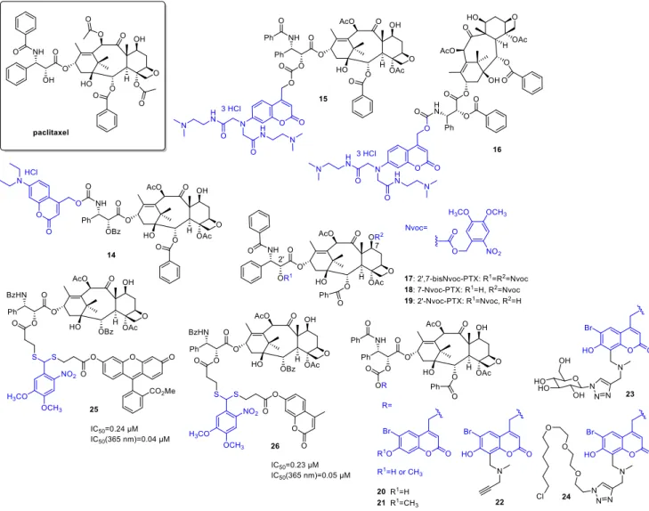
Paclitaxel(PTX) has a broad antitumor activity spectrum (e.g., breast, ovarian, head and neck cancers, malignancies resistant to conventional chemotherapy). Its action is based on affecting tubulin polymerization and microtubule dynamics, thereby disrupting cell division and interphase mechanisms and leading to cell death. Given the widespread therapeutic applications of PTX and the challenges limiting its use (e.g., resistance mecha- nisms, neuro-, and hematological toxicities), it is not surprising that several research groups proposed different photoactivatable prodrug designs. As one of the first examples, Kiso and coworkers linked a 7-N,N-diethylamino-4-hydroxymethyl coumarin (DECM) PPG to theO-acyl isoform of the parent drug (14) (Figure5) [49]. Previous SAR studies suggested that the introduction will render the drug inactive (i.e., the structural modification is carried out at a key pharmacophore). Due to solubility reasons, the photolysis was studied in a 1/1 mixture of PBS and methanol at 430 nm (diode laser, 10 mW), monitored by UV/VIS absorption and HPLC (recovery rate: 69% after 30 min irradiation). The photorelease prod- uct subsequently yields the parentpaclitaxelthrough O-N acyl migration at a reasonable rate under physiological conditions. To solve the solubility issue, a new coumarin PPG with improved water solubility was designed in a follow-up study by the same group [50]
and linked topaclitaxelas carbonate (15), and 30-N-debenzoylpaclitaxel (as carbamate,16) respectively (solubility over 100 mg/mL vs. 0.00025 mg/mL forpaclitaxel). Despite the suitable water solubility, the carbonate prodrug was not stable enough under physiological conditions; therefore, it was not studied further (20%paclitaxelrelease after 8 h incubation at pH 7.4, 37◦C). In vitro photorelease from the carbamate, prodrug was studied in PBS, depending on the light source providing 24% (355 nm pulse laser, 10 Hz, 5 mJ, 4 min) or 69% (365 nm 6W UV-lamp) recovery rate of the parentpaclitaxel. The lower recovery rate in the former case was ascribed to the sensitivity of the protecting group to intense light irradiation, besides the wavelength. No further biological assays were pursued, however.
Del Campo and coworkers used a 4,5-dimethoxy-2-nitrobenzyloxycarbonyl (Nvoc) PPG for preparing photocaged paclitaxels by blocking either one or both of two reactive hydroxyls (C2’, C7) critical for bioactivity (17,18,19) [51]. Photolysis of the caged probes was studied in ACN (5% water, 1 mM) solutions, with UV and HPLC monitoring (360 nm, 2.7 mW/cm2, chemical yields of PTX release: 17—61%,18—89%,19—52%). In in vitro microtubule polymerization assay under dark and lit conditions, 7-Nvoc-PTX (18) showed residual activity, whereas 20-Nvoc-PTX (19) and 20,7-bisNvoc-PTX (17) operated only after UV exposure. However, for the double-protected drug, higher doses were necessary to obtain the same amount of released drug (according to HPLC studies, a sequential deprotection occurs). In cellular assay on HeLa cells, both monoprotected probes showed cytotoxicity, normal cell morphology was detected only following 20,7-bisNvoc-PTX (17) treatment. Treatment with pre-irradiated probes led to similar results as observed with free
Cancers2021,13, 3237 10 of 49
PTX (i.e., the activity could be restored). Assessing cell viability in liquid HeLa cell culture, 20,7-bisNvoc-PTX (17) was not cytotoxic till 10µM, whereas 20-(19) and 7-Nvoc-PTX (18) showed ~50% viability at 0.1µM. A further demonstration of the need for double protection.
Masking of activity and restoring it upon pre-irradiation was also confirmed in in vitro assays of microtubule catastrophes and mitotic index (HeLa cells). In situ photolysis by a short light pulse under a fluorescent microscope resulted as well in the reorganization of microtubules.
Cancers 2021, 13, x FOR PEER REVIEW 10 of 50
was studied in ACN (5% water, 1 mM) solutions, with UV and HPLC monitoring (360 nm, 2.7 mW/cm2, chemical yields of PTX release: 17—61%, 18—89%, 19—52%). In in vitro mi- crotubule polymerization assay under dark and lit conditions, 7-Nvoc-PTX (18) showed residual activity, whereas 2′-Nvoc-PTX (19) and 2′,7-bisNvoc-PTX (17) operated only after UV exposure. However, for the double-protected drug, higher doses were necessary to obtain the same amount of released drug (according to HPLC studies, a sequential depro- tection occurs). In cellular assay on HeLa cells, both monoprotected probes showed cyto- toxicity, normal cell morphology was detected only following 2′,7-bisNvoc-PTX (17) treat- ment. Treatment with pre-irradiated probes led to similar results as observed with free PTX (i.e., the activity could be restored). Assessing cell viability in liquid HeLa cell culture, 2′,7-bisNvoc-PTX (17) was not cytotoxic till 10 µ M, whereas 2′-(19) and 7-Nvoc-PTX (18) showed ~50% viability at 0.1 µ M. A further demonstration of the need for double protec- tion. Masking of activity and restoring it upon pre-irradiation was also confirmed in in vitro assays of microtubule catastrophes and mitotic index (HeLa cells). In situ photolysis by a short light pulse under a fluorescent microscope resulted as well in the reorganization of microtubules.
Figure 5. Photocaged paclitaxel derivatives, the PPG indicated in blue.
To broaden the structural diversity of PPGs, Furuta and coworkers designed a mod- ular 6-bromo-7-hydroxycoumarin-4-ylmethyl (Bhc) group that has a terminal alkyne moi- ety allowing further functionalization via click chemistry and used it for the synthesis of Figure 5.Photocagedpaclitaxelderivatives, the PPG indicated in blue.
To broaden the structural diversity of PPGs, Furuta and coworkers designed a modular 6-bromo-7-hydroxycoumarin-4-ylmethyl (Bhc) group that has a terminal alkyne moiety allowing further functionalization via click chemistry and used it for the synthesis of caged PTXs [52]. Besides a Bhc cage-PTX conjugate (20,21), two further probes were prepared exploiting the alkyne chemical handle (22), one functionalized with a sugar to improve water solubility (23), and another with a halo tag ligand for cellular targeting (24). PTX was released from all novel probes under in vitro conditions at 350 nm (KMOPS containing 0.1%
DMSO, 10 mJ/s lamp,Φdis= 3.5–14), the improved water solubility of compound23also translating to more efficient photolysis, presumably by promoting ion-pair intermediates implicated in the reaction. Masking and UV-triggered restoring of the activity of the parent drug was assessed in an in vitro tubulin polymerization assay (porcine brain tubulin, turbidity assay at 340 nm), confirming an irradiation-dependent effect (350 nm, 10 mJ/cm2, 60 s) and comparable maximum activity of activated caged compounds withpaclitaxel;
however, no animal studies were undertaken.
Cancers2021,13, 3237 11 of 49
For the thioacetalortho-nitrobenzaldehyde dual-arm photocages described in the previ- ous section, PTX was used as another study drug (paclitaxel-TNB-fluorescein (25, ester linkage), paclitaxel-TNB-coumarin (26, ester linkage)). Efficient photorelease was also detected for the PTX-constructs (25/26:Φuncaging= 0.05/0.07). Cellular uptake and reactivation after uptake of25were studied by fluorescence with flow cytometry in FAR(+) KB cells. The results confirmed an intracellular accumulation of the PTX-TNB- fluorescein conjugate25and an efficient 365 nm photorelease of the fluorescent reporter intracellularly (365 nm, 2 min). The correlation between the cytotoxicity and fluores- cence was studied in a microplate assay on FAR(+) KB cells (XTT assay). Dose-dependent cytotoxicity was recorded both for irradiated and non-irradiated conjugates; however, cyto- toxicity prior irradiation was modest compared to the parent drug (before irradiation25/26:
IC50= 0.24/0.23µM; after irradiation25/26: IC50 = 0.04/0.05 vs. PTX: IC50= 0.02µM).
Moreover, the cytotoxicity was in correlation with the increase in fluorescence upon irradi- ation, paving the way toward quantification and effective real-time monitoring of the drug release process. Real-time fluorescence in cellular systems was recorded over extended periods of time following irradiation to study the release kinetics (24 h monitoring, 0.5 or 2 min irradiation). The gradual, continuous increase in fluorescence was ascribed to the conversion of transient dye intermediates to parent fluorescent probes. On the other hand, a certain degree of increase in fluorescence was observed for the conjugates even without UV irradiation after extended periods, i.e., a dark release was occurring.
Dobber et al. designed a photocaged small-molecule tubulin inhibitor starting from the previously described inhibitor CMPD1 and using DMNB as PPG [53], linked to a phenolic OH group, the crucial role of which has been confirmed by previous SAR stud- ies. First, the UV-stability of the parent inhibitor was ascertained at 365 nm, followed by measuring the kinetics of the photorelease from the cage monitored by HPLC (2.7 W, τ= 0.61 min, maximum parent drug concentration reached after 2 min irradiation). For as- sessing the pharmacological effects, cell viability assays on U251 and patient-derived RN1 glioblastoma cells were used, after first checking their UV-irradiation tolerance (1.8 mW till 5 min, maximum tolerated light exposure: U251—1 min, RN1—30 s). A 55- and 125-fold difference was recorded between the activity of the parent and the caged drug without UV irradiation. UV irradiation of the cage-treated cells could recover al- most totally the cytotoxicity (photocaged drug after irradiation—EC50(U251) = 2.1µM, EC50(RN1) = 1.2µM). Additionally, in control experiments blocking the pharmacophoric OH with a bulky aromatic group and cytotoxicity of the released DMNB were addressed (released DMNB did not change cell viability till 100µM concentration in U251 cells). Loss of activity of the caged drug and recovering the activity upon UV irradiation were further addressed in in vitro tubulin polymerization (porcine brain tubulin) and tubulin binding assays (competitive colchicine binding assay), as well as via assessing the effect on the tubulin network and cell morphology in U251 glioblastoma cells (immunofluorescence staining ofβ-tubulin) and measuring the apoptotic effect on RN1 glioblastoma cells (An- nexin V staining). All the pharmacological studies pointed toward the release of a bioactive molecule from the photocage upon UV exposure and inactivity without UV irradiation.
2.4. DNA Alkylating Agents
Alkylating agents cause direct DNA damage (consequent fragmentation, crosslinking or mutations due to mispairing) and thereby disrupt DNA replication and cell division.
The nitrogen mustards were among the first anticancer chemotherapeutics, and alkylating agents have widespread use against a variety of hematological and solid tumors. Nitrogen mustards typically have a bis(2-chloroethyl) group important for action, e.g., also inchlo- rambucil. Schuberth et al. developed photoactivatable prodrugs for the highly cytotoxic duocarmycin [54]. Despite their cytotoxic potential (activity in multidrug-resistant cells), no duocarmycins are yet approved for therapy. (+)-Duocarmycin SA contains a DNA recognition and a DNA alkylating unit, the latter implicated in forming DNA adducts. In the cited study, a seco- and a dimeric duocarmycin analog was used as the parent drug, and
Cancers2021,13, 3237 12 of 49
the PPG (3,4-methylenedioxy-ONB—with an additionaltert-butyl ester or carboxylic acid atα-position, overall 5 novel probes) was linked to a phenolic OH to hinder the formation of the spiro-cyclopropyl unit critical for action (Figure6). Photorelease was studied in PBS buffer (1/10% DMSO, 365 nm, 1.1 mW/cm2) with LC-MS monitoring. For compounds27, 28,the photoreactions proceeded via the expected pathway, i.e., PPG cleavage and subse- quent cyclization (28:Φ= 0.45). For the29–31series, however, the photorelease was slow and led to the formation of several side products. Cellular cytotoxicity was assessed on A549 bronchial carcinoma cells (HTCFA-derived test) with pre-irradiated samples (365 nm, 1.1 mW/cm2, 30 min) or without UV exposure. For27,a more than 106-fold difference of activity was recorded vs. the parent drug, and the activity could be efficiently restored with UV exposure. Interestingly, for28,the PPG construct itself was found to have enhanced cytotoxicity vs. the free drug. No proper phototherapeutic index values were found for the 29–31series.
Cancers 2021, 13, x FOR PEER REVIEW 12 of 50
2.4. DNA Alkylating Agents
Alkylating agents cause direct DNA damage (consequent fragmentation, crosslink- ing or mutations due to mispairing) and thereby disrupt DNA replication and cell divi- sion. The nitrogen mustards were among the first anticancer chemotherapeutics, and al- kylating agents have widespread use against a variety of hematological and solid tumors.
Nitrogen mustards typically have a bis(2-chloroethyl) group important for action, e.g., also in chlorambucil. Schuberth et al. developed photoactivatable prodrugs for the highly cytotoxic duocarmycin [54]. Despite their cytotoxic potential (activity in multidrug-re- sistant cells), no duocarmycins are yet approved for therapy. (+)-Duocarmycin SA con- tains a DNA recognition and a DNA alkylating unit, the latter implicated in forming DNA adducts. In the cited study, a seco- and a dimeric duocarmycin analog was used as the parent drug, and the PPG (3,4-methylenedioxy-ONB—with an additional tert-butyl ester or carboxylic acid at α-position, overall 5 novel probes) was linked to a phenolic OH to hinder the formation of the spiro-cyclopropyl unit critical for action (Figure 6). Photore- lease was studied in PBS buffer (1/10% DMSO, 365 nm, 1.1 mW/cm2) with LC-MS moni- toring. For compounds 27, 28, the photoreactions proceeded via the expected pathway, i.e., PPG cleavage and subsequent cyclization (28: Φ = 0.45). For the 29–31 series, however, the photorelease was slow and led to the formation of several side products. Cellular cy- totoxicity was assessed on A549 bronchial carcinoma cells (HTCFA-derived test) with pre- irradiated samples (365 nm, 1.1 mW/cm2, 30 min) or without UV exposure. For 27, a more than 106-fold difference of activity was recorded vs. the parent drug, and the activity could be efficiently restored with UV exposure. Interestingly, for 28, the PPG construct itself was found to have enhanced cytotoxicity vs. the free drug. No proper photothera- peutic index values were found for the 29–31 series.
Figure 6. Photocaged duocarmycin derivatives, the PPG indicated in blue.
Several photoactivatable prodrug approaches were described for the aromatic nitro- gen mustard chlorambucil (Figure 7), used for chronic lymphocytic leukemia, Hodgkin lymphoma, and non-Hodgkin lymphoma. Push-pull stilbenes were devised with the aim to develop visible light-activated PPGs with turn-on fluorescent real-time monitoring abil- ity [55]. The PPG was based on a trans-4-(N,N-dimethylamino)-4′-nitrostilbene unit, plac- ing the substrate (an alcohol or a carboxylic acid) to be liberated at the point of photocy- clization, serving as a leaving group upon the process. By formation of the cyclized phe- nanthrene product, a blue fluorescence appears that was used for monitoring of the pho- torelease. Besides simple model substrates, as model drug molecules for photorelease studies, chlorambucil was selected (32). Due to the presence of appropriately placed elec- tron-donor and –acceptor groups and consequent charge transfer, the novel caged deriv- atives had an absorption maximum typically above 400 nm, whereas the fluorescence be- came quenched in polar solvents. The uncaging was studied in ACN (medium-pressure mercury lamp (125 W)), with NMR, UV/VIS, HPLC, and MS monitoring with ≥410 nm Figure 6.Photocaged duocarmycin derivatives, the PPG indicated in blue.
Several photoactivatable prodrug approaches were described for the aromatic nitro- gen mustardchlorambucil(Figure7), used for chronic lymphocytic leukemia, Hodgkin lymphoma, and non-Hodgkin lymphoma. Push-pull stilbenes were devised with the aim to develop visible light-activated PPGs with turn-on fluorescent real-time monitoring ability [55]. The PPG was based on a trans-4-(N,N-dimethylamino)-40-nitrostilbene unit, placing the substrate (an alcohol or a carboxylic acid) to be liberated at the point of pho- tocyclization, serving as a leaving group upon the process. By formation of the cyclized phenanthrene product, a blue fluorescence appears that was used for monitoring of the photorelease. Besides simple model substrates, as model drug molecules for photorelease studies,chlorambucilwas selected (32). Due to the presence of appropriately placed electron- donor and –acceptor groups and consequent charge transfer, the novel caged derivatives had an absorption maximum typically above 400 nm, whereas the fluorescence became quenched in polar solvents. The uncaging was studied in ACN (medium-pressure mercury lamp (125 W)), with NMR, UV/VIS, HPLC, and MS monitoring with≥410 nm irradiation, confirming an efficient liberation of the caged substrates accompanied by an increase in fluorescence (450 nm) (chlorambucil: Φp = 0.10). Modeling physiological conditions, uncaging ofchlorambucilwas also measured in ACN/HEPES (1/19) mixture (λ≥410 nm, 75% release after 1 h,Φp= 0.02). Real-time monitoring of photorelease was studied in MCF-7 cells by confocal microscopy after different light exposure periods (λ≥410 nm, 0–30 min). Irradiation-dependent cytotoxicity of cagedchlorambucilwas assessed on MCF-7 cells (MTT assay), confirming a significant drop of cell viability in the irradiated samples vs.
the unlit ones (dark sample: more than 75% cell viability (100µM), after 30 min irradiation:
15% viability, IC50= 230µM).
Cancers2021,13, 3237 13 of 49
Cancers 2021, 13, x FOR PEER REVIEW 13 of 50
irradiation, confirming an efficient liberation of the caged substrates accompanied by an increase in fluorescence (450 nm) (chlorambucil: Φp = 0.10). Modeling physiological condi- tions, uncaging of chlorambucil was also measured in ACN/HEPES (1/19) mixture (λ ≥ 410 nm, 75% release after 1 h, Φp = 0.02). Real-time monitoring of photorelease was studied in MCF-7 cells by confocal microscopy after different light exposure periods (λ ≥ 410 nm, 0–
30 min). Irradiation-dependent cytotoxicity of caged chlorambucil was assessed on MCF-7 cells (MTT assay), confirming a significant drop of cell viability in the irradiated samples vs. the unlit ones (dark sample: more than 75% cell viability (100 µ M), after 30 min irradi- ation: 15% viability, IC50 = 230 µ M).
Figure 7. Photoactivatable chlorambucil derivatives, the PPG indicated in blue.
As a second approach, a photoactivatable double prodrug strategy was developed to increase the selectivity of drug release (“dually locked photoresponsive DDS”) [56]. Chlo- rambucil was linked to a first 7-hydroxycoumarin, that was itself masked with an o-nitro- benzyl PPG quenching its fluorescence (fluorescent quantum yields: coumarin-chlorambu- cil (CC)-Φf = 0.49, ONB-CC-Φf = 0.010). To provide tumor-targeting ability, a biotin ligand was used as well (33). Photolysis was studied with 1H NMR, HPLC, and fluorescence monitoring (10 μM, water/ACN 4/1, 125 W, ≥365 nm). The fluorescence (546 nm) increased within 5 min irradiation and reached a maximum in 30 min, confirming the turn-on ability of the construct. After 5 min irradiation, CC release (90%) and nitrosobenzaldehyde for- mation occurred. The second uncaging (chlorambucil release, 70%) necessitated a longer, 20 min irradiation (however, less than 7% release with diode laser, 730 nm, 30 mW/cm2).
Incubating MDA-MB-231 (breast adenocarcinoma) cells with ONB-CC and subjecting them to a UV-irradiation regime, a gradually intensifying green fluorescence appeared, confirming intracellular accumulation and CC release. Cytotoxicity was studied on the MDA-MB-231 cell line with UV irradiation (30 min, ≥365 nm). At 50 μM concentration, a 50% cell viability was observed (vs. chlorambucil IC50 = 67 µ M). Without light irradiation, no significant effect was detected for the drug-PPG construct. Higher apoptotic activity observed for the biotin-tagged construct in cell cycle analysis pointed toward a contribu- tion of accumulation to the increased cytotoxicity.
The same group used PPGs for the design of dual drug delivery systems [57]. As a photoactivatable and fluorescent unit allowing imaging besides caging, an acetly-carba- zole was used, from which single and dual-arm cages were prepared (34, 35). In the latter Figure 7.Photoactivatablechlorambucilderivatives, the PPG indicated in blue.
As a second approach, a photoactivatable double prodrug strategy was developed to increase the selectivity of drug release (“dually locked photoresponsive DDS”) [56].
Chlorambucilwas linked to a first 7-hydroxycoumarin, that was itself masked with an o-nitrobenzyl PPG quenching its fluorescence (fluorescent quantum yields: coumarin- chlorambucil(CC)-Φf= 0.49, ONB-CC-Φf= 0.010). To provide tumor-targeting ability, a biotin ligand was used as well (33). Photolysis was studied with1H NMR, HPLC, and fluorescence monitoring (10µM, water/ACN 4/1, 125 W,≥365 nm). The fluorescence (546 nm) increased within 5 min irradiation and reached a maximum in 30 min, con- firming the turn-on ability of the construct. After 5 min irradiation, CC release (90%) and nitrosobenzaldehyde formation occurred. The second uncaging (chlorambucilrelease, 70%) necessitated a longer, 20 min irradiation (however, less than 7% release with diode laser, 730 nm, 30 mW/cm2). Incubating MDA-MB-231 (breast adenocarcinoma) cells with ONB-CC and subjecting them to a UV-irradiation regime, a gradually intensifying green fluorescence appeared, confirming intracellular accumulation and CC release. Cytotoxicity was studied on the MDA-MB-231 cell line with UV irradiation (30 min,≥365 nm). At 50µM concentration, a 50% cell viability was observed (vs. chlorambucilIC50 = 67µM).
Without light irradiation, no significant effect was detected for the drug-PPG construct.
Higher apoptotic activity observed for the biotin-tagged construct in cell cycle analysis pointed toward a contribution of accumulation to the increased cytotoxicity.
The same group used PPGs for the design of dual drug delivery systems [57]. As a photoactivatable and fluorescent unit allowing imaging besides caging, an acetly-carbazole was used, from which single and dual-arm cages were prepared (34,35). In the latter case, either two of the same or two different substrates could be linked as esters. Photorelease was first studied in ACN/water 7/3 solutions at 365 nm using various acid model sub- strates, confirming a clean process (i.e., formation of the parent 3-(hydroxyacetyl) 9-ethyl 9Hcarbazole and the uncaged free acid substrate) and suitable recovery of the free acid (medium-pressure mercury lamp (125 W),Φp~0.1). Importantly, dual-arm cages released two equivalents of the linked carboxylic acids, and a simultaneous release occurred in the case of dual-arm cages with two different acid substrates. For a proof-of-concept study of dual drug delivery systems, caffeic acid andchlorambucilwere selected as the bioactive antitumor compounds. Photorelease of the parent drugs was verified in ACN/water 7/3 solution (medium-pressure mercury lamp (125 W), 0.1 mM, 60 min irradiation, caffeic acid:
Cancers2021,13, 3237 14 of 49
91% release,Φp= 0.046;chlorambucil: 94%,Φp= 0.051). The cellular uptake of the dual-arm drug-PPG conjugate was studied in glial cancer cells (U87MG) by confocal fluorescence at 10µM concentration (3 h treatment), confirming an internalization of the conjugate.
The in vitro cytotoxicity of the conjugate without and after irradiation was determined on normal HaCaT cells, confirming feasible biocompatibility (80% cell viability before irradiation, 40% cell viability after irradiation at 80µM concentration). The anticancer activity of single-arm PPG-caffeic acid, PPG-chlorambucil,and the dual-arm PPG-conjugate was studied on U87MG cells by MTT assay. A significant difference in cell viability was recorded between the experiments without and after UV irradiation (more than 70%
cell viability for all constructs at 25µM concentration, after irradiation: PPG-caffeic acid:
IC50 = 15 µM, PPG-chlorambucil 34: IC50 = 15 µM, PPG-caffeic acid-chlorambucil 35:
IC50= 9µM).
Further development led to the modifications of thep-hydroxyphenacyl PPG to render it fluorescent and activatable above 400 nm [58]. A 2-(2’-hydroxyphenyl)benzothiazole unit was added, known to have a fast and efficient excited-state intramolecular proton transfer (ESIPT) effect, contributing to interesting fluorescent properties potentially allowing real- time monitoring of photorelease processes. The novel PPG system was tested for caging chlorambucil(36). In vitro photorelease was assessed in ACN/HEPES 1/19 (0.1 mM, 410 nm, medium-pressure mercury lamp (125W),Φu= 0.46, 90% release after 15 min irradiation).
The ESIPT-assisted photorelease mechanism was further addressed with computational chemistry, supporting a singlet-state ESIPT followed by intersystem crossing (ISC) and triplet-state photo-Favorskii rearrangement. The in vitro photoreaction was accompanied by a fluorescent color change from green (517 nm) to blue (450 nm). In the cellular en- vironment (MDA-MB-231 cells), after internalization of the PPG-chlorambucilconstructs, the photorelease could be followed by confocal microscopy (≥410 nm, 15 min irradia- tion). Cytotoxicity of the parent and caged drug was assessed on MDA-MB-231 cells with MTT assay. Without irradiation, the cell viability remained above 90% for different PPG- chlorambucilconcentrations; however,≥410 nm irradiation efficiently restored the activity of the parent drug. To address tissue penetration issues, ap-hydroxyphenacyl-based two- photon (TP) responsive system for cagingchlorambucil[59] was also designed. The novel PPG system showed ESIPT and aggregation-induced emission (AIE) phenomenon. Upon in vitro photolysis (ACN/PBS buffer) of the PPG-chlorambucilconjugate (37) (≥365 nm), a fluorescence color change was observed (550→430 nm blue shift), potentially allow- ing real-time monitoring of the photorelease (0.1 mM, ACN/PBS 1/9, medium-pressure mercury lamp (125 W),≥365 nm, 15 min,Φu= 0.49). Drug release could be effectuated sequentially as well with an on-off irradiation regime, thus verifying the light-dependency of the process. Uncaging occurred from the aggregate state, as confirmed with irradiation of the conjugate in the presence of different water fractions. Two-photon photorelease was confirmed experimentally at 700 nm (0.1 mM in DMSO/water 1/9, 25% release after 3 h) with an open aperture Z-scan technique with a pulsed laser. In vitro cytotoxicity studies were conducted in MCF-7 (breast cancer) cells, confirming first the cellular uptake of the conjugate. Upon 15 min≥365 nm irradiation, the fluorescent color change due to photorelease could be monitored by confocal microscopy, and a concentration- and irradiation-dependent cytotoxic effect was recorded using the MTT assay (cell viability above 95% before irradiation, after irradiation IC50= 31µM).
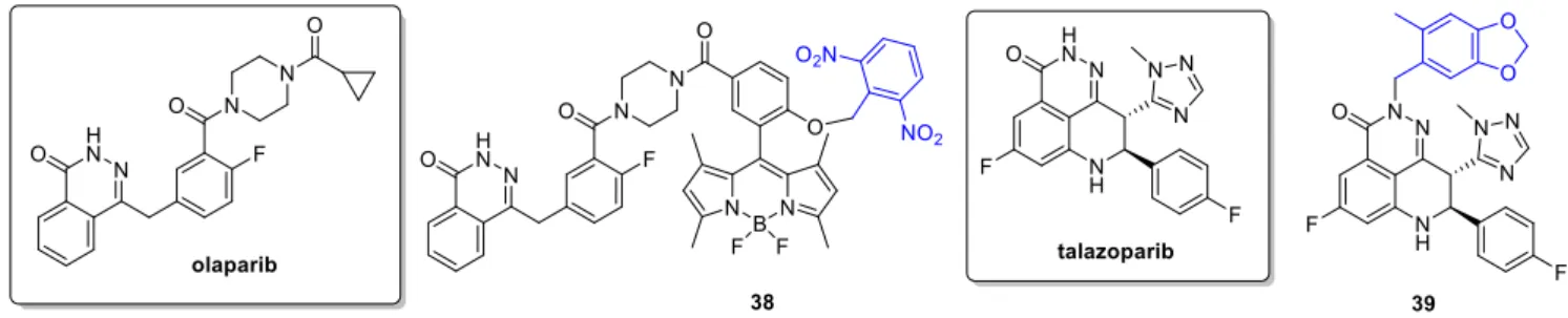
2.5. PARP Inhibitors
Poly (ADP-ribose) polymerase (PARP) inhibitors target DNA damage response, partic- ularly in cancer cells with high replication stress and genomic instability. The first in class olaparibwas approved in 2014,talazoparibin 2018. Indications include BRCA mutated ovar- ian and breast cancer, and clinical trials addressing further indications and combination therapies were launched. Weissleder and coworkers aimed to develop a construct allowing real-time monitoring of intracellular drug transport using a photocaging concept [60]. As fluorophore, a BODIPY unit was selected, a 2,6-dinitrobenzyl (DNB) as PPG and as the