Phosphatases (Phosphomonoesterases)
Kurt Linhardt and Klaus Walter
Phosphatases catalyse the hydrolytic cleavage of phosphoric acid esters. Depending on their p H optimum they are classified as "alkaline" or " a c i d " phosphatases. Alkaline phosphatases occur in practically all animal and human tissues. Bile and osteoblasts contain especially large amounts.
The determination o f "alkaline" serum phosphatase is of great importance in the diagnosis and differential diagnosis of many diseases of man. Raised serum values are due either to an increased osteoblast activity (Recklinghausen's Disease, Paget's Disease, osteoblastic and osteoclastic skeletal metastases, osteomalacia, rickets, etc.) or to a disease of the liver or bile duct (cholangitis, obstructive jaundice, hepatitis, etc.). In children the normal serum values are considerably higher than those o f adults.
Acid phosphatase is found in large amounts in the prostate and in erythrocytes. The activity, in human semen is also very high. However, the sera of healthy men and w o m e n have the same normal values. The acid phosphatase in erythrocytes is selectively inhibited by formaldehyde and that in serum by fluoride ions. According to Jacobsson
1
) the prostatic phosphatase is inhibited by an 0.02 M tartrate solution between p H 4.9 and 5.7. Prostatic phosphatase in serum can therefore be deter
mined separately. This determination is important for the diagnosis o f metastasizing prostatic carcinoma (depends on the extent of the increase) or for following the progress of hormone therapy in this disease.
To determine the activity of acid and alkaline phosphatases measurements are made of either the phosphate liberated from the substrate or of the dephosphorylated residue. A s normal serum contains inorganic phosphate, relatively high blank values are obtained in the determination of phos
phate (see for example
2
)). King and Armstrong
3
^ have used phenylphosphate as substrate; the activity of the enzyme is measured by the amount o f phenol liberated. The method is simplified by use of substrates, whose phosphate-free residues are coloured at alkaline p H (phenolphthalein diphos
p h a t e
4
^ ) , /7-nitrophenylphosphate
5
)). Deproteinization at the end of the incubation is unnecessary.
The assay mixture is made strongly alkaline, the colour is formed and the enzyme is completely inhibited.
Determination in Serum with Phenolphthalein Diphosphate
4)Principle
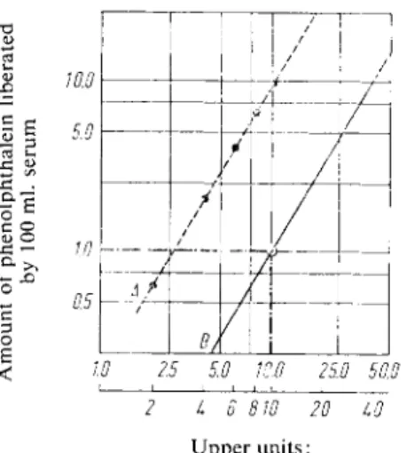
The optimum hydrolysis of phenolphthalein diphosphate is at p H 5.4 with acid phosphatases and at p H 9.7 with alkaline phosphatases. The phenolphthalein liberated is determined colorimetric- ally. There is a linear relationship between the logarithm of the enzyme activity and the log of the amount o f phenolphthalein liberated per unit time (Fig. 1), because both phosphate groups must be split off before the phenolphthalein colour can be formed in the alkali.
Reagents
1. Diethylbarbituric acid (veronal), sodium salt 2. Sodium acetate, CH 3 COONa • 3 H 2 0 3. Acetic acid, glacial
D K. Jacobsson, Scand. J. clin. Lab. Investigation 1959, 358.
2
) M. Bodansky, J. biol. Chemistry 101, 93 [1933].
3
) E. J. King and M. D. Armstrong, Canad. med. Assoc. J. 31, 376 [1934].
4) C. Huggins and P. Talalay, J. biol. Chemistry 159, 399 [1945].
4a) K. Linhard and K. Walter, Hoppe-Seylers Z. physiol. Chem. 289, 245 [1952].
5) O. A. Bessey, O. H. Lowry and M. J. Brock, J. biol. Chemistry 164, 321 [1946].
4. Sodium hydroxide, A. R., 0.1 N 5. Glycine
6. Sodium pyrophosphate, Na4P2C>7-10 H2O
7. Phenolphthalein diphosphate, pyridine salt*) (C2oH ] 6 OioP2'C5H 5 N) 8. Phenolphthalein
9. Ethanol, 96% (w/v) pure 10. Sodium chloride, A. R.
11. Chloroform 12. Formaldehyde
Preparation of Solutions (for ca. 100 determinations) I. Acid buffer-substrate solution (pH 5.4):
Dissolve 5.85 g. sodium acetate • 3 H2O in a 500 ml. volumetric flask with doubly distilled water, add 0.279 g. phenolphthalein diphosphate (pyridine salt) and 0.4 ml, glacial acetic acid, and make up to the mark with doubly distilled water. Add 4 ml.
chloroform to preserve the solution.
II. Alkaline buffer-substrate solution (pH 9.7):
Dissolve 10.3 g. sodium veronal and 0.279 g. phenolphthalein diphosphate (pyridine salt) in doubly distilled water, add 6.5 ml. 0.1 N NaOH and make up to 500 ml.
Add 4 ml. chloroform to preserve the solution.
III. Buffer for the colour reaction (pH 11.2):
Dissolve 4.59 g. glycine and 3.58 g. NaCl in 50 ml. doubly distilled water and dissolve in this solution 20 g. Na4P2O7«10 H2O with gentle warming. After cooling, make up to 500 ml. with doubly distilled water. Check the pH.
IV. Phenolphthalein standard solution (1 mg./ml.):
Dissolve 100 mg. phenolphthalein in 96% ethanol and make up to 100 ml.
V. Neutral formaldehyde solution (10%):
Neutralize a 10% formalin solution with a few drops of 0.1 N NaOH using methyl red as indicator.
Stability of the solutions
The buffer-substrate solutions (I and II) are stable for about a m o n t h * * ) when stored at 0—4°C.
It is advisable to fill up a separate container with the required amount of buffer-substrate solution just before use. The buffer for the colour reaction (solution III) should not be stored in a refrigerator
because the constituents crystallize out at a low temperature.
Procedure
Enzymatic reaction
Use only fresh serum, and for measurements of acid phosphatase it must be free from haemolysis
+ ) .
*) C. F. Boehringer & Soehne G m b H , Mannheim, Germany.
**) According to
6
) substrate solutions prepared with the tetracolamine salt of phenolphthalein di
phosphate are stable for up to eight weeks.
+) With slightly haemolytic serum add 0.1 ml. formaldehyde solution (V) to tubes A and B to inhibit the acid phosphatase liberated from the erythrocytes
7 6
).) H. Mattenheimer, Naturwissenschaften 40, 460 [1953].
7) K.Linhardt and K. Walter. Rontgen- u. Lab.-Praxis 7, 113 [1954].
Wavelength: 530 mu, or an adjacent wavelength (e.g. Photometer Elko III, filter S53);
light path: 1 cm.; temperature: 37°C; final volume: 10 ml.
Prepare for each sample an experimental tube (A) and a control tube (B).
Pipette into test tubes:
A B
buffer-substrate solution (I or II) 5.0 ml. 5.0 ml.
Incubate at 37° C (water bath) for 10 min. Add
serum 0.5 ml. — note the time, place a rubber or glass stopper in the tube A and mix the contents by inverting several times. Incubate at 37° C for exactly 120 min. Add
colour reaction buffer (soln. Ill) 4.5 ml. 4.5 ml.
serum — 0.5 ml.
and mix. Measure the optical density of solution A against solution B. The red colour is stable for several hours.
Standard curve and calculations
The phosphatase units cannot be defined in the usual way by the amount of dephosphorylated substrate liberated (phenolphthalein) under standard conditions (see p. 32). The definition of a unit given by Huggins and Talalay*) is only a point on the line of the logarithmic plot which corresponds to the relation between the amount o f phenolphthalein liberated and the phosphatase units:
10 phosphatase units is the amount of enzyme contained in 100 ml. serum, which liberates 1 mg.
phenolphthalein in 60 min. at 37° C.
A standard curve is prepared directly from the standard phenolphthalein solution (IV). Pipette into 1000 ml. volumetric flasks
2, 4, 6 and 10 ml. phenolphthalein standard solution (IV) and dilute with doubly distilled water up to the mark.
Pipette into test tubes:
5.0 ml. dilute phenolphthalein standard solution 0.5 ml. doubly distilled water
4.5 ml. colour reaction buffer (solution III).
Place a rubber or glass stopper in each tube and mix the contents by inverting several times. Immediate
ly measure the red colour of the solutions at 530 mu. in 1 cm. cuvettes against a mixture of 4.5 ml.
colour reaction buffer (solution III) and 5.5 ml. doubly distilled water. The optical densities corres
pond to 1, 2, 3 and 5 mg. phenolphthalein in 1000 ml. solution. Plot the optical densities (abscissa) against the amounts of phenolphthalein (ordinate) (phenolphthalein standard curve); the line passes through the origin.
T o obtain the ratio (enzyme activity): (phenolphthalein liberated), carry out the following measure
ments :
Dilute a solution containing high phosphatase activity (pathological serum or 1 : 1 500 diluted human semen) 2, 4, 6, 8 and 10-fold with doubly distilled water. Treat these solutions as described under
"Enzymatic reaction". Obtain the mg. phenolphthalein/1 000 ml. corresponding to the measured
optical densities from the phenolphthalein standard curve and multiply these values by 2 *). Plot the products (ordinate) against the corresponding enzyme dilutions (abscissa) o n double log graph paper. This gives a straight line A. Draw a line parallel so that it cuts the point enzyme dilution 10 =
10 phosphatase units = 1 mg. phenolphthalein (see Fig. 1, enzyme standard curve). This parallel line B does not pass through the origin.
The division of the abscissa of this standard curve is arbitrary, corresponding to the arbitrary phos
phatase test preparation. The standard measurements serve to determine the slope of the standard curve. The orientation o f the lines o n the graph is also arbitrary, but corresponds to the definition of the unit according t o
4
) .
T o calculate the results for the measured optical densities obtain the amount of phenolphthalein liberated from the phenolphthalein standard curve, multiply by 2, read off from the enzyme standard curve (Fig. 1) the phosphatase units corresponding to the amounts of phenolphthalein and divide by 2 (because the incubation was for 2 hours).
The two standard curves can be combined to give a single (non-linear) standard curve; ordinate:
phosphatase units, abscissa: optical densities of the phenolphthalein standard curve.
Fig. 1. Enzyme standard curve for the determination of acid and alkaline phosphatase with phenolphthalein di
phosphate as substrate.
< W 2.5 5.0 10.0 25.0 50.0
' ~2
1
6 810To To
Upper units:
Phosphatase units according to Muggins and Talalay.
Lower units:
Proportionate enzyme dilution.
Example
Normal serum. Solution A was measured against solution B. Optical density: 0.600. According to the phenolphthalein standard curve this optical density corresponds to 2.6 mg. phenolphthalein/1 000 ml.
or 2 . 6 x 2 = 5.2 mg. phenolphthalein/100 ml. serum. From the enzyme standard curve (Fig. 1) 5.2 mg. phenolphthalein/100 ml. corresponds to 28 phosphatase units. Divided by 2 = 14 phosphatase units (according to Huggins and Talalay)/100 ml. serum.
Normal values
Alkaline serum phosphatase 10 (5 to 15) units according to Huggins and Talalay.
Acid serum phosphatase 4.5 (3 to 10) units according to Huggins and Talalay.
*) The phenolphthalein standard curve gives the mg. phenolphthalein/1000 ml. solution correspon
ding to the measured optical densities. In the 10 ml. reaction mixture only 1/100 of that amount is liberated; also the unit is defined for 100 ml. serum, but only 0.5 ml. serum, i.e.
J
/200 of 100 ml.
is taken for the assay. The factors
!
/ioo
a n
d 200 give the factor 2.
Determination in Serum with p-Nitrophenylphosphate
5 8^
Principle
p-Nitrophenylphosphate is used as the substrate for the determination of the activity of acid and alkaline phosphatase. After 30 min. incubation the phosphatases are completely inhibited by N a O H and the p-nitrophenol liberated by the phosphatases forms a yellow anion. The phospha
tase activity is directly proportional to the amount of p-nitrophenol liberated per unit time.
Reagents *>
1. Citric acid, A . R . 2. S o d i u m citrate, A . R . 3. G l y c i n e
4. M a g n e s i u m c h l o r i d e , A . R . , M g C h - 6 H2O 5. S o d i u m h y d r o x i d e , A . R . ; 0.1 N a n d 0 . 0 2 N 6. p - N i t r o p h e n y l p h o s p h a t e , s o d i u m s a l t * * ) 7. / 7 - N i t r o p h e n o l
Preparation of Solutions
I. A c i d buffer-substrate s o l u t i o n ( 0 . 0 5 M citrate buffer; 5 . 5 x l O
_ 3
M p - n i t r o p h e n y l - p h o s p h a t e p H 4 . 8 ) :
D i s s o l v e 0.41 g. citric a c i d , 1.125 g. s o d i u m citrate a n d 165 m g . N a - p - n i t r o p h e n y l p h o s - p h a t e (ca. 7 5 % p u r e ) in 1 0 0 m l . d o u b l y distilled w a t e r .
II. A l k a l i n e bufTer-substrate s o l u t i o n ( 0 . 0 5 M g l y c i n e buffer; 5.5 x 1 0
-3
M /?-nitrophenyl- p h o s p h a t e ; p H 1 0 . 5 ) :
D i s s o l v e 3 7 5 m g . g l y c i n e , 10 m g . MgCi2-6 H2O a n d 165 m g . N a - / ? - n i t r o p h e n y l p h o s p h a t e (ca. 7 5 % p u r e ) in 4 2 m l . 0.1 N N a O H a n d dilute t o 1 0 0 m l . w i t h d o u b l y distilled water. C h e c k t h e p H w i t h a glass e l e c t r o d e .
III. p - N i t r o p h e n o l s t a n d a r d s o l u t i o n (5 x 1 0
-5
M ) :
D i s s o l v e 6 9 6 m g . /?-nitrophenol in 0 . 0 2 N N a O H a n d m a k e u p t o 1 0 0 0 m l . D i l u t e 10 m l . o f this s o l u t i o n t o 1 0 0 0 m l . w i t h 0 . 0 2 N N a O H .
Stability of the solutions
p-Nitrophenylphosphate is not stable indefinitely. Solutions keep for about a week at 0 to 4 ° C without the reagent blank becoming too high due to hydrolysis of the p-nitrophenylphosphate (optical densities < 0.150 measured against water).
Procedure
Enzymatic reaction
U s e o n l y fresh s e r u m , a n d for t h e m e a s u r e m e n t s o f a c i d p h o s p h a t a s e it m u s t b e free f r o m h a e m o l y s i s * * * ) .
W a v e l e n g t h : 4 0 5 mu, ( o r a w a v e l e n g t h b e t w e e n 3 9 0 mJJL a n d 4 2 0 m ( j i ) ; light p a t h : 1 c m . ; t e m p e r a t u r e : 3 7 ° C . Prepare a c o n t r o l for e a c h s e r u m a n d carry o u t t h e c o l o r i m e t r i c m e a s u r e m e n t s a g a i n s t this c o n t r o l .
*) Complete reagent kits are available commercially, see p. 1037.
**) e.g. from Eastman Kodak, Rochester 4, N . Y. U S A .
***) See footnote+) on p. 780 a n d
7
) .
8) M. A. Andersch and A. J. Szcypinski, Amer. J. clin. Pathol. 17, 571 [1947]
Pipette into test tubes:
For the determination of
acid phosphatase alkaline phosphatase
Sample Control Sample Control
buffer-substrate soln. (I) 1.0 ml. 1.0 ml. — — buffer-substrate soln. (II) — — 1.0 ml. 1.0 ml.
Equilibrate 5 — 10 min. (37° C) and add
serum 0.2 ml. — 0.1ml. — Mix the contents of the tubes by inverting several times and incubate for 30 min. at 37°C.
Then add
0.1 N NaOH 4.0 ml. 4.0 ml.
0.02 N NaOH - - 10.0 ml. 10.0 ml.
serum — 0.2 ml. — 0.1ml.
Mix the tubes by inverting several times and measure the optical density of the blank against the control.
Calculations
1 Phosphatase unit is the amount of enzyme contained in 1 000 ml. serum, which liberates 1 mmole (139.11 mg.) /7-nitrophenol at 37°C
5
>8) (mmole units).
The extinction coefficient for /?-nitrophenol in alkaline solution is 1S.8X 1 0
6
c m .
2
/ m o l e at 400 mu,.
The results are calculated as follows:
Acid phosphatase: With an assay volume of 5.2 ml., 0.2 ml. serum and an incubation period of 30 min. a unit corresponds to an optical density of 0.362 at 400 mu..
Therefore
— — - - = E400X2.76 = phosphatase units (acid) (mmole units). E o 0.362
Alkaline phosphatase: With an assay volume of 11.1 ml., 0.1 ml. serum and an incubation period of 30 min. a unit corresponds to an optical density of 0.084 at 400 mu,. Therefore
E4
" 0~5^T
=
^
4 0 0 X =
p h
0 S
P h
a t a s e
units (alkaline) (mmole units)
At 405 mu. the factors are 2.81 for acid phosphatase (instead of 2.76) and 12.00 for alkaline phosphatase (instead of 11.82).
For other wavelengths prepare a standard curve with the />-nitrophenol solution (III): dilute 1 , 2 , 3 , 5 and 7 ml. of solution III (corresponding to 0.05, 0.10, 0.15, 0.25 and 0.35 [jimoles /?-nitrophenol) with 0.02 N N a O H to 11.1 ml. and measure the optical density against 0.02 N N a O H . Plot the optical densities (ordinate) against the [j.moles /?-nitrophenol (abscissa).
According to the definition 1 unit of alkaline phosphatase is the amount of enzyme which liberates 0.05 umoles /?-nitrophenol under the described conditions (0.1 ml. serum, 11.1 ml. assay volume, 30 min.). To obtain the units of alkaline phosphatase read off from the standard curve the umoles corresponding to the measured optical densities and multiply by 20.
For calculation of the acid phosphatase units allowance must be made for the fact that the assay volume is 5.2 ml. with 0.2 ml. serum, but that the standard curve is prepared with a volume o f 11.1 ml.
The umoles read off from the standard curve must therefore be multiplied by 20 X (5.2/11.1) X V2 = 4.68 to obtain the units of acid phosphatase.
Normal values
Acid serum phosphatase: 0.0 to 0.4 (0.5) m m o l e units Alkaline serum phosphatase: 1.0 to 2.5 mmole units
Stability of the Enzyme in the Serum Sample
The alkaline phosphatase is stable for at least 8 days at room temperature. Serum samples can therefore be sent by mail. On the other hand acid phosphatase rapidly loses activity at r o o m temperature. It is therefore recommended to centrifuge blood immediately after coagulation and to cool the serum as quickly as possible. According to Rosenmund
9
) 5 mg. sodium hydrogen sulphate ( N a H S 0 4 - H 2 0 ) per 5 ml. serum should be added immediately to serum samples for dispatch. If alkaline phosphatase is also to be determined, then a second sample free o f sodium hydrogen sulphate should be sent.
Details for Measurements in Other Biological Fluids, Cell Suspensions and Tissue Homo- genates
The determination of phosphatase activity in urine and semen can be carried out as described above.
N o reliable results are obtained with duodenal juice and bile, because these fluids contain substances which interfere with the enzyme reaction and therefore proportionality is no longer obtained between amount of sample and the measured activity.
T o determine the phosphatase activity in leucocyte suspensions (important for the differential diagnosis of diseases of the b l o o d
1 0
.
1 1
) ) , the leucocytes are isolated from freshly collected blood, washed repeatedly and suspended in physiological saline ( 5 0 0 0 — 1 0 0 0 0 cells/ml.). It is recommended that the mixture of leucocytes and buffer-substrate is shaken mechanically during the incubation.
Before the colorimetric measurements the leucocytes must be removed by centrifuging; the colour intensity of the supernatant is measured.
Phosphatase activity in tissue homogenates is usually determined with glycerophosphate as the sub
strate. The p H of the buffer-substrate solution must be adjusted to give optimum activity according to the type of t i s s u e
1 2
) . In principle, the two procedures described here are also suitable for the assay of activity in tissue homogenates. Whole homogenates
1 2
>
1 3
) or a u t o l y s a t e s
1 2
.
1 4
) can be used. The intensity of the colour in the supernatant obtained by high speed centrifugation (after adding alkali at the end of the incubation) is measured. For the measurements of alkaline phosphatase, for example, in mouse kidney, 10 to 30 mg. tissue (wet weight) is sufficient.
Determination in Milk with Phenylphosphate
1 5>
1 6)
Hans-Ulrich Bergmeyer and Erich Bernt
The phosphate activity in milk is measured mainly to detect whether the milk has been heated suffi
ciently (pasteurization process). The samples must be deproteinized before the colorimetric measure-
9
) H. Rosenmund, Habilitationsschrift, Medizinische Fakultat, Universitat Zurich (Switzerland) 1953.
10) A. G. Meislin, St. Lee and L. R. Wasserman, Cancer 12, 760 [1959].
n ) H Merker and L. Heilmeyer, Schweiz. med. Wschr. 89, 1051 [1959].
12) / . Roche in J. B. Sumner and K. Myrbdck: The Enzymes. Academic Press, N e w York 1950, Vol. I, 1, p. 473.
1
3
) K. Walter and H. Achtnich in H. Nowakowski: Die endokrine Behandlung des Mamma- und Prostatacarcinoms; Endokrine Regulationen des Kohlenstoffwechsels. Springer-Verlag, Berlin, Gottingen, Heidelberg 1961, p. 241.
1
4
) C. D. Kochakian, Amer. J. Physiol. 46, 118 [1945].
is) H. Scharer, J. Dairy Sci. 21, 21 [1938]; Lait 19, 154 [1939].
16) G. Schwarz and O. Fischer, Milchwiss. 3, 41 [1948].
ments because of their high protein content. In principle, the methods with phenolphthalein phos
phate (see p. 779)
1 7
> and p-nitrophenylphosphate (see p. 783)
1 8
> are also suitable. For a review, s e e
1 9 )
.
Principle
Phenylphosphate is used as substrate. After a two hour incubation at 37° C the reaction mixture is deproteinized and the phenol liberated is determined colorimetrically with 2,6-dibromoquinone- 4-chlorimide (blue colour).
Reagents
1. S o d i u m c a r b o n a t e , a n h y d r o u s *) 2. S o d i u m h y d r o g e n c a r b o n a t e , NaHCC>3 3. S o d i u m h y d r o x i d e , A . R., 0.25 N 4. D i s o d i u m p h e n y l p h o s p h a t e • 2 H2O
free from phenol *).
5. Z i n c s u l p h a t e , a n h y d r o u s
6. 2 , 6 - D i b r o m o q u i n o n e - 4 - c h l o r i m i d e *) 7. E t h a n o l , 9 6 % ( w / v )
Preparation of Solutions
I. Substrate-buffer s o l u t i o n ( p H 9 . 3 ) :
D i s s o l v e 0.1 g. d i s o d i u m p h e n y l p h o s p h a t e • 2 H
2
0 , 0.1 g. N a2
C 03
a n d 0.9 g. N a H C 03
in distilled w a t e r . C h e c k t h e p H . II. Z i n c s u l p h a t e (0.1 % w / v ) :
D i s s o l v e 10 g. ZnSC>4 in distilled w a t e r a n d m a k e u p t o 100 m l . III. 2 , 6 - D i b r o m o q u i n o n e - 4 - c h l o r i m i d e (0.1 % w / v ) :
D i s s o l v e 10 m g . 2 , 6 - d i b r o m o q u i n o n e - 4 - c h l o r i m i d e in 10 ml. e t h a n o l . Stability of the s o l u t i o n s
Prepare the substrate-buffer solution (I) and dye solution (III) freshly each day. Store solution III in a brown bottle protected from light. Protect the N a O H solution from atmospheric CO2.
Procedure
A s s a y
W a v e l e n g t h : ca. 6 0 0 mu. ( Z e i s s filter 5 6 1 , 5 5 9 E o r H g 5 7 8 ) ; light p a t h : 1 c m . ; i n c u b a t i o n t e m p e r a t u r e : 3 7 ° C . M e a s u r e at r o o m t e m p e r a t u r e a g a i n s t a r e a g e n t b l a n k c o n t a i n i n g distilled w a t e r i n s t e a d o f s a m p l e . F o r e a c h m e a s u r e m e n t p r e p a r e a c o n t r o l c o n t a i n i n g m i l k w h i c h h a s b e e n h e a t e d for 2 m i n . at 90° C .
P i p e t t e i n t o test t u b e s :
0.5 m l . m i l k (or distilled w a t e r for t h e r e a g e n t b l a n k ) 10.0 m l . substrate-buffer s o l u t i o n (I).
*) e.g. from E. Merck, Darmstadt, Germany.
>7) St. Janecke and W. Diemaier, Z. analyt. Chem. 130, 56 [1949].
18) R. Aschaffenburg and J. E. C. Mullen, J. Dairy Res. 16, 58 [1949].
19) H. Stetter Enzymatische Analyse. Verlag Chemie, Weinheim/Bergstr. 1951, p. 179.
Mix thoroughly and incubate at 37°C in an incubator. After exactly 2 hours add 1.0 ml. Z n S 0 4 solution (III),
mix and add
1.5 ml. 0.25 N NaOH.
Mix and filter through an extra hard filter paper (e.g. Schleicher and Schiill No. 605). The filtrate must be clear. Pipette into test tubes:
5.0 ml. clear filtrate 0.5 ml. 0.25 N NaOH
10 drops dibromoquinone-4-chlorimide solution (II).
Mix and after 15 min. read the optical densities.
Calculations
It is generally not customary to calculate the phosphatase units in dairy science. However, standard curves analogous to those described on p. 784 can be prepared with a standard phenol solution and the units obtained with the aid of these.
The optical density o f the reagent blank (ca. 0.100) must be subtracted from all the other measured values. Heated milk (control tube) gives optical densities of from 0.030 to 0.070 according to the wavelength at which the measurements are made. Higher values (experimental tubes) indicate that the milk has not been heated sufficiently.
Standard curves prepared with heated milk which contains 0 . 1 — 1 0 % of raw milk are linear. Plot the optical densities after subtraction of the optical density of the control (ordinate) against the % o f raw milk (abscissa). Optical densities of 0.100 to 0.130 correspond to the presence of 1 % raw milk or its equivalent in milk which has not been heated sufficiently. The presence of 0.1 to 0 . 2 % raw milk in heated milk can be detected easily.
Sources of Error
If the pH of the reaction mixture is below 9.0, low values are obtained. Consequently, acid milk should be neutralized with 0.25 N N a O H before the measurements.